Embed Size (px)
Citation preview

1
Version 18.09.2013
Characterization of crocodile teeth: Correlation of composition,
microstructure, and hardness
Joachim Enaxa, Helge-Otto Fabritiusb, Alexander Rackc, Oleg Prymaka, Dierk Raabeb,
Matthias Epplea,*
a Institute of Inorganic Chemistry and Center for Nanointegration Duisburg-Essen
(CeNIDE), University of Duisburg-Essen, Universitaetsstr. 5-7, 45117 Essen, Germany
b Microstructure Physics and Alloy Design, Max-Planck-Institut für Eisenforschung, Max-
Planck-Str. 1, 40237 Düsseldorf, Germany
c European Synchrotron Radiation Facility (ESRF), 6 Rue Jules Horowitz, 38000
Grenoble Cedex, France
* Correspondence to:
Matthias Epple, Institute of Inorganic Chemistry and Center for Nanointegration
Duisburg-Essen (CeNIDE), University of Duisburg-Essen, Universitaetsstr. 5-7, 45117
Essen, Germany
Telephone: + 49 (0) 201 183-2413
Fax: + 49 (0) 201 183-2621
E-mail: [email protected]
2
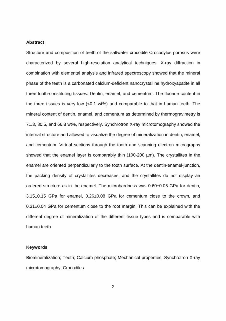
Abstract
Structure and composition of teeth of the saltwater crocodile Crocodylus porosus were
characterized by several high-resolution analytical techniques. X-ray diffraction in
combination with elemental analysis and infrared spectroscopy showed that the mineral
phase of the teeth is a carbonated calcium-deficient nanocrystalline hydroxyapatite in all
three tooth-constituting tissues: Dentin, enamel, and cementum. The fluoride content in
the three tissues is very low (<0.1 wt%) and comparable to that in human teeth. The
mineral content of dentin, enamel, and cementum as determined by thermogravimetry is
71.3, 80.5, and 66.8 wt%, respectively. Synchrotron X-ray microtomography showed the
internal structure and allowed to visualize the degree of mineralization in dentin, enamel,
and cementum. Virtual sections through the tooth and scanning electron micrographs
showed that the enamel layer is comparably thin (100-200 µm). The crystallites in the
enamel are oriented perpendicularly to the tooth surface. At the dentin-enamel-junction,
the packing density of crystallites decreases, and the crystallites do not display an
ordered structure as in the enamel. The microhardness was 0.60±0.05 GPa for dentin,
3.15±0.15 GPa for enamel, 0.26±0.08 GPa for cementum close to the crown, and
0.31±0.04 GPa for cementum close to the root margin. This can be explained with the
different degree of mineralization of the different tissue types and is comparable with
human teeth.
Keywords
Biomineralization; Teeth; Calcium phosphate; Mechanical properties; Synchrotron X-ray
microtomography; Crocodiles

3
1. Introduction
Crocodiles belong to a very old phylogenetic group that has prevailed for millions of
years (Erickson et al., 2012; Janke et al., 2005). Unlike human teeth, reptile teeth
including crocodile teeth are continuously replaced (Kieser et al., 1993; Osborn, 1974;
Poole, 1961). For an approximately 4 m long crocodile (13 ft), it was estimated that each
tooth was replaced 45 times during the lifetime of the animal (Poole, 1961). Crocodiles
possess so-called thecodont teeth which are attached in sockets in the jaw (Dauphin
and Williams, 2008). Compared with other animals, crocodiles exert extraordinarily high
bite-forces and tooth pressures (Erickson et al., 2012). Like all vertebrate teeth,
crocodile teeth consist of a crown and a root.
In general, the interior bulk of the tooth crown consists of softer, less mineralized bone-
like dentin covered by an external layer of harder, highly mineralized enamel. The root,
however, consists of dentin (interior) that is covered by an external layer of cementum.
The inorganic mineral of human enamel is a calcium-deficient carbonated
hydroxyapatite, simplified: Ca5(PO4)3(OH), with small amounts of an organic matrix
(Busch et al., 2001; Dorozhkin and Epple, 2002; Fincham et al., 1999; Lowenstam and
Weiner, 1989). For details on teeth in general see, e.g., Teaford et al. (2000).
In contrast to human teeth and shark teeth (which are fully replaced upon loss) (Marks
Jr. and Schroeder, 1996; Smith et al., 2012), the root of a crocodile tooth is hollow. Each
mature functional tooth is accompanied by a small initial replacement tooth on the
lingual side of the root that grows from a bud formed by a specialized dental lamina.
Together, they form a tooth family unit. Crocodylian teeth cycle continuously. While the
new tooth grows, it is moving outward and induces the resorption of the root of the old
tooth which is then shed (Wu et al., 2013). The human tooth eruption follows a similar

4
pattern. During the change from deciduous tooth to permanent tooth, the root of the old
tooth is resorbed by osteoclasts and the crown erupts as a compact object (Marks Jr.
and Schroeder, 1996).
Studies of the structure of reptile enamel were reported by Dauphin (1987), Sahni
(1987), and Sander (1999). In general, reptile teeth have not been as thoroughly
investigated as the teeth of other large animals; one reason is that their enamel does not
consist of defined prisms such as mammalian teeth which can be more easily analyzed
(Sander, 2000). Because reptile enamel is lacking prisms, it is typically denoted as
"prismless enamel", a feature which is common to most non-mammalian amniotes
(reptiles) (Sander, 2000). Currently, there are only a few reports about the structures of
crocodile and alligator teeth (Dauphin and Williams, 2007; Dauphin and Williams, 2008;
Erickson et al., 2012; Osborn, 1998; Sander, 1999; Sato et al., 1993; Sato et al., 1990;
Shimada et al., 1992).
To close this gap, we have analyzed the chemical and crystallographic composition, the
ultrastructure, and the microhardness of dentin, enamel, and cementum, of teeth of the
saltwater crocodile Crocodylus porosus with the aim to correlate all parameters, namely,
structure, hardness, and biological function, to gain an integral view. Additionally, we
compare these properties with human teeth.
2. Materials and methods
2.1 Sample preparation and analytical methods
Teeth of the recent crocodile species C. porosus were stored in dry state at room
temperature. We used five different teeth to produce fine powders of dentin, enamel,
and cementum (several mg per sample) by mechanical abrasion with a Proxxon fine

5
drilling and polishing tool FBS 230/E, equipped with a diamond-coated drill. The mineral
phase and the size of the crystalline domains of these powder samples were determined
by infrared (IR) spectroscopy and X-ray powder diffraction (XRD) measurements.
Fourier-transform infrared spectroscopy (FTIR) was carried out with a Bruker Vertex 70
instrument in potassium bromide (KBr) pellets (range 400-4000 cm-1 and 2 cm-1
resolution). XRD measurements were carried out with a Panalytical Empyrean
diffractometer equipped with a furnace (XRK 900, Anton Paar) using a silicon single
crystal as sample holder to minimize scattering. First, a diffractogram was measured at
30 °C. Then, the sample was heated to 750 °C and he ld at this temperature for 2 h
before another diffractogram was measured. The measurements at 750 °C were
performed to identify the conversion products of the mineral phase after thermal
treatment. Rietveld refinement for the calculation of the lattice parameters and the size
of the crystalline domains was performed using the Bruker software TOPAS 4.2. The
correction for instrumental peak broadening as determined with an LaB6 powder sample,
National Institute of Standards and Technology (NIST), as standard reference material
(SRM 660b), was included. As reference, we used the pattern of hydroxyapatite (#9-
0432) from the International Centre for Diffraction Data (ICDD) database.
A part of the remaining powdered sample material was used to perform elemental
analysis to determine the overall chemical composition and to confirm the identity of the
mineral phases. Calcium, magnesium, and sodium were determined with atomic
absorption spectroscopy (AAS), fluoride with ion-selective potentiometry, and phosphate
with ultraviolet (UV) spectroscopy. All measurements were carried out using several mg
of powdered dentin, enamel, and cementum which were dissolved in concentrated
hydrochloric acid. For fluoride analysis we used ion-selective potentiometry (ion-
6
selective electrode, ISE; pH/ION 735, WTW; the measurements were performed by
Analytische Laboratorien GmbH, Lindlar, Germany). Atomic absorption spectroscopy
was performed with a Thermo Electron M-Series instrument. Phosphate was determined
with a Varian Cary 300 UV-Vis spectrophotometer as phosphate-molybdenum blue
complex.
Thermogravimetry (TG) was used to determine the contents of water, organic matrix,
and carbonated apatite in the remaining powder samples of dentin, enamel, and
cementum from five different teeth. The experiments were carried out in a Netzsch STA
449 F3 Jupiter instrument in dynamic oxygen atmosphere at a heating rate of 2 K min-1
from 25 to 1200 °C in open alumina crucibles. For V icker’s microhardness tests, the
teeth were axially cut with a jeweler’s saw (for the convention of axial and transversal
denomination see Figure 1). Subsequently, the samples were embedded in one-
component UV-curable methyl methacrylate CEM 4000 Lightfix resin (Cloeren
Technology GmbH, Wegburg) that was cured in a Struers UV-Box using the bottom
source only for 3 min and with bottom and top source together for 6 min. The surfaces of
interest were polished using successively abrasive papers with decreasing grit sizes
(120, 220, 400, 600, 1000, 2500, and 4000; Hermes) followed by polishing with a 3 µm
diamond suspension (Struers), and finally with a 0.1 µm silica suspension (Buehler;
Saphir 320/330 instrument, ATM). In addition to polished samples, parts of teeth
fractured to expose either cross sections or axial sections were also prepared for
scanning electron microscopy (SEM). All SEM samples were mounted on standard
aluminum holders, rotary shadowed with 4 nm of platinum using a Gatan PECS 682
sputter coater, and observed in a high resolution scanning electron microscope (Zeiss
Gemini 1540XB) at acceleration voltages of 5-10 kV using a small aperture (30 µm) and

7
either an in-lens secondary electron (SE) detector or a backscattered electron (BSE)
detector for compositional contrast. For a clearer view on the microstructure, selected
samples were superficially etched using aqueous EDTA solution (0.15 M and 2.5%
glutaraldehyde for 20 min) followed by a quick rinse by double-distilled H2O and 100%
methanol for 1 s each. Where necessary, contrast and brightness of the digital images
were adjusted using Adobe Photoshop CS3 (Adobe Inc.).
Synchrotron X-ray microtomography (SRµCT) is a very useful technique for the
visualization of microstructures because it provides 3D data sets in a widely non-
destructive manner. This technique was already successfully used to study biological
materials, e.g., bone microstructures (Bonse et al., 1994; Larrue et al., 2011; Sanchez et
al., 2012) and human teeth (Dowker et al., 2004; Neues et al., 2009; Sanchez et al.,
2012).
SRµCT analysis was used to evaluate the gray values as indication of the local density
of the material and thus the degree of mineralization as well as to create virtual 3D
sections of the tooth. SRµCT analyses were carried out at beamline ID19 of the
European Synchrotron Radiation Facility (ESRF), Grenoble, France. Experimental
details of the beamline and on the evaluation procedure can be found in Weitkamp et al.
(2010). The 3D images and virtual sections were rendered with the software VGStudio
MAX 2.1. The gray values were identified by the graphic software ImageJ 1.45s
(Schneider et al., 2012). For the measurements, the sample was placed 935 mm
upstream of an indirect X-ray imaging detector operating with an effective pixel size of
5 µm (ESRF inhouse CCD camera FReLoN type F_7899 in combination with a 125 µm-
thick LuAG:Ce single-crystal scintillator and a 2.8× magnifying optical system,
2048·2048 pixels). Polychromatic radiation was chosen, i.e. the beamline U17.6

8
undulator was combined with only 2.8 mm Al and 0.4 mm Cu absorbers at a gap of 15
mm in order to gain a sufficient photon flux density (the ESRF operated in 4-bunch
mode). The resulting spectrum was dominated by the 2nd harmonic of the undulator
around an X-ray photon energy of 35 keV. 2000 projection images were acquired over a
tomographic scan range of 180 degrees with an exposure time of 2 s each. Single-
distance phase retrieval was applied in order to establish a direct correlation between
the gray levels of the voxels and the materials associated with them before the
tomographic reconstruction by means of filtered-back projection was performed
(Weitkamp et al., 2011). The chosen approach is robust and allows to work with a single
propagation distance only but as well only for a single component: The dentin-enamel
junction (dej) was fringe-free after this phase-retrieval while other interfaces such as
dentin-air remained with fringes (Paganin et al., 2002).
Vicker’s microhardness tests were carried out on the exposed axial sections of two teeth
in four designated areas of interest: The crown enamel, crown dentin, the distal
cementum layer close to the crown and the cementum layer close to the proximal
margin of the tooth. In every area of interest on each sample, 20 indentations were
made with a Leco M-400-H1 microhardness testing device. The location of each
indentation was manually chosen and a weight of 10 g (HV0.01) was applied for 15 s.
These experiments, including the distance between two indentations, were performed
according to DIN EN ISO 6507-1 and DIN EN ISO 6507-4 and the indentations were
manually controlled and evaluated. Vicker’s hardness HV0.01 was converted into
Berkovich hardness H according to HV0.01/kg mm-2 = 92.65 s2 m-1·H/GPa.
3. Results
9
A typical tooth of the crocodile species C. porosus has a cone-shaped crown and a
hollow, roughly cylindrical root (Figure 1). The absolute size of each tooth varies
depending on the age and size of individual animals.
The transition between dentin and enamel is marked by the so-called dentin-enamel-
junction (dej) and is clearly visible on scanning electron micrographs of axially polished
cross sections through the teeth (Figures 2A, 2B, and 3A). The crocodile enamel has a
thickness of about 100-200 µm at the crown (Figure 3A), becomes thinner towards the
root and disappears completely just before the onset of the hollow part (Figure 2A). In
this region, an enamel-cementum interface can be observed where a thin cementum
layer is present that covers the enamel layer (Figure 2A). The uniform contrasts
observed in composition-sensitive backscattered electron (BSE) contrast micrographs
(Figure 2) indicate a homogeneously distributed mineral content in all three layers.
Surfaces of fractured enamel show that it consists of small elongated mineral crystallites
that are mainly oriented with their long axes perpendicular to the tooth surface (Figures
3C and 4). These crystallites are very densely packed and therefore the shape and
dimensions of individual crystallites are difficult to determine. Superficial etching of
polished enamel surfaces revealed the needle-like shape of the mineral structures. The
crystallites in the enamel have a length of a few µm. No defined crystallite bundles as
they occur in prismatic enamel of e.g., mammalians were visible (Figure 4). Throughout
the enamel, a fine horizontal (parallel to the tooth surface) striation can be observed
(Figure 4A). At the dentin-enamel-junction (dej) the packing density of the crystallites
decreases and their orientation becomes more random (Figures 3B, 4B). The dej is
further characterized by a relatively high content of randomly arranged organic fibers
(Figures 2B, 3B). The mineral phase of the cementum is not as clearly structured as in

10
the enamel in terms of regularly arranged crystallites. The cementum contains a loose
network of randomly arranged organic fibers (Figures 2C, 3D). These fibers disappear in
the transition between cementum and dentin (dentin-cementum-junction, dcj), which is
not as sharp as the dej (Figures 2C, 3D). The dentin of crocodile teeth is pervaded by
numerous µm-sized dentin tubuli (Figure 2D). The SEM-micrographs of fractured teeth
indicate that the mineral phase of dentin also consists of needle-shaped crystallites
(Figure 2B). However, a well-defined structural organization as found in enamel could
not be observed with the techniques used in our study.
Elemental analysis of the crocodile teeth shows that the enamel contains more calcium,
phosphate, and sodium but less magnesium than dentin (Table 1). Sodium is present in
dentin and enamel but absent in the cementum. The fluoride concentration in crocodile
teeth altogether is very low (<0.1 wt%). The cementum contains significantly more
fluoride than dentin and enamel. The magnesium content is very high, especially in
dentin, and it was included to compute the (Ca+Mg)/P molar ratio in the apatite mineral
phase. In all cases, the stoichiometry of a calcium-deficient hydroxyapatite was found,
i.e. around n(Ca+Mg):n(P)=1.67:1 (Dorozhkin and Epple, 2002).
The mineral phase of the teeth was analyzed by X-ray powder diffraction in combination
with Rietveld analyses (Figure 5 and Table 2). The diffractograms of dentin, enamel, and
cementum show the typical peak pattern of hydroxyapatite. While the diffractograms of
dentin and cementum show broad diffraction peaks, those of the enamel are slightly
sharper. The average size of the crystalline domains is 8-9 nm, and the lattice
parameters and cell volumes differ just a little between dentin, enamel, and cementum.
Heating of the samples to 750 °C in vacuum resulted in a mixture of hydroxyapatite and
β-tricalcium phosphate, β-Ca3(PO4)2, in comparable amounts indicating the presence of

11
a calcium-deficient hydroxyapatite in the initial phase (i.e. before calcination) (Dorozhkin
and Epple, 2002).
The IR-spectra recorded for dentin, enamel, and cementum (Figure 6) show the
absorption bands specific for phosphate (490-640 cm-1 and 900-1220 cm-1), carbonate
(875 cm-1 and 1360-1590 cm-1), and water (3010-3660 cm-1). Additional bands that
indicate the organic matrix (mainly proteins) appear in all three layers at 2940 cm-1 (C-H)
and 1600-1700 cm-1 (amide I band of proteins) (Preston et al., 2011).
Thermogravimetry of dentin, enamel, and cementum shows three main regions of mass
loss: Release of water (<200 °C), the combustion of the organic matrix (200-500 °C),
and finally the release of CO2 from carbonated apatite (>500 °C) (LeGeros, 1981; Peters
et al., 2000) (Figure 7). The residue is the decarboxylated mineral phase, i.e. pure
calcium phosphate. Enamel has a much higher mineral content than dentin and
cementum with a low content of organic matrix (Table 3). Cementum has a lower
mineral content than dentin. The content of water is comparable in all three tissues. Note
that the content of water may be variable, depending on the storage conditions.
SRµCT analyses show the exact geometry of the crocodile teeth and the spatial
arrangement of the constituting layers (Figure 8). The enamel layer (whitish color) is
confined to the crown and generally very thin with the thickest regions located at the
tooth tip. From the 3D images, it is difficult to distinguish between dentin and the very
thin cementum layer that covers the root due to a very similar X-ray absorption contrast.
In axial sections, it becomes obvious that the hollow root takes up over two thirds of the
total tooth size, and that the crown is comparatively small. Moreover, the cavity formed
by the root has the shape of a distally pointed cone, and the root’s wall thickness is
decreasing in proximal direction.
12
In the SRµCT-3D images, the enamel has a white color, and the dentin has a gray color.
Evaluation of the gray values using line scans through the tooth crown (Figures 9A and
10A) shows that the enamel has a much higher gray value (~2.8) than the dentin (~1.8).
The root (Figures 9B and 10B), however, shows a lower gray value for the dentin (~1.3)
compared to the crown dentin. Cementum has a slightly higher gray value than the
dentin in the root.
Representative Vicker’s microhardness tests were performed at different positions of
polished tooth samples of C. porosus. For better comparability, the results were
converted into Berkovich hardness. We found 0.60±0.05 GPa for dentin, 3.15±0.15 GPa
for enamel, 0.26±0.08 GPa for cementum close to the crown, and 0.31±0.04 GPa for
cementum close to the margin of the root (Table 4).
4. Discussion
The teeth of the saltwater crocodile C. porosus are mainly characterized by their specific
shape, a small crown with a comparably large root, and the three constituting tissues
dentin, enamel, and cementum that differ in microstructure, composition, and resulting
mechanical properties. The major part of both the crown and the root consists of dentin
which contains dentin tubuli that are also present in human teeth (Marten et al., 2010).
Both exterior layers, the cementum covering the root and the enamel covering the
crown, are separated from the dentin by a structurally distinct layer, the dentin-
cementum-junction (dcj) and the dentin-enamel-junction (dej), respectively. The dentin-
enamel-junction is also present in mammalian teeth (Line and Novaes, 2005; Walker
and Fricke, 2006). Within the enamel, the mineral crystallites are all oriented parallel,

13
hence, this type of enamel is typically denoted as "parallel crystallite enamel" (Sander,
2000). A similar enamel microstructure was found for teeth of Alligator mississippiensis
(Sato et al., 1990). The horizontal striation observed on etched enamel surfaces
indicates the presence of incremental growth lines that have also been described in
teeth of other ectotherm animals (Line and Novaes, 2005).
X-ray diffractograms of dentin and enamel showed broad diffraction peaks which
indicate a comparable size of crystalline domains between these tissues. That is
remarkable because in mammalian enamel and shark enameloid the apatite rods have a
larger size of crystalline domains than the apatite nanocrystals in dentin (LeGeros, 1994;
Xue et al., 2008). The lattice parameters of the mineral in crocodile teeth are very similar
to those of human teeth and of geological hydroxyapatite and confirm the presence of
apatite. The small differences in the lattice parameters of dentin, enamel, and cementum
can be ascribed to different amounts of incorporated ions, e.g., magnesium, sodium, and
carbonate into the apatite lattice, as it is well known for biological apatite (Dauphin and
Williams, 2007; LeGeros, 1981). Magnesium as substituent is known to reduce the
crystallinity of apatites while sodium has no significant influence (Elstnerova et al., 2010;
LeGeros, 1994). Especially magnesium strongly influences the lattice parameters.
Apatites with high contents of magnesium become amorphous (Masayuki et al., 1986).
The fluoride content of crocodile teeth is very low like in human teeth (Aoba, 1997;
LeGeros, 1981). Overall, the results of elemental analysis agree well with microprobe
analyses of recent reptile teeth (Dauphin and Williams, 2007; Dauphin and Williams,
2008). Our results indicate that dentin, enamel, and cementum all contain
nanocrystalline hydroxyapatite. The size of the crystalline domains of the different tooth
layers is comparable.

14
The amide I band of proteins in the IR-spectra in enamel is less intense compared to
dentin and cementum, probably due to the lower content of organic matrix. This is
supported by thermogravimetry. Human dentin and cementum are known to have a high
content of organic matrix which consists mainly of collagen (Wiesmann et al., 2005). The
thermogravimetric analysis of human dentin showed similar results as found here for
crocodile dentin (Lim and Liboff, 1972). In general, the IR spectra of dentin, enamel, and
cementum are all very similar to each other and confirm the results of X-ray diffraction.
They are also very similar to human teeth (LeGeros, 1981), synthetic hydroxyapatite
(Zhou et al., 1993), and also shark teeth which do not consist of hydroxyapatite but of
fluoroapatite, Ca5(PO4)3F (Enax et al., 2012).
The results of X-ray diffraction correspond well with the microstructure analysis by
scanning electron microscopy. The prismless enamel of crocodile teeth contains no
crystallite bundles and no complex microstructures but only needle-like mineral
structures which are ordered perpendicularly to the tooth surface. Human teeth show a
especially high fracture toughness (Padmanabhan et al., 2010; Yilmaz et al., 2013) due
to the presence of crystallite bundles (enamel prisms) which are hierarchically ordered in
a mm-thick enamel layer (Ang et al., 2010; Dunlop and Fratzl, 2010; He and Swain,
2008). In contrast, the enamel of crocodile teeth is very thin compared to mammalian
teeth, which is consistent with their specific function. Crocodiles do not use their teeth for
cutting and chewing, but only for gripping and securing their prey, which is then pulled
into the water and killed by drowning. Generally, their teeth are not used to dismember
prey animals; instead crocodiles use violent body movements to tear pieces off which
are then swallowed. The enamel cap on mammalian teeth can be up to 5 mm thick
(Lucas et al., 2008). Here, SRµCT showed clearly that the enamel thickness of crocodile

15
teeth reaches its maximum at the tooth tip and is rapidly thinning towards the root. This
indicates that crocodile teeth must be very resistant at the tip area, probably optimized
for snapping their prey. Crocodiles are ambush hunters which attack with a very fast bite
where they exert extraordinarily high bite-forces and tooth pressures (Erickson et al.,
2012). The thick enamel at the tip of the teeth might help to prevent damage upon
impact on hard parts of the prey, e.g. bones, during the attack. Interestingly, the
arrangement of teeth in the upper and lower jaw is such that opposing teeth will not get
in contact if the bite misses the target. This is also corroborated by the fact that the root
dentin shows a lower gray value than the crown dentin, corresponding to a lower mineral
content and thus a higher potential for deformation without suffering a brittle fracture.
The comparably higher deformability and lower hardness together with the large size of
the root and thus large contact area with the jawbone via the teeth-ridge further helps to
dissipate the pressure and thus the kinetic energy acting on individual teeth during the
impact on prey animals.
Thus, the structural and compositional organization of the crocodile tooth is ideally
suited for its function. Nevertheless, the hunting mode using very fast and powerful bites
presumably increases the risk of teeth getting damaged compared to other predatory
animals like mammalian carnivores that use similarly shaped canine teeth for killing their
prey. However, these animals use their teeth in a much more controlled, almost surgical
mode by biting into specific neuralgic points of their prey. The constant replacement of
crocodile teeth may thus represent an additional adaptation to their predation technique
that was necessary to compensate the higher risk of damage. Vicker’s microhardness
tests of crocodile teeth gave a hardness close to the hardness of human teeth and shark
teeth. This is surprising because crocodile teeth do not have hierarchically organized

16
crystallite bundles like human teeth. The different hardness values of the different
tissues found in crocodile teeth can be explained by different degrees of mineralization
and thus by different mineral contents which was shown by SRµCT, SEM, elemental
analysis, and thermogravimetry. This is in good agreement with results for human teeth.
It was shown by spatially resolved studies that an increasing calcium content (and thus
mineral content) leads to a higher hardness in human enamel (Cuy et al., 2002; Jeng et
al., 2011). Note that the microhardness of teeth is lower than that of pure (geological)
hydroxyapatite which amounts to 5.4±1.3 GPa (White et al., 2001). This is because a
geological hydroxyapatite crystal does not contain an organic matrix and is therefore
much more brittle (and less elastic).
5. Conclusions
The hardness of human teeth and crocodile teeth is comparable, although their
microstructures are significantly different. The prismless enamel layer of crocodile teeth
is very thin, with a maximum thickness at tooth tip and consists of nanocrystalline
hydroxyapatite, in striking contrast to mammalian enamel and shark tooth enameloid.
Structure and composition of crocodile teeth are well suited for their biological function.
In contrast to most mammals, crocodiles do no not need their teeth for cutting but mainly
for holding their prey. Crocodiles are changing their teeth continuously during their
lifetime, possibly because of their way of hunting, i.e. very fast and powerful bites. As
consequence, crocodiles have a higher risk of tooth-damage than other animals like
mammalian carnivores. The construction of their teeth is well adapted to their biological
function.

17
Acknowledgements
We thank C. Fischer, Essen, Germany, for help with the tooth preparation, M. Ruiz,
Grenoble, France, for support during the beamtime at the ESRF, and A. Gillis, Halifax,
Canada, for helpful discussions. We thank the Deutsche Forschungsgemeinschaft for
support within the priority program SPP 1420.

18
References
Ang, S.F., E.L. Bortel, M.V. Swain, A. Klocke, and G.A. Schneider, 2010. Size-dependent elastic/inelastic behavior of enamel over millimeter and nanometer length scales. Biomaterials 31: 1955-1963.
Aoba, T., 1997. The effect of fluoride on apatite structure and growth. Crit. Rev. Oral Biol. Med. 8: 136-153.
Bonse, U., F. Busch, O. Gunnewig, F. Beckmann, R. Pahl, et al., 1994. 3D computed X-ray tomography of human cancellous bone at 8 µm spatial and 10-4 energy resolution. Bone Miner. 25: 25-38.
Busch, S., U. Schwarz, and R. Kniep, 2001. Morphogenesis and structure of human teeth in relation to biomimetically grown fluoroapatite-gelatine composites. Chem. Mater. 13: 3260-3271.
Cuy, J.L., A.B. Mann, K.J. Livi, M.F. Teaford, and T.P. Weihs, 2002. Nanoindentation mapping of the mechanical properties of human molar tooth enamel. Arch. Oral. Biol. 47: 281-291.
Dauphin, Y., 1987. Implications of preparation processes on the interpretation of reptilian enamel structure. Palaentolog. Z. 61: 331-337.
Dauphin, Y., and C.T. Williams, 2007. The chemical compositions of dentine and enamel from recent reptile and mammal teeth-variability in the diagenetic changes of fossil teeth. CrystEngComm. 9: 1252-1261.
Dauphin, Y., and C.T. Williams, 2008. Chemical composition of enamel and dentine in modern reptile teeth. Mineral. Mag. 72: 247-250.
del Pilar Gutierrez-Salazar, M., and J. Reyes-Gasga, 2003. Microhardness and chemical composition of human tooth. Mater. Res. 6: 367-373.
Dorozhkin, S.V., and M. Epple, 2002. Biological and medical significance of calcium phosphates. Angew. Chem. Int. Ed. 41: 3130-3146.
Dowker, S.E.P., J.C. Elliott, G.R. Davis, R.M. Wilson, and P. Cloetens, 2004. Synchrotron x-ray microtomographic investigation of mineral concentrations at micrometre scale in sound and carious enamel. Caries Res. 38: 514-522.
Dunlop, J.W.C., and P. Fratzl, 2010. Biological composites. Annu. Rev. Mater. Res. 40: 1-24. Elstnerova, P., M. Friak, H. Fabritius, L. Lymperakis, T. Hickel, et al., 2010. Ab initio study of
thermodynamic, structural, and elastic properties of Mg-substituted crystalline calcite. Acta Biomater. 6: 4506-4512.
Enax, J., O. Prymak, D. Raabe, and M. Epple, 2012. Structure, composition, and mechanical properties of shark teeth. J. Struct. Biol. 178: 290-299.
Erickson, G.M., P.M. Gignac, S.J. Steppan, A.K. Lappin, K.A. Vliet, et al., 2012. Insights into the ecology and evolutionary success of crocodilians revealed through bite-force and tooth-pressure experimentation. PLoS One 7: 1-12.
Fincham, A.G., J. Moradian-Oldak, and J.P. Simmer, 1999. The structural biology of the developing dental enamel matrix. J. Struct. Biol. 126: 270-299.
He, L.H., and M.V. Swain, 2008. Understanding the mechanical behaviour of human enamel from its structural and compositional characteristics. J. Mech. Behav. Biomed. Mater. 1: 18-29.
Janke, A., A. Gullberg, S. Hughes, R.K. Aggarwal, and U. Arnason, 2005. Mitogenomic analyses place the gharial (Gavialis gangeticus) on the crocodile tree and provide pre-K/T divergence times for most crocodilians. J. Mol. Evol. 61: 620-626.

19
Jeng, Y.R., T.T. Lin, H.M. Hsu, H.J. Chang, and D.B. Shieh, 2011. Human enamel rod presents anisotropic nanotribological properties. J. Mech. Behav. Biomed. Mater. 4: 515-522.
Kieser, J.A., C. Klapsidis, L. Law, and M. Marion, 1993. Heterodonty and patterns of tooth replacement in Crocodylus niloticus. J. Morphol. 218: 195-201.
Larrue, A., A. Rattner, Z.-A. Peter, C. Olivier, N. Laroche, et al., 2011. Synchrotron radiation micro-CT at the micrometer scale for the analysis of the three-dimensional morphology of microcracks in human trabecular bone. PLoS One 6: e21297.
LeGeros, R.Z., 1981. Apatites in biological systems. Prog. Cryst. Growth Charact. 4: 1-45. LeGeros, R.Z., 1994. Biological and synthetic apatites, p. 3-28, in: P. W. Brown and B.
Constantz, Eds.), Hydroxyapatite and related materials, CRC Press, Boca Raton. Lim, J.J., and A.R. Liboff, 1972. Thermogravimetric analysis of dentin. J. Dent. Res. 51: 509-
514. Line, S.R.P., and P.D. Novaes, 2005. The development and evolution of mammalian enamel:
Structural and functional aspects Braz. J. morphol. Sci. 22: 67-72. Lowenstam, H.A., and S. Weiner, 1989. On biomineralization, Oxford University Press, New
York. Lucas, P., P. Constantino, B. Wood, and B. Lawn, 2008. Dental enamel as a dietary indicator in
mammals. BioEssays 30: 374-385. Malek, S., M.A. Darendeliler, and M.V. Swain, 2001. Physical properties of root cementum: Part
I. A new method for 3-dimensional evaluation. Am. J. Orthod. Dentofacial Orthop. 120: 198-208.
Marks Jr., S.C., and H.E. Schroeder, 1996. Tooth eruption: Theories and facts. Anat. Rec. 245: 374-393.
Marten, A., P. Fratzl, O. Paris, and P. Zaslansky, 2010. On the mineral in collagen of human crown dentine. Biomaterials 31: 5479-5490.
Masayuki, O., T. Junzo, and K. Hiroshi, 1986. Comparison of crystallographic properties of magnesium, iron, sodium carbonate, fluoride, and chloride-containing apatites. J. Osaka Univ. Dent. Sch. 26: 79-89.
Neues, F., A. Klocke, F. Beckmann, J. Herzen, J.P. Loyola-Rodriguez, et al., 2009. Mineral distribution in highly fluorotic and normal teeth: A synchrotron microcomputer tomographic study. Mat.-wiss. u. Werkstofftech. 40: 294-296.
Osborn, J.W., 1974. On the control of tooth replacement in reptiles and its relationship to growth. J. Theor. Biol. 46: 509-527.
Osborn, J.W., 1998. Relationship between growth and the pattern of tooth initiation in alligator embryos. J. Dent. Res. 77: 1730-1738.
Padmanabhan, S.K., A. Balakrishnan, M.-C. Chu, T. Kim, and S.J. Cho, 2010. Micro-indentation fracture behavior of human enamel. Dent. Mater. 26: 100-104.
Paganin, D., S.C. Mayo, T.E. Gureyev, P.R. Miller, and S.W. Wilkins, 2002. Simultaneous phase and amplitude extraction from a single defocused image of a homogeneous object. J. Microscopy 206: 33-40.
Peters, F., K. Schwarz, and M. Epple, 2000. The structure of bone studied with synchrotron X-ray diffraction, X-ray absorption spectroscopy and thermal analysis. Thermochim. Acta 361: 131-138.
Poole, D.F.G., 1961. Notes on tooth replacement in the nile crocodile Crocodilus niloticus. Proc. Zool. Soc. Lond. 136: 131-140.

20
Preston, L.J., M.R.M. Izawa, and N.R. Banerjee, 2011. Infrared spectroscopic characterization of organic matter associated with microbial bioalteration textures in basaltic glass. Astrobiology 11: 585-599.
Saenger, A.T., and W.F. Kuhs, 1992. Structural disorder in hydroxyapatite. Z. Kristallogr. 199: 123-148.
Sahni, A., 1987. Evolutionary aspects of reptilian and mammalian enamel structure. Scanning Microsc. 1: 1903-1912.
Sanchez, S., P.E. Ahlberg, K.M. Trinajstic, A. Mirone, and P. Tafforeau, 2012. Three-dimensional synchrotron virtual paleohistology: A new insight into the world of fossil bone microstructures. Microsc. Microanal. 18: 1095-1105.
Sander, P.M., 1999. The microstructure of reptilian tooth enamel: Terminology, function, and phylogeny, München.
Sander, P.M., 2000. Prismless enamel in amniotes: terminology, function, and evolution in: M. F. Teaford, et al., Eds.), Development, function and evolution of teeth, Cambridge University Press, Cambridge.
Sato, I., K. Shimada, H. Ezure, T. Sato, and V.A. Lance, 1993. Distribution of calcium-ATPase in developing teeth of embryonic American alligators (Alligator mississippiensis). J. Morphol. 218: 249-256.
Sato, I., K. Shimada, A. Yokoi, J.C. Handal, N. Asuwa, et al., 1990. Morphology of the teeth of the American Alligator (Alligator mississippiensis): Fine structure and chemistry of the enamel. J. Morphol. 205: 165-172.
Schneider, C.A., W.S. Rasband, and K.W. Eliceiri, 2012. NIH Image to ImageJ: 25 years of image analysis. Nat. Methods 9: 671-675.
Shimada, K., I. Sato, and H. Moriyama, 1992. Morphology of the tooth of the American alligator (Alligator mississippiensis): The fine structure and elemental analysis of the cementum. J. Morphol. 211: 319-329.
Smith, M.M., Z. Johanson, C. Underwood, and T.G.H. Diekwisch, 2012. Pattern formation in development of chondrichthyan dentitions: a review of an evolutionary model. Hist. Biol. 25: 127-142.
Teaford, M.F., M.M. Smith, and M.W.J. Ferguson, 2000. Development, function and evolution of teeth, Cambridge University Press, Cambridge.
Walker, M.P., and B. Fricke, 2006. Dentin-enamel junction of human teeth, in: M. Akay, (Ed.), Wiley Encyclopedia of Biomedical Engineering, John Wiley & Sons, Inc., New York.
Weitkamp, T., D. Haas, D. Wegrzynek, and A. Rack, 2011. ANKAphase: software for single-distance phase retrieval from inline X-ray phase-contrast radiographs. J. Synchrotron Radiat. 18: 617-29.
Weitkamp, T., P. Tafforeau, E. Boller, P. Cloetens, J.-P. Valade, et al., 2010. Status and evolution of the ESRF beamline ID19, p. 33-38, in: M. A. Denecke and C. T. Walker, Eds.), X-Ray Optics and Microanalysis, Proceedings.
White, S.N., W. Luo, M.L. Paine, H. Fong, M. Sarikaya, et al., 2001. Biological organization of hydroxyapatite crystallites into a fibrous continuum toughens and controls anisotropy in human enamel. J. Dent. Res. 80: 321-326.
Wiesmann, H.P., U. Meyer, U. Plate, and H.J. Hoehling, 2005. Aspects of collagen mineralization in hard tissue formation. Int. Rev. Cytol. 242: 121-156.
Wu, P., X.S. Wu, T.X. Jiang, R.M. Elsey, B.L. Temple, et al., 2013. Specialized stem cell niche enables repetitive renewal of alligator teeth. Proc. Nat. Acad. Sci. USA 110: E2009-E2018.

21
Xue, J., L. Zhang, L. Zou, Y. Liao, J. Li, et al., 2008. High-resolution X-ray microdiffraction analysis of natural teeth. J. Synchrotron Radiat. 15: 235-238.
Yilmaz, E.D., S. Bechtle, H. Oezcoban, A. Schreyer, and G.A. Schneider, 2013. Fracture behavior of hydroxyapatite nanofibers in dental enamel under micropillar compression. Scr. Mater. 68: 404-407.
Zhou, J., X. Zhang, J. Chen, S. Zeng, and K. De Groot, 1993. High temperature characteristics of synthetic hydroxyapatite. J. Mater. Sci.: Mater. Med. 4: 83-85.

22
Figure captions
Figure 1: Image of a crocodile tooth (C. porosus), including the convention of axial and
transversal directions, with an additional view into the hollow root (insert).
Figure 2: Scanning electron micrographs in back-scattered electron mode (BSE) of a
polished tooth surface of C. porosus, showing the dentin-enamel-junction and also the
dentin-cementum-junction (A), the dentin-enamel-junction in higher magnification (B),
the dentin-cementum-junction in higher magnification (C), and the dentin with its tubuli
(D). (d dentin, e enamel, c cementum, dej dentin-enamel-junction, dcj dentin-
cementum-junction).
Figure 3: Scanning electron micrographs of fractured tooth samples of C. porosus. (A)
The overview of an axially fractured tooth tip shows a clear border between dentin and
enamel. (B) The organization of the dentin-enamel-junction at higher magnification. (C)
Area at the tooth surface where the outermost layer of enamel was chipped off,
revealing the constituting small elongated mineral crystallites that are mainly oriented
with their long axes perpendicular to the tooth surface. (D) The organization of the
dentin-cementum-junction at higher magnification. (d dentin, e enamel, c cementum, dej
dentin-enamel-junction, dcj dentin-cementum-junction).
Figure 4: Scanning electron micrographs recorded in BSE contrast of the exposed axial
surface of enamel from teeth of C. porosus that was gently etched using EDTA to
expose the shapes and arrangement of the mineral crystallites. (A) The outermost

23
enamel consists of horizontal layers of parallel needle-shaped crystallites that are all
oriented with their long axes perpendicular to the tooth surface. The different contrasts,
especially of the horizontal bands, originates from slight differences in composition. (B)
Close to the dentin-enamel-junction, the horizontal layers of needle-shaped crystallites
are still visible, but frequently interrupted by the cavities of small canaliculae. (e enamel,
dej dentin-enamel-junction).
Figure 5: X-ray powder diffractograms of dentin (A), enamel (B), and cementum (C) of
teeth of C. porosus in comparison to pure hydroxyapatite (#9-0432 from the ICDD
database; computed as nanocrystalline phase) (D).
Figure 6: IR spectra obtained from dentin, enamel, and cementum of teeth of C.
porosus.
Figure 7: Thermogravimetric analysis of dentin, enamel, and cementum from teeth of C.
porosus: <200 °C: Release of water ( 1), 200-500 °C: Combustion of the organic matrix
(2), and >500 °C: Release of CO 2 from carbonated apatite (3).
Figure 8: SRµCT-3D images of the complete tooth (A) and of two virtual axial sections
(B, C) through a tooth of C. porosus.
Figure 9: Line scans on transverse sections through the crocodile tooth obtained by
SRµCT to generate the gray value profiles shown in Figure 10. A representative virtual
section of the crown (A) and a section through the root just below the altitude where the

24
enamel has been replaced by cementum (B) are shown. Interference fringes between
enamel and air as well as dentin and air remain due to the phase-retrieval chosen
algorithm (Paganin et al., 2002).
Figure 10: Comparison of the gray values of two virtual transversal sections through a
tooth of C. porosus. Linescan of the tooth crown (A) and linescan of the tooth root (B).

25
Table 1: The chemical composition of dentin, enamel, and cementum of teeth of C.
porosus (in wt%), in comparison to human teeth.
C. porosus Human teeth
LeGeros (1981)
Dentin Enamel Cementum Dentin Enamel
Ca2+ 20.56 28.56 23.01 27.0 36.0
PO43- 38.80 44.05 32.95 39.9 a 54.3 a
Ca/P molar
ratio 1.26:1 1.55:1 1.65:1 1.60:1 1.57:1
(Ca+Mg)/P
molar ratio 1.51:1 1.63:1 1.74:1 1.72:1 1.60:1
Na+ 0.78 1.01 -- 0.3 0.5
Mg2+ 2.47 1.10 0.75 1.1 0.44
F- 0.06 0.09 0.21 0.05 0.01
a Calculated from the content of phosphorus.

26
Table 2: Crystallographic properties of the mineral phase in teeth of C. porosus
compared to geological hydroxyapatite crystals and to human teeth. a Saenger and Kuhs
(1992), b LeGeros (1981) and c Enax et al. (2012). The standard deviation is given in
parentheses.
Sample a-axis
/ Å
c-axis
/ Å
V
/ Å3
Size of
crystalline
domains
/ nm
Crocodile dentin 9.43(1) 6.857(9) 528(2) 9
Crocodile enamel 9.451(8) 6.883(7) 532(1) 8
Crocodile cementum 9.409(8) 6.873(6) 527(1) 8
Geological hydroxyapatite a 9.4249(4) 6.8838(4) 529.56 -
Human dentin b
Human enamel b
9.421(3)
9.441(3)
6.887(3)
6.880(3)
529.3
531.1
-
-
Shark dentin c
Shark enameloid (fluoroapatite) c
9.404(5)
9.385(2)
6.842(10)
6.883(2)
524,0(7)
525,1(1)
2..13
34..75

27
Table 3: Results of the thermogravimetric analysis of dentin, enamel, and cementum of
teeth of C. porosus.
Dentin
/ wt%
Enamel
/ wt%
Cementum
/ wt%
Release of water
(<200 °C) 2.7 3.3 3.9
Combustion of the organic matrix
(200-500 °C) 24.2 13.8 27.7
Release of CO2 from carbonated apatite
(>500 °C) 1.8 2.4 1.6
Mineral content (residual) 71.3 80.5 66.8

28
Table 4: Results of Vicker’s microhardness tests (HV0.01) of C. porosus teeth
compared to values obtained for human teeth and shark teeth from literature. The values
are averages of all tested specimens including the standard deviation. They were
converted into Berkovich hardness in GPa. a del Pilar Gutierrez-Salazar and Reyes-
Gasga (2003), b Malek et al. (2001), and c Enax et al. (2012).
Crocodile t eeth
/ GPa
Human teeth
/ GPa
Shark teeth
/ GPa
Dentin 0.60±0.05 0.5..0.6 a 0.5..0.7 c
Enamel 3.15±0.15 2.9..3.9 a 3..4 c
Cementum
(distal, close to crown) 0.26±0.08
0.2..0.6 b - Cementum
(proximal, close to root margin) 0.31±0.04