Embed Size (px)
Citation preview

CHARACTERIZATION AND CONTROL OF MICROPROPAGATION
PROBLEMS IN ALOE, DEVIL’S CLAW AND BANANA
By
MICHAEL WOLDAY BAIRU
(MSc University of KwaZulu-Natal)
Submitted in fulfilment of the requirements for the degree of
DOCTOR OF PHILOSOPHY
Research Centre for Plant Growth and Development, School of Biological
and Conservation Sciences, University of KwaZulu-Natal, Peitermaritzburg
January 2008
South Africa

i
Contents
DECLARATION ....................................................................................................... v
ACKNOWLEDGEMENTS ...................................................................................... vi
PUBLICATIONS FROM THIS STUDY .................................................................. vii
CONFERENCE CONTRIBUTIONS ..................................................................... viii
LIST OF FIGURES .................................................................................................. x
LIST OF TABLES ................................................................................................... xi
LIST OF ABBREVIATIONS .................................................................................. xiii
ABSTRACT ........................................................................................................... xv
1. General introduction and background to the problem ......................................... 1
1.1 Introduction .................................................................................................... 1
1.2 Background to the problem ............................................................................ 2
1.2.1 Comparative study of the biological activity of selected cytokinins using
the soybean callus bioassay ................................................................... 3
1.2.2 Micropropagation and hyperhydricity in Aloe polyphylla.......................... 4
1.2.3 Shoot-tip necrosis in Harpagophytum procumbens ................................ 5
1.2.4 Somaclonal variation in banana .............................................................. 6
1.3 Significance of the study ................................................................................ 6
1.4 Objectives of the study .................................................................................. 7
2. Literature review .................................................................................................. 8
2.1 Cytokinins: biosynthesis, metabolism and activity ......................................... 8
2.1.1 Introduction ............................................................................................. 8
2.1.2 Cytokinin biosynthesis and metabolism .................................................. 9
2.1.3 Cytokinin activity ................................................................................... 12
i) The general effect of ring substitution of aminopurines ........................... 13
ii) The effect of the hydroxyl group on the side chain ................................. 13
iii) The effect of a double bond on the side chain ....................................... 14
iv) The effect of the methyl and amino groups on the side chain ................ 14
v) The effect of side chain configuration (geometrical and optical isomers)15
vi) The effect of position of the substituent on the purine ring .................... 15
vii) The effect of halogen substituents ........................................................ 16
2.2 Hyperhydricity .............................................................................................. 17

ii
2.2.1 Introduction ........................................................................................... 17
2.2.2 Definition and cause of hyperhydricity ................................................... 17
i) Plant growth regulators ............................................................................ 18
ii) Agar quality and concentration ............................................................... 19
iii) Relative humidity and ventilation ........................................................... 20
iv) Light and temperature ............................................................................ 20
v) Organic and inorganic components ........................................................ 20
2.2.3 Physiological and biochemical reactions leading to hyperhydricity ....... 21
i) Water content .......................................................................................... 21
ii) Lignification and cell wall components .................................................... 22
iii) Chlorophyll and photosynthesis ............................................................. 22
2.2.4 Ultrastractural aspects of hyperhydricity ............................................... 22
2.3 Shoot-tip necrosis (STN) ............................................................................. 23
2.3.1 Introduction ........................................................................................... 23
2.3.2 Factors contributing to shoot necrosis in plants .................................... 24
i) The role of plant growth regulators .......................................................... 24
ii) The role of calcium ................................................................................. 25
iii) The role of boron.................................................................................... 27
iv) Association between Ca, B and PGR .................................................... 28
v) The role of physiological processes associated with rooting .................. 29
vi) Effect of aeration.................................................................................... 30
vii) Other factors ......................................................................................... 31
2.4 Somaclonal variation ................................................................................... 31
2.4.1 Introduction ........................................................................................... 31
2.4.2 Definition and types of somaclonal variation ......................................... 32
2.4.3 Origin and sources of somaclonal variation .......................................... 33
i) Pre-existing variation ............................................................................... 34
a) Use of chimeras .................................................................................. 34
b) Chromosome aberration and rearrangements .................................... 35
c) The role of the cell cycle ..................................................................... 35
d) Activation of cryptic transposable elements ........................................ 36
ii) Tissue culture-induced variation ............................................................. 37
a) Methods of vegetative propagation used ............................................ 37
b) Types of tissue or starting material used ............................................ 38

iii
c) Types and concentration of PGR in use ............................................. 38
d) Number and time of sub-culturing ....................................................... 39
e) Effect of stress and genotype ............................................................. 40
2.4.4 Description and occurrence of variants ................................................. 41
2.4.5 Detection and/or characterization of variants ........................................ 42
i) Morphological detection ........................................................................... 42
ii) Physiological characterization ................................................................ 43
iii) Molecular detection ................................................................................ 43
a) Proteins and isozymes ........................................................................ 43
b) Restriction Fragment Length Polymorphism (RFLP) .......................... 44
c) The polymerase chain reaction (PCR) ................................................ 45
2.5 Concluding remarks ..................................................................................... 47
3. Comparative study of the biological activity of selected cytokinins using the
soybean callus bioassay ..................................................................................... 48
3.1 Introduction .................................................................................................. 48
3.2 Materials and methods ................................................................................ 50
3.3 Results and discussion ................................................................................ 51
4. Micropropagation and hyperhydricity in Aloe polyphylla .................................... 60
4.1 Introduction .................................................................................................. 60
4.2 Material and methods .................................................................................. 62
4.2.1 Source material and bulking of explants ............................................... 62
4.2.2 Shoot multiplication ............................................................................... 62
4.2.3 DNA extraction and quantification ......................................................... 63
4.2.4 Acclimatization ...................................................................................... 64
4.3 Results and discussion ................................................................................ 64
4.3.1 Effect of meta-topolin on shoot multiplication ........................................ 64
4.3.2 Effect on hyperhydricity ......................................................................... 67
4.3.3 Effect on fresh weight (FW) ................................................................... 68
4.3.4 Effect on acclimatization ex vitro on A. polyphylla ................................. 72
4.3.5 Effect of hyperhydricity on total DNA content of A polyphylla ................ 72
5. Shoot-tip necrosis in Harpagophytum procumbens ........................................... 75
5.1 Introduction .................................................................................................. 75
5.2 Materials and methods ................................................................................ 77
5.2.1 Effect of media type and strength on STN ............................................ 77

iv
5.2.2 Effect of type and concentration of cytokinins on STN .......................... 78
5.2.3 Effect of type and concentration of sugars on STN ............................... 78
5.2.4 Effect of calcium and boron on STN...................................................... 79
5.2.5 Rooting and acclimatization of tissue cultured H. procumbens ............. 79
5.3 Results and discussion ................................................................................ 80
5.3.1 Effect of media composition and strength on STN ................................ 80
5.3.2 Effect of cytokinins on STN ................................................................... 81
5.3.4 Effect of sub-culturing on STN .............................................................. 83
5.3.5 Effect of sucrose concentration and type of sugars on STN ................. 85
5.3.6 Effect of Ca and B on STN .................................................................... 88
5.3.7 Aeration and rooting effects .................................................................. 90
5.3.8 Rooting and acclimatization of tissue cultured H. procumbens ............. 91
6. Micropropagation and somaclonal variation in banana ..................................... 93
6.1 Introduction .................................................................................................. 93
6.2 Material and methods .................................................................................. 94
6.2.1 Micropropagation .................................................................................. 94
6.2.2 Analysis of somaclonal variation ........................................................... 95
6.3 Results and discussion ................................................................................ 97
6.3.1 Micropropagation .................................................................................. 97
6.3.2 Analysis of somaclonal variation ......................................................... 101
7. Conclusions and recommendations ................................................................ 103
References .......................................................................................................... 105
Appendices ......................................................................................................... 137
Appendix – 1 .................................................................................................... 137
Appendix – 2 .................................................................................................... 138

v
DECLARATION
I hereby declare that, except where acknowledged, the contents of this thesis are
the results of my own investigation. The study was done at the Research Centre
for Plant Growth and Development, School of Biological and Conservation
Sciences, University of KwaZulu-Natal, Pietermaritzburg campus under the
supervision of Professor Johannes van Staden and co-supervision of Doctor
Wendy A. Stirk.
____________________
Michael Wolday Bairu
I declare that the above statement is correct
____________________
Professor J. van Staden
Supervisor
____________________
Dr WA Stirk
Co-supervisor

vi
ACKNOWLEDGEMENTS
The lion’s share of my sincere thanks and gratefulness goes to my supervisor
Professor J. van Staden and my co-supervisor Dr WA Stirk for their continuous
encouragement, advice and support. Special thanks to my supervisor who
believed in me and gave me the confidence to do things in my own way; and
always had time when I needed to see him. Thank you Prof, I learned a great deal
from you.
A very big thank you to all members of the Research Centre for Plant Growth and
Development for making the centre such a pleasant place to work and of course
for all the laughter and constructive discussions we have had during the course of
this study.
I am very grateful to Dr Karel Doležal; Laboratory of Growth Regulators, Palacky
University and Institute of Experimental Botany AS CR, 783 71 Olomouc-Holice,
Czech Republic for his continuous advice and timely supply of the topolins I used
throughout the study. Karel, I haven’t forgotten about the microbreweries and the
caves you helped me visit while I was there, thank you.
Many thanks to the Eritrean and Ethiopian community in Pietermaritzburg for
making my time outside research and science enjoyable and meaningful. Special
thanks to my closest friends Angesom Emahatsion and Michael Gebrekidan for
the life I shared with you.
Special thanks to my parents for your patience and understanding. I miss you a lot
and think every day about you and I pray for the barrier between me and you to
break.
Last but not least, Glory is to The Almighty - the Three in one - who made the
beginning and ending of this journey possible.

vii
PUBLICATIONS FROM THIS STUDY
MW BAIRU, WA STIRK, K DOLEZAL and J VAN STADEN (2007). Optimizing the
micropropagation protocol for the endangered Aloe polyphylla: can meta-
topolin and its derivatives serve as replacement for benzyladenine and
zeatin? Plant Cell Tissue and Organ Culture 90:15-23
IN PREPARATION
MW BAIRU, WA STIRK, K DOLEZAL and J VAN STADEN. Comparative study of
the biological activity of some selected cytokinins using the soybean callus
bioassay
MW BAIRU, J NEERU, WA STIRK, and J VAN STADEN. Characterization and
control of shoot-tip necrosis in micropropagated Harpagophytum
procumbens: I - effect of type and concentration of media, cytokinins and
carbon sources
MW BAIRU, J NEERU, WA STIRK, and J VAN STADEN. Characterization and
control of shoot-tip necrosis in micropropagated Harpagophytum
procumbens: II - effect of calcium, boron and duration in culture
MW BAIRU, WA STIRK, K DOLEZAL and J VAN STADEN. The role of topolins in
micropropagation and somaclonal variation of banana cultivars ‘Williams’
and ‘Grand Naine’ (Musa spp. AAA)
RELATED PUBLICATION
MW BAIRU, CW FENNELL and J VAN STADEN (2006). The effect of plant growth
regulators on somaclonal variation on Cavendish banana (Musa AAA cv.
‘Zelig’). Scientia Horticulturae 108:347-351

viii
CONFERENCE CONTRIBUTIONS
1. International conferences
MICHAEL W BAIRU, WENDY STIRK, KAREL DOLEZAL AND JOHANNES VAN
STADEN. 2007. Characterization and control of shoot-tip necrosis in
micropropagated harpagophytum procumbens, 7th International Symposium
in the Series; RECENT ADVANCES IN PLANT BIOTECHNOLOGY, PLANT
BIOTECHNOLOGY: IMPACT ON HIGH QUALITY PLANT PRODUCTION.
Stara Lesna, June 10-16, 2007, High Tatras, Slovak Republic
MICHAEL W. BAIRU, WENDY STIRK, KAREL DOLEZAL AND JOHANNES VAN
STADEN. 2007. Optimizing the micropropagation protocol for the
endangered aloe polyphylla: can meta-topolin and its derivatives serve as
replacement for benzyladenine and zeatin? 7th International Symposium in
the Series; RECENT ADVANCES IN PLANT BIOTECHNOLOGY, PLANT
BIOTECHNOLOGY: IMPACT ON HIGH QUALITY PLANT PRODUCTION.
Stara Lesna, June 10-16, 2007, High Tatras, Slovak Republic
MICHAEL W BAIRU, KAREL DOLEZAL, WENDY STIRK, MIROSLAV STRNAD
and JOHANNES VAN STADEN. Aromatic cytokinins: Their biological
activity, endogenous occurrence and use in the micropropagation of
selected plant species. Joint symposium of the International Organization
for Chemical Sciences in Development (IOCD) and International Society for
the Development of Natural products (ISDNP), 25-29 February 2008,
Mowana Safari Lodge/Chobe River, Botswana
2. National conferences
MW BAIRU, WA STIRK and J VAN STADEN. Optimizing the micropropagation
protocol for the endangered Aloe polyphylla: can meta-topolin and its
derivatives serve as replacement for benzyladenine and zeatin? 32nd

ix
Congress of the South African Association of Botanists, Nelson Mandela
Metropolitan University, 16-19 January 2006, Port Elizabeth, South Africa
MW BAIRU, WA STIRK and J VAN STADEN. Characterization and control of
shoot-tip necrosis in micropropagated Harpagophytum procumbens. 33rd
Congress of the South African Association of Botanists, University of Cape
Town, 14-18 January 2007, Cape Town, South Africa
MW BAIRU, W STIRK, K DOLEŽAL AND J VAN STADEN. The effect of meta-
topolins on micropropagation of ‘Williams’ banana (Musa AAA sub group
Cavendish). 34th Congress of the South African Association of Botanists,
14-18 January 2008, Drakensberg, South Africa
INVITED PRESENTATION
MICHAEL W. BAIRU, WENDY STIRK, KAREL DOLEZAL AND JOHANNES VAN
STADEN. 2007. Topolins and problems of micropropagation on some
African plants. Laboratory of Growth Regulators, Palacky University and
Institute of Experimental Botany, The Czech Academy of Sciences, June
20, Oloumuc, Czech Republic

x
LIST OF FIGURES
Figure 3.1: Relative biological activities of cytokinins in the soybean callus bioassay (mean
callus yield in grams). Means denoted by the same letters are not significantly
different statistically…………………………………………………………………...…57
Figure 3.2: Molecular structure of the cytokinins tested in the soybean callus bioassay.
Please note that compounds oTR and pTR were not studied but included for
comparison………………………………………………………………………………59
Figure 4.1: The effect of the type and concentration of cytokinin on shoot multiplication
(A), total multiplication rate (B) and multiplication rate of shoots greater than
1.5 cm in length (C). Note that meta-topolin gave more shoots per jar and a
larger number of shoots big enough for acclimatization. The 5 µM mT
treatment was selected as an optimum concentration due to the good
balance between shoot and root growth……………………………………………...69
Figure 4.2: Effect of the various cytokinin treatments on shoot and root growth and incidence
of abnormality of A. polyphylla (A = BA; B = MemT; C = Control; D = mT; E =
MemTR; F = Zeatin; G = 15 µM mT, H = 15 µM BA and I = 7.5 µM mTR). Note the
mT-treated plants had healthy shoot growth and numerous roots as opposed to the
abnormal growth (BA) and failed rooting (zeatin). Also note the relatively less toxic
effect of mT compared to BA (G and H)………………………………………………70 Figure 4.3: The effect of the selected optimum cytokinin concentration (5 µM) on
hyperhydricity of A polyphylla cultures. TNS = total number of shoots; NNS =
number of normal shoots; NHS = number of hyperhydric shoots. Note that
hyperhydricity was totally controlled using mT. See Table 4.2 for statistical
details……………………………………………………………………………………..71
Figure 4.4: Effect of the selected optimum cytokinin concentration on fresh weight of A
polyphylla cultures. See Table 4.3 for statistical details……………………………..71
Figure 5.1: In vitro grown H. procumbens (A) necrotic symptoms and development of basal
callus on four-week-old culture; (B) trimmed basal callus; (C) two-month-old
plantlets cultured in hormone free medium; (D) rooted plants ready for
acclimatization; (E) fully acclimatized and greenhouse-grown plants and (F)
example of STN at its worst…………………………………………………………….84
Figure 5.2: The effect of BA, mT and mTR (5 and 10 µM) with (2.5 µM) and without IAA
on the development of basal callus……………………………………………………84
Figure 6.1: An example of amplification products of the control samples separated by agarose
gel electrophoresis. A = 1500 bp fragment of the MWM; B = the dwarf specific
marker (OPC-151500) consistently amplified by normal plants but absent in dwarfs
and C = dwarf off-type sample lacking the OPC-151500 fragment the normal
samples have…………………………………………………………………………...102

xi
LIST OF TABLES
Table 3.1: Details of the cytokinins tested in the soybean callus bioassay……………………55
Table 3.2: Comparison of the biological activity of different cytokinins using the soybean
callus bioassay…………………………………………………………………………...56
Table 4.1; The effects of various cytokinin treatments on overall shoot and root growth.
Observations for the control had no variation among replicates and hence the
statistical test was not applied………………………………………………………….65
Table 4.2: Effect of different types and concentrations of cytokinins on percentage of
hyperhydric shoots (HS) and total number of shoots (TNS)………………………...66
Table 4.3: Effect of type and concentration of cytokinin on mean fresh weight (g) of A
polyphylla…………………………………………………………………………………70
Table 4.4: Effect of various cytokinin treatments on acclimatization of A polyphylla. Aloe
polyphylla can easily be acclimatized to ex vitro growth conditions………………..73
Table 4.5: Effect of the different cytokinins tested on ex vitro growth. Fresh weight of ten
(random) fully acclimatized (two-month-old) plants per treatment was used for this
analysis. Only those treatments that produced plantlets with both shoots and roots
were considered………………………………………………………………………….73
Table 4.6: Effect of hyperhydricity on DNA quantity and quality………………………………..74
Table 5.1: Sub-species of the two commercial Harpagophytum species. Differences
between taxa are based on flower and fruit characteristics (STEWART and
COLE, 2005)……………………………………………………………………………..75
Table 5.2: Effect of media composition and strength of media on STN of H. procumbens….81
Table 5.3: Effect of BA and Kinetin on growth and STN of H. procumbens………………….. 83
Table 5.4: Effect of mT and mTR with and without IAA on growth and STN of H.
procumbens. NNoST = number of normal shoot-tips; NNeST = number of
necrotic shoot-tips………………………………………………………………...……..86
Table 5.5: Effect of sub-culturing on growth and STN of H. procumbens……………………...87
Table 5.6: Effect of sucrose concentration on growth and STN of H. procumbens…………. 87
Table 5.7: Effect of sugars on growth and STN of H. procumbens……………………………. 87
Table 5.8: The effect of calcium and boron concentration on growth and STN of H.
procumbens………………………………………..……………………………………..89
Table 6.1: The role of topolins in the micropropagation and incidence of abnormality of
‘Williams’ banana. TNS = total number of shoots; NNS = number of normal
shoots; NAS = number of abnormal shoots; Abnormality index = the ratio of
abnormal to normal shoots and MR = multiplication rate……………………………98
Table 6.2: The role of topolins in the micropropagation and incidence of abnormality of ‘Grand
Naine’ banana. TNS = total number of shoots; NNS = number of normal shoots;

xii
NAS = number of abnormal shoots; abnormality index = the ratio of abnormal to
normal shoots and MR = multiplication rate…………………………………..……..99
Table 6.3: The effect of the different cytokinins on banana micropropagation……...……….100
Table 6.4: The effect of mT and mTR on somaclonal variation of ‘Williams’ banana….....…101
Table A1-1: MS basal medium full strength (MURASHIGE and SKOOG, 1962)……………...137
Table A2-1: Differences in electrophoresis and visualization techniques of AP-PCR
procedures………………………………………………………………………………142
Table A2-2: Characteristics of the different MAAP techniques (CAETANO-ANOLLES, 1994)142
Table A2-3: Comparison of concentrations of PCR buffers used in this study with
concentrations recommended by INNIS and GELFAND (1990)………………….144

xiii
LIST OF ABBREVIATIONS
3FBA……………………. Fluorobenzylamino purine
AFLP……………………. Amplified fragment length polymorphism
BA………………………...6-benzylamino purine
B………………………….Boron
Ca………………………...Calcium
CBP……………………... Cytokinin binding protein
CDK……………………... Cycline Dependent Kinase
C/N……………………… Carbon nitrogen ratio
CTAB…………………….Cetyltrymethyl ammonium bromide
dNTP……………………. Deoxynucleoside triphosphate set
Fv/Fm……………………..Chlorophyll fluorescence ratio
GA3……………………… Gibberellic acid
IAA………………………. Indole-3-acetic acid
IBA………………………. Indole-3-butyric acid
Ip………………………… iso-pentenyladenine
MemT…………………… meta-Methoxytopolin
MemTR…………………. meta- Methoxytopolin riboside
MeoT……………………. ortho-Methoxytopolin
MeoTR………………….. .ortho- Methoxytopolin riboside
MepT……………………. para-Methoxytopolin
MepTR…………….……..para- Methoxytopolin riboside
MS………………………. Murashige and Skoog (1962) basal medium
mT………………………. meta-topolin
mTR…………………….. meta-topolin riboside
NAA……………………... ά-Naphthaleneacetic acid
NB………………………..Nitsch and Nitsch (1969) basal medium
oT……………………….. ortho-topolin
oTR……………………… ortho-topolin riboside
PAL……………………… Phenylalanine ammonia-lyase
PCR……………………... Polymerase chain reaction
PGR…………………….. Plant growth regulator

xiv
pT……………………….. para-topolin
pTR……………………… para-topolin riboside
RAPD…………………… Randomly amplified polymorphic DNA
RFLP……………………. Restriction Fragment Length Polymorphism
STN……………………... Shoot-tip necrosis
WPM……………………. Woody plant medium

xv
ABSTRACT
The development of the science of micropropagation from the very initial concept
of totipotency to the modern day advancement and sophistication has been
affected by a wide range of problems such as hyperhydricity, shoot-tip necrosis
and somaclonal variation. These problems are largely the result of the obvious fact
of trying to grow plants in an environment that is different from the one plants are
used to naturally. The extent of these problems ranges from minor technical
inconvenience to significant economic loss. Characterization and control of
micropropagation problems has been one of the priorities of plant tissue culture
research due to the enormous contribution of this discipline for plant production,
improvement and conservation.
The prevalence and severity of these tissue culture problems varies widely among
plant species. The rationale of this research project was therefore, to identify plant
species most affected by the problems studied, characterize the problem and find
mechanism(s) to control or minimize the damage caused by the problem. The
literatures reviewed provide sufficient background information for the experimental
chapters. Due to the different nature of the problems and variation in the plant
species they affect, the model plant, the methodologies used and parameters
analysed were also different. The findings of these investigations, in their own
different way, addressed certain problems that individually and collectively pose
difficulties to the micropropagation industry. The difference in the content of the
experimental chapters is therefore the result of the broader objective of the
research project to tackle such difficulties.
The success and failure of tissue culture system greatly depends on the choice of
PGR’s. This choice can be made based on comparative study of their biological
activity. Some promising reports on the role of topolins in micropropagation led to
the idea of testing these cytokinins for their potential in tissue culture. As a
prerequisite to subsequent investigations, the biological activity of some selected
topolins and BA derivatives was tested using the soybean callus bioassay. The
activity of the cytokinins tested varied significantly. The results demonstrated that

xvi
the structure of a cytokinin dictates its activity. Modifications of side-chain
improved the activity of oT but had no effect on pT. The presence of the methyl
group had an enhancing effect on cytokinin activity of topolins or at least it did not
reduce it. BA derivatives BA9THP (conjugated at N9 position), 3FBA and
2Cl6(3OHBA)R (halogenated derivatives) also showed good cytokinin activity and
hold good promise for future research.
In an attempt to alleviate hyperhydricity in Aloe polyphylla and optimize the
micropropagation protocol, meta-topolin and its derivatives were tested at various
concentrations together with BA and zeatin. Of all the cytokinins tested mT
produced the best results in terms of shoot and root growth. Five µM was found to
be the optimum concentration at which complete control of hyperhydricity was
achieved without compromising shoot and root growth. Plantlets rooted in a
multiplication media. BA generally had a negative effect on growth and
development both in vitro and ex vitro. Acclimatization of plantlets was achieved
easily by initially transferring plantlets to a mist house (for three weeks) followed
by transfer to the greenhouse. The type of cytokinin also had an effect on ex vitro
growth with BA-treated plants producing the lowest shoot and root biomass.
Various experiments were conducted to characterize and control factors affecting
STN in Harpagophytum procumbens. Media type and strength, PGR, carbon
sources, sub-culturing, calcium and boron were tested. Results indicated that all of
the tissue culture components tested affected STN. From the different media types
tested, half strength was MS found to be the preferred medium. Increasing
cytokinin concentration increased the incidence of STN and the problem was
aggravated by the addition of auxin to the multiplication medium. Optimum shoot
multiplication was achieved by omitting auxin and using the cytokinin mTR.
Plantlets produced basal callus which interfered with rooting. The quantity of this
basal callus was minimum when mTR was used.
Sub-culturing plantlets onto fresh medium every two weeks helped minimize STN.
Off all the sugars tested 3% sucrose was optimum. Other sugars either
aggravated STN or inhibited growth when compared at equi-molar concentration.
Increasing the concentration of either Ca or B prevented the development of

xvii
necrotic shoots. When the concentration of both elements is increased
simultaneously negative effects on both growth and STN were observed. Using 6
mM Ca in half strength MS medium was optimum. B was toxic at higher
concentrations. Plantlets rooted readily in half strength cytokinin-free MS media
supplemented with 2.5 µM IAA. Rooted plantlets produced using the optimized
protocol were acclimatized successfully by transferring directly to a greenhouse in
a 1:1 ratio of sand and soil mixture.
The effect of meta-toplins on micropropagation and somaclonal variation of
banana was investigated. Tissue cultured explants of cultivars ‘Williams’ and
‘Grand Naine’ were cultured in MS media containing the cytokinins BA, mT,
MemT, MemTR and mTR at various concentrations. Results of the investigation
revealed that superior multiplication and lower abnormality index was recorded
from the mTR and mT treatments at 22.2 µM concentration. These treatments,
however, had an inhibitory effect on rooting. The effect of these treatments (22.2
µM mT and mTR) in comparison with equi-molar concentration of BA on
somaclonal variation of ‘Williams’ banana was tested using RAPD-PCR at the 7th
multiplication cycle. No significant difference was found between the treatments. It
should however be highlighted that cultures were initially maintained for three
multiplication cycles in media containing BA. The inherent stability and initial effect
of BA could have influenced the results.

xviii

1
1. General introduction and background to the probl em
1.1 Introduction
With the fast rate of population growth, human pressure on plants for food and raw
material for industry is growing at an alarming rate. This severe pressure has
resulted in many species becoming endangered and (in some cases) extinct. The
lengthy list of endangered and threatened species presented by the Southern
African Botanical Diversity Network (GOLDING, 2002) is an example even though
the problem is international. Thousands of plant-derived products across the world
originate from the harvest of wild species. The lack of basic knowledge of the
ecology of the harvested species, and the absence of production and yield data
undoubtedly create further complications to the problem (SHACKLETON and
GAMBIZA, 2007). There is a move towards advocating sustainable use of natural
resources however, some economic, social and political problems remain as
obstacles. PFAB and SCHOLES (2004) stated that “sustainable use of resources”
is one of the most commonly misunderstood and misused concepts in
contemporary conservation. They further emphasized that without ex situ
cultivation and farming of these widely used wild resources and addressing human
population growth, sustainability can never be achieved.
Mankind has been and is using plants for food, medicinal, horticultural and
traditional purposes. Lack of sufficient knowledge of the use of plants coupled with
the absence of regulatory mechanisms results in excessive and destructive
harvesting of plants. This destructive harvesting in most cases involves the use of
whole plants or underground parts that are essential for the survival and
reproduction of the plant. In addition to the unsustainable use of plants,
urbanization and the development of associated infrastructures causes serious
damage to the ecosystem. Due to the change in ecosystems, some plant species
such as Aloe polyphylla have lost their pollinators; leaving them with little or no
chance of natural perpetuation. Furthermore, some species are very sensitive to
adult plant harvesting. PFAB and SCHOLES (2004), for example, demonstrated
that harvesting only one adult plant annually from a population of 100 Aloe
2
peglerae produces an extinction probability of 100%. This means, harvesting of
only 0.12% or less of the mature plants of A. peglerae per year can be considered
sustainable – virtually impossible to control. These and other facts necessitate the
need to develop the heavily used wild plants as farming crops – thereby ensuring
their conservation by solving the problems associated with plants being produced
commercially.
1.2 Background to the problem
The development and applications of the techniques of plant biotechnology in
general and plant tissue culture systems in particular has revolutionized the
production, improvement and conservation of plants of economic importance.
Rapid shoot multiplication and development followed by acclimatization and
establishment of plantlets for field planting is now possible for several species
through clonal propagation. The application of these techniques is particularly
significant for plants with low regeneration potential due to recalcitrant seeds,
problem with pollination and/or specific habitat requirements. The efficiency and
use of the technique of micropropagation, however, is greatly affected by
associated problems such as hyperhydricity, shoot-tip necrosis and somaclonal
variation. These physiological and genetic anomalies of tissue cultured plants can
be observed during in vitro and greenhouse growth stages.
Comparison of the relative biological activity of some cytokinins, micropropagation
and hyperhydricity in spiral aloe (Aloe polyphylla), shoot tip necrosis in devil’s claw
(Harpagophytum procumbens) and somaclonal variation in banana (Musa
acuminata) were investigated in this study. The following subsections of this part
of the thesis are presented as separate chapters or sections of this thesis and
hence are only intended to give an insight to the forthcoming contents of this
research project. They also address the importance of the research project and
pose questions relevant to the general objectives of the research.

3
1.2.1 Comparative study of the biological activity of selected cytokinins
using the soybean callus bioassay
Any attempt to develop successful micropropagation protocols depend largely on
the precision of the choice of the type and concentration of plant growth
substances. This requirement varies between plant species due to their diverse
requirements. This diversity between plants makes it a necessity to develop and/or
optimize the tissue culture protocols for individual species that are being tissue
cultured. The knowledge of the relative biological activity of plant growth regulators
(PGR) is therefore, an integral part of this whole process.
VAN STADEN and DREWES (1991) emphasized the importance of bioassays in
cytokinin research despite the presence of some sophisticated techniques for the
estimation and identification of cytokinins. The selection criterion for any type of
bioassay, however, remains largely subjective. The sensitivity of the active
substance, morphological variability, and seasonal changes in storage tissues are
some of the factors noted to influence selection criteria for bioassays (SKOOG and
ARMSTRONG, 1970). According to LETHAM (1967) the relative activities of
various cytokinins can be very different and even in reverse order (sometimes) in
different bioassays (see chapter 3).
After showing promising results in some tissue culture systems (WERBROUCK et
al., 1996), topolins have increasingly become the subject of investigations. The
soybean callus bioassay employed in this study provides information on the
relative biological activities of selected topolins. Apart from the use of these results
in the subsequent sections of this project, it provides additional knowledge on
these previously unstudied classes of cytokinins. Some previously studied
cytokinins are also included as a reference for comparison. Some BA derivatives
of great agricultural and horticultural importance are also included (see table 3.1
for description of these cytokinins).

4
1.2.2 Micropropagation and hyperhydricity in Aloe polyphylla
Populations of Aloe polyphylla Schonland ex Pillans, commonly called spiral aloe,
in its wild habitat have diminished mainly due to threats to its specific habitat
requirement, overgrazing (associated with change in water regime), unsustainable
harvesting for horticultural and medicinal purposes and increasing rarity of its
pollinator, the Malachite Sunbird. As a result, this plant is currently registered in
the Red Data List of threatened species by SABONET (TALUKDAR, 2002).
Cooperative efforts between conservationists and nurseries to propagate the plant
for commercial trade is having some success in reducing the number of spiral
aloes collected from its wild habitat (AUBREY, 2002). Attempts have also been
made to propagate A. polyphylla by tissue culture. However, hyperhydricity of
regenerated shoots is proving to be a limitation for tissue culturing of this species
(ABRIE and VAN STADEN, 2001; CHUKWUJEKWU et al., 2002).
Hyperhydricity of tissue cultured plants has been reported as early as 1960’s
(HACKETT and ANDERSON, 1967) in carnation cultures. With the increased
application of tissue culture, the magnitude of the problem increased from a minor
loss to a serious limitation in the use of tissue culture with an estimated loss
(sometimes) of more than 50% (ZIV et al., 1983). Hyperhydric cultures loose their
ability to produce normal shoots and roots, which result in poor multiplication rates
of healthy plantlets and failure to acclimatize to ex-vitro conditions.
An enormous amount of work has been done on hyperhydricity during the past
three decades (HACKETT and ANDERSON, 1967; DEBERGH, 1983; LESHEM,
1983; ZIV et al., 1983; Von ARNOLD and ERIKSSON, 1984; GASPAR, 1986;
ORLIKOWSKA, 1987; PAQUES and BOXUS, 1987; LESHEM et al., 1988;
PAQUES, 1991; PIQUERAS et al., 2002 and many more). However, failure of total
control over the problem coupled with an ever growing need and application of
tissue culture to modern science and the emergence of more sophisticated
biotechnological practices for plant development, necessitate an investigation
towards the control of this problem.

5
1.2.3 Shoot-tip necrosis in Harpagophytum procumbens
Harpagophytum procumbens [(Burch) de Candolle ex], also known as Devil’s
Claw, is one of the several southern African medicinal plants recently placed in the
red data list of endangered species (GOLDING, 2002) because of high demand
and subsequent overexploitation in their habitat. It is found in arid areas of
Botswana, Namibia and South Africa (CRAVEN and LOOTS, 2002). The tubers of
this plant are harvested for the anti-inflammatory properties of the extracts used in
the treatment of arthritis, lumbago and muscular pain (VAN HAELEN et al., 1983).
The total trade of this plant for all southern African countries was ca. 700 tonnes
(RAIMONDO and DONALDSON, 2002).
This destructive practice, combined with poor natural regeneration, contributes to
gradual depletion of the natural populations of devil’s claw. High levels of
dormancy and low germination rates (<20%) are characteristics of H. procumbens.
From the limited number of germinated seeds, few seedlings survive the first year.
This necessitates the need for better methods of mass propagation and cultivation
in order to satisfy the growing demand of this species. Micropropagation of this
valuable plant followed by transfer or cultivation of plants in their natural habitat
would be a viable alternative. However, in vitro propagation of this species suffers
from the serious problem of shoot-tip necrosis (An observation made in the
Research Centre for Plant Growth and Development, University of KwaZulu-Natal,
South Africa). Characterizing and controlling this problem will, therefore, be a
major contribution towards any conservation efforts of this species.
In vitro shoot-tip necrosis (also known as apical necrosis or non-pathogenic
dieback) is a common physiological disorder in the micropropagation of many
plants. The symptoms result from the senescence and death of tissues in the
apical bud, which subsequently proceeds basipetally. There is a transitional zone
between the dead apical part and the unaffected lower part of the plant. Lower
branches and sometimes the whole plant are affected in severe cases
(BARGHCHI and ALDERSON, 1996).

6
1.2.4 Somaclonal variation in banana
The growth of plant cells in vitro and their regeneration into whole plants is an
asexual process, involving only mitotic division of the cells and hence it should,
theoretically, not cause variation. Clonal multiplication of genetically uniform plants
is the expectation. This expectation is usually considered as the basis for the
micro-propagation industry. The occurrence of uncontrolled and random
spontaneous variation during the culture process is, therefore, unexpected and
undesired (KARP, 1994).
Bananas are one of the most important fruit crops in the world in terms of
production and consumption. Cavendish bananas comprise approximately 47% of
global banana production and are the most important of all bananas (ARIAS et al.,
2003). Micropropagation of banana cultivars from the Cavendish subgroup
accelerated the production of good quality planting material which in turn
transformed the industry by minimizing the time required by farmers to produce
planting material conventionally (BAIRU, 2004). Despite its huge economic
importance, tissue culture of bananas often results in severe genetic defects
known as somaclonal variation (LARKIN and SCOWCROFT, 1981). This high rate
of somaclonal variation is limiting the use of in vitro techniques for banana
propagation in large-scale commercial operations and prevents the widespread
acceptance of tissue-cultured planting material in the banana industry (SMITH and
DREW, 1990a). In banana tissue culture, variation rates less than 5% are
commercially acceptable (STOVER, 1987). Therefore, avoiding or reducing the
variation rate to the commercially acceptable rate is very important.
1.3 Significance of the study
It is becoming increasingly evident that we can no longer rely completely on nature
for plants used in our daily lives. It is therefore, imperative to develop some means
of production for both sustainable use and conservation purposes. Plant tissue
culture offers a viable alternative in this regard. Tissue culture protocols developed
in this study and the findings of the associated problems and their solutions will be

7
a significant addition to the ever growing knowledge in this discipline of science.
The utilization of this knowledge will help reduce the pressure mankind is putting
on the plant species studied in this research project and plants in general.
1.4 Objectives of the study
The objectives of this project could be categorized into four main categories
• To compare the relative biological activity of some cytokinins (mainly
topolins) using the soybean callus bioassay;
• To optimize the micropropagation protocol of A. polyphylla and investigate
the use of mT and its derivatives as potential replacements to BA and
zeatin to improve shoot multiplication and control hyperhydricity;
• To characterize and control shoot tip necrosis in H. procumbens, enabling a
micropropagation and acclimatization protocol to be developed;
• To investigate the role of mT and its derivatives in tissue culture and
somaclonal variation of William’s banana

8
2. Literature review
2.1 Cytokinins: biosynthesis, metabolism and activi ty
2.1.1 Introduction
Different categories of plant growth regulators (PGR’s) have been discovered and
are available for use. Cytokinins, N6-substituted adenines with potent plant growth
regulatory activity, are among those PGR’s that play a major role in plant growth
and development. They are defined as ‘substances which, in combination with
auxin, stimulate cell division in plants and interact with auxin in determining the
direction in which differentiation of cells takes place’ (McGAW and BURCH, 1995).
Due to the diverse effects they induce, cytokinins are considered to be involved in
almost all phases of plant development (HORGAN, 1992).
The search for new growth regulatory molecules or compounds is ongoing due to
the limitations of the existing phytohormones. Identifying new compounds, their
effect and mechanism of action greatly depends on in vitro techniques and their
applications. In vitro culture of plants have been used as closely linked tools in
identification and characterization of the role of PGR’s in plant growth regulation
(KRIKORIAN, 1995).
Since the discovery of cytokinins, the most research has been concentrated on
members of the isoprenoid group represented by zeatin, isopentenyladenine, and
related compounds (STRNAD, 1997). The aromatic cytokinin BA and its
derivatives were considered to be synthetic until the discovery of cytokinins with
an aromatic side chain. HORGAN et al. (1975) isolated, for the first time, the
aromatic cytokinin 6-(2-hydroxybenzylamino)-9-β-D-ribofuranosylpurine from
poplar leaves. JONES et al. (1996) reported the occurrence of aromatic cytokinins,
BA, meta-topolins (mT) and ortho-topolins (oT) in various tissues of oil palm
(Elaeis guineensis Jacq.). STRNAD et al. (1997) isolated N6- (meta-
hydroxybenzyl) adenine, a highly active aromatic cytokinin, from poplar leaves
9
(Populus x canadensis Moench, cv.Robusta) and proposed the trivial name ‘meta-
topolin’.
2.1.2 Cytokinin biosynthesis and metabolism
A considerable amount of work has been done on plant hormone physiology.
However, the biosynthesis of cytokinins is not fully understood. TAYLOR et al.
(2003) stated the following factors as obstacles to the understanding of the
biosynthetic pathways:
• Extremely low levels of endogenous cytokinins;
• The central role of the likely precursors in cellular metabolism;
• The existence of numerous native substances with more or less
pronounced cytokinin activity and;
• The reliance on incorrect or partially correct assumptions about cytokinin
biosynthesis.
The absence of a common metabolic pattern for cytokinins makes it difficult to
draw a general conclusion about the role of cytokinin metabolites. The improbable
condition of all the cytokinins being converted to a common metabolite, which is
responsible for growth responses observed, has also been highlighted (FORSYTH
and VAN STADEN, 1986). According to PALMER et al. (1981) this variation in the
metabolism of applied cytokinins could be due to differences between stages of
development, physiological condition and plant species analyzed. PALNI et al.
(1984) noted that while 9-ribosylation of zeatin enhanced activity and O-
glucosylation did not significantly reduce it, 7-and 9- glucosylation essentially
abolished cytokinin activity.
LETHAM and PALNI (1983) reviewed the biochemical and physiological effects of
cytokinins in plants. VENIS (1985) elaborated the structure function relationships
of cytokinins and their receptor sites. However, there is no clear cut understanding
of the cytokinin action in plants (VAN STADEN and CROUCH, 1996). VENIS
(1985) explained that changes in tissue sensitivity could be related to some
changes in receptor properties such as their number and/or hormone binding
affinity. While emphasizing structure–activity considerations, VENIS (1985)

10
indicated that the precision of the recognition process can probably only be
accommodated in a macromolecular structure and all known hormone receptors
are proteins. A detailed study on protein profile and change in polypeptides in
response to cytokinin action could, therefore, lead to a better understanding of
structure function relations.
Plant tissues metabolize exogenous cytokinins to different types of metabolites
such as products of ring substitution (ribosides, nucleotides, N-glucosides) and
products of side chain cleavage (adenine, adenosine, adenosine-5’-
monophosphate (LETHAM and PALNI, 1983; VAN STADEN and CROUCH,
1996). Although the functional significance of these metabolites is said to be
obscure (WAGNER and BECK, 1993), there is a suggestion (LETHAM and PALNI,
1983) that these compounds could be:
1. Active forms of cytokinin, i.e. the molecular form that induces growth or
physiological response;
2. Translocation forms;
3. Storage forms which would release free (active) cytokinins when required;
4. Detoxification products formed following exogenous cytokinin application at
toxic levels;
5. Deactivation products formed to lower endogenous (active cytokinin) levels;
and
6. Post-activation products, formation of which is coupled with cytokinin action.
In an extensive review on benzyladenine and derivatives VAN STADEN and
CROUCH (1996) emphasised the absence of a common metabolic pattern when
compared to the knowledge on isoprenoid cytokinin metabolism and reviewed the
factors that could have contributed to this complexity. The stage of plant
development, physiological condition, organ type, plant species used,
concentration of supplied compounds and method of application have all been
indicated to have an effect on the metabolism of exogenous and endogenous
cytokinins. These authors also emphasized the need to determine the active
form(s) of the cytokinins.

11
A study of cytokinin action at the molecular and cellular levels needs to be done in
terms of signal perception, transduction and response. This could be performed by
looking for proteins which may serve as signal receiving molecules followed by
studying the signal transduction pattern(s) (STRNAD, 1997). Very little is known
about the molecular mechanism by which target cells for plant hormones translate
the signals to specific responses. LIBBENGA and MENNES (1995) stated that in a
hormonal system, cells of different tissues and organs not only transmit signals,
but they are also capable of detecting signals which they receive from other parts
and respond to those signals in their own characteristic way.
In his review STRNAD (1997) explained the specificity and complexity of cytokinin
binding. The existence of two related groups of cytokinin binding proteins (CBPs),
one with low affinity to zeatin but with strong affinity to BA and the other with an
opposite character, indicates that there is a distinction between aromatic and
isoprenoid cytokinins with respect to nature of binding and receptor response. The
role of modified adenines in cytokinin-binding interaction has been elucidated
using X-ray crystallographic structural studies (STRNAD, 1997). IWAMURA et al.
(1980) however, proposed that differences found in the electron structure and
hydrophobicity of N6-substituent could be due to the cytokinin binding site. The N6-
side chains are undoubtedly important in the binding of cytokinins to their
appropriate receptors.
More recently, MOK et al. (2005) reported that topolins and hydroxylated
thidiazuron derivatives are substrates for cytokinin O-glucosyltransferase with
position specificity related to receptor recognition. They demonstrated that the m-
OH and o-OH (meta and ortho respectively) derivatives are preferred substrates of
ZOF1 (enzyme encoded by P. lunatus) and cisZOG1 (enzyme encoded by Zea
mays) respectively. They also noted correlation between the activity of the
cytokinins and their ability to serve as a substrate for glucosyltransferase which
lead to the conclusion that there may be similarity between cytokinin-binding sites
on the enzyme and cytokinin receptors. They found support for their interpretation
from cytokinin recognition studies involving the Arabidopsis CRE1/WOL/AHK4 and
maize ZmHK1 receptors. The AHK4 receptor responded to trans-zeatin and meta-
topolin while the ZmHK1 receptors responded to cis-zeatin and ortho-topolin.

12
Cytokinins, in combination with auxins, are also known to affect the basic
mechanism of cell proliferation and differentiation. HARRAR et al. (2003)
demonstrated that this hormonal control of cell proliferation and differentiation
requires PASTICCINO (PAS) genes. These authors determined the role of the
PAS gene by analysing the expression profiles of several genes involved in cell
division and meristem function and found that differentiated and meristematic cells
of the PAS mutants were more competent for ectopic cell division, and were
especially enhanced by cytokinins. They further demonstrated that disorganised
cell divisions were associated with the deregulation of cell cycle marker genes like
cyclin-dependent kinase A (CDKA) and cyclin B1 (CYCB1).
2.1.3 Cytokinin activity
The concept of cytokinin structure-activity relationship has attracted the attention
of researchers for more than five decades. Now, research interest and focus on
the same topic is as fresh as it was earlier due to the growing application of our
knowledge of PGRs. It is well documented that the activity of cytokinins is highly
affected by their structure; with the structure activity relationship of cytokinins
being affected in a number of different ways. LETHAM (1978) reviewed the
influence of structural modifications on the activity of natural and synthetic
cytokinins and listed the following important features for high activity; an intact
adenine moiety with an N6-substituent of moderate molecular length (SKOOG and
ARMSTRONG, 1970), an intact purine ring, unsubstituted 1- and 3- positions,
optimum side chain length of five carbon atoms, unsaturation in the side-chain, a
substituent which is planar (HECHT et al., 1970a; NISHIKAWA et al., 1989) and
hydrophobic, an electron rich nitrogen group located opposite to the substituent
and linkage atoms or a group that connects the purine ring with the side-chain and
restrict the molecular configuration (NISHIKAWA et al., 1989), 4-hydroxylation of
the iso-pentenyl side chain (MATSUBARA, 1980) have been mentioned as a
structural requirement for high cytokinin activity. CHEN and KRISTOPEIT (1981)
on the other hand, suggested that activity depends on the interconversion between
the free bases and their ribosides. MATSUBARA (1980) attributed the structure
activity relationship of cytokinins to a number of factors which could be placed in

13
one or more of the above mentioned categories. Of particular interest to this study
are the following structural and functional requirements for cytokinin activity. They
are discussed briefly: -
i) The general effect of ring substitution of amino purines
Much of the pioneering work on the effect of ring substitution on cytokinin activity
dates back to the 1950s and 60s (for review see MATSUBARA, 1980). Replacing
the furfuryl group of kinetin by a wide range of other side-chains was possible
without considerable loss of cytokinin activity. BA, arguably the most active
synthetic cytokinin owes its discovery to this form of ring substitution – replacing
the furfuryl group by a benzyl group. BA was found to be more active than kinetin
in the tobacco callus assay. The activity of ring substituted aminopurines is also
affected by the degree of saturation. Generally aminopurines with an unsaturated
ring are more active than the saturated ones (SKOOG et al., 1967; SKOOG and
ARMSTRONG, 1970; MATSUBARA, 1980).
ii) The effect of the hydroxyl group on the side ch ain
Hydroxylation of the trans methyl group in the N6 side-chain of N6-(∆2-iso-
pentenyl)adenosine increased the biological activity but the activity was not
affected or decreased when the cis methyl group was hydroxylated (KAMINEK et
al., 1979). Regiospecific hydroxylation of aromatic cytokinins is believed to
regulate their activity (KAMINEK et al., 1987a). This form of hydroxylation is likely
to have an effect on the dihedral angle and the formation of hydrogen bond with
the nitrogen at N1 position - both known to affect the activity of aromatic cytokinins
depending on the position of the hydroxyl group. Taking this fact in to account one
would expect mT to be more active than oT – which is indeed the case (for review
see STRNAD, 1997).
The position of the hydroxyl group in the side-chain has a significant effect on the
biological activity of the parent cytokinins. Substitution on the phenyl ring
enhanced the activity in the order meta > ortho > para in most bioassays for
purinyl and urea-type cytokinins (KAMINEK et al., 1987a). HORGAN et al. (1975)

14
noted a decrease in activity of N6-benzyladenosine after hydroxylation of the
phenyl ring at the ortho position. MOK and MOK (1985) demonstrated that activity
increased in order of meta > ortho > para in the Phaseolus lunatus callus assay for
the hydroxylated derivatives of N-phenyl-N’-1,2,3-thidiazol-5-ylurea (thidiazuron-
TDZ).
iii) The effect of a double bond on the side chain
The presence of a double bond on the side chain is believed to be one of the
structural requirements for high cytokinin activity. High cytokinin activity can be
achieved or maintained by keeping side-chain planarity. Addition of substituents to
the double bond disturbed the side-chain planarity, thereby reducing cytokinin
activity (HECHT et al., 1970a). The position of the double bond also affects
cytokinin activity. LEONARD et al. (1968) demonstrated that the 2,3-position of the
side-chain as in 6-(3-methyl-2-butenylamino)purine enhanced activity while shifting
the double bond to the 3,4-position as in 6-(3-methyl-3-butenylamino)purine
reduced the activity in the tobacco callus bioassay. By synthesizing and testing the
biological activity of two 6-substituted purines, one with an ά-double bond and one
without, NISHIKAWA et al. (1986) revealed that the compound with the ά-double
bond was twice as active as the one without, indicating the importance of the
double bond in cytokinin activity.
iv) The effect of the methyl and amino groups on th e side chain
Cytokinin activity is affected by the methyl group(s) and their position on the side-
chain of N6-substituted adenines. Lengthening the bridge or removal of the methyl
group between the ring and the amino group on the 6-position of the purine ring of
ring substituted adenines decreases cytokinin activity (MATSUBARA, 1980).
Disubstitution with two methyl groups at position 3- produced a highly active 6-(γ,γ-
dimethylallylamino)purine whereas addition of a methyl group to the 1, 2, or 3-
positions of the carbon atom on the side-chain did not affect the activity. However,
putting these two methyl groups at position 1- of the side-chain reduced the
activity a 100 fold. Shifting the methyl group from position 3- to 2- of the side-chain
of cis and trans zeatin or removing it from cis zeatin resulted in a significant
15
decrease in activity (SKOOG et al., 1967; MATSUBARA, 1980). Replacing the
amino group of kinetin by a sulphur atom resulted in a considerable decrease in
activity suggesting the importance of the amino group on the N6-position for
cytokinin activity (MATSUBARA, 1980).
v) The effect of side chain configuration (geometri cal and optical isomers)
The side-chain configuration affects cytokinin activity. The trans isomer of zeatin is
more active than the cis isomer in stimulation of cucumber cotyledon expansion,
retention of chlorophyll in detached leaf pieces, induction and stimulation of
chlorophyll synthesis in cucumber cotyledon and betacyanin synthesis in
Amaranthus caudatus seedlings grown in the dark (KAMINEK et al., 1979). The
difference in the side-chain configuration of geometrical isomers affects the side-
chain planarity, which in turn affects activity (HECHT et al., 1970a). The interaction
between the cytokinin molecule and its receptor site is also influenced by the
absolute configuration around the asymmetric carbon (MATSUBARA, 1980).
vi) The effect of position of the substituent on th e purine ring
The position of the substituents in the purine ring affects cytokinin activity. This
position of attachment also gives the cytokinins their respective identities. For
example 6-benzylaminopurine (BA) indicates that the side-chain is attached on
position 6 of the purine ring. Much research has been done on the position effect
of the substituent across the purine ring (For review see MATSUBARA, 1980).
Substitution at N1 lowered activity but the activity was enhanced after the
compound was autoclaved. The assumption was made that autoclaving might
have helped the conversion to the active N6 isomers. Further investigations,
however, suggested that the reactive N1 position must remain free to make the
cytokinin active (MATSUBARA, 1980). This concept further elaborates the fact that
oT is less active than mT due to the formation of the hydrogen bond with the
nitrogen at N1 position (see part ii of this section).
The effect of substitution at N2 varied depending on the type of assay and the
nature of the substituents. An example of a compound with substitution at N3

16
position is triacanthine – a naturally occurring adenine derivative. Like the isomers
with N1 substitution, triacanthine was active only following autoclaving leading to
the suggestion of producing N6-benzyladenine (LEONARD and HENDERSON,
1975). These authors demonstrated that when autoclaved, 3-substituted adenines
undergo rearrangement and convert in low quantity to N6-substituted adenines
(BA). N6 substituted adenines are the most active of all N-substituted adenines
and generally the monosubstituted ones are more active than their corresponding
disubstituted (SKOOG et al., 1967). Substitutions at positions N7, N8 and N9
produce inactive or slightly active, depending on the nature of the substituent,
molecules (MATSUBARA, 1980).
vii) The effect of halogen substituents
The addition of electron withdrawing and hydrophobic substituents improved the
activity of phenylurea cytokinins (RICCI et al., 2005). This however, was greatly
affected by the position of substitution and number of substituents. For instance
monosubstitution with Cl or Br at ortho, para or meta positions on the phenyl ring
decreased activity though the decrease caused by meta substitution was minimal.
Substitution at the 2-position of the pyridyl ring with halogens increased activity as
opposed to substitution with CH3, OH or NH2 which did not affect the activity
(SHUDO, 1994).
The activity enhancing effect of halogen substitution on isoprenoid
(CLEMENCEAU et al., 1996; HAIDOUNE et al., 1998) and aromatic (DOLEŽAL et
al., 2006, 2007a) cytokinins was also reported. Different authors explained the
activity enhancing effect of halogen substitution differently, among others: high
electronegativity and small size of the halogens (CROCKER et al., 2007), the
ability of organo-halogen compounds to form structural motifs via inter-molecular
interactions (EMMERLING et al., 2007), decrease sensitivity of the compounds to
cytokinin oxidase (CLEMENCEAU et al., 1996; HAIDOUNE et al., 1998), and
forming an hydrogen bond with electron donors of a cytokinin receptor (DOLEŽAL
et al., 2007a) are thought to be contributing factors.
The structural variability of organic compounds in general and cytokinins in
particular offered a wide range of possibilities to modify their structure and
17
enhance their application spectrum. The variations in the biological activity of the
same cytokinins in various bioassay systems are logical indications of the diverse
recognition patterns and/or signalling mechanisms that may operate in cytokinin-
dependent physiological responses. It also highlights the possibility of designing
specific compounds to modulate a particular cytokinin-dependent response
(DOLEŽAL et al., 2006; DOLEŽAL et al., 2007a). This makes interpretation of
biological activity test results relative to the nature of the assay and plant material
used.
2.2 Hyperhydricity
2.2.1 Introduction
A variety of terminologies has been used to describe this abnormal physiological
and morphological condition. These include; hyperhydricity, translucency,
hyperhydration, succulency and glassiness (ZIV, 1991). The most often used term
to describe such abnormalities in vitro is vitrification. This term however, is said to
be misused as it refers to physical and not biological processes (ZIV, 1991). These
abnormalities have been redefined as hyperhydricity (DEBERGH, et al., 1992).
High relative humidity, poor gaseous exchange between the internal atmosphere
of the culture vessel and its environment, and the accumulation of ethylene that
may induce physiological disorders, are characteristic features of the tissue culture
environment. Given the importance of optimum culture conditions for successful
clonal propagation, it is imperative to undergo an investigation towards the type
and cause of the abnormalities induced by these culture conditions.
2.2.2 Definition and cause of hyperhydricity
Hyperhydricity is defined as the morphological and physiological disorder of plants
grown in vitro, which results in a loss of ability to grow normally and impede in situ
acclimatization (PAQUES and BOXUS, 1987). Hyperhydricity can be of two types,
one resulting from the passive diffusion of water into tissues and the other from an
active phenomenon related to a disturbance in metabolic processes (PAQUES,
1991). Possible causes of hyperhydricity (GASPAR et al., 1987) include
18
environmental factors, gelling agents, growth regulators and others. Hyperhydricity
is a complex response and tissues having this problem show a wide variety of side
effects namely, oxidative stress, anoxia and mineral deficiencies, alteration of
peroxidase and failure in lignin biosynthesis (PIQUERAS et al., 2002). Some of
these factors are discussed below.
i) Plant growth regulators
Plant growth regulators are among the most frequently reported causes of
hyperhydricity (ORLIKOWSKA, 1987; LESHEM et al., 1988). PARK et al. (2004)
reported that application of an ethylene scrubber (perlite treated with KMnO4)
resulted in the normal growth of potato shoots without any sign of hyperhydricity.
They also found no ethylene in a completely sealed vessel in the presence of the
ethylene scrubber. GASPAR (1986) explained the possible role of ethylene on
hyperhydricity and suggested an hypothetical sequence of reactions leading to
hyperhydricity. He suggested that there is an increase in peroxidases and high
initial production of ethylene during the first few days due to stress conditions in
culture. This and other culture conditions impede lignification and encourage
increased water uptake.
TURGEON (1982) compared abnormal plants in culture with Crown Gall
teratomata having many short stems and irregularly arranged thick leaves. These
teratomata led to abnormal synthesis of auxins and cytokinins (AMASINO and
MILLER, 1982). LESHEM and SACHS (1985) also suspected a possible change in
the synthesis of auxin and cytokinin when shoot tips are excised and cultured in
vitro. Results of these investigations indicated that a high concentration of NAA
increased the proportion of hyperhydric plants. The effect was pronounced in the
absence or low concentrations of cytokinins and resulted in disorganised
development and callus formation. Cytokinins counteracted the effect of high
auxin. During sub-culturing, only shoot tips from abnormal plants were able to
grow in the absence of any auxin. LESHEM and SACHS (1985) suggested this to
be due to growth factor(s) imbalance. At equivalent concentration NAA caused
more abnormality than IAA.

19
Reports indicate that the cytokinin BA causes hyperhydricity in vitro (LESHEM and
SACHS, 1985; LESHEM et al., 1988; TERAMOTO et al., 1993). LESHEM et al.
(1988) demonstrated that there was no hyperhydricity in the absence of BA in
melon cultures, but 0.01 mg l-1 BA was enough to induce 100% hyperhydricity.
The auxin indoleacetic acid alone did not cause hyperhydricity. They further noted
that none of the other accepted factors inducing hyperhydricity such as a low agar
concentration, high relative humidity and a high concentration of ammonium
caused hyperhydricity without the presence of BA in the medium.
ii) Agar quality and concentration
Different reports indicate that it is possible to reduce the incidence of
hyperhydricity, with a negative impact on shoot multiplication and rooting, by
increasing the concentration of agar (DEBERGH, 1983; LESHEM, 1983; ZIV et al.,
1983; Von ARNOLD and ERIKSSON, 1984; ORLIKOWSKA, 1987). Both the
concentration and brand of agar affected the chemical and physical characteristics
of the culture medium (DEBERGH, 1983). He also noted that the availability of
labelled kinetin was decreased with an increased concentration (8-15 g) of Difco
Bacto agar in tissue cultured globe artichoke. The concentration of Difco Bacto
agar was found to have a significant effect on the levels of Ca2+, K+, Na+, Mg2+,
and Mn2+; the higher the agar concentration the higher the levels of these ions.
This was also related to impurities introduced with the agar (DEBERGH, 1983).
BORNMAN and VOGELMANN (1984) reported a significant inverse correlation
between radiolabelled N6-benzyladenine (14C-BA) accumulation and degree of gel
stiffness for both agar (Tayio) and none-agar (Gelrite) media. They found
significantly greater numbers of adventitious buds induced on media at low to
medium levels of rigidity. However, at extremely low concentrations of gelling
agents (<5.0 g Tayio l-1 and 1 and 1.5 g Gelrite l-1), a high level of hyperhydricity
nullified the results. BROWN et al. (1979) reported that the water potential of the
medium was highly influenced by agar.

20
iii) Relative humidity and ventilation
PARK et al. (2004) investigated the effect of ventilation on hyperhydricity of potato
shoots with and without the ethylene inhibitor KMnO4 and found that shoots grown
in a gas permeable vessel were normal as opposed to shoots grown in completely
sealed vessels. They also indicated that the levels of ethylene and CO2 were
significantly higher in the completely sealed vessels and little ethylene was
detected in the gas permeable vessels. ZIV et al. (1983) found that lowering the
relative humidity of the medium helped reduce hyperhydricity. GRIBBLE (1999)
stated the possibility of manipulating the relative humidity of the culture media to
produce plantlets of a particular morphology.
iv) Light and temperature
Optimum light and temperature are among the critical requirements of clonal
propagation. Hyperhydricity could be reversed (up to 80%) by lowering the
temperature of the culture medium by means of a bottom cooling device with no
additional change in other culture conditions (PIQUERAS et al., 2002). They also
indicated that spontaneous reversions of hyperhydricity never exceed 20%.
GRIBBLE (1999) argued that the efficiency of a bottom cooling device could easily
be affected by small changes in the in vitro temperature distribution. The
placement of culture vessels in relation to light is one of the factors that could bring
a change in culture temperature.
v) Organic and inorganic components
A variety of organic and inorganic components play a role in the normal growth
and development of a plant and factors governing it. Any irregularity in the
production, function and uptake of these components is likely to affect the overall
physiological activities of the plant. In tissue culture, plants are exposed to an
artificial supply of organic and inorganic compounds that could upset the natural
balance. Different reports, for example, indicate that the large quantities of
ammonium ions in tissue culture increase hyperhydricity by causing toxification to
the activities of some enzymes involved in the synthesis of lignin and by lowering

21
the C/N ratio of the media (KEVERS et al., 1984; VIEITEZ et al., 1985; DAGUIN
and LETOUZE, 1986; PHAN and HEGEDUS, 1986).
2.2.3 Physiological and biochemical reactions leadi ng to hyperhydricity
DEBERGH et al. (1981) stated that the source and the physiological stage of the
mother plants at the time of culture initiation do not interfere with hyperhydricity.
KEVERS et al. (1984) made a comparative study of the physiological and
biochemical events leading to hyperhydricity. Water content, peroxidase activities
and isoperoxidases, phenylalanine ammonia-lyase (PAL) and phenolic contents
were comparatively analysed in tissue of normal and hyperhydric plants of
different species cultured in vitro. Results of these investigations indicated that
hyperhydricity results from a burst in ethylene controlled by the peroxidase-IAA-
oxidase system. There was considerably higher soluble peroxidase activity in
hyperhydric plants. Electophoretic zymogrames of all analysed plants uniformly
showed a general increase in the activity of basic isoenzymes and a decrease in
the activity of acidic ones. PAL activity and phenol content were consistently lower
in hyperhydric plants (KEVERS et al., 1984).
i) Water content
Hyperhydricity is highly influenced by the total water potential of the medium and
the availability of water (BROWN et al., 1979). VIEITEZ et al. (1985); WERKER
and LESHEM (1987) explained cell hypertrophy observed in hyperhydric plantlets
in terms of an enhanced diffusion of water into the cell. Hyperhydricity in plant
tissue culture has been associated with reduced transpiration and increased water
uptake. This phenomenon plays a role in reducing the total level of oxygen within
plant tissues, which in turn leads to other biochemical disorders (PIQUERAS et al.,
2002). Comparative analysis of dry weight percentage in normal and hyperhydric
plants of seven species was compared and resulted in a 0-43% lower dry weight
and a correspondingly higher fresh weight in hyperhydric samples (KEVERS et al.,
1984).
22
ii) Lignification and cell wall components
It is believed that in initial cultures stress caused by excess PGR’s (mainly
cytokinins) or NH4+ enhances the activity of soluble and membrane-bound
peroxidase through a rapid modification of the phenolic level (KEVERS et al.,
1984; GASPAR et al., 1987). Phenylalanine ammonia-lyase (PAL) and acidic
peroxidase are involved in the process of cell wall lignification (EGLEY et al.,
1983; GOLDBERG et al., 1983). The excess of ethylene in the atmosphere of
stressed plants inhibits its own biosynthesis. This causes a decrease in the
activities of PAL and acidic peroxidase which in turn impedes the lignification
processes (KEVERS et al., 1984).
The biosynthesis of lignin and cellulose are closely associated (KEVERS et al.,
1984). A lower C/N ratio that is caused by an excess of nitrogen, results in the
deficiency of both lignin and cellulose. Lignin, in addition to cellulose, plays a
significant role in cell wall rigidity. Deficiency of both lignin and cellulose would
allow greater water uptake due to reduced wall pressure. This results in
hyperhydricity.
iii) Chlorophyll and photosynthesis
Lower photosynthetic efficiency is a characteristic feature of in vitro plants.
Hyperhydric plants have less chlorophyll than normal plants. The ratio chlorophyll
a/chlorophyll b, however, remains the same (ZIV et al., 1983). LESHEM (1983)
however, indicated that the photosynthetic rate of hyperhydric plants is about half
that of normal plants.
2.2.4 Ultrastractural aspects of hyperhydricity
Studies using scanning electron microscopy (WERKER and LESHEM, 1988)
revealed that thickening of the stem and retardation of elongation are the first
changes observed in hyperhydric carnation plantlets. More recently, OLMOS and
HELLIN (1998), made a comparative study of ultrastractural differences of
hyperhydric and normal leaves of carnation plantlets using scanning and

23
transmission electron microscopy. Their results indicated that large vacuolated
mesophyll cells (showing hypertrophy of cells and large intercellular spaces), lack
of cuticular wax and presence of abundant plastoglobuli on chloroplasts were
characteristic features of hyperhydric leaves. They also noted differences in the
morphology of guard cells with X-ray microanalysis revealing high levels of K+ on
abnormal plants. Stomatal density was significantly greater in normal leaves with
the crystalline structure of the epicuticular wax absent in hyperhydric leaves. An
irregular assortment of organelles and unorganised spongy mesophyll were also
observed in hyperhydric leaves.
There are reports which indicate that hyperhydricity is a reversible process
(PIQUERAS et al., 2002). OLMOS and HELLIN (1998), however, suggested the
possibility of this being an ontogenic process. DELARUE et al. (1997) also isolated
a new class of recessive Arabidopsis mutant that display genetically heritable
hyperhydric symptoms such as translucent and wrinkled cotyledons and leaves,
abnormal chloroplast organization, a reduced amount of chlorophyll, a reduced dry
weight and a decreased number of palisade cells. They mapped the recessive
gene (cristal or cri1) on chromosome 4 close to the DHS1 marker. These findings
trigger the need for molecular studies of hyperhydricity that involves the nucleic
acids.
2.3 Shoot-tip necrosis (STN)
2.3.1 Introduction
At the first glance of the symptom of STN, browning of buds and youngest leaves,
one would automatically assume the cause to be nutrient deficiency. This is
because of the general understanding that symptoms of nutrient deficiency of less
mobile minerals such as Ca and B (RAVEN, 1977) first appear on the
meristematic regions and young leaves whereas symptoms of excessive presence
of these minerals are observed on the older leaves (BARGHCHI and ALDERSON,
1996). STN in vitro, however, is the function of a complex set of factors.
PIAGNANI et al. (1996), for example, suggested that in addition to Ca, the

24
involvement of a complex set of other factors in STN could have contributed to the
discrepancies found by different authors. They, however, stated that hormonal
balance could be of greater importance.
2.3.2 Factors contributing to shoot necrosis in pla nts
The problem of non-pathogenic dieback or shoot-tip (apical) necrosis in vitro has
been associated with various culture conditions such as salt formulation and
growth regulators (VIEITEZ et al., 1989; KATAEVA et al., 1991; MACKAY et al.,
1995; PIAGNANI et al., 1996; GRIGORIADOU et al., 2000; PEREZ-TORNERO
and BURGOS, 2000), use of activated charcoal (MACKAY et al., 1995; WANG
and VAN STADEN 2001), duration in culture (GRIGORIADOU et al., 2000), type
of tissue culture medium (GENEVE et al., 1992; MACKAY et al., 1995;
GRIGORIADOU et al., 2000), exudation of phenolics in tree species
(BELLAROSA, 1988), physiological disorder associated with rooting (KATAEVA et
al., 1991), sulphur content and NH4/NO3 ratio (LAKSHMI and RAGHAVA, 1993),
fructose (KULKARNI and D'SOUZA, 2000) and pH fluctuations (De BLOCK,
1990). These are discussed in detail below.
i) The role of plant growth regulators
PIAGNANI et al. (1996) cited the main cause of STN to be the general tendency
of most protocols to leave or reduce the quantity of cytokinins to a very low level
because of their anti-rooting activity. MACKAY et al., (1995) reported that plants
cultured on a medium supplemented with iP or kinetin showed excessive yellowing
of leaves and necrosis at the higher concentrations. At higher concentration TDZ
also showed similar results. GRIGORIADOU et al., (2000) on the other hand
reported that BA has no effect on shoot tip necrosis of the pear cultivars ‘William’s’
and ‘Highland’ grown in vitro. However, they indicated that STN increased with BA
concentration in Quercus ruber emphasizing that response to BA could be highly
genotype dependent. PEREZ-TORNERO and BURGOS (2000) found that dipping
the shoot tips in BA solution prior to transfer to rooting medium helped overcome
apical necrosis in cultures of apricot cultivars; a finding similar to that of PIAGNANI
et al., (1996) where BA was used to control STN of chestnut in vitro.

25
Unlike reports on the different effect of cytokinin on STN, the effect of auxins on
STN is more consistent. According to PEREZ et al. (1985), IBA added to the
rooting media was responsible for the apex degeneration of Corylus avellana
shoots. BARGHCHI and ALDERSON (1996) highlighted the possibility of auxins
added to culture media altering the production or availability of endogenous
cytokinins which could have prevented STN. This argument could also apply for
other growth substances or requirements. PIAGNANI et al. (1996), for example,
showed that Ca treatments which prevent necrosis significantly affected rooting.
This result could suggest a possible role of Ca on auxin activity and/or availability.
It has long been reported that Ca mediates the effect of auxins and cytokinins (for
review see HEPLER and WAYNE, 1985; HEPLER, 2005; HIRSCHI, 2004).
ii) The role of calcium
Calcium is a relatively large essential divalent cation found in large quantities in
plant tissues. In contrast to other macronutrients, a high proportion of total calcium
is present in the cell walls (some associated with the cell wall and some
exchangeable at the plasma membrane). Calcium can also be found in significant
quantities in the vacuole. Without causing harmful effects to plant growth, calcium
could represent as high as 10% of the total dry weight in some plant species. What
is interesting though is despite the large concentration of cellular calcium, the
cytosolic-free Ca2+ activity within cells remains around the 0.1 to 0.2 µM level
(MARSCHNER, 1995; HIRSCHI, 2004). It is believed that this low intracellular
concentration gives calcium an advantage over the other abundant cations to act
as a carrier of information (HEPLER and WAYNE, 1985 and references therein;
ZOCCHI and MIGNANI, 1995).
In his well-written review, HEPLER (2005) elaborated the multiple roles Ca2+ plays
in plants and emphasized the crucial nature of calcium as a regulator of plant
growth and development. HIRSCHI (2004) discussed that versatility and specificity
are two contrasting roles Ca2+ plays in plants. This is mainly due to the fact that
plants rely on the unique properties of Ca2+ for their structural, enzymatic and
signalling functions. Calcium is involved in cell elongation and cell division,

26
influences the pH of the cells in addition to its role as a regulatory ion in the
source-sink translocation of carbohydrates. Ca2+ strengthens cell walls and
provides stress tolerance (biotic and abiotic). This is why calcium deficiency in
plants, in most cases, is associated with diseases and post harvest problems. It
stabilizes cell membranes by connecting various proteins and lipids at membrane
surfaces (MARSCHNER, 1995; HIRSCHI, 2004). DYSON and DIGBY (1975) also
indicated the importance of Ca in maintaining the apical dominance of potato
sprouts and to prevent some of the changes which could attribute to physiological
aging.
Ca2+ deficiency also causes serious damage to plant cell ultrastructures. In plants
deficient in calcium, the nuclear envelope, the plasma and vacuolar membranes
deteriorate and structure-less areas appear in the cell resulting in the
disorganization of other structures such as the golgi apparatus, mitochondria and
plastids (SHA et al., 1985). This disorganization in turn results in rapid
deterioration of metabolically active tissues such as the shoot apices of shoot
cultures resulting in STN.
There are many reports on the role of Ca in shoot-tip necrosis both in vitro and ex
vitro. These reports however, lack consistency as some report that the use of
higher concentrations of calcium alleviates or controls the problem (BARGHCHI
and ALDERSON, 1996; CHANG and MILLER, 2005; DYSON and DIGBY, 1975;
MARTIN et al., 2007; PIAGNANI et al., 1996; SINGHA et al., 1990; VIEITEZ et al.,
1989; WANG and VAN STADEN, 2001). GRIGORIADOU et al., (2000) on the
other hand reported that high calcium concentrations in culture media significantly
increased the percentage of STN. BHALLA and MULWA (2003) also reported that
increasing Ca concentration in macadamia rooting experiments failed to stop STN;
concentrations more than 6 mM even aggravated the problem. This is opposite to
ex vitro plant growth conditions where calcium deficiency causes symptoms like
leaf-tip burn and blossom-end rot in fruits (SHEAR, 1975). In plant tissue culture
media, it is also known that calcium and boron deficiency may cause STN and
death of shoot-tip meristems respectively. Although many reports support the
association between low Ca levels and shoot necrosis, the effect of Ca could vary
from species to species.

27
PIAGNANI et al. (1996) and WANG and VAN STADEN (2001) demonstrated that
the addition of Ca to the media alleviated the problem of STN in vitro in chestnut
and tree peonies respectively. The former however, indicated that the effect of
calcium was indirectly due to its role as a mediator of hormone activity. They also
demonstrated that the genotype with higher affinity for calcium was less sensitive
to STN. Genotype dependent response for both ion uptake and translocation was
indicated as Ca appear to accumulate at the basal and middle part of the shoot
with a very slow movement to the apex which possibly causes Ca starvation to the
shoot-tip (PIAGNANI et al., 1996).
BARGHCHI and ALDERSON (1996) demonstrated the effects of the form in which
calcium is supplied in Pistacia vera cultures. Higher concentrations of calcium
supplied in the form of calcium chloride reduced STN and had no inhibitory effect
on shoot multiplication or elongation whereas calcium supplied in the form of
calcium acetate had severe inhibitory effects on shoot elongation. BORCHERT
(1986) reported that calcium absorption by Gleditsia leaf tissue was affected by
the form in which calcium was applied. He noted that calcium absorption was very
fast when applied as calcium acetate as opposed to calcium chloride. The
inhibitory effect of calcium acetate, according to BARGHCHI and ALDERSON
(1996), could therefore be due to excessive absorption of calcium or a resultant
change in the pH of the medium.
iii) The role of boron
Boron requirements differ widely among plant species and it is known to have a
narrow range between deficiency and toxicity when compared to other mineral
nutrients (ABDULNOUR et al., 2000). In addition, what is optimum for one species
could be toxic for another (BARGHCHI and ALDERSON, 1996). HU and BROWN
(1994) provided cytological evidence for the structural role of boron in the cell wall.
They also noted that one of the effects of boron deficiency was a rapid inhibition of
growth of root and shoot meristems; an observation that underlines the importance
of boron for growing tissues. Boron toxicity symptoms on the other hand occur on
the margins of old leaves (SHELP et al., 1995). These observations for many
years led to the general belief that boron is an immobile element; an
28
understanding that has now been changed. In their review BROWN and SHELP
(1997) classified plants in two categories based on their B mobility; those with
restricted B mobility and those in which B is highly mobile. OERTLI (1993), in an
experiment made on tomato plants, showed that little boron was remobilized and
transported to plant tops while small but adequate amounts were remobilized and
transported to the roots. Apart from the normal transpiration stream, boron
transport and remobilization involves a number of other factors. Among others the
effect of sugar alcohols on this microelement is discussed below.
BROWN and HU (1996) demonstrated that phloem mobility of boron is species
dependent. Their investigation revealed that in sorbitol-rich species (Pyrus, Malus
and Prunus) boron is freely mobile as opposed to sorbitol-poor Pistachio – known
to have the problem of STN (BARGHCHI and ALDERSON, 1996) and where
boron is largely immobile. They suggested that in such species boron mobility is
mediated by the formation and transport of B-sorbitol complexes. By isolating and
characterizing B-sugar alcohol complexes HU et al., (1997) underlined that the
main factor that confers phloem boron mobility in plants is the synthesis of sugar
alcohols and the subsequent transport of the B-sugar alcohol complexes.
According to BROWN et al. (1999) tobacco plants, genetically engineered to
synthesize sorbitol, showed a marked increase in within-plant boron mobility and
increased growth and yield when grown with limited or interrupted soil boron
supply in contrast to their wild counterparts (the controls). They further elaborated
that growth of these transgenic tobacco plants was maintained by reutilization of
boron present in matured leaves as opposed to the controls. Using isotopic boron,
they verified that boron was phloem mobile in the transgenic lines (have sorbitol)
but immobile in the control lines. LEHTO et al., (2000) reported that boron is re-
translocated in some species as a B-sugar alcohol complex. It was suggested that
the B-manitol and B-pinitol complexes serve as a possible means of transport.
iv) Association between Ca, B and PGR
Ex vitro experiments on boron fertilization (WOJCIK and WOJCIK, 2003) showed
some association between boron and calcium in plant metabolism. Foliar boron

29
application on pear trees increased calcium in fruitlets, fruits and leaves but
showed no effect on nitrogen, phosphorus, potassium and magnesium in plant
tissues. ABDULNOUR et al., (2000) studied the effect of boron on Ca uptake in
micropropagated potatoes and found that excess boron (0.1 and 0.3 mM) could
adversely affect calcium uptake. Media with high boron levels decreased the Ca
content in shoots and leaves but media with a boron content of 0.025 mM, four
times less than the control MS, enhanced calcium uptake. REDONDO-NIETO et
al., (2003) demonstrated the relationship between boron and calcium in nitrogen
fixation. High calcium enhanced tissue invasion by Rhizobium was inhibited by
boron deficiency and increased boron concentration in the nodules of plants grown
in boron free media, suggesting that calcium promoted boron import to the
nodules. One could therefore assume that the role of these factors on the problem
of STN in vitro could be due to the effect they exert on one another.
BOHNSACK and ALBERT (1977) demonstrated that boron deficiency increased
the level and activity of IAA oxidase of squash root tips thereby inhibiting cell
elongation. This in turn, could result in the total or partial disruption of some
physiological processes related to rooting. In their review HEPLER and WAYNE
(1985) outlined the role Ca plays in mediating cytokinin action. Ca deficiency can
apparently block mitosis due to its effect on cytokinins which are involved in
mitotic-cytokinetic processes.
v) The role of physiological processes associated w ith rooting
Contrary to reports by GRIGORIADOU et al. (2000), VIEITEZ et al., (1989)
indicated that calcium deficiency, lack of cytokinins and presence of auxins caused
STN in European chestnuts and oaks during the rooting stage. KATAEVA et al.,
(1991) elaborated on this report by associating STN with rooting. They explained
that cultivation of micro shoots of apple varieties with low rooting ability in
cytokinin-free medium leads to depletion of cytokinin from the shoots. This means
that in the absence of roots, shoots had no source of endogenous cytokinin. This
in turn led to cellular necrosis due to termination of cell division in the apical
meristems. XING et al., (1997) overcame necrosis by using a calcium-rich rooting
medium with reduced cytokinin and no auxin for the American chestnut. LAKSHMI

30
and RAGHAVA (1993) on the other hand reported that doubling calcium strength
in the medium did not help prevent STN in rosewood culture. They controlled the
disorder by using a sulphur enriched medium and modifying NH4/NO3 ratios.
PIAGNANI et al. (1996) on the other hand found that various levels of NH4+, K+
and SO4- had no significant effect.
STN, however, is not always associated with the rooting phase and occurs even at
shoot induction stages. GRIGORIADOU et al., (2000) noted STN during the first
week of culture initiation and the severity increased with increased duration in
culture. ABOUSALIM and MANTELL (1994) noted STN in potato and Pistacio
cultures during the multiplication stage which was linked with calcium deficiency.
KULKARNI and D'SOUZA (2000) also reported that STN occurred during shoot
induction in Butea. They controlled it by supplementing the media with 10 mg l-1
fructose.
vi) Effect of aeration
Tissue culture systems generally involve the use of tightly closed culture vessels
to prevent evaporation, desiccation of tissues and to keep cultures sterile. These
tightly closed culture vessels however, create some physical and chemical
conditions that make normal developmental processes difficult. The first of these is
the high relative humidity in the culture vessels. This reduces the transpiration
stream thereby affecting the efficiency of transport and translocation of nutrients
(SHA et al., 1985). Accumulation of some gases such as CO2 and ethylene is
another feature of sealed culture vessels (DUNWELL, 1979). SHA et al. (1985)
elaborated that the accumulation of these gases could lead to some physiological
disorders such as reduced respiration rates, low or abnormal metabolic activities,
and an accumulation of some metabolites like amino acids and oxalates. These
metabolites in turn, could bind with some important elements like Ca; making it
unavailable to the plant.
BHALLA and MULWA (2003) reported that STN in macadamia was linked with
inadequate aeration of the cultures. OGASAWARA (2003) discussed the effects of
ventilation and noted that ventilation conditions affected the water-vapour pressure

31
and concentration of organic volatile substances such as ethylene and CO2 inside
the culture vessel. This effect on volatile substances in turn affected the
physiology (including the translocation of photosynthates and essential elements)
and morphology of plants (OGASAWARA, 2003 and references therein). MARIN
(2003) indicated that exposing plants to low relative humidity before transplanting
ex vitro improved survival rate of acclimatized plants. This effect could be by
triggering some changes (functional and structural) that helped the plant become
autonomous. The deposition of more crystalline epicuticular wax on the leaf
surface of plants (BENZIONI et al., 2003) is one of the advantages ventilated
plants have over non-ventilated ones.
vii) Other factors
MACKAY et al. (1995) reported that the addition of activated charcoal reduced or
eliminated chlorosis and callus growth in Mexican redbud (Cercis canadensis var.
mexicana) regardless of the gelling agent used. MARTIN et al., (2007) reported
that timing and frequency of shoot necrosis in micropropagated bananas was
influenced by the cultivar after a varying number of sub-cultures. PIAGNANI et al.
(1996) also reported genotype dependent response to treatments against STN.
According to PIAGNANI et al. (1996) this difference in genotype could be due to
the difference in the ability of plants to absorb and translocate Ca, inherent
difference in Ca requirement and difference in Ca transport at the membrane level.
Increasing the agar and Ca concentration of the media were also reported to have
positive effect on STN and vitrification of quince cultures up to a certain level
whereafter growth was retarded (SINGHA et al., 1990).
2.4 Somaclonal variation
2.4.1 Introduction
Bananas, staple food and important source of carbohydrate for millions of people
in the tropical and subtropical parts of the world, are one of the most studied plants
in the discipline of plant tissue culture and biotechnology. Their huge economic
importance, problems associated with conventional propagation (see section 6.1),

32
the demand for large quantities and good quality planting material and the
presence of somaclonal variants in tissue culture systems makes them one of the
highly prioritized research crops. After the first notable report on banana tissue
culture by Ma and Shii in 1972 (see VUYLSTEKE, 1998 for detail), an upsurge in
research on banana tissue culture and related studies was observed from all
corners of the world. Research outputs of more than three decades on banana
tissue culture are reviewed and documented by VUYLSTEKE (1998). This review
will therefore, pay special emphasis only on one of the most limiting factors
(problems) of the banana micropropagation industry, somaclonal variation.
2.4.2 Definition and types of somaclonal variation
After the first observation and report by BRAUN (1959), somaclonal variation has
been and still is the major problem of many tissue cultured plants, banana being
among the most affected (STOVER, 1987; REUVENI and ISRAELI, 1990; BAIRU
et al., 2006). It is defined as variation originating in cell and tissue cultures
(LARKIN and SCOWCROFT, 1981). Currently this definition and/or name is
universally adopted or used although there are terms like protoclonal and
gametoclonal variation used to describe variants of protoplast and anther culture
respectively (KARP, 1994). Somaclonal variation involves all forms of variation in
tissue culture. Some scientists add another aspect to the definition and require
that somaclonal variation be heritable through a sexual cycle. Unfortunately, it is
not always possible or feasible to demonstrate heritability because of complex
sexual incompatibilities, seedlessness, and polyploidy or long generation times.
Therefore, explaining the heritable nature of somaclonal variation for these types
of crops could be difficult or impossible (SKIRVIN et al., 1994).
In contrast to spontaneous mutations in vivo, in vitro generated variations seem to
occur more frequently, and are detected more easily since variants can be readily
spotted in a limited space and within a short time. This can be verified by
considering the occurrence of an albino variant in a Petri dish as compared to that
in a field of spaced plants. The exposure of unprotected genetic material to
chemicals in the medium and survival of the resulting variants in a non-selective
environment increases the mutation rate several fold over that in glasshouse or
33
field grown plant populations. Even if the rate of mutagenesis is the same in cell
and tissue cultures as in field-grown plants, the sheer number of occurrences in a
cell population (106 after 20 cell divisions) would make accumulation of mutants far
greater than in field-grown plants. Hence, somaclonal variants can be detected
more frequently in cell cultures than mutations in field grown populations
(AHLOOWALIA, 1986).
The in vitro culture of plant material can induce or reveal variation between cells,
tissues and organs thereby creating variation within cultures, or between the
plants derived from them. Some, or all, of the regenerated plants may be
physically different from the stock plants from which the culture was derived.
Variability of this kind, which usually occurs spontaneously and is largely
uncontrolled or directed, can be of two different kinds. These are, firstly, changes
caused by cells having undergone persistent genetic change and secondly, those
caused by temporary changes to cells or tissues, which is either genetically or
environmentally induced (PIERIK, 1987; KARP, 1994).
There are many types of somaclonal variation. These include individuals
exhibiting:
• Physical and morphological changes in undifferentiated callus;
• Differences in the ability to organize and form organs in vitro;
• Changes manifested among differentiated plants; and
• Chromosomal changes (SKIRVIN et al., 1993).
Off-type plants differ from the source plant permanently or temporarily. Temporary
changes are an epigenetic or physiological effect and are non-heritable and
reversible. The permanent variants referred to, as somaclonal variants are
heritable and represents an expression of pre-existing variation in the source plant
or are due to the de novo variation via an undetermined genetic mechanism
(LARKIN and SCOWCROFT, 1981).
2.4.3 Origin and sources of somaclonal variation Definitively, the causes of somaclonal variation are not always well understood
and have not been elucidated. Although studied extensively, the causes remain

34
largely theoretical or unknown (SKIRVIN et al., 1993; 1994). EVANS and SHARP
(1986) stated that among the heritable types of variation, single base-pair
changes, chromosome deletion, translocation and changes in ploidy levels have
been encountered. Generally variation in tissue culture could either be pre-existing
or tissue culture induced (GEORGE, 1993). GENGENBACH and UMBECK (1982)
demonstrated that somaclonal variation is not limited to nuclear DNA. By using
restriction enzyme analysis of isolated mitochondrial DNA they showed variation in
mitochondrially controlled male sterility. This could suggest that variation could
occur in the whole plant genome.
i) Pre-existing variation
Heritable cellular variation could result from mutations, epigenetic changes, or a
combination of both mechanisms. The distinction between the two mechanisms is
an important one, since genetic mutations are essentially irreversible and are likely
to persist in the progeny of regenerated plants, whereas epigenetic changes are
not transmitted by sexual reproduction (GEORGE, 1993). Use of chimeric plants
(GEORGE, 1993; McPHEETERS and SKIRVIN, 1983), variation in ploidy level
(BRIGHT et al., 1983), tissue culture-induced chromosome aberrations and
rearrangement (BRYANT, 1976; LEE and PHILLIPS, 1988), mechanisms
regulating the cell cycle (KAPLAN, 1992; BEEMSTER et al., 2003), activation of
cryptic transposable elements (PESCHKE et al., 1987) are some of the factors
thought to induce pre-existing variations and are briefly discussed below.
a) Use of chimeras Chimeras are a source of pre-existing variation in vitro (GEORGE, 1993). The
arrangement of the genetically different tissues within the plant meristem affects
chimera stability. For instance McPHEETERS and SKIRVIN (1983) reported that
nearly half of the tissue obtained from tissue culture of a chimeral thornless
blackberry were dwarfed and pure thornless. This can best explain the
importance of the inherent genetic composition and genome uniformity of the
mother plant that is used as starting material for tissue culture. It is more likely that
more than one explant is taken from one plant which could cause variation.

35
Therefore, it is imperative to assess the entire plant for genetic uniformity before
using it for tissue culture.
b) Chromosome aberration and rearrangements
Thorough characterization and classification of tissue culture-induced
chromosome aberrations have led to a more complete understanding of
somaclonal variation. Variation in chromosome number and structure has been
observed among cultured cells and regenerated plants. Detailed studies have
indicated that structural chromosome changes most accurately reflect the
frequency and extent of karyotypic changes. In cultured cells the predominant type
of aberration is the result of changes in chromosome structure. Therefore events
leading to chromosome breakage, and in some instances subsequent exchange or
reunion of fragments, appear to be of fundamental importance (LEE and
PHILLIPS, 1988).
BRYANT (1976) indicated that late – replicating heterochromatin and nucleotide
pool imbalance are two possible origins of chromosome rearrangement in tissue
culture. The former involves the mitotic cell cycle of higher organisms. This cell
cycle consists of four phases, G1 (gap), S (synthesis of DNA), G2 (gap), M
(mitosis consisting of prophase, metaphase, anaphase, and telophase), each with
a species specific and cell type specific duration. Any perturbation affecting the
synchrony between chromosome replication during S phase and cell division
would likely result in chromosome aberration. Because heterochromatic regions
replicate later than euchromatic segments, their integrity may be particularly
vulnerable to fluctuations in the cycle (LEE and PHILLIPS, 1988).
c) The role of the cell cycle
The regulatory mechanism of the cell cycle can play a direct role in plant growth
and morphogenesis. KAPLAN, (1992) proposed two opposing views; the ‘cell
theory’ and the ‘organismal theory’ to address the function of the cell cycle in the
growth process. The former considers cells as the building blocks of an organism
any increase in cell number causes growth. While the latter considers cell division

36
as a consequence rather than cause of growth. In their review BEEMSTER et al.
(2003), however, explained that ‘cells are not autonomous and their growth and
development is governed by a variety of signalling and growth substances that
are, in turn, synthesised by individual or groups of cells, either within the same or
in a different organ’. As a result they proposed an integrated model of the two
theories. This suggests that any effect on a cell or organ could possibly induce an
effect to the whole organism.
In view of the complicated role of the cell cycle in controlling the growth and
morphogenesis of plants, the problems in cell cycle control can possibly create
errors during tissue culture that may alter the normal life phenomenon. KARP
(1994) explained that during protoplast cultures, for example, a high frequency of
errors in microtubule synthesis, spindle formation, spindle orientation, chromatid
segregation and cross wall formation occurs which, result in variation in
chromosome number and structure. This suggests that any operation that alters
the normal process of the cell cycle can cause somaclonal variation.
Evidence supporting the importance of mitotic recombination in generating
somaclonal variation is lacking. However, with a few noteworthy exceptions,
mitotic recombination including somatic crossing-over and sister-chromatid
exchange could produce several types of chromosome rearrangements observed
in tissue culture, especially if the exchanges were symmetric or between non-
homologous chromosomes (LARKIN and SCOWCROFT, 1981). There are other
avenues of chromosome rearrangement and many forms of somaclonal variation.
Some examples of somaclonal variation such as morphology of regenerated
plants, position effects, qualitative variation and chromosome rearrangements,
changes in sequence copy number and gene amplification are involved with
chromosome rearrangements (LEE and PHILLIPS, 1988).
d) Activation of cryptic transposable elements
Activation of cryptic transposable elements is another source of chromosome
based somaclonal variation. The discovery of activation of maize transposable
elements in tissue culture suggested a possible relationship between somaclonal

37
variation and mobile elements. Chromosome breakage is a means for initiating
activity of maize transposable elements (PESCHKE et al., 1987). To test for pre-
existing somaclonal variation, regenerants may be subjected to another round of in
vitro regeneration. Clones with pre-existing variation should yield more variability
in the first generation than in the second and thereafter variation should be
eliminated or stabilized. Subsequent variation is more likely to be tissue culture-
derived (SKIRVIN et al., 1994).
ii) Tissue culture-induced variation
Although the causes of somaclonal variation are not well understood, it is
generally agreed that the method of vegetative propagation used, using chimeral
plants, the type of growth regulator, the type of tissue and starting material used,
genotype and number and time of sub-cultures are factors determining the chance
and frequency of variation during in vitro culture (PIERIK, 1987).
a) Methods of vegetative propagation used
Cellular organisation is important in terms of describing origin and cause of
somaclonal variation. Tissue culture involves disorganised growth at various
levels, from those systems which least disturb the cellular organization such as
meristem tip culture to systems such as protoplasts and explant cultures where
regeneration is achieved through the formation of adventitious meristems after a
phase of disorganised callus or cell suspension culture. Systems subject to
instability and disorganised growth suggested that cellular organization is a critical
feature and that somaclonal variation is related to disorganised growth (KARP,
1994). Generally, the more the organizational structure of the plant is broken
down, the greater the chance of mutations occurring. It is possible to preserve
genetic stability to a certain extent by using a single-node or the axiliary-bud
method for in vitro propagation. If adventitious shoot formation occurs as a result
of the use of regulators then the chance of mutations occurring is increased
(PIERIK, 1987). Although the direct formation of plant structures from cultured
plant tissue, without any intermediate callus phase, minimizes the chance of
instability, the stabilizing influence of the meristem is usually lost when plants are
grown in culture (KARP, 1994).
38
b) Types of tissue or starting material used The use of undifferentiated tissue such as the pericycle, procambium and
cambium as starting material for tissue culture reduces the chance of variation.
Gross changes in the genome including endo-polyploidy, polyteny and
amplification or diminution of DNA sequences could also occur during somatic
differentiation in normal plant growth and development (D’AMATO, 1977). Tissue
source therefore can affect the frequency and nature of somaclonal variation. The
process of de-differentiation and re-differentiation may involve both qualitative and
quantitative changes in the genome and different DNA sequences may be
amplified or deleted during these changes in the state of the cell that is related to
the original tissue source and regeneration system. Somaclonal variation,
therefore, can arise from somatic mutations already present in the donor plant
(KARP, 1994)
c) Types and concentration of PGR in use
In a review, KARP (1994) explained that evidence for direct mutagenic action of
growth regulators is somewhat contradictory and most evidence point to a more
indirect effect through stimulation of rapid disorganised growth. Growth regulators
can also cause transient modifications of phenotypes, although inherited
mitotically during plant growth, and not sexually transmitted they are therefore,
epigenetic. Inadequate gas exchange in a closed vessel in tissue culture may also
result in accumulation of growth regulators such as ethylene which cause
epigenetic changes (KARP, 1994).
BAYLISS (1980) reported that plant growth regulators might preferentially increase
the rate of division in those cells already genetically abnormal. D’AMATO (1975)
also stated that the genetic composition of a cell population can be influenced by
the relative levels of both cytokinins and auxins and cells of normal ploidy are
often seen to be at a relative advantage in media where these chemicals are
present in low concentrations or totally absent. In cultures of unorganised calli or
cell suspension, auxin was found to increase genetic variation by increasing the
rate of DNA-methylation (LoSCHIAVO et al., 1989). The synthetic auxin 2,4-D that
is frequently used in callus and cell cultures is often associated with genetic

39
abnormalities such as polyploidy and the stimulation of DNA synthesis that may
result in endoreduplication (SWARTZ, 1991). According to SWARTZ (1991)
unbalanced concentrations of auxins and cytokinins are most likely to induce
polyploidy. OONO 1982, cited in GEORGE (1993) reported that high levels of BA
(30 mg l-1) greatly increased the genetic variability of rice callus cultures compared
to that found in cultures incubated with 2 mg l-1 BA.
REUVENI et al. (1993) indicated that variation in other medium components used
in vitro did not directly affect the rate of somaclonal variation in ‘Cavendish’
bananas. Even with high levels of cytokinins in the medium, which induces the
formation of adventitious meristems, the main effect was that of genotype. Despite
the recognized importance of hormone levels on shoot regeneration in vitro and
ex-vitro, very few exogenous hormone studies have been reported in banana
micropropagation (ZAFFARI et al., 2000). Sub- and supra-optimal levels of plant
growth substances in banana tissue culture media have been associated with
somaclonal variation (STOVER, 1987). However, REUVENI and ISRAELI (1990)
studied the effect of growth regulators and rate of multiplication and found that
these do not affect somaclonal variation in banana. SCOWCROFT (1984) also
found that culture media and growth regulators appear not to be mutagenic.
d) Number and time of sub-culturing SWARTZ (1991) stated that increasing the number of sub-cultures and their
duration favours the rate of somaclonal variation. Somaclonal variation is high
when a high rate of proliferation is achieved, the period between sub-cultures is
short and more sub-cultures are performed in a given time (ISRAELI et al., 1995).
REUVENI and ISRAELI (1988) stated that with a relatively constant period of time
between sub-cultures, the rate of somaclonal variation increases with generation
number. RODRIGUES et al. (1998) also showed that somaclonal variants
appeared from the fifth sub-culture (1.3%) onwards and increased to 3.8% after 11
sub-cultures. This variation in culture also differs among cultivars. HWANG and
KO (1987) for instance reported an overall variation rate of 3% among banana
cultures, but the rate of variation for ‘Cavendish’ banana went as high as 20%, a
rather disconcerting development.

40
In a review LEE and PHILLIPS (1988) explained that plant cells in tissue culture
might be especially susceptible to nucleotide pool imbalances (dNTP) because
they can be serially transferred from depleted to fresh media almost indefinitely.
Intracellular deoxyribonucleotide pools have an important influence on the fidelity
of several components of prokaryotic and eukaryotic DNA metabolism; including
precursor biosynthesis, replication, DNA repair, recombination, and possibly
degradation. Imbalances in the dNTP pools may have serious consequences such
as nuclear chloroplast and mitochondrial mutation, mitotic recombination,
chromosome (structural) aberrations, aneuploidy, sister chromatid exchange,
increased sensitivity to mutagens and oncogenic transformation.
e) Effect of stress and genotype
A stress during tissue culture can also induce somaclonal variation. Different
genomes however, respond differently to this stress-caused variation indicating
that somaclonal variation has genotypic components. The differences in stability
are related to differences in genetic make-up whereby some components of the
plant genome make them unstable during the culture process. This could be better
explained by the repetitive DNA sequences, which can differ in quality and quantity
between plant species (LEE and PHILLIPS, 1988).
DAMASCO et al. (1998a) reported that the inherent instability of a cultivar is
another major factor that influences dwarf off-type production in banana tissue
culture. They found that cv. ‘New Guinea Cavendish’ has a higher level of
instability in vitro than cv. ‘Williams’. They further demonstrated that the dwarf off-
types were stable in vitro, and the tissue culture conditions that induced dwarfism
in normal plants did not induce reversion of the dwarf off-type trait. Multiplication of
the already existing dwarf off-types on the subsequent sub-cultures, along with the
irreversible nature of the dwarf off-types in vitro, plays a significant role in raising
the percentage of the dwarf population. This situation stimulates interest to study
factors governing or influencing genetic stability in vitro.
ETIENNE and BERTRAND (2003) reported that embryogenic cell suspension age
and genotype affected the frequency and phenotype of variants produced
41
significantly in Coffea arabica. The severity of variation also increased with cell
suspension age. MEHTA and ANGRA (2000) indicated that there was variation in
disease resistance among regenerants of somaclones of wheat cultivars. In
another report POPESCU et al. (1997) demonstrated that both genotype and type
of explant strongly influenced occurrence of somaclonal variation in callus cultures
of strawberry.
2.4.4 Description and occurrence of variants Dwarfism, accounting for 75% (STOVER, 1987) to 80% (REUVENI and ISRAELI,
1990) of the total variants, is the most common variation observed in the
‘Cavendish’ banana sub-group. Variation in stature is a means of characterization
for various variants. For example in ‘Williams’ and ‘Grand Naine’ the off-types are
very similar to ‘Dwarf Cavendish’, while in the Israeli selection ‘Nathan’ (derived
from ‘Dwarf Cavendish’) the most common variant is an extra-dwarf type (ISRAELI
et al., 1991) which, is an unwanted quality in terms of uniformity of production and
yield parameters.
Variation in foliage is another common type of variation, the mosaic type being the
most common (REUVENI and ISRAELI, 1990). Thick, rubbery, narrow leaves
characterize it, with different degrees of pale-green mottling resembling a virus
infection to the inexperienced eye (ISRAELI et al., 1991). There is also variation in
colour and morphology of pseudostems such as height, circumference and
spacing between petioles. DANIELLS and SMITH (1991) reported an extremely
thin pseudostem. ISRAELI et al. (1991) and DANIELLS and SMITH (1991) also
indicated changes in colour involving various degrees of black, reddish, pale-green
and brown associated with colour changes of petioles and midribs. Variation in
reproductive organs was also reported. Nipple-like tips, bunches with only male
flowers (STOVER, 1987), persistent flowers, split fingers (ISRAELI et al., 1991),
changes in bract colour, and in the shape and colour of male buds (STOVER,
1987; DANIELLS and SMITH, 1991) were noticed. There are also unexposed
variants some of which are detected for their usefulness. SMITH and DREW
(1990b) for example, indicated variants of ‘Mons Mari’ having extra-long fingers
and a dwarf type with no obvious choke-throat problems. HWANG and KO (1987)
42
also screened and selected a somaclonal variant resistant to race 4 of Fusarium
wilt.
2.4.5 Detection and/or characterization of variants Present commercial procedures reduce the proportion of off-type plants that reach
the farmer to the commercially acceptable rate, below 5%, (HWANG and KO,
1987) even though percentages as high as 29% (REUVENI and ISRAELI, 1988),
80% (RODRIGUES et al., 1998) and 90% (SMITH and DREW, 1990b) were
reported. Early detection and elimination of off-types or reducing the rate to below
the commercially acceptable rate during the process of in vitro production is
crucial. Optimising in vitro techniques (REUVENI and ISRAELI, 1990),
morphological parameters for selecting off-types during the hardening stage, early
detection of off-types with molecular markers (GRILLO et al., 1998) and
physiological characterization (DAMASCO et al., 1997) were proposed as means
to detect and reduce the rate of variants.
In addition to its use in commercial propagation of tissue cultured plants to remove
or minimize off-types before field establishment, understanding the cause and
origin of somaclonal variation is important for its realistic potential application to
crop impovement.
i) Morphological detection
In field conditions it is possible to detect dwarf off-types by observing the plant
stature and leaf index (leaf length/width) 3 to 4 months after establishment
(ISRAELI et al., 1991). However, SMITH and DREW (1990b) indicated that
sometimes dwarf off-types could not be detected until the flowering and fruiting
stage. Detection at this stage is very costly and too late to replace variants.
Therefore, it becomes very important to detect off-types at earlier stages in vitro
and in the nursery. Dwarf off-types, for example, can be detected by measuring
differences in plant height, petiole length and leaf morphology carefully in the
nursery (DANIELLS and SMITH, 1991; ISRAELI et al., 1991). ISRAELI et al.
(1991) indicated that in the nursery dwarf off-types are approximately 5 cm and 10
cm shorter than the normal plant for ‘Grand Naine’ and ‘Williams’ respectively.
43
Though it is not convenient for routine detection, SANDOVAL (1994) reported that
detection of dwarf and giant off-types of ‘Grand Naine’ was possible using a
combination of leaf characteristics in vitro.
ii) Physiological characterization
DAMASCO et al. (1996a) demonstrated the application of gibberellic acid (GA3)
and photo-inhibition responses to detect somaclonal variants. The former involves
an exogenous application of GA3 at the in vitro or acclimatization stages to
enhance morphological differences between dwarf off-types and normal plants.
‘Cavendish’ dwarf off-types are considered to be GA3 non-responsive (REUVENI,
1990). SANDOVAL et al. (1995) also demonstrated that dwarf off-types have lower
levels of endogenous gibberellins compared to normal plants. For effective
utilization of this technique it is necessary to determine the GA3 concentration and
timing of application that gives maximum discrimination between normal and dwarf
off-types (DAMASCO et al., 1998b).
DAMASCO et al. (1997) indicated that leaf yellowing, due to photo-oxidation of
chlorophyll, was more severe in normal plants than in dwarf off-types in the field.
The decrease in chlorophyll fluorescence ratio Fv/Fm was also significantly greater
in normal plants than dwarf off-types. ‘Cavendish’ banana leaves are susceptible
to photoinhibition under controlled experimental conditions. In both attached and
detached leaves, the decrease in Fv/Fm was significantly higher in normal plants
than in dwarf off-types (DAMASCO et al., 1997).
iii) Molecular detection
a) Proteins and isozymes Proteins are the most abundant organic molecules found in cells and have many
different functions. The most abundant class of proteins, and those used for
electrophoretic analysis, are the enzymes, which catalyse metabolic reactions.
These enzymes are highly specific both in the reaction they catalyse and the
substrates that they modify (ERASMUS, 2001).

44
This technique involves analysing clones for protein and enzyme polymorphism
(JARRET and LITZ, 1986). It detects naturally occurring genetic variability that had
arisen for many enzyme proteins, which can be used to type, or fingerprint,
individuals with respect to the isozyme variants present (JARRET and LITZ, 1986;
ERASMUS, 2001). Subjecting tissue extracts to various types of gel
electrophoresis and subsequently incubating in solutions containing enzyme-
specific stains reveals isozymes. A good example of the use of isozyme based
characterization of Musa clones, as mentioned by JARRET and LIZT (1986), is a
resolution for heterozygosity obtained after electrophoresis for isozymes of malate
dehydrogenase (MDH), phosphoglucomutase (PGM), glutamate oxaloacetate
transmaninase (GOT), shikimate dehydrogenase (SKDH) and peroxidase (PRX).
Polymorphism was detected in all 5 enzyme systems.
According to JERRET and GAWEL (1995) total protein and isozyme analysis has
also been used in Musa for species and cultivar differentiation. But this technique
does not reveal differences between normal and dwarf plants and mosaic-like
variants in ‘Williams’. It has also very limited potential in detecting somaclonal
variants in Musa. Since isozyme markers represent only a small portion of the
genome, there is a very low chance that a mutation event would involve a
particular enzyme-coding gene. They are unstable markers (DAMASCO et al.,
1998b).
b) Restriction Fragment Length Polymorphism (RFLP)
Restriction Fragment Length Polymorphism (RFLP) is a technique in which
organisms may be differentiated by analysis of patterns derived from cleavage of
their DNA. If two organisms differ in the distance between sites of cleavage of a
particular restriction endonuclease, the length of the fragments produced will differ
when the DNA is digested with a restriction enzyme. The similarity of the patterns
generated can be used to differentiate species (and even strains) from one
another (BROWN, 1995).
Restriction endonucleases are enzymes that cleave DNA molecules at specific
nucleotide sequences depending on the particular enzyme used. Enzyme

45
recognition sites are usually 4 to 6 base pairs in length. Generally, the shorter the
recognition sequence, the greater the number of fragments generated. If
molecules differ in nucleotide sequence, fragments of different sizes may be
generated. The fragments can be separated by gel electrophoresis. Restriction
enzymes are isolated from a wide variety of bacterial genera and are thought to be
part of the cell's defenses against invading bacteria and viruses. These enzymes
are named by using the first letter of the genus, the first two letters of the species,
and the order of discovery (BROWN, 1995).
RFLP analysis has been used in Musa species to detect genetic diversity and to
differentiate between A and B genomes (JARRET and GAWELL, 1995).
Characterization of Musa somaclonal variants using this method has not been
undertaken due to the following technical limitations (KARP et al., 1996;
DAMASCO et al., 1998b):
• A good supply of probes is needed and, if heterologous probes are
unavailable, cDNA or genomic DNA probes must be developed;
• The blotting and hybridisation steps are time consuming and difficult to
automate; and
• Sufficient quantities of good quality DNA (up to 10 µg per digestion) are
required. RFLPs are, thus, not applicable where very limited amounts of
source material (such as in vitro plants) or preserved tissues are available.
c) The polymerase chain reaction (PCR)
The development of the PCR for amplifying DNA brought an advancement in the
applicability of molecular methods and a range of new technologies were
developed which can overcome many of the technical limitations of RFLPs. PCR is
a technique for amplifying a specific region of DNA, defined by a set of two short
oligonucleotides at which DNA synthesis is initiated by a thermo-stable DNA
polymerase. Usually, at least a million-fold increase of a specific section of a DNA
molecule can be realized and the PCR product can be detected by gel
electrophoresis. The most common version of PCR is RAPD (random amplified
polymorphic DNA) analysis, in which the amplification products are separated on
agarose gels in the presence of ethidium bromide and visualized under ultraviolet

46
light. The regions amplified are usually between 150-3,000 base pairs in length
(BROWN, 1995; KARP et al., 1996).
Polymerase Chain Reaction (PCR)-based techniques offer much potential for
detecting somaclonal variants in micropropagated bananas. The advantages of
PCR – based genetic markers over other markers such as allozymes and RFLPs
include: (MORELL et al., 1995; KARP et al., 1996; DAMASCO et al., 1998b)
• The PCR assay is easy to perform and requires small amounts of DNA (as
small as 10 ng);
• For RAPD (random amplified polymorphic DNA), no prior sequence
information is required since a number of commercially available arbitrary
primers can be used;
• The technique is convenient to use for screening large numbers of plants;
• Since the procedure involves no blotting or hybridising steps, it is quick,
simple and automatable. It is absolutely critical, however, to maintain strictly
constant PCR reaction conditions in order to achieve reproducible profiles;
• PCR involves fewer steps compared to RFLPs and it is technically straight
forward;
• PCR does not require the use of radioactivity;
• At most stages PCR is automated from DNA extraction to data collection
and analysis; and
• The vast range of potential primer sequences gives the PCR great
diagnostic power.
HOWELL et al. (1994) reported that RAPD analysis did not reveal variation
between normal and off-type plants from micropropagated ‘Cavendish’ banana
cultivar ‘Valery’. DAMASCO et al. (1996b), however, found a RAPD marker using
primer OPJ-04, 5’-CCGAACACGG-3’ specific to ‘Cavendish’ dwarf off-types.
GRAJAL-MARTIN et al., (1998) also reported that the primers OPC-15 (5’-
GACGGATCAG-3’) and OPJ-04 revealed variability in in vitro plants.
Due to its cost effectiveness and suitability for small projects (RAGOT and
HOISINGTON, 1993), RAPD has been used for a number of studies in the
47
assessment of genetic variability, determining varietal purity and assessing
germplasm (WEEDEN et al., 1992). Identification and mapping of polymorphism in
wheat (WEINING and LANGRIDGE, 1991), somaclonal variation of pear and
apple (CABONI et al., 2000), Populus deltoids (RANI et al., 1995), strawberry
(POPESCU et al., 1997), coffee (ETIENNE and BERTRAND, 2003), maize
(EDWARDS, 2000), turfgrass breeding (PEAKALL, 1997), study of clonality in
Haloragodendron lucasii (SYDES and PEAKALL, 1998) and disease resistance in
wheat (MEHTA and ANGRA, 2000) are among the few examples that utilized
RAPD analysis. For more details about PCR and its technical requirements refer
to Appendix 2 c.
2.5 Concluding remarks
The reviews discussed in four sections above intend to provide some scientific
background and the required knowledge to understand and explain the results of
the following experimental chapters in addition to identifying the gap in knowledge.
Due to the diverse nature of the problems investigated, at most effort was made to
highlight only the relevant literatures and limit the scope of the review within the
range of the topics. The common elements of the chapters, the cytokinins, are
discussed in relation to the micropropagation problems studied and the basic
knowledge of cytokinins including their biosynthesis, metabolism and activity. The
complex nature of problems related to micropropagation made it imperative to
cover as many interrelated topics as possible to try and understand the
mechanisms by which each problem affects plant growth and development. It is
therefore, important to highlight that the experimental chapters of this thesis do not
cover each and every topic in the review. But the contents of the review are used
in one way or another, to at least explain some of the results. Factors affecting
cytokinin activity, hyperhydricity, STN and somaclonal variation are emphasised.
48
3. Comparative study of the biological activity of selected
cytokinins using the soybean callus bioassay
3.1 Introduction
The past half a century witnessed a tremendous upsurge in research in structure-
activity relationship studies of small organic molecules in a wide range of
disciplines including the study of plant growth regulators (CORSE et al., 1989),
Included cytokinins (LEONARD et al., 1969; HECHT et al., 1975; IWAMURA et al.,
1980; MATSUBARA, 1980; KAMINEK et al., 1987a; HOLUB et al., 1998; and
many more). It however, remains important to compare the biological activity of
particular cytokinins and their derivatives before embarking to more applied
research such as plant tissue cultures. Information from the study of structure-
activity relationships helps to elucidate the mechanism of cytokinin action and
provides clues on the receptor sites involved in the various physiological
processes (CORSE et al., 1989).
The discovery of aromatic cytokinins as naturally occurring compounds (HORGAN
et at., 1975) has revolutionized cytokinin research both with respect to the
chemistry and application in tissue culture of these compounds. There is now
growing interest to research the potential of aromatic cytokinins. The presence of a
wide range of natural analogues of these aromatic cytokinins with potent activity
has made the search and research more interesting. Hydroxybenzyladenines, now
commonly called topolins (STRNAD et al., 1997), are members of these
interesting classes of compounds. Research on topolins and their derivatives is
based on the working hypothesis set by STRNAD et al. (1992) which states that
‘hydroxybenzyladenines and other related plant-derived compounds with some
biological activity are natural analogues of the cytokinin BA. Further research is
now focused on identification, synthesis and activity analysis of these analogues
(TARKOWSKÁ et al., 2003; DOLEŽAL et al., 2006; DOLEŽAL et al., 2007a).
49
It has long been known that cytokinin activity depends on the side chain
configuration. For example HECHT et al. (1975) attributed the higher activity of
trans-zeatin compared to its cis isomer to the co-planarity of the side chain.
Differential influence of the position of the hydroxyl group on the N6 side-chain was
also reported with the 3,4-hydroxy derivatives being more active than their 2,3-
hydroxy counterparts (LEONARD et al., 1969). Slight structural differences within
the topolins affected the biological activity of these compounds (HOLUB et al.,
1998). Major differences in cytokinin activity of different topolin isomers can be
explained by differences in their molecular structures. (TRÁVNÍČEK et al., 1997),
The presence of the hydrogen bond between the N(1) atom and OH group of the
benzyl ring entails a considerable decrease in the dihedral angle between the
benzyl ring and purine skeleton of the ortho-topolin molecule. This dihedral angle
is equal to 62.7°. Similar deformation (65.3°) was recently found for the inactive
para-derivative due to the presence of several intermolecular hydrogen bonds. In
contrast, the same angle in cytokinin active BAPR is 98.9° (KORSZUN et al.,
1989). It is also clear, that similar hydrogen bonds can not exist in case of methoxy
derivatives, because of the lack of free OH groups in their molecules.
Structural modification with the addition of halogens also influences cytokinin
activity. It is now possible to synthesize these analogues by replacing specific
protons by halogens such as chlorine and fluorine. Apart from their use to study
plant physiology and biochemistry and use as commercial growth regulators, these
halogenated derivatives are playing an important role in the study of some
structural requirements and biological activity of plant growth regulators
(REINECKE and OZGA, 1999). Fluorinated iP and zeatin are more active than
their corresponding free bases. Two derivatives of N-6-isopentenyladenine,
bearing either a vinylic fluorine atom or a trifluoromethyl group, were synthesized
from known fluorinated precursors (CLEMENCEAU et al., 1996). Both derivatives
were found to be more active than the parent compounds, zeatin and N-6-
isopentenyladenine. Two such compounds are included in this study for
comparison (Figures 3.1 M and O).
Cytokinin structure-activity relationships are well researched. The biological
activity of cytokinins varies considerably with change in structure, which in most

50
cases is the result of the modification of the side chain. It should however, be
noted that the activity of cytokinins could vary from bioassay to bioassay
depending on the type of biological response examined and the plant material
selected for the study. Differences in cytokinin uptake, metabolism and difference
in structural requirements for activity at the site of action could be possible causes
for a difference in structure activity relationships (MOK et al., 1978). Hydroxylation
of the benzyl ring on the meta position increased the activity in tobacco callus and
chlorophyll retention bioassays compared to ortho-hydroxylation but decreased
activity in the Amaranthus bioassay (HOLUB et al., 1998). A general trend in
biological activity of aromatic substituents is in the order of meta > para > ortho for
both phenyl- and diphenyl-urea derivatives (IWAMURA et al., 1980). Conversely,
KAMÍNEK et al. (1987a) reported that the activity of hydroxylated derivatives of BA
is in the order of free base > meta > ortho > para. Care should therefore, be taken
during interpreting and analysing activity results of the various bioassay systems.
It should also be noted that there may not always be a direct correlation between
activity in bioassay systems and effects on morphogenesis or lateral bud growth of
a given cytokinin (KAMÍNEK et al., 1987a and b).
3.2 Materials and methods
All the cytokinins (except BA, kinetin and zeatin) were prepared as published
previously (TARKOWSKÁ et al., 2003; DOLEŽAL et al., 2006; DOLEŽAL et al.,
2007a and b; SZÜČOVÁ et al., 2007). BA, kinetin and zeatin were purchased from
SIGMA. The soybean callus bioassay developed by MILLER (1965), as used by
VAN STADEN and DREWES (1991), was employed to determine the biological
activity of the different cytokinins. Seventeen cytokinins (Table 3.1 and Figure 3.2)
were each used in a range of concentrations (0.001, 0.01, 0.1, 1, 10 and 100 µM
serial dilution) in 15 ml Miller’s media solidified with 1% agar (Oxoid No. 1). The
control contained Miller’s media with no added cytokinin. There were four
replicates per treatment. Three pieces of soybean callus (approximately 10 mg)
were then transferred to each flask aseptically. Cultures were maintained for 28
days at 25±2°C under a low light intensity of 0.5 µmol m-2 s-1. Data were collected
as fresh mass of callus per flask and subjected to analysis of variance (SPSS
version 13.0). Activity was expressed as mean mass of callus per treatment per

51
concentration and significance test at 5% probability level. Results were then
analyzed from two perspectives; considering the different types of cytokinins as
sources of variation (Table 3.2) and the different concentrations as sources of
variation (Figure 3.1).
3.3 Results and discussion
The results of the soybean callus bioassay are presented in Table 3.2 and Figure
3.1 representing differences between the various cytokinins and between the
concentrations respectively. The cytokinins tested varied considerably in their
activity. Of all the topolins tested pT, its methoxy (MepT) and methoxy riboside
(MepTR) derivatives and oT showed the least activity. This is in agreement with
previous reports (MATSUBARA, 1980; KAMINEK et al., 1987a). Neither the
methoxy nor the methoxy riboside forms of this compound improved activity. The
effect of a terminal hydroxy group and position specificity in cytokinin activity has
long been understood. Significant effects on the activity of cytokinins have been
observed by altering the position of the hydroxyl group on the side chain
(KAMINEK et al., 1979; IWAMURA et al., 1980).
The methoxy (MeoT) and methoxy riboside (MeoTR) forms of oT significantly
improved activity compared to oT. These derivatives of oT showed the highest
activity at a concentration of 10 µM. This significant increase in activity can be
explained in two ways. First oT forms a hydrogen (H) bond between the OH group
on the benzyl ring and the nitrogen at the N-1 position. This H bond is thought to
be responsible for the intermolecular inactivation. The addition of the methoxy
group prevents the formation of this bond, thereby increasing its activity. Secondly,
the H bond between the N-1 and H-16 atoms imposes a considerable decrease in
the dihedral angle between least-square planes fitted through the purine and
phenyl ring. This reduction in dihedral angle reduces activity (TRAVNICEK et al.,
1997). It is also clear that the presence of the methyl group in the molecular
structures of methoxytopolins prevents the formation of the H bond thereby
maintaining the angle. In addition, MATSUBARA (1980) mentioned that removing
the methyl group from cis-zeatin reduced its activity more than five-fold. This is an

52
indication that the methyl group plays a major role in increasing the activity of
MeoT and MeoTR. The methyl group is very important in determining cytokinin
activity, with activity being highly affected by the position of the methyl group
(MATSUBARA, 1980). The shift of the methyl group from the 3- to 2- position in
cis- and trans-zeatin resulted in deceased activity, of up to 100 times, and
removing it completely from cis-zeatin resulted in significant loss of activity
(SCHIMTZ et al 1972). The high activity of this compound in the soybean callus
bioassay could therefore, be attributed to the methyl group.
Results of mT and derivatives also showed a similar trend with exception of MemT
which had slightly lower activity (Figure 3.1B - E). The activity trend was MemTR >
mTR > mT > MemT. This result and results from the pT and derivatives show that
the methyl group per se is not responsible for the overall increase or decrease in
activity. It is also important to note the relatively higher activity observed by these
compounds at the highest concentration whereas BA was toxic at this
concentration (Figure 3.1 A - E). This indicates that mT and derivatives are less
toxic than BA at higher concentration. This is in agreement with the literature. It
was suggested by DOLEŽAL et al. (2006) that this BA toxicity is at least partially
caused by inhibition of endogenous cycline dependent kinase (CDK) -like kinases
which play regulatory roles in cell division. Similar results are found in tissue
culture systems (see chapter 4). These results could have great practical
significance should there be a need to use higher hormone doses. Such use has
been reported in an ex vitro experiments on shoot and lateral bud induction in
Poinsettia and gerbera daisy (KAMINEK et al., 1987b).
KAMINEK et al., (1979) demonstrated that the hydroxylation of the trans methyl
group in the N6 side chain of N6-(∆2-isopentenyl) adenosine increased the
biological activity. This activity either decreased or was not significantly affected
when the cis methyl group was hydroxylated in four bioassays namely, stimulation
of cucumber cotyledon expansion, retention of chlorophyll in detached leaf pieces,
induction and stimulation of chlorophyll synthesis in cucumber cotyledons and
betacyanin synthesis in Amaranthus caudatus seedlings. The soybean callus
bioassay employed in this study showed that hydroxylation of BA at the ortho and
para positions (as in oT and pT) decreased activity. However, activity increased

53
when the hydroxylation was on the meta position (as in mT) (Table 3.2 and Figure
3.1A-K). This result is similar to that of KAMINEK et al., (1987a). They tested
cytokinin activity of BA and its derivatives hydroxylated on the side chain phenyl
ring at the ortho, meta and para positions in four bioassays namely, stimulation of
growth of tobacco callus, retention of chlorophyll in excised wheat leaves, dark
induction of betacyanin synthesis in Amaranthus cotyledons and release of lateral
buds of pea from apical dominance and found that activity was decreased in the
ortho and para positions but increased in the meta position.
Of particular interest is the high biological activity recorded with the derivatives of
BA, with BA9THP having the highest activity at 1 µM (Figures 3.1 L - N). ZHANG
and LETHAM (1989) tested the senescence retarding activity of 9-substituted
derivatives of BA and found that BA99THP (Figure 3.1L) and BA9THF (a
compound not included in this assay) were considerably more effective than BA in
retarding soybean leaf senescence. These authors attributed their results to the
ability of these derivatives to release free BA and their ability to be debenzylated to
9THP-adenine and 9THF-adenine respectively, unlike the weakly active 9-alanine
conjugate which was not metabolized in leaf discs. In addition these active
derivatives had greater stability and the BA they released was less susceptible to
inactivation by alanine conjugate formation as opposed to exogenously applied BA
(ZHANG and LETHAM, 1989). These findings could therefore, be logical
explanations when interpreting the results of this study. The toxic effect observed
at 100 µM by BA9THP could be the result of continuous release of BA by this
stable derivative.
3FBA also showed good activity at 1 µM unlike most of the cytokinins tested where
highest activity was recorded at 10 or 100 µM. The effect of fluorine substitution
on the activity of zeatin analogues had been studied (HAIDOUNE et al., 1998 and
references therein). The fluorine atom improved the activity of the fluoro-analogue
of cis-zeatin but had no effect on the fluoro-analogue of zeatin itself
(CLEMENCEAU et al., 1996; HAIDOUNE et al., 1998). These authors suggested
that the presence of a vinylic fluorine atom might have made these molecules less
sensitive to oxidative cleavage of the aliphatic chain by cytokinin oxidases. They
however, indicated that this assumption may not explain the situation with the

54
fluoro-analogues of zeatin. Instead, they attributed the change in response to the
existence of an internal hydrogen bond between the fluorine and the OH hydrogen
atoms since they observed no loss of side-chain planarity, which is essential to
cytokinin activity (HECHT et al., 1970a). High electronegativity and small size are
among the special properties in fluorine chemistry (CROCKER et al., 2007). The
ability of organo-halogen compounds to form structural motifs via inter-molecular
interactions is well known. Fluorine makes higher intermolecular interaction than
the other halogens (EMMERLING et al., 2007). The higher activity observed in
3FBA could, therefore, be due to the better interacting ability of the fluorine atom.
2Cl6(3OHBA)R showed less activity compared to the BA derivatives (Figures 3.1
L-N) although it was slightly more active than both kinetin and zeatin at 10 µM
(Table 3.2). The substitution with electron-withdrawing groups can increase
cytokinin activity (RICCI et al., 2005 and references therein). 6-(3-chloro-trans-2-
butenylamino)purine was more active than the cis isomer possibly due to the
improved ability of the chloro-group to maintain side-chain planarity compared to
the cis isomer (HECHT et al., 1970a). It has also been suggested that the absolute
configuration around the asymmetric carbon may contribute to the interaction
between the cytokinin molecule and its receptor site (MATSUBARA, 1980) thereby
affecting the activity.
HECHT et al. (1970b) studied the effect of substitution at position 2 of the purine
ring. They found that the chloro-derivatives were consistently more active than
their unsubstituted counterparts. The activity of 2-chloro-zeatin was greater or
equal to zeatin itself. The results from the present study are contrary to these
findings since the chlorine substitution at position 2 reduced the activity compared
to that of the free base (BA). One positive observation about this compound
however, was the absence of a toxic effect at the highest concentration tested
(100 µM), unlike its free base and the other BA derivatives (Figure 3.1 A and L-O).

55
Table 3.1: Details of the cytokinins tested in the soybean callus bioassay
Name Symbol Chemical name Molecular
weight Type N6 -Benzyladenine BA [6 – benzylaminopurine] 225.25 Aromatic
Kinetin K [6 – furfurylaminopurine] 215.22 Aromatic meta-Topolin mT [6-(3-hydroxybenzylamino)purine] 241.25 Aromatic
meta-Topolin riboside mTR [6-(3-hydroxybenzylamino)-9-β-D-ribofuranosylpurine] 373.37 Aromatic
ortho-Topolin oT [6-(2-hydroxybenzylamino)purine] 241.25 Aromatic para-Topolin pT [6-(4-hydroxybenzylamino)purine] 241.25 Aromatic
meta-Methoxytopolin MemT [6-(3-methoxybenzylamino)purine] 255.28 Aromatic ortho-Methoxytopolin MeoT [6-(2-methoxybenzylamino)purine] 255.28 Aromatic para-Methoxytopolin MepT [6-(4-methoxybenzylamino)purine] 255.28 Aromatic
meta-Methoxytopolin riboside MemTR [6-(3-methoxybenzylamino) -9-β-D-ribofuranosylpurine] 387.40 Aromatic
ortho-Methoxytopolin riboside MeoTR [6-(2-methoxybenzylamino) -9-β-D-ribofuranosylpurine] 387.40 Aromatic
para-Methoxytopolin riboside MepTR [6-(4-methoxybenzylamino) -9-β-D-ribofuranosylpurine] 387.40 Aromatic
Fluorobenzylamino purine 3FBA 6-(3-fluorobenzylamino)purine 243.25 Aromatic
N/A 2Cl6(3OHBA)R 2-chloro-6-(3-hydroxybenzylamono) purine-9-roboside 407.82 Aromatic
N/A BA9THP 6-benzylamino-9-tetrahydropyranylpurine 309.37 Aromatic
trans-Zeatin Z [6-((E)-4-hydroxy-3-mehtylbut-2-enylamino)purine] 219.25 Isoprenoid

56
Table 3.2: Comparison of the biological activity of different cytokinins using the soybean callus bioassay
Cytokinins
tested
Mean mass of callus (g) at various concentrations (µM)
0 0.001 0.01 0.1 1 10 100
BA 0.06±0.03a 0.14±0.07bcd 0.22±0.05bcd 0.41±0.01de 2.41±0.09a 2.66±0.29abc 0.65±0.30cd
mT 0.06±0.03a 0.28±0.21abc 0.32±0.06b 0.37±0.09de 1.06±0.26cdef 1.91±0.45abcde 1.77±0.32abc
mTR 0.06±0.03a 0.39±0.06a 0.14±0.05bcd 0.34±0.10de 2.08±0.22ab 2.63±0.39abc 2.12±0.37abc
MemT 0.06±0.03a 0.06±0.02cd 0.08±0.03cd 0.53±0.30cde 0.85±0.30defg 1.70±0.64bcdef 2.59±0.12ab
MemTR 0.06±0.03a 0.42±0.08a 0.67±0.14a 0.98±0.05abc 1.30±0.49bcde 2.95±0.24ab 2.44±0.52ab
oT 0.06±0.03a 0.03±0.01cd 0.07±0.02d 0.37±0.06de 0.57±0.11efg 1.21±0.57def 0.86±0.31cd
pT 0.06±0.03a 0.05±0.02cd 0.04±0.01d 0.02±0.004e 0.06±0.02g 0.51±0.23f 1.77±0.13abc
MeoT 0.06±0.03a 0.11±0.07cd 0.025±0.03bcd 0.33±0.16de 1.70±0.44abcd 3.15±0.16a 2.97±0.47a
MepT 0.06±0.03a 0.02±0.003d 0.03±0.008d 0.15±0.07e 0.29±0.05fg 0.53±0.08f 0.65±0.22cd
MeoTR 0.06±0.03a 0.04±0.002cd .031±0.04bc 1.04±0.04abc 0.91±0.09defg 3.02±0.06a 1.98±0.57abc
MepTR 0.06±0.03a 0.05±.0.01cd 0.03±0.002d 0.04±0.008e 0.04±0.006g 0.65±0.10ef 0.04±0.01d
BA9THP 0.06±0.03a 0.03±0.003d 0.72±0.15a 0.77±0.13bcd 2.18±0.09ab 2.01±0.61abcd 0.02±0.003d
3FBA 0.06±0.03a 0.04±0.01cd 0.34±0.13b 1.16±0.31ab 1.97±0.65abc 1.32±0.69def 0.71±0.10cd
2Cl6(3OHBA)R 0.06±0.03a 0.03±0.008cd 0.04±0.005d 0.26±.07de 0.61±0.25efg 1.74±0.16bcdef 1.99±0.91abc
Kinetin 0.06±0.03a 0.04±0.02cd 0.02±0.003d .0.08±0.04e 0.45±0.20efg 1.62±0.20cdef 1.45±0.63bcd
Zeatin 0.06±0.03a 0.22±0.03abcd 0.70±0.04a 0.82±0.07abcd 0.91±0.40defg 1.65±0.44cdef 1.21±0.06bcd
Means denoted by the same letters have no statistically significant difference. Please note that this table is made to give readers a closer look at the
figures and make comparison between the cytokinins at a given concentration (columns) and/or across the concentrations (rows) and compliments Figure
3.1.

0 1 2 3 4 5 6 7 8
Cal
lus
yiel
d (g
)
0
1
2
3
4 A) BA
aa
bbcbcbcc
0 1 2 3 4 5 6 7 8
B) mT
aab
bc
cdcdcdd0 1 2 3 4 5 6 7 8
C) mTR
a
aa
bb
bb
0 1 2 3 4 5 6 7 8
Cal
lus
yiel
d (g
)
0
1
2
3
4 D) MemT
a
c c c
cbc
ab
0 1 2 3 4 5 6 7 8
F) oT
a a aa
a
a a
0 1 2 3 4 5 6 7 8
Cal
lus
yiel
d (g
)
0
1
2
3
4 G) MeoT
b
a a
c c c c
c
0 1 2 3 4 5 6 7 8
H) MeoTR
c cd
ab
dee e
0 1 2 3 4 5 6 7 8
I) pT
c c c c c
b
a
0 1 2 3 4 5 6 7 8
Cal
lus
yiel
d (g
)
0
1
2
3
4J) MepT
c c c c bcab
a
0 1 2 3 4 5 6 7 8
K) MepTR
b b b b b
a
b
0 1 2 3 4 5 6 7 8
Cal
lus
yiel
d (g
)
0
1
2
3
4 M) 3FBA
b bb
abab
a
b
0
Concentration (µM)
0 1 2 3 4 5 6 7 8
Cal
lus
yiel
d (g
)
0
1
2
3
4
0 0.001 0.01 0.1 1 10 100
P) Kinetin
b b b b
b
aa
0.001
Concentration (µM)
0 1 2 3 4 5 6 7 80.01 0.1 1 10 100
N) 2Cl6(3OHBA)R
o
b b bb
b
a
a
Concentration (µM)
0 1 2 3 4 5 6 7 80.001 0.01 0.1 1 10 100
O) Zeatin
d cdbc bc
bc
a
ab
0 1 2 3 4 5 6 7 8
E) MemTR
cbc
bcbc
b
a a
0 1 2 3 4 5 6 7 8
L) BA9THP
b b
b b
b
aa
Figure 3.1: Relative biological activities of cytokinins in the soybean callus bioassay (mean
callus yield in grams). Means denoted by the same letters are not significantly
different statistically.
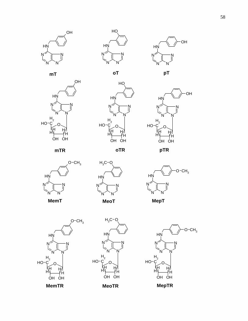
58
N
N
N
N
NH
OH
mT oT
N
N
N
N
NH
OH
pT
N
N
N
N
NHOH
OCH
2
H
OH
H
OH
HH
OH
N
N
N
N
NH
OH
mTR
OCH
2
H
OH
H
OH
HH
OH
N
N
N
N
NH
OH
oTR
OCH
2
H
OH
H
OH
HH
OH
N
N
N
N
NHOH
pTR
N
N
N
N
NH
OCH3
MeoT MemT
N
N
N
N
NH
O CH3
N
N
N
N
NHO CH3
MepT
OCH
2
H
OH
H
OH
HH
OH
N
N
N
N
NH
OCH3
MeoTR
OCH
2
H
OH
H
OH
HH
OH
N
N
N
N
NH
O CH3
MemTR
OCH
2
H
OH
H
OH
HH
OH
N
N
N
N
NHO CH3
MepTR

59
Figure 3.2: Molecular structure of the cytokinins tested in the soybean callus bioassay.
Please note that compounds oTR and pTR were not studied but included for
comparison.
N
N
N
N
NH
F
3FBA
OCH
2
H
OH
H
OH
HH
OH
N
N
N
N
NH
OH
Cl
2Cl6(3OHBA)R
O
N
N
N
N
NH
BA9THP
CH2 C
H
C
CH3
CH2 OHHN
N7
N9
N3
N1
Zeatin
CH2HN
N7
N9
N3
N1
O
Kinetin
N
N
N
N
NH
O H
BA
60
4. Micropropagation and hyperhydricity in Aloe polyphylla
4.1 Introduction
Aloe polyphylla Schonland ex Pillans, commonly called spiral aloe, is a member of the
Asphodelaceae. It is a beautiful threatened plant species native to the Maluti
Mountains in Lesotho (Southern Africa). The Thaba Putsoa Range and the Maseru
areas of the Drakensberg Mountains are the places where this species is found in
greatest number (CHUKWUJEKWU et al., 2002). It grows in basalt rock crevices on
high altitude slopes (2230 - 2720m) and is the only alpine member of the genus Aloe.
It thrives well in loose rocks, which facilitate good drainage (AUBREY, 2002;
CHUKWUJEKWU et al., 2002). This plant is grown mainly for horticultural
(ornamental) and medicinal uses. Julian Mathews, garden and plant writer, describes
this plant as “an excellent example of a plant which is a living work of art - the spirals
of closely packed, thick, fleshy leaves are a wonderful example of symmetry and the
soft green colour is a delight” MATHEWS, 2004).
The perfect spiral pattern in which the leaves are arranged is the most fascinating
feature of this plant. Five ranks of leaves (containing 15-30 leaves each) form the
spiral clockwise or anti-clockwise. Leaves are broad and have a grey-green colour
with sharp tips, purplish brown in colour. It has approximately 75 – 150 leaves with
irregularly toothed margins (EMANOIL, 1994; AUBREY, 2002). This species has
attractive flowers with varied colour from dull red to salmon – pink and rarely yellow.
Inflorescence is branched with a compact flower head. Flowering occurs from August
(spring) through to December, peaking in September and October (summer)
(EMANOIL, 1994).
Both sexual (seed) and asexual (vegetative) propagation are possible. Despite the
production of large numbers of seeds, there is limited viability due to the endangered
pollinator. Vegetative propagation by offshooting of pups is also very rare. To ensure

61
conservation of this species a micropropagation protocol has been developed (ABRIE
and VAN STADEN, 2001; CHUKWUJEKWU et al., 2002). Incidence of hyperhydricity,
the need for an additional rooting step and the use of zeatin (a very expensive
cytokinin) are limitations of these protocols.
Currently benzyladenine is the most widely used cytokinin in the micropropagation
industry due to its effectiveness and affordability. It, however, has disadvantages in
some crops. In micropropagated Spathiphylum floribundum [9G]BA accumulated at
the base of the plant (WERBROUCK et al., 1995). A slow release of this BA
derivative caused heterogeneity in growth and inhibition of rooting during
acclimatization. BA is also reported to causes hyperhydricity in many species
(LESHEM and SACHS, 1985; LESHEM et al., 1988; TERAMOTO et al., 1993).
Therefore, it is imperative to try and find an alternative to BA while maintaining
reasonable multiplication rates and acceptable plant quality.
When WERBROUCK et al., (1996) compared the types and effects of BA and mT
derivatives in tissue culture of S. floribundum, they found that the main metabolite of
BA, [9G]BA, was more stable but had a negative impact on rooting and
acclimatization while, with the main metabolite of mT, mTOG, was easily degraded
during acclimatization. When comparing the post vitro effect of different
concentrations of BA and mT on rooting after an acclimatization period of four weeks,
plants treated with mT produced a significantly higher number and greater length of
roots than those treated with BA. Addition of the aromatic cytokinin mTR to the culture
medium of potato significantly improved its survival in culture (BAROJA-FERNANDEZ
et al., 2002). KAMINÉK et al. (1987b) compared the activities of BA and mT in the
induction of growth of lateral buds in Poinsettia and gerbera daisy and found that mT
was more active. These results indicate that the slight structural difference between
BA and mT could have a profound impact on plants during micropropagation. The aim
of this section of the research project was therefore, to investigate the role of topolins
in the micropropagation of A. polyphylla and control of hyperhydricity.

62
4.2 Material and methods
4.2.1 Source material and bulking of explants
In vitro grown cultures of A. polyphylla, initiated from seeds, were obtained from the
Research Centre for Plant Growth and Development, University of KwaZulu-Natal
Pietermaritzburg, South Africa. Bulking of stock material was done using full strength
MS media (Murashige and Skoog, 1962) supplemented with 30 g l-1 sucrose, 0.1 g l-1
myo-inositol, 0.5 mg l-1 IBA, 1.0 mg l-1 zeatin and solidified with 1% agar
(Bacteriological agar Oxoid Ltd., Basingstoke, Hampshire, England). Cultures were
incubated in a growth room with continuous cool fluorescent tubes (Osram L75
W/20X) with a light intensity of 45 µmol m-2 s-1 and a temperature of 24±1ºC for nine
weeks (CHUKWUJEKWU et al., 2002).
4.2.2 Shoot multiplication
After bulking sufficient explants, experiments to investigate the effect of meta-topolins
on shoot multiplication were designed. The meta-topolin derivatives mT, MemT and
MemTR along with BA and zeatin were investigated. The same type of growth media
as used for bulking was used for the multiplication experiments. Concentrations 0.5,
2.5, 5.0, 7.5 and 15.0 µM were tested with five replicates and the control plants
cultured with no cytokinin. Five shoot tip explants with 2-4 young leaves attached
were cultured in screw cap jars (300 ml) containing 50 ml of media. A total of 25
explants per treatment were used. Cultures were then incubated under the same
growth conditions mentioned above. After nine weeks, growth parameters including
total number of shoots, multiplication rate, multiplication rate for shoots >1.5 cm,
number of hyperhydric shoots, number of normal shoots, number of shoots >1.5 cm,
number of shoots <1.5 cm, fresh weight, rooting ability (scored as 0 = absence, 1 =
moderate rooting and 2 = excessive rooting) and leaf growth (scored as 1 = reduced
leaf growth, 2 = average leaf growth and 3 = excessive leaf growth) were measured.
The experiment was repeated three times.

63
4.2.3 DNA extraction and quantification
To study the effect of hyperhydricity on DNA synthesis, hyperhydric and normal
shoots were produced using the optimized protocol. Induction of hyperhydricity was
achieved by replacing the agar with gelrite and keeping the other media components
the same. For observation purposes, two sets of cultures were prepared using BA
and mT as cytokinins and agar and gelrite as gelling agents. DNA extraction and
quantification was done by the protocols outlined in Appendix 2. Ten samples each of
normal and hyperhydric plants were used. Results on ratio and DNA yield were then
subjected to analysis of variance (GenStat 9th edition).
Before the start of DNA extraction and quantification the following assumptions were
made to help explain and validate the results;
1. Since equal mass of tissue was used for DNA extraction variation in DNA
quantity could be due to the inherent high water content of hyperhydric shoots;
and
2. If assumption one is true, the difference between fresh and dry weight of both
normal and hyperhydric samples should fully or partly explain the difference in
DNA yield. To support this hypothesis equal mass of tissue of normal and
hyperhydric plants treated with mT (with agar and gelrite respectively) was
taken at the time of DNA extraction. Fresh weight and dry weight was then
compared and the difference in change in mass between the normal and
hyperhydric samples was noted;
The remaining approximately 50 plants of each of normal and hyperhydric cultures
treated with BA and mT were left in the culture room for observation as to their
capacity to recover and to see if the plants were able to reverse hyperhydricity without
additional treatment.

64
4.2.4 Acclimatization
On the basis of results of the multiplication experiments, only those treatments that
gave acceptable multiplication rates and the optimum cytokinin concentration that
induced better morphogenesis were considered for the acclimatization experiments.
Plants treated with 2.5 and 5.0 µM of mT and zeatin and 2.5 µM BA were used. BA
failed to produce healthy shoots but was included for comparison purposes as it is the
most widely used cytokinin in tissue culture. Shoot clusters were carefully separated
from each other in such a way that all had at least one functional root. Individual
plants were then washed thoroughly and extra large roots cut back. Plants were
planted in a potting mixture with a 1:1:1 ratio of soil, sand and vermiculite, treated with
fungicide (Benlate, 0.01%) and transferred to a mist house with 90% relative humidity.
After four weeks in the mist house, plants were transferred to a greenhouse in the
same potting mix (CHUKWUJEKWU et al., 2002). After two months of growth in the
greenhouse, fresh weight of shoots and roots was analyzed (GenStat 9th edition).
4.3 Results and discussion
4.3.1 Effect of meta -topolin on shoot multiplication
The choice of cytokinin to use in tissue culture is determined by its cumulative
efficiency in inducing an acceptable rate of shoot multiplication, normal shoots and
roots and the eventual ability of plants to acclimatize easily. The shoot and root
growth responses and variations observed and analyzed in this study are due to
treatment effects as indicated in Table 4.1. Means of total number of shoots showed
significant differences among the different cytokinins and concentrations tested. At
lower concentrations, BA produced more shoots. As the concentration increased
larger numbers of shoots were recorded with the meta-topolin treatments (Figure 4.1
A-C). Both total multiplication rate (Figure 4.1 B) and multiplication rate of shoots
greater than 1.5 cm in length (Figure 4.1C) were recorded and analyzed separately.

65
Results of the analysis showed that treatment means were significantly higher than
the control for most cytokinin levels tested. Multiplication rates increased with an
increase in concentration up to 15.0 µM, where a decline in multiplication rate and
excessive abnormal growth were observed. A better multiplication rate, spontaneous
rooting and healthy explants were found at 5.0 µM level for the mT treatment (the
quality not found in any of the other treatments), hence this was selected as the
optimum cytokinin level. At this level a multiplication rate of eight shoots (shoots
greater than 1.5 cm in length) per explant was found with the mT treatment. This rate
was significantly higher than all the other treatments and better than previous reports
of a total of seven shoots per explant (CHUKWUJEKWU et al., 2002). A few reports
on the use of topolins indicate that this group of cytokinins could be a new source of
cytokinins with high morphogenetic activity. KAMINEK et al. (1987b) found that mT
was nearly twice as effective as BA in the induction of shoot growth of cuttings.
KUBALAKOVA and STRNAD (1992) compared the effects of aromatic and isoprenoid
(zeatin) cytokinins on micropropagation and organogenesis of sugar beet cultures
and found higher activity (greater number of shoots per explant) with mT. They also
observed abnormal growth in BA-treated plants during subculturing, but not with mT.
Table 4.1 : The effect of various cytokinin treatments on overall shoot and root growth.
Observations for the control had no variation among replicates and hence the
statistical test was not applied.
Treatments
(µM
concentration)
Shoot growth Root growth
Pearson chi-
square value
Probability
level
Pearson chi-
square value
Probability
level
control - - - -
0.5 31.50 <0.001 17.53 0.063ns
2.5 35.56 <0.001 24.00 0.008
5.0 30.77 <0.001 34.29 <0.001
7.5 19.44 0.035 40.80 <0.001
15.0 28.26 0.002 43.20 <0.001 ns indicates non-significant treatment effect. This table shows that the observed differences in the
parameters analysed are due to treatment effect.

66

67
The use of mTR for improving survival of potato cultures has been reported
(BAROJA-FERNANDEZ et al., 2002). It was also observed that mTR (which was not
included in this experiment) had a comparable effect with that of mT when applied at
higher concentrations than the optimum level of mT (5 µM) in A. polyphylla. Between
5-15 µM concentrations mTR produced a comparable number of shoots and
improved the rooting ability of the plants (with longer duration in culture) unlike mT,
which caused inhibition of rooting and abnormal growth at concentrations greater than
5 µM (Figure 4.2 I). mTR was tested after the whole experiment was completed.
The slight structural difference between BA and mT (Figure 3.2) had a notable effect
on cultures of S. floribundum (WERBROUCK et al., 1996). In this study, it was found
that A polyphylla plants treated with mT were superior in quality and quantity
compared to plants treated with BA (Figure 4.2) indicating that meta-topolin would be
a good replacement for zeatin in the tissue culture of A. polyphylla. Given the
structural difference between zeatin and mT, an explanation of this result would be
the pattern of receptor recognition as position specificity is related to receptor
recognition (MOK et al., 2005). In cytokinin recognition studies using the Arabidopsis
CRE1, WOL, AHK4 and maize ZmHK1 receptors, the authors found that AHK4
responded to trans-zeatin and mT while ZmHK1 responded to cis-zeatin and oT
(MOK et al., 2005). This similar affinity in receptor recognition could be the possible
reason why mT and zeatin had comparable effects in tissue culture of A. polyphylla.
Spontaneous rooting of plants in multiplication media, a better multiplication rate and
lower cost however, make mT the preferred cytokinin.
4.3.2 Effect on hyperhydricity
Generally, the incidence of hyperhydricity increased with an increase in the
concentration of cytokinins (Table 4.2), a fact well documented in the literature. At an
optimum concentration (5.0 µM), no hyperhydric shoots were recorded for the mT and
MemT treatments. Hyperhydricity was most severe with BA treatments although all
treatments caused hyperhydricity at higher concentrations (>5 µM). Although there

68
was not a significant difference among the treatments (except for BA), the higher
multiplication rate and good rooting in a multiplication media makes mT the preferred
cytokinin. Apart from hyperhydricity, BA-treated plants failed to root, were yellowish in
colour and had excessive abnormal shoot growth (Figure 4.2). Type of gelling agent
and cytokinin play a major role in the control of hyperhydricity in A. polyphylla with BA
and gelrite causing the most hyperhydricity.
4.3.3 Effect on fresh weight (FW)
Total fresh mass per jar was measured and analyzed. There was a strong treatment
effect at all concentrations for all the cytokinins tested. Mean mass of treatments were
significantly higher than the control. At lower concentrations, BA and zeatin gave
higher FW. Unlike other treatments where a decline was observed at higher
concentrations, (Table 4.3) there was a consistent increase of FW with mT treatment.
At optimum concentration (5 µM) zeatin gave significantly higher FW but had lower
multiplication rates compared to mT (Figure 4.4). This indicated that FW was not a
reliable measure of multiplication and normal growth as it was affected more by
growth of individual plants than by multiplication rate. It has also been observed that
hyperhydricity and an undifferentiated mass of tissue due to abnormal growth
contributed to an increase in FW.
At higher cytokinin concentrations, plants treated with BA and zeatin had lower
multiplication rates, more pronounced abnormal growth and smaller mass of fresh
weight compared to plants treated with mT (Tables 4.2 and 4.3). This indicates that at
higher equimolar concentrations mT was less toxic to A. polyphylla compared to BA
and zeatin (see Figure 4.2 G and H for comparison between BA mT). KAMINEK et al.
(1987b) reported that mT promoted shoot formation of stem cuttings of Ephorbia
pulcherrima Wild. and Gerbera jamesonii Hook at 10-4 mol L-1 concentration. At this
concentration BA inhibited shoot formation. They attributed this effect to the faster
translocation of mT in plant tissues which prevents its localized accumulation.

69
Tot
al n
umbe
r of
sho
ots/
jar
0
20
40
60
80
100
120ControlBAmTMemTMemTRZeatin
AT
otal
mul
tiplic
atio
n ra
te
0
5
10
15
20 B
Concentration (µM)
0 5 10 15
Mul
tiplic
atio
n ra
te o
f big
ger
shoo
ts
0
2
4
6
8
10 C
Figure 4.1: The effect of the type and
concentration of cytokinin on
shoot multiplication (A), total
multiplication rate (B) and
multiplication rate of shoots
greater than 1.5 cm in length
(C). Note that meta-topolin
gave more shoots per jar
and a larger number of
shoots big enough for
acclimatization. The 5 µM
mT treatment was selected

70
Figure 4.2: Effect of the various cytokinin treatments on shoot and root growth and incidence of
abnormality of A. polyphylla (A = BA; B = MemT; C = Control; D = mT; E = MemTR; F
= Zeatin; G = 15 µM mT, H = 15 µM BA and I = 7.5 µM mTR). Note the mT-treated
plants had healthy shoot growth and numerous roots as opposed to the abnormal
growth (BA) and failed rooting (zeatin). Also note the relatively less toxic effect of mT
compared to BA (G and H)
Table 4.3: Effect of type and concentration of cytokinin on mean fresh weight (g) of A polyphylla
Treatments Cytokinin concentration (µM)
0.5 2.5 5.0 7.5 15
Control 4.00±0.46 4.00±0.46 4.00±0.46 4.00±.46 4.0 0±.46
BA 17.52±0.84 18.85±1.95 15.27±1.4 11.86±1.62 14.98±1.33
mT 11.16±2.20 15.6±1.66 17.75±2.04 20.58±0.62 22.96± 1.08
MemT 9.11±2.16 10.77±0.46 8.88±1.17 9.84±1.85 14.55±1. 60
MemTR 12.10±1.86 11.84±1.76 7.54±1.48 9.12±1.86 11.37± 1.81
Zeatin 16.85±2.32 18.76±1.2 22.06±1.05 16.71±1.25 16.55±.026
LSD (5%) 3.564 4.410 3.729 4.281 3.334
F Probability <0.001 <0.001 <0.001 <0.001 <0.001
Control (no cytokinin)Control (no cytokinin)
F
C
E
B
D
A
H G I

71
0
10
20
30
40
50
60
70
Control BA mT MemT MemTR Zeatin
Num
ber
of s
hoot
s
TNS
NNS
NHS
Figure 4.3: The effect of the selected optimum cytokinin concentration (5 µM) on hyperhydricity of
A polyphylla cultures. TNS = total number of shoots; NNS = number of normal shoots;
NHS = number of hyperhydric shoots. Note that hyperhydricity was totally controlled
using mT. See Table 4.2 for statistical details.
0
5
10
15
20
25
Contro
lBA mT
MemT
MemTR
Zeatin
Fre
sh w
eigh
t (g)
Figure 4.4: Effect of the selected optimum cytokinin concentration on fresh weight of A polyphylla
cultures. See Table 4.3 for statistical details.
Cytokinin treatments
Cytokinin treatments

72
4.3.4 Effect on acclimatization ex vitro on A. polyphylla
More than 91% of the plants treated with mT acclimatized successfully compared to
65% survival rate recorded with BA-treated plants (Table 4.4). Results ex vitro showed
that the effect of mT is not only limited to in vitro growth with significant treatment
effects noted during the acclimatization process. Acclimatized plants treated with mT
showed faster growth and superior rooting compared to BA and zeatin-treated plants as
indicated by mean fresh weight of roots and shoots (Table 4.5). Under normal
conditions of growth, root and shoot growth complement one another. The poor ex vitro
growth observed with plants treated with BA and zeatin (although statistically not
significant when zeatin was compared to mT) could therefore be attributed to a
treatment effect. The negative effect of BA on ex vitro growth could be due to the
accumulation of the more stable derivative, [9G]BA. Accumulation of [9G]BA on the
basal portion of Spathyphylum plants resulted in slow release of BA during
acclimatization, which caused different problems such as heterogeneity in growth and
inhibition of rooting. The concentration of [9G]BA was about ten times more than the
BA concentration in the medium after just two weeks of growth. Apart from its high
concentration, this derivative is produced and stored at the plant base rather than being
transported and could have contributed to its negative effect (WERBROUCK et al.,
1995).
4.3.5 Effect of hyperhydricity on total DNA content of A polyphylla
The effect of hyperhydricity on the quality and quantity of DNA was studied to verify if
this abnormality affects DNA synthesis. Results of the experiment indicated that
hyperhydric samples produced three times less DNA than the normal samples (Table
4.6). This result can be explained based on two assumptions. Firstly, low DNA levels
could be due to the high level of water content in hyperhydric shoots which could have
affected the DNA yield. Secondly, it could be as a result of a negative effect of
hyperhydricity on DNA synthesis.

73
Table 4.4: Effect of various cytokinin treatments on acclimatization of A polyphylla. Aloe
polyphylla can easily be acclimatized to ex vitro growth conditions.
Treatments
Total number
transferred to mist
house
Total number
survived in the
greenhouse
Survival rate (%)
2.5 BA 45 30 66.67
2.5 mT 45 42 93.33
2.5 zeatin 45 39 86.67
5.0 mT 45 41 91.11
5.0 zeatin 45 38 84.44
Table 4.5: Effect of the different cytokinins tested on ex vitro growth. Fresh weight of ten
(random) fully acclimatized (two-month-old) plants per treatment was used for this
analysis. Only those treatments that produced plantlets with both shoots and roots
were considered.
Cytokinin concentration (µM) Mean fresh weight (g)
Shoot Root
2.5 BA 5.9±1.37 0.56±0.12
2.5 mT 19.3±2.83 1.75±0.26
2.5 zeatin 13.2±2.23 1.00±0.14
5.0 mT 23.6±7.03 1.66±0.44
5.0 zeatin 14.9±2.65 0.82±0.17
LSD (5%) 10.87 0.726
F Prob . 0.028 0.006
The first assumption failed to fully explain the result since the difference in change in
mass between fresh and dry weight of the normal and hyperhydric sample was less
than two fold leading to the conclusion that hyperhydricity does affect DNA synthesis.
Very little has been done on the molecular biology of hyperhydricity, especially on
aspects of DNA synthesis. Currently, there is only one report which demonstrated that
hyperhydricity is linked with abnormal DNA content in grass pea Lathyrus sativus L.
(OCHATT et al., 2002).
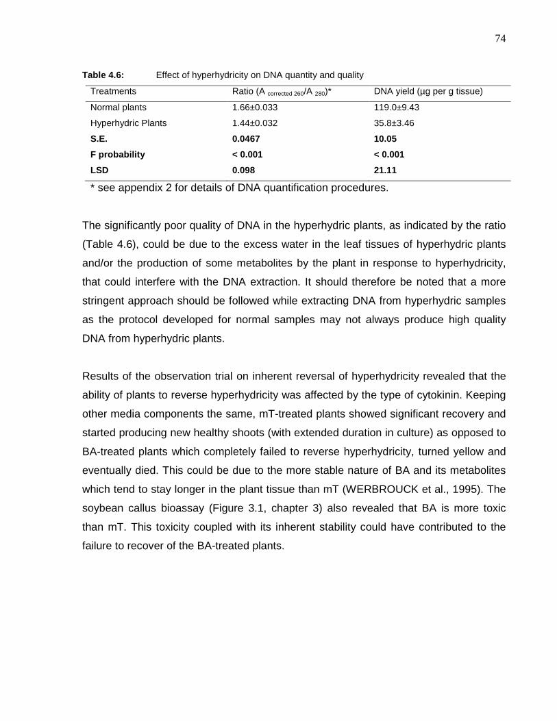
74
Table 4.6: Effect of hyperhydricity on DNA quantity and quality
Treatments Ratio (A corrected 260/A 280)* DNA yield (µg per g tissue)
Normal plants 1.66±0.033 119.0±9.43
Hyperhydric Plants 1.44±0.032 35.8±3.46
S.E. 0.0467 10.05
F probability < 0.001 < 0.001
LSD 0.098 21.11
* see appendix 2 for details of DNA quantification procedures.
The significantly poor quality of DNA in the hyperhydric plants, as indicated by the ratio
(Table 4.6), could be due to the excess water in the leaf tissues of hyperhydric plants
and/or the production of some metabolites by the plant in response to hyperhydricity,
that could interfere with the DNA extraction. It should therefore be noted that a more
stringent approach should be followed while extracting DNA from hyperhydric samples
as the protocol developed for normal samples may not always produce high quality
DNA from hyperhydric plants.
Results of the observation trial on inherent reversal of hyperhydricity revealed that the
ability of plants to reverse hyperhydricity was affected by the type of cytokinin. Keeping
other media components the same, mT-treated plants showed significant recovery and
started producing new healthy shoots (with extended duration in culture) as opposed to
BA-treated plants which completely failed to reverse hyperhydricity, turned yellow and
eventually died. This could be due to the more stable nature of BA and its metabolites
which tend to stay longer in the plant tissue than mT (WERBROUCK et al., 1995). The
soybean callus bioassay (Figure 3.1, chapter 3) also revealed that BA is more toxic
than mT. This toxicity coupled with its inherent stability could have contributed to the
failure to recover of the BA-treated plants.
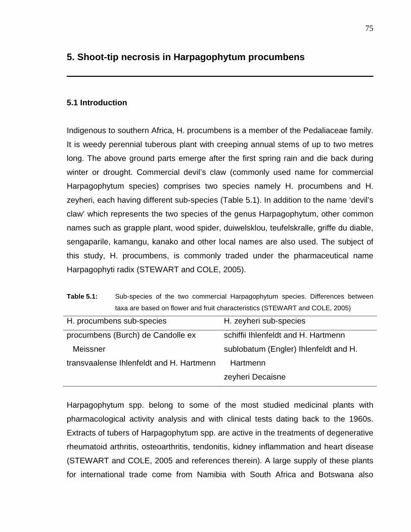
75
5. Shoot-tip necrosis in Harpagophytum procumbens
5.1 Introduction
Indigenous to southern Africa, H. procumbens is a member of the Pedaliaceae family.
It is weedy perennial tuberous plant with creeping annual stems of up to two metres
long. The above ground parts emerge after the first spring rain and die back during
winter or drought. Commercial devil’s claw (commonly used name for commercial
Harpagophytum species) comprises two species namely H. procumbens and H.
zeyheri, each having different sub-species (Table 5.1). In addition to the name ‘devil’s
claw’ which represents the two species of the genus Harpagophytum, other common
names such as grapple plant, wood spider, duiwelsklou, teufelskralle, griffe du diable,
sengaparile, kamangu, kanako and other local names are also used. The subject of
this study, H. procumbens, is commonly traded under the pharmaceutical name
Harpagophyti radix (STEWART and COLE, 2005).
Table 5.1: Sub-species of the two commercial Harpagophytum species. Differences between
taxa are based on flower and fruit characteristics (STEWART and COLE, 2005)
H. procumbens sub-species H. zeyheri sub-species
procumbens (Burch) de Candolle ex
Meissner
transvaalense Ihlenfeldt and H. Hartmenn
schiffii Ihlenfeldt and H. Hartmenn
sublobatum (Engler) Ihlenfeldt and H.
Hartmenn
zeyheri Decaisne
Harpagophytum spp. belong to some of the most studied medicinal plants with
pharmacological activity analysis and with clinical tests dating back to the 1960s.
Extracts of tubers of Harpagophytum spp. are active in the treatments of degenerative
rheumatoid arthritis, osteoarthritis, tendonitis, kidney inflammation and heart disease
(STEWART and COLE, 2005 and references therein). A large supply of these plants
for international trade come from Namibia with South Africa and Botswana also

76
contributing their share (RAIMONDO and DONALDSON, 2002). Despite the huge
economic importance, the cultivation of these species is facing some problems.
Attempts to commercially propagate these species from seeds failed due to low
germination rates. Plants propagated by cuttings failed to produce primary roots
resulting in a single harvest (KATHE et al., 2003).
An in vitro propagation method of Devil’s claw was reported (LEVIEILLE et al., 2000;
LEVIEILLE and WILSON, 2002) and gave a coefficient of multiplication of four plants
per month per explant. This technique, using single node explants, consisted of root
induction treatments with IAA, in the dark, followed by plant development in growth
regulator-free medium, under light (LEVIEILLE and WILSON, 2002). An in vitro
method was also developed in Pietermaritzburg, South Africa by the Research Centre
for Plant Growth and Development where a plant development step using BA as the
sole growth regulator was included. Developed plantlets spontaneously rooted in the
same medium, after two months of culture. This second method seems attractive as it
eliminates sub-culturing to rooting medium. However, shoot-tip necrosis (STN)
occurred during elongation of the explants. This rapid necrosis of the apical shoot
triggered growth of axillary buds. This mode of development produced plants with
many branches before acclimatization and transplantation into soil. With longer
duration in culture and repeated sub-culturing, the problem of STN became severe
with plants accumulating basal callus and eventually losing their ability to root in a
multiplication medium (RAMAROSANDRATANA, personal communication).
Apical shoot necrosis has been reported due to pathogenic infection (BENTUR and
KALODE, 1996; GUTHRIE et al., 2001) and calcium deficiency (MORARD et al.,
1996; SELING et al., 2000). However, H. procumbens naturally shows dieback of
shoots between April and June as a prelude to winter dormancy (RAIMONDO and
DONALDSON, 2002). The problem of non-pathogenic dieback or shoot necrosis in
vitro has been associated with various culture conditions such as salt formulation and
growth regulators (VIEITEZ et al., 1989; KATAEVA et al., 1991; MACKAY et al.,
1995; MORARD et al., 1996; PIAGNANI et al., 1996; GRIGORIADOU et al., 2000;

77
PEREZ-TORNERO and BURGOS, 2000; SELING et al., 2000), use of activated
charcoal (MACKAY et al., 1995; WANG and VAN STADEN 2001), duration in culture
(GRIGORIADOU et al., 2000), type of tissue culture medium (GENEVE et al., 1992;
MACKAY et al., 1995; GRIGORIADOU et al., 2000), exudation of phenolics in tree
species (BELLAROSA, 1988), physiological disorder associated with rooting
(KATAEVA et al., 1991), sulphur content and NH4/NO3 ratio (LAKSHMI and
RAGHAVA, 1993), fructose (KULKARNI and D'SOUZA, 2000) and pH flactuations
(De BLOCK, 1990). These factors are discussed in detail in Chapter Two Section 2.3.
5.2 Materials and methods
Previously maintained cultures of Harpagophytum procumbens were used as the
source of nodal explants. Cultures were sub-cultured to fresh medium after every 3-4
weeks to maintain the stock. Cultures were incubated in a growth room with cool
fluorescent tubes (Osram L75 W/20X) with a light intensity of 45 µmol m-2 s-1 and a
temperature of 24±1ºC at 16-hour light and 8-hour dark photoperiod. The nodal
explants (1 cm) were implanted into medium supplemented with 0.9 % (w/v) agar and
3% (w/v) sucrose. These culture conditions were also used for all experiments.
5.2.1 Effect of media type and strength on STN
To investigate the effect of type and strength of nutrient media on STN, MURASHIGE
and SKOOG (1962), NITSCH and NITSCH (1969) Basal Medium (NB) and Woody
Plant Medium (WPM) (LLOYD and McCOWN, 1980) supplemented with 0.9 % (w/v)
agar, 3% (w/v) sucrose and 1.0 mg l-1 BA and were tested in a completely
randomized design at three levels; full strength, half-strength and quarter-strength.
The pH of the media was adjusted to 5.8 with 1N HCl or 1 N NaOH, before addition of
agar. Thirty ml of medium was poured into 250 ml screw cap jars which were then
autoclaved at 1.05 KPa at 121 °C for 20 min. Cultures were maintained in the
conditions described above. Each treatment had four replicates with six nodal
explants each. After four weeks of growth, growth parameters such as average

78
number of shoots per explant, percent necrotic shoots and average shoot length were
measured and analyzed using the MINITAB version 14 (Minitab Ltd., USA). Fisher's
least significant difference (LSD) at the 5% level was used to analyze the differences
between the means.
5.2.2 Effect of type and concentration of cytokinin s on STN
The effect of cytokinins on STN was assessed using half strength MS media
containing media supplements indicated in section 5.2.1 above. Initially BA and
kinetin (Sigma-Aldrich) were tested at various concentrations (2.22, 4.44, 6.66 and
8.88 µM) and the control plants were cultured in hormone free half-strength MS
media. Each treatment had four replicates with six nodal explants each. After four
weeks of growth, growth parameters such as average number of shoots per explant,
percent necrotic shoots and average shoot length were measured and analyzed using
the MINITAB version 14 (Minitab Ltd., USA). Fisher's least significant difference
(LSD) at the 5% level was used to analyze the differences between the means.
In an attempt to find an alternative to BA and kinetin, the effect of mT and mTR with
and without auxin on STN was also investigated and results analysed using SPSS
package version 15.0. Cultures from these experiments were also used to assess the
effect of sub-culturing. Uniformly grown cultures were divided into two categories.
Cultures in the first category were sub-cultured every four weeks whereas cultures in
category two were sub-cultured every two weeks, keeping other media components
constant. Growth parameters were then recorded and analysed.
5.2.3 Effect of type and concentration of sugars on STN
Various concentrations of sucrose (1, 2, 3 or 4%) and equi-molar concentrations
(0.086 M) of different sugars (glucose, fructose, sucrose or maltose) were also tested.
A completely randomized block design with four replicates having 25 explants each
was used. The culture responses were expressed in terms of number of shoots per

79
explant, percentage of necrotic shoots and average length of shoots. Percentages of
response data were analyzed using one-way analysis of variance (ANOVA) after
arcsine transformation using the MINITAB package.
5.2.4 Effect of calcium and boron on STN
To assess the effect of calcium and boron on STN, an experiment having three steps
was designed. Treatments in step one contained constant boron and varied levels of
calcium (6, 9, 12, and 15 mM), step two contained constant calcium and varied levels
of boron (0.2, 0.3, 0.4 and 0.5 mM) and step three had proportionally varied levels of
both calcium and boron (6 mM Ca + 0.2 mM B, 9 mM Ca + 0.3 mM B, 12 mM Ca +
0.4 mM B and 15 mM Ca + 0.5 mM B). The concentrations of both elements in MS
media (3 mM for Ca and 0.1 mM for B) were taken as standard concentrations. The
control plants were cultured in full strength MS media. Other media components were
kept constant. Each treatment had four replicates with 40 ml of media having 6
explants per screw cap jar each. After four weeks of growth, growth parameters such
as average shoot length per jar (cm), fresh and dry weight (g) and average number of
infections per jar were recorded and analysed using SPSS version 15.0 and mean
separation by least significant difference (LSD) was done using Duncan’s Multiple
Range Test.
5.2.5 Rooting and acclimatization of tissue culture d H. procumbens
Based on the results of the above experiments, a protocol to successfully multiply the
plants was established. This protocol involved the use of auxin-free half strength MS
media with 6 mM calcium, 3 % sucrose, 0.1 g l-1 myo-inositol and 5 µM mTR solidified
with 1 % agar after adjusting the pH to 5.8. Forty ml of the media were added to 250
ml screw-cap jars after autoclaving. Six to ten nodal explants per jar were used
without notable effect on growth and multiplication. Regenerated plantlets were then
transferred to cytokinin-free rooting media containing half strength MS media
supplemented with 2.5 µM IAA and the same supplements as above. The transfer to
80
rooting media, was preceded by careful trimming of the basal callus, after three
weeks when the first symptom of tissue browning was observed. This initial attempt to
root plantlets was successful and hence no additional rooting experiment was done.
Two sets of rooted plantlets were then potted in 1:1 mixture of sand and soil for ex
vitro acclimatization. The first set was transferred to the mist house and the second
set transferred directly to the greenhouse. Fully acclimatized plants were then
transferred to bigger pots containing the same potting mixture to evaluate their ability
to produce tubers and left to grow for one year with a watering frequency of once per
week.
5.3 Results and discussion
5.3.1 Effect of media composition and strength on S TN
Three different basal media (MS, NB and WPM) were used for raising cultures of
Harphagophytum. The three basal media differed in their ionic strength. Of the three
media tried, MS exhibited the maximum (98 %) necrotic shoots while WPM had the
least number (88 %) of necrotic shoots. Half-strength and quarter-strength of all three
basal media resulted in less necrotic shoots compared to their full-strength
counterparts.
Lowering the concentration of salts in media reduced the shoot elongation capacity.
On the half- and quarter-strength WPM as well as on quarter-strength NB medium,
shoot elongation was drastically affected. In some cases, the shoot buds failed to
open even after a prolonged culture period of 8 weeks (Table 5.2). The number of
shoots developed per explant varied significantly on full- and half-strength medium (P
≤ 0.05).

81
Table 5.2: Effect of media composition and strength of media on STN of H. procumbens
Media Average number of
shoots/explant
Necrotic shoots
(%)
Average shoot
length (cm)
MS
Full-strength 1.68±0.09a 88a 5.08±0.09a
Half-strength 1.59±0.07a 29b 5.08±0.10a
Quarter-strength 1.60±0.07a 14c 3.26±0.08b
NB
Full-strength 1.64±0.07a 90a 3.69±0.09b
Half-strength 1.62±0.06a 27b 1.53±0.05c
Quarter-strength 1.64±0.07a 26b *
WPM
Full-strength 1.49±0.05b 86a 3.31±0.16b
Half-strength 1.43±0.04b 27b *
Quarter-strength 1.23±0.06c 28b *
* shoot buds failed to attain measurable length
Values represent mean ± standard error. Values followed by the same letter in each column are not
significantly different (p ≤ 0.05).
5.3.2 Effect of cytokinins on STN
Plants cultured in media containing BA were affected by STN at all concentrations.
The control plants (with no plant growth regulators) showed no symptoms of necrosis
but failed to grow and remained stunted (Figure 5.1c). On kinetin-supplemented
medium, a higher percentage of necrotic cultures were obtained compared to that of
BA-supplemented media. Among the various concentrations of BA tested, 4.44 µM
favoured maximum number of shoot development per explant, while 6.66 µM
favoured the elongation of shoots.
The symptom of shoot-tip necrosis started randomly in all cultures after nine days in
culture. With increased duration in culture, the number of plants affected increased
and all plants were affected by the end of the sixth week. The severity of the problem
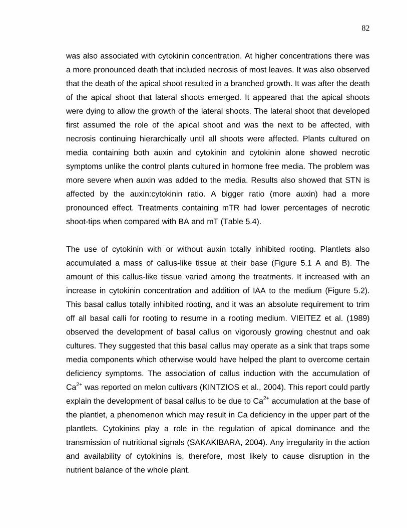
82
was also associated with cytokinin concentration. At higher concentrations there was
a more pronounced death that included necrosis of most leaves. It was also observed
that the death of the apical shoot resulted in a branched growth. It was after the death
of the apical shoot that lateral shoots emerged. It appeared that the apical shoots
were dying to allow the growth of the lateral shoots. The lateral shoot that developed
first assumed the role of the apical shoot and was the next to be affected, with
necrosis continuing hierarchically until all shoots were affected. Plants cultured on
media containing both auxin and cytokinin and cytokinin alone showed necrotic
symptoms unlike the control plants cultured in hormone free media. The problem was
more severe when auxin was added to the media. Results also showed that STN is
affected by the auxin:cytokinin ratio. A bigger ratio (more auxin) had a more
pronounced effect. Treatments containing mTR had lower percentages of necrotic
shoot-tips when compared with BA and mT (Table 5.4).
The use of cytokinin with or without auxin totally inhibited rooting. Plantlets also
accumulated a mass of callus-like tissue at their base (Figure 5.1 A and B). The
amount of this callus-like tissue varied among the treatments. It increased with an
increase in cytokinin concentration and addition of IAA to the medium (Figure 5.2).
This basal callus totally inhibited rooting, and it was an absolute requirement to trim
off all basal calli for rooting to resume in a rooting medium. VIEITEZ et al. (1989)
observed the development of basal callus on vigorously growing chestnut and oak
cultures. They suggested that this basal callus may operate as a sink that traps some
media components which otherwise would have helped the plant to overcome certain
deficiency symptoms. The association of callus induction with the accumulation of
Ca2+ was reported on melon cultivars (KINTZIOS et al., 2004). This report could partly
explain the development of basal callus to be due to Ca2+ accumulation at the base of
the plantlet, a phenomenon which may result in Ca deficiency in the upper part of the
plantlets. Cytokinins play a role in the regulation of apical dominance and the
transmission of nutritional signals (SAKAKIBARA, 2004). Any irregularity in the action
and availability of cytokinins is, therefore, most likely to cause disruption in the
nutrient balance of the whole plant.
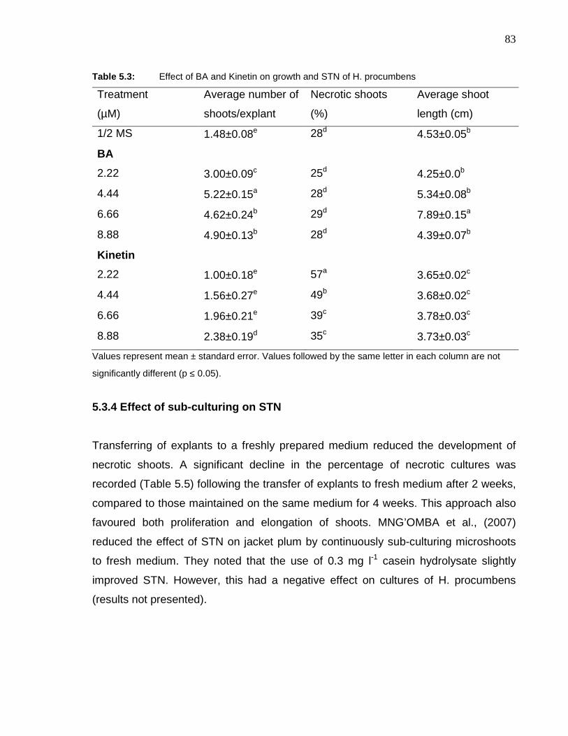
83
Table 5.3: Effect of BA and Kinetin on growth and STN of H. procumbens
Treatment
(µM)
Average number of
shoots/explant
Necrotic shoots
(%)
Average shoot
length (cm)
1/2 MS 1.48±0.08e 28d 4.53±0.05b
BA
2.22 3.00±0.09c 25d 4.25±0.0b
4.44 5.22±0.15a 28d 5.34±0.08b
6.66 4.62±0.24b 29d 7.89±0.15a
8.88 4.90±0.13b 28d 4.39±0.07b
Kinetin
2.22 1.00±0.18e 57a 3.65±0.02c
4.44 1.56±0.27e 49b 3.68±0.02c
6.66 1.96±0.21e 39c 3.78±0.03c
8.88 2.38±0.19d 35c 3.73±0.03c
Values represent mean ± standard error. Values followed by the same letter in each column are not
significantly different (p ≤ 0.05).
5.3.4 Effect of sub-culturing on STN
Transferring of explants to a freshly prepared medium reduced the development of
necrotic shoots. A significant decline in the percentage of necrotic cultures was
recorded (Table 5.5) following the transfer of explants to fresh medium after 2 weeks,
compared to those maintained on the same medium for 4 weeks. This approach also
favoured both proliferation and elongation of shoots. MNG’OMBA et al., (2007)
reduced the effect of STN on jacket plum by continuously sub-culturing microshoots
to fresh medium. They noted that the use of 0.3 mg l-1 casein hydrolysate slightly
improved STN. However, this had a negative effect on cultures of H. procumbens
(results not presented).

84
Figure 5.1: In vitro grown H. procumbens (A) necrotic symptoms and development of basal callus
on four-week-old culture; (B) trimmed basal callus; (C) two-month-old plantlets cultured
in hormone free medium; (D) rooted plants ready for acclimatization; (E) fully
acclimatized and greenhouse-grown plants and (F) example of STN at its worst.
0
0.2
0.4
0.6
0.8
1
1.2
Control 5 BA 5 mT 5 mTR 5 BA +IAA
5 mT +IAA
5 mTR+ IAA
10 BA +IAA
10 mT+ IAA
10 mTR+ IAA
Cytokinin treatments (µM)
Mas
s of
cal
lus-
like
tissu
e (g
)
Figure 5.2: The effect of BA, mT and mTR (5 and 10 µM) with (2.5 µM) and without IAA on the
development of basal callus
E
B A
D F
C

85
5.3.5 Effect of sucrose concentration and type of s ugars on STN
Sucrose is an important source of carbohydrate in the culture medium. It also acts as
an osmoticum. Various concentrations of sucrose were tried. At 1% level, shoot
proliferation as well as elongation was very poor. Increase in concentration of sucrose
favoured shoot development with a maximum of 5.4 shoots per explant at 3%.
Increased concentrations of sucrose concomitantly increased the number of necrotic
shoots (Table 5.6).
Although sucrose is the most widely used carbohydrate and carbon source in tissue
culture, some reports indicate that it may cause hypoxia and ethanol accumulation
due to fast metabolism. The addition of carbon source could also bring a significant
decrease in the media osmotic potential (NETO and OTONI, 2003 and references
therein). These conditions could in turn interfere with the nutrient uptake process.
This interference would most likely result in the failure of absorption or diffusion of
some important elements. Increasing sucrose concentration increased the incidence
of STN and improved shoot multiplication. Since the ultimate goal of any tissue
culture system is to have an improved multiplication rate of healthy plants, an
alternative solution to overcome STN must be found.
To study the effect of different sugars (two monosaccharides: glucose and fructose;
two disaccharides: maltose and sucrose) on shoot proliferation and necrosis, equi-
molar (0.086 M) concentration of different sugars were tried. Fructose completely
inhibited the development as well as elongation of shoots. On glucose containing
medium, abnormal shoots were produced, they were thick and stunted.
Supplementation of maltose to the culture medium resulted in the development of
maximum number of necrotic shoots (Table 5.7).

86
Table 5.4: Effect of mT and mTR with and without IAA on growth and STN of H. procumbens. NNoST = number of normal shoot-tips;
NNeST = number of necrotic shoot-tips
Treatment
(µM)
Shoot length
(cm)
No. of branches NNoST NNeST Fresh
weight (g)
Necrotic
shoots (%)
Control 3.03±0.24ab 1.5±0.16c 1.5±0.16bc 0 c 0.14±0.02cd 0
5 mT 4.05±0.16ab 5.9±0.84a 3.8±0.63a 2.1±0.46b 0.99±0.22bc 35.59
5 mTR 4.00±0.27ab 4.4±0.43b 3.3±0.32a 1.1±0.22bc 0.56±0.07c 25
5 BA 4.81±0.35a 4.3±0.47b 2.9±0.48ab 1.4±0.29bc 1.69±0.32ab 32.56
5 BA + IAA 3.43±0.17ab 6.2±0.66a 2.4±0.62ab 3.8±0.56a 2.26±0.13a 61.29
5 mT + IAA 3.75±0.14ab 6.9±0.44a 2.6±0.57ab 4.3±0.58a 1.39±0.15b 62.32
5 mTR+ IAA 3.60±0.43ab 5.3±0.43ab 3.3±0.58a 2±0.37b 1.31±0.09b 37.73
10 BA + IAA 6.78±2.9a 4.2±0.46b 2.1±0.36ab 2.1±0.49b 2.03±0.18a 50
10 mT + IAA 3.35±0.16ab 7.2±0.59a 4.3±0.39a 2.9±0.74ab 2.1±0.19a 40.28
10 mTR + IAA 2.90±0.22ab 6.9±0.48a 4.6±0.55a 2.3±0.37b 2.45±0.36a 33.33
Values represent mean ± standard error. Values followed by the same letter in each column are not significantly different (p ≤ 0.05).

87
Table 5.5: Effect of sub-culturing on growth and STN of H. procumbens
Treatment Average number
of shoots/explant Necrotic shoots
(%)
Average shoot
length (cm)
Continuous for 4
weeks
4.92±0.13b 28a 5.09±0.09b
Sub culturing after 2
weeks
6.84±0.22a 19b 6.97±0.08a
Values represent mean ± standard error. Values followed by the same letter in each column are not
significantly different (p ≤ 0.05).
Table 5.6: Effect of sucrose concentration on growth and STN of H. procumbens
Sucrose treatment Average number of
shoots/explant
Necrotic shoots
(%)
Average shoot
length (cm)
1% 2.16±0.11c 18b 2.45±0.02c
2% 4.36±0.13b 21ab 3.88±0.07b
3% 5.36±0.09a 25a 4.54±0.05a
4% 5.10±0.15a 26a 4.64±0.06a
Values represent mean ± standard error. Values followed by the same letter in each column are not
significantly different (p ≤ 0.05).
Table 5.7: Effect of sugars on growth and STN of H. procumbens
Sugars
(0.086 M)
Average number of
shoots/explant
Necrotic shoots (%) Average shoot
length (cm)
Sucrose 6.01±0.17a 29d 6.08a
Glucose 1.82±0.11b 37c 3.93b
Maltose 1.52±0.13b 76a 2.65c
Fructose 0.59±0.15c 64b *
* shoot buds failed to attain measurable length
Values represent mean ± standard error. Values followed by the same letter in each column are not
significantly different (p ≤ 0.05).

88
5.3.6 Effect of Ca and B on STN
Increasing the concentration of Ca and B separately reduced the incidence of STN.
Simultaneous addition of higher levels of these ions however, had a detrimental effect
on growth. Although high concentrations of Ca reduced STN without affecting growth
and regeneration, an elevated B concentration was toxic to the plant and inhibited
growth considerably. There are a number of reports that indicate that increasing the
concentration of Ca and/or B in the culture medium reduces the problem of STN
(VIEITEZ et al., 1989; KATAEVA et al., 1991; BARGHCHI and ALDERSON, 1996;
PIAGNANI et al., 1996).
Other reports indicate that Ca is involved in cellular growth and differentiation,
enzymatic activity and membrane permeability (see HIRSCHI, 2004 and HEPLER,
2005 for reviews). Thus Ca deficiency could result in the disturbance of metabolic
activities of growing tissues; which in turn results in growth abnormality such as STN.
ABDULNOUR et al., (2000) studied the effect of B on Ca uptake in micropropagated
potatoes and found that excess B can adversely affect Ca uptake. Medium with a
high B level decreased Ca content in shoots and leaves but medium with B content
four times less than MS enhanced Ca uptake. This could therefore, provide an
explanation for our results where increasing the concentration of both B and Ca had
an inhibitory effect on the growth of H. procumbens plantlets.
Another explanation for the effect of Ca in STN would be its mode of transport in the
plant system. HIRSCHI (2004) in his review stated that Ca in the xylem sap needs a
transpiration system for upward transport and translocation and indicated the
immobile nature of this ion once deposited. The apparent lack of an efficient
transpiration stream in culture vessels and in explants (see section 5.3.7) coupled
with the limited mobility of this ion could therefore be the causes for the deficiency
symptoms observed in the form of STN in young tissues. The fact that STN was
observed in cultures with the highest concentration of Ca (up to 15 mM, five times
more than the normal full strength MS medium), indicated that the problem of
89
transport of this ion could have contributed more to STN than its quantity in the
medium.
Table 5.8: The effect of calcium and boron concentration on growth and STN of H. procumbens.
Treatments
(mM)
Average shoot
length (cm)
Fresh weight
(g)
Dry weight
(g)
Mean No. of necrotic
shoots per jar
Control* 5.26±0.28a 6.98±0.26ab 0.23±0.02ab 4.80±2.60b
6 Ca 4.36±0.31ab 6.35±0.70abc 0.23±0.01ab 1.60±0.67bcde
9 Ca 4.97±0.25a 5.89±1.33abc 0.20±0.06ab 3.20±1.35bcde
12 Ca 4.27±0.30ab 7.57±1.17a 0.33±0.06a 1.40±0.93bcde
15 Ca 4.70±0.37ab 5.98±1.21abc 0.18±0.05ab 1.00±0.63cde
0.2 B 4.58±0.41ab 7.43±0.82a .032±0.03a 4.40±0.93bc
0.3 B 4.37±0.27ab 7.16±1.42ab 0.30±0.06ab 3.20±0.92bcde
0.4 B 4.08±0.53ab 4.81±1.26abc 0.24±0.06ab 0.40±0.40de
0.5 B 3.68±0.36b 3.69±0.84c 0.16±0.03b 0.00±0.00e
6 Ca + 0.2 B 4.47±0.28ab 4.81±1.21abc 0.25±0.04ab 8.00±1.14a
9 Ca + 0.3 B 3.68±0.45b 4.04±0.64bc 0.24±0.04ab 4.00±1.41bcd
12 Ca + 0.4 B 3.50±0.28b 3.46±0.67c 0.22±0.03ab 1.20±0.37bcde
15 Ca + 0.5 B 3.63±0.51b 4.31±0.61abc 0.24±0.03ab 1.80±0.49bcde
* The control plants were cultured in half-strength MS media.
Values represent mean ± standard error. Values followed by the same letter in each column are not
significantly different (p ≤ 0.05).
Increasing the amount of B in the medium to more than 200 µM significantly reduced
STN but it also reduced shoot multiplication (BARGHCHI and ALDERSON, 1996).
This result is in agreement with our findings. These authors further indicated that B
deficiency in vascular plants (characterized by cessation of cell division in the apical
meristem) could lead to a number of secondary effects such as perturbation of auxin
metabolism, increased lignification and phenol accumulation (BARGHCHI and
ALDERSON, 1996 and references therein).

90
5.3.7 Aeration and rooting effects
Throughout this study it was observed that cultures in culture tubes sealed with
Parafilm were more prone to STN, excessive branching (reduced elongation) and
hyperhydricity as opposed to those cultures in loosely closed screw-cap jars where
plants were much healthier. SHA et al., (1985) also reported similar results in
‘Norland’ shoot cultures (Solanum spp) with this symptom being particularly evident in
cultures with low Ca levels. They also observed that plants grown in Parafilm-sealed
vessels had reduced weight and increased branching. It has long been known that
aeration plays a considerable role in plant growth and development via its effect on
the transpiration stream. Transpiration stream on the other hand plays a pivotal role in
the translocation of mineral nutrients through the xylem. In an experiment on Dianthus
microplants, CASSELLS and WALSH (1994) demonstrated that allowing high
moisture vapour loss from culture vessels increased microplant establishment and
calcium accumulation in the leaves and enhanced stomatal functioning, presumably
by increasing transpiration in vitro. Therefore, increased rates of transpiration could
most likely result in an increased rate of nutrient flow to the actively growing
meristematic regions (BIDDULPH et al., 1961; KOHL and OERTLI, 1961; BOWEN,
1972; BARGHCHI and ALDERSON, 1996). Thus a reduced transpiration stream
means reduced nutrient flow to growing shoots, resulting in the deficiency of some
mineral nutrients. This problem is particularly serious in tissue culture systems where
culture vessels are characterized by a very humid atmosphere which suppresses the
transpiration stream of cultured shoots. In such cases other mechanisms such as root
pressure become limited means for plantlets to absorb nutrients from the culture
medium. In the present study, rooted plantlets of H. procumbens in a rooting medium
slowly overcome the problem of STN. This effect was particularly pronounced with
plants treated with mT and mTR while BA-treated plants failed to show a similar
effect. BARGHCHI and ALDERSON (1985) reported that STN did not occur in rooted
shoots of Pistacia vera L.; a result identical to our findings. MNG’OMBA et al., (2007)
also published a similar finding for jacket plum.

91
Our findings suggest that plants in a multiplication stage (having no or only few roots)
could suffer from mineral deficiency due to a reduced transpiration stream and
absence of root pressure. The problem becomes particularly serious in the case of
less mobile minerals such as Ca and B (RAVEN, 1977) where the demand by the
plantlets exceeds the supply from the medium. Although this problem can partly be
alleviated by improving aeration in the culture vessels as was noted in H. procumbens
cultures, it should be done carefully as excessive reduction of vessel humidity can
result in reduction in, or total inhibition of, shoot multiplication as a result of tissue
desiccation due to increased evaporation. Excessive aeration could also compromise
the sterility of the culture environment by giving access to pathogens (SHA et al.,
1985).
5.3.8 Rooting and acclimatization of tissue culture d H. procumbens
The initial attempt to root the plantlets produced 100% rooting in just 10 days after
transfer to rooting media (Figure 5.1 D). An interesting observation was that plantlets
with necrotic symptoms to the rooting media were capable of reverting it or at least,
their growth and rooting in vitro was not affected. This observation is contrary to some
reports which suggest that the possible cause of STN is the omission of cytokinins
from the rooting media to counteract their anti-rooting activity (VIEITEZ et al., 1989;
KATAEVA et al., 1991; PIAGNANI et al., 1996). These reports explain that the
omission or reducing to very low level of cytokinin leads to depletion of cytokinin from
the shoots resulting in shoot degeneration due to failed cell division. Results of this
study therefore, suggest that STN in H. procumbens is a more complex phenomenon
controlled by multiple factors. The fact that STN increased with an increase in
cytokinin concentration also indicated that cytokinins play a negative role in the
process. There are consistent reports indicating that inclusion of auxin in culture
media aggravates STN (see section 2.3). In this study plantlets cultured in rooting
media containing IAA alone did not suffer from STN; suggesting that the effect of the
different media components on STN could be species dependent.

92
When transferring rooted plantlets to pots, it was found that roots were very weakly
attached to the plant and easily fell off. It was therefore, imperative to carefully
separate the plants from the agar media with at least some of their roots attached.
This problem led to the idea of using liquid rooting media. This was tried but resulted
in serious problem of hyperhydricity.
After carefully removing plants from the rooting media using water, plants were potted
in a 1:1 ratio of sand and soil. All of the plants kept in the mist house were dead within
one week after transfer. Plants transferred directly to the greenhouse, with daily light
watering for the first week, once every three days for the second week and a watering
frequency of once a week thereafter had a survival rate of more than 70%. After six
weeks growth, acclimatized plants were re-potted to bigger pots for tuber formation.
These plants were fully established, grew vigorously and produced flowers and tubers
(Figure 5.1 E). The tubers were kept for a comparative pharmacological study for the
presence of the bioactive compounds in comparison with tubers from the natural
habitat.

93
6. Micropropagation and somaclonal variation in ban ana
6.1 Introduction
Bananas and plantains (Musa spp. L.) are giant perennial herbs widely cultivated in
the humid and sub-humid tropics as main source of carbohydrate. The present day
bananas are believed to be the evolution products of intra- and inter-specific hybrids
of the two diploid wild species, Musa acuminata Colla. and Musa balbisiana Colla., in
the EuMusa series of the genus Musa of the family Musaceae (VUYLSTEKE, 1998
and references therein).
For centuries, conventional propagation of bananas was done using suckers.
Increased population growth and the spread of some serious diseases and pests of
this crop, necessitated the need for modern propagation techniques. In addition to the
obvious reasons for increasing productivity and uniformity, VUYLSTEKE (1998)
outlined the following reasons for the importance of banana micropropagation: -
• Problems in germplasm exchange due to the lengthy quarantine period
required to clear conventional propagules;
• The risk of disseminating disease and pests which affects the quality of
germplasm conservation in the field;
• The bulkiness of conventional propagules and poor phytosanitary quality
compromises distribution of selected planting material; and
• Complexity of genetic improvement by conventional hybridization due to low
seed fertility and the trisomic pattern of gene inheritance.
The use of shoot-tip culture for the propagation of banana is well established. The
first banana tissue culture report appeared in 1972 from Taiwan (MA and SHII, 1972
cited in VUYLSTEKE, 1998). The past three decades thereafter, resulted in a
tremendous increase of research both in the development and improvement of this
modern technique. The low multiplication rate and the occurrence of a high level of

94
somaclonal variation limited the widespread use of this method (for details and
references see section 2.4).
Banana micropropagation has long been optimized (see VUYLSTEKE, 1998 for
details). This section of the research project will therefore, focus only on assessing
the potential of topolins as alternatives to BA in banana tissue culture. This attempt
results from some recent promising results on the role of toplins in tissue culture. The
ability of mT to improve acclimatization and ex vitro survival rate in Spathiphylum spp.
(WERBROUCK et al., 1996), mTR improving survival rate of potato cultures
(BAROJA-FERNANDEZ et al., 2002), the histogenic stability and anti-senescent
effects observed by mT derivatives on petunia and rose cultures respectively
(BOGAERT et al., 2006) are some examples of these promising results.
6.2 Material and methods
6.2.1 Micropropagation
Tissue cultured ‘Williams’ and ‘Grand Naine’ bananas at the third multiplication cycle
were purchased from African Biotechnologies Pty. Ltd., South Africa. Explants were
prepared according to the protocol outlined by KRIKORIAN and CRONAUER (1984)
with some modification. Aseptically established in vitro plantlets were cut transversely
to separate leaves and produce a section of pseudostem approximately 1 cm in
length, including an intact vegetative bud. The lower part of the pseudostem was
trimmed to remove darkened or necrotic tissues and the sheath removed carefully by
peeling. The explants were then cut in half longitudinally. These explants were then
cultured in a screw-cap jar containing 50 ml of modified MURASHIGE and SKOOG
(1962) macro and microelements (VUYLSTEKE, 1998), supplemented with sodium
dihydrogen orthophosphate (0.38 g l-1), ascorbic acid (0.18 g l-1), adenine sulphate
(0.2 g l-1), sucrose (30 g l-1), Gelrite (2 g l-1), and growth the regulators auxin; 1 mg l-1
and cytokinins; BA, MemT, MemTR, mT and mTR at 7.5, 15 and 30 µM
concentration. After adjusting the pH to 5.8 the media were autoclaved at 121oC and
95
103 kpa for 20 min. Filter sterilized ascorbic acid was then added when the media
were just above the solidification temperature (about 50oC). Cultures were incubated
in a growth room having 16 h light/8 h dark conditions with an irradiance of 43 µmol
m-2 s-1 at a temperature of 26±1oC. There were five replicates per treatment each
having three explants. Sub-cultures were made at six-week intervals. After initial
observations of growth responses, BA, mT and mTR were compared for multiplication
efficiency at 22.2 µM (other components were kept constant) (VUYLSTEKE, 1998).
Data on growth parameter were analyzed using either GenStat 9th edition or the
SPSS 15.0 version. Abnormality index was calculated by taking the ratio of abnormal
to normal shoots. The lower the abnormality index values the better the treatments
are.
6.2.2 Analysis of somaclonal variation
Sufficient leaf samples from each of BA, mT and mTR-treated ‘Williams’ cultivar were
collected at the 7th multiplication cycle. The control samples were collected from the
initial plantlets at the 3rd multiplication cycle before being subjected to any treatment.
Genomic DNA was extracted and quantified using protocols described by RICHARDS
(1997) and (HILLS and VAN STADEN, 2002) with slight modifications (see Appendix
2 a-c for details). DNA (2 µg) was diluted in 300 µl of TE buffer for all DNA samples to
make a final uniform concentration of 0.006 µg µl-1 of DNA. Diluted DNA template was
then used to add 54 ng of DNA to the 25 µl of PCR reaction mixture (BAIRU et al,
2006).
DNA amplification reactions were performed in volumes of 25 µl containing reaction
mixture as described by DAMASCO et al. (1996b) with slight modifications. These
included 2.5 µl PCR Manufacturer’s Reaction Buffer (10 mM Tris-HCl, 1.5 mM MgCl2,
50 mM KCl, 0.1 mg ml-1 gelatine, pH 8.3; Roche Diagnostics GmbH, Mannheim,
Germany), 0.2 mM each of dATP, dCTP, dGTP and dTTP (Roche Diagnostics
GmbH, Mannheim, Germany), 0.2 µM of OPC-15 random decamer primer (Operon
Technologies, Alameda, CA, USA), 54 ng of banana genomic DNA and 1.5 units of

96
Taq DNA Polymerase (Roche Diagnostics GmbH, Mannheim, Germany). Sterile
HPLC grade water was used to adjust the final volume to 25 µl and overlaid with 50 µl
of paraffin oil. Amplification was performed in a Hybaid Thermal Reactor (HYBAID
Ltd., UK, HYBAID 1991 Model). The amplification reaction was performed at an initial
denaturation temperature of 95oC for 1 min, 36oC for 20 sec and 72oC 2 min (1 cycle)
followed by 45 cycles of each 95oC for 10 sec, 36oC for 20 sec and 72oC for 2 min
with a final elongation step (1 cycle) of 72oC for 5 min and 35oC for 1 min.
The amplification products were analysed by gel electrophoresis in 1.5% agarose
(HISPANAGAR, Burgos, Spain) containing 0.25 µg ml-1 of ethidium bromide. After
thermocycling, 3 µl of gel-loading buffer were mixed with the 25 µl reaction mixture,
and 15 µl were loaded onto a 1.5% agarose gel. The gel was run in 1 x TAE (running
buffer) at 50V (5 V/cm) for 3.5 h (HOEFER SCIENTIFIC INSTRUMENTS, USA). DNA
molecular weight marker XIV (100 bp) (Roche Diagnostics GmbH, Mannheim,
Germany) was included on each gel. Visualization of the amplification product was
made under UV light, 6x8 W – 312 nm tube, (UVItec Limited, Cambridge, UK , BTS-
20. M, model). The image was then saved for statistical analysis.
The analysis was done using three replicates, having 20 samples each, to give a total
of 60 samples per treatment. The analysis was made based on the presence of the
dwarf off-type due to the high prevalence of this variant (SMITH and DREW, 1990b;
RAMAGE et al., 2004) in the tissue culture of ‘Williams’ cultivar (Musa spp. AAA, sub
group Cavendish) and the absence of other known markers to detect other variants.
The amplification products (the gel images) of each sample were then scored for the
presence or absence of the dwarf specific marker OPC-151500 (BAIRU et al., 2006)
using the molecular weight marker. An example of PCR products separated by gel
electrophoresis is presented in Figure 6.1. Percentage variation from each replicates
was then Arc-sign transformed and analyzed for significance using the MINITAB
package.

97
6.3 Results and discussion
6.3.1 Micropropagation
Results of the micropropagation experiment revealed that mT produced the largest
number of shoots followed by mTR (Tables 6.1 and 6.2). An higher gross
multiplication rate (total number of shoots divided by initial number of explants) was
obtained from the mT and mTR treatments at 30 µM concentration for ‘Williams’ and
at 15 µM for ‘Grand Naine’. The high abnormality index recorded at 30 µM for all the
treatments makes this treatment unusable. The 6.13 shoots per explant obtained for
‘Williams’ at 15 µM mT was in fact comparable to the 6.4 shoot per explant at 30 µM
mT (Table 6.1). Shoot clusters produced by MemT and MemTR were very small in
size compared to BA, mT and mTR treatments for both cultivars. The topolin
treatments inhibited rooting at all concentrations compared to BA and the control.
Previous reports (CRONAUER and KRIKORIAN, 1984; JARRET et al., 1985;
VUYLSTEKE and DE LANGHE, 1985) indicated that 5 mg l-1 (22.2 µM) is the
optimum cytokinin concentration for most banana tissue cultures. The shoot
multiplication and rooting effect of BA, mT and mTR at this concentration was
therefore, examined. Results indicated that mTR produced a significantly larger
number of shoots (5.3 normal shoots per initial explant) compared to BA. mT was
also superior to BA but the difference was not statistically significant (Table 6.3).
These results were consistent with those presented in Tables 6.1 and 6.2 although
the total number of normal shoots was higher (5.3) at the 22.2 µM concentration of
mTR as opposed to 4.9 and 2.8 respectively of 15 and 30 µM mTR. A similar trend
was observed for mT treatment. The optimum concentration (22.2 µM) reported by
many authors also applies for the topolins tested in this study. It could, therefore, be
concluded that the topolins tested can be used as alternatives to BA due to their
better shoot multiplication effect at equi-molar concentrations.

98
Table 6.1: The role of topolins in the micropropagation and incidence of abnormality of ‘Williams’ banana. TNS = total number of shoots;
NNS = number of normal shoots; NAS = number of abnormal shoots; Abnormality index = the ratio of abnormal to normal
shoots and MR = multiplication rate
Treatments TNS per jar NNS per jar NAS per jar Abnormality
index MR
Control 5.6±0.51f 5.2±0.37e 0.4±0.24e 0.07±0.04d 1.87±0.17e
7.5 µM BA 9.6±1.53ef 7.4±1.21cde 2.2±0.66cde 0.32±0.13bcd 3.20±0.51ef
7.5 µM MemT 14.0±1.09bcde 11.6±1.16abc 2.4±0.40cde 0.22±0.05cd 4.67±0.36bcde
7.5 µM MemTR 11.4±0.67de 9.8±0.58bcde 1.6±0.40e 0.17±0.04cd 3.80±0.22de
7.5 µM mT 14.2±2.11bcde 12.4±1.91ab 1.8±0.37de 0.15±0.02cd 4.73±0.70bcde
7.5 µM mTR 11.8±1.32de 11.4±1.08abc 0.4±0.24e 0.03±0.02cd 3.93±0.44de
15 µM BA 12.2±1.24cde 6.0±0.45de 6.2±1.11ab 1.05±0.18ab 4.07±0.41cde
15 µM MemT 15.0±1.48abcd 9.6±0.40bcde 5.4±1.21abc 0.55±0.12abcd 5.00±0.49abcd
15 µM MemTR 17.0±1.55abc 9.6±0.51bcde 7.4±1.63a 0.79±0.18abcd 5.67±0.51abc
15 µM mT 18.4±2.16ab 10.2±2.03abcd 7.8±2.50a 1.09±0.58ab 6.13±0.72ab
15 µM mTR 18.0±1.30ab 14.8±1.59a 3.2±0.53bcde 0.23±0.05cd 6.00±0.43ab
30 µM BA 11.8±0.97de 5.4±0.67e 6.4±1.29ab 1.32±0.32a 3.93±0.32de
30 µM MemT 16.0±2.34abcd 10.6±2.96abcd 5.4±1.33abc 0.86±0.41abc 5.33±0.78abcd
30 µM MemTR 12.8±1.46cde 6.6±1.03de 6.2±1.16ab 1.07±0.29ab 4.27±0.48bcde
30 µM mT 19.8±1.80a 12.4±1.86ab 6.8±1.28ab 0.65±0.20abcd 6.40±0.60a
30 µM mTR 13.8±1.83bcde 8.6±1.96bcde 5.2±0.49abcd 0.89±0.37abc 4.60±0.61bcde
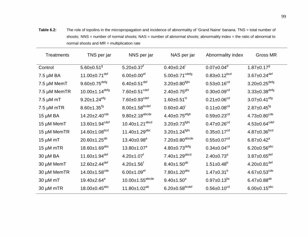
99
Table 6.2: The role of topolins in the micropropagation and incidence of abnormality of ‘Grand Naine’ banana. TNS = total number of
shoots; NNS = number of normal shoots; NAS = number of abnormal shoots; abnormality index = the ratio of abnormal to
normal shoots and MR = multiplication rate
Treatments TNS per jar NNS per jar NAS per jar Abnormality index Gross MR
Control 5.60±0.51g 5.20±0.37f 0.40±0.24i 0.07±0.04d 1.87±0.17g
7.5 µM BA 11.00±0.71def 6.00±0.00ef 5.00±0.71cdefg 0.83±0.12bcd 3.67±0.24def
7.5 µM MemT 9.60±0.75defg 6.40±0.51def 3.20±0.80fghi 0.53±0.16cd 3.20±0.25defg
7.5 µM MemTR 10.00±1.14defg 7.60±0.51cdef 2.40±0.75ghi 0.30±0.09cd 3.33±0.38defg
7.5 µM mT 9.20±1.24efg 7.60±0.93cdef 1.60±0.51hi 0.21±0.06cd 3.07±0.41efg
7.5 µM mTR 8.60±1.35fg 8.00±1.58bcdef 0.60±0.40i 0.11±0.08cd 2.87±0.45fg
15 µM BA 14.20±2.40cde 9.80±2.18abcde 4.40±0.75efgh 0.59±0.23cd 4.73±0.80cde
15 µM MemT 13.60±1.94cdef 10.40±1.21abcd 3.20±0.73fghi 0.47±0.26cd 4.53±0.64cdef
15 µM MemTR 14.60±1.08bcd 11.40±1.29abc 3.20±1.24fghi 0.35±0.17cd 4.87±0.36bcd
15 µM mT 20.60±1.25ab 13.40±0.98a 7.20±0.80abcde 0.55±0.07cd 6.87±0.42a
15 µM mTR 18.60±1.69abc 13.80±1.07a 4.80±0.73defg 0.34±0.04cd 6.20±0.56abc
30 µM BA 11.60±1.94def 4.20±1.07f 7.40±1.29abcd 2.40±0.73a 3.87±0.65def
30 µM MemT 12.60±2.44def 4.20±1.56f 8.40±1.50ab 1.51±0.48b 4.20±0.81def
30 µM MemTR 14.00±1.58cde 6.00±1.09ef 7.80±1.20abc 1.47±0.31b 4.67±0.53cde
30 µM mT 19.40±2.64a 10.00±1.55abcde 9.40±1.50a 0.97±0.13bc 6.47±0.88ab
30 µM mTR 18.00±0.45abc 11.80±1.02ab 6.20±0.58bcdef 0.56±0.10cd 6.00±0.15abc

100
Table 6.3: The effect of different cytokinins on banana micropropagation
Treatments
22.2 µM
No. of normal
shoots/explant No. of roots
Average
shoot
length (cm)
Control 1.5b 11.8a 7.85
BA 3.0ab 10.4a 5.25
mT 4.8a 3.3b 6.20
mTR 5.3a 0.4c 6.45
SE 0.909 1.473 0.942
F prob. 0.001 0.001 0.072
LSD 1.866 3.022 Ns*
*ns - indicates non significant treatment effects
BA was the most toxic cytokinin based on the higher abnormality index recorded
for both cultivars (Tables 6.1 and 6.2). The inhibitory and/or toxic effects observed
at higher concentrations are not new to banana tissue culture. Although
concentrations as high as 10 mg l-1 were used (DORE SWAMY et al., 1983),
higher concentrations generally inhibited shoot multiplication (WONG, 1986;
VUYLSTEKE, 1998 and references therein). ‘Williams’ banana was more sensitive
than ‘Grand Naine’ to higher cytokinin concentrations. It is well documented that
bananas show a wide range of dose dependent responses among and within the
genomic groups of EuMusa series. VUYLSTEKE (1998) suggested that
differences in sensitivity could be due to cultivar dependent responses to the
different cytokinin concentrations.
In contrast to previous findings with topolins (WERBROUCK et al., 1996; chapter
four of this thesis), mT and mTR had an inhibitory effect on rooting when
compared to BA and the controls (Table 6.3). Particularly striking is the near
complete inhibition of rooting by mTR, a compound that promoted rooting of Aloe
polyphylla shoots at concentrations where both BA and mT completely inhibited
the process. In his banana shoot-tip culture manual, VUYLSTEKE (1998)
emphasized the importance of a high cytokinin-auxin ratio for efficient shoot
multiplication. The author also indicated that best multiplication was achieved

101
without associated rooting. Therefore, based on the existing knowledge the
following assumptions may explain the results obtained: -
1. The physiological effects of topolins on root formation could be species
dependent;
2. In the presence of a constant auxin concentration, the difference in the
molecular weight of the cytokinins could have significantly influenced the
cytokinin-auxin ratio to the extent of inhibition of rooting. For example 22.2
µM BA equals 5 mg/L whereas 22.2 µM mTR equals 8.3 mg/L; and
3. There appears to be an inverse correlation between shoot multiplication
and root formation.
6.3.2 Analysis of somaclonal variation
Results of the analysis of somaclonal variation (Table 6.4) revealed that there was
no significant difference between treatments. Nevertheless all the treatments were
significantly different from the control (plants from the 3rd multiplication cycle).
There was a significant increase in variation rate between the 3rd and 7th
multiplication cycles. Results of the amplification product revealed the presence of
the polymorphic 1.6 kb RAPD marker previously reported using OPC-15 primer
(BAIRU et al., 2006) and OPJ-04 primer (DAMASCO et al., 1996b). The present
result is therefore additional proof of the reliability of the dwarf specific marker. The
fact that previous authors reported this variant from different banana cultivars
could suggest the presence of only one type of dwarf off-type.
Table 6.4: The effect of mT and mTR on somaclonal variation of ‘Williams’ banana
Treatments No. of samples analyzed Variation (%)
Control 60 5b
BA 60 23a
mT 60 19a
mTR 60 21a
The absence of any statistically significant difference between the treatments
could be explained in two ways. Firstly, all the cytokinins tested at equi-molar
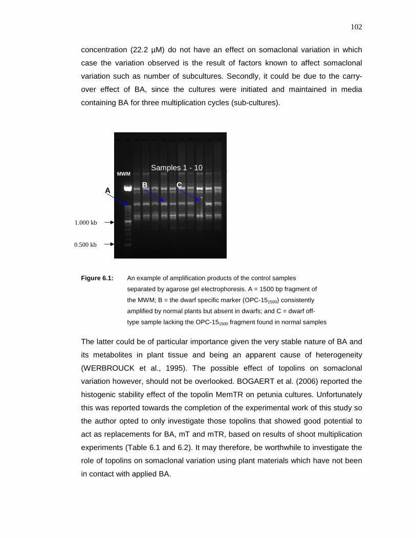
102
concentration (22.2 µM) do not have an effect on somaclonal variation in which
case the variation observed is the result of factors known to affect somaclonal
variation such as number of subcultures. Secondly, it could be due to the carry-
over effect of BA, since the cultures were initiated and maintained in media
containing BA for three multiplication cycles (sub-cultures).
Figure 6.1: An example of amplification products of the control samples
separated by agarose gel electrophoresis. A = 1500 bp fragment of
the MWM; B = the dwarf specific marker (OPC-151500) consistently
amplified by normal plants but absent in dwarfs; and C = dwarf off-
type sample lacking the OPC-151500 fragment found in normal samples
The latter could be of particular importance given the very stable nature of BA and
its metabolites in plant tissue and being an apparent cause of heterogeneity
(WERBROUCK et al., 1995). The possible effect of topolins on somaclonal
variation however, should not be overlooked. BOGAERT et al. (2006) reported the
histogenic stability effect of the topolin MemTR on petunia cultures. Unfortunately
this was reported towards the completion of the experimental work of this study so
the author opted to only investigate those topolins that showed good potential to
act as replacements for BA, mT and mTR, based on results of shoot multiplication
experiments (Table 6.1 and 6.2). It may therefore, be worthwhile to investigate the
role of topolins on somaclonal variation using plant materials which have not been
in contact with applied BA.
A
MWM Samples 1 - 10
C B
1.000 kb
0.500 kb

103
7. Conclusions and recommendations
The results of the soybean callus bioassay are useful additions to the ever growing
knowledge of synthesis and use of cytokinins. The results demonstrated the
importance of structure activity relationships in cytokinin research. The differential
effects of the relative biological activity of different cytokinins in different bioassay
system highlight the importance of activity profiling of these cytokinins using the
different bioassay systems. With time, this activity profiling should include the
entire family of topolins including their corresponding nucleotides, nucleosides,
glucosides as well as related derivatives.
The micropropagation protocol developed for A. polyphylla has been tested on a
larger scale and can be recommended for commercial propagation of this species.
The complete control of hyperhydricity using mT and the absence of the negative
ex vitro effect frequently observed with BA-treated cultures warrants related
research for species with similar problems. Currently there are ongoing trials to
test the protocol on different aloes.
In view of the shoot multiplication potential of H. procumbens, all cuttings of nodal
segments and the apical shoots were capable of regeneration. It is therefore
possible to generate a very large number of plants in a very short period for
commercial propagation. Virtually, all of the tissue culture conditions tested
affected STN in some way. The optimum protocol developed, following the
investigations made in this study contains the results from each experiment.
Auxin-free half strength MS media supplemented with 3% sucrose, 3-5 µM mTR,
1% agar, 6 mM Ca, 0.1 g l-1 myo-inositol, pH 5.8 and a sub-culture period of two
weeks are requirements for shoot multiplication. The very complex nature of STN
in this species necessitates further investigations on boron transport and the role
of sugar alcohols in this process as well as investigating the interaction between
Ca, B and PGR’s using labelled compounds.
104
Low multiplication and high somaclonal variation rates are characteristics of the
existing banana micropropagation protocol. The higher multiplication rate obtained
using topolins is therefore, an important addition to our knowledge for application
to banana tissue culture. The inhibitory effect of these cytokinins on rooting of
banana cultures indicates that the rooting response is species specific. The
physiological effects of topolins on rooting of plants require detailed investing-
ations. The results reported on the effect of topolins on somaclonal variation
should not lead to general conclusions mainly due to the nature of the
experimental plants used and associated carry-over effects. Recent reports on
histogenic stability effects of some topolins also warrant detailed investigations
starting right from the culture initiation stage.
The present results demonstrate the potential topolins hold in alleviating and/or
controlling some tissue culture-related problems. The positive effect of topolins on
hyperhydricity, STN and banana micropropgation are the first of their kind and
serve as foundation for future research. This study aimed at understanding factors
contributing to the problems investigated and seeking solutions to alleviate or
control them. More stringent research using labelled cytokinins could lead to a
better understanding of the mode of action and physiological effects of these
compounds.
105
References
ABDULNOUR JE, DONNELLEY DJ and BARTHAKUR NN. 2000. The effect of
boron on calcium uptake and growth in micropropagated potato plantlets.
Potato Research 43:287-295
ABOUSALIM A and MANTELL SH. 1994. A practical method for alleviating shoot-
tip necrosis symptoms in in vitro shoot cultures of Pistacia vera cv. Matheur
Journal of Horticultural Science 69:357-365
ABRIE AL and VAN STADEN J. 2001. Micropropagation of the endangered Aloe
polyphylla. Plant Growth Regulation 33:19-23
AHLOOWALIA BS. 1986. Limitations to the use of somaclonal variation in crop
improvement. In: SEMAL, J. (ed.). Somaclonal Variation and Crop
Improvement. pp 14-27. Martinus Nijhoff publishers
AMASINO RM and MILLER CO. 1982. Hormonal control of tobacco crown gall
tumor morphology. Plant Physiology 69:389-392
ARIAS D, DANKERS C, LIU P and PILKAUSKAS P. 2003. The World Banana
Economy 1985-2002. Food and Agricultural Organization of the United
Nations, Rome
AUBREY, A. 2002. Aloe polyphylla. Available online at
http://www.plantzafrica.com/plantab/aloepoly.htm
BAIRU MW. 2004. Plant growth regulators and somaclonal variation in Cavendish
banana (Musa AAA cv. ‘Zelig’). MSc thesis, University of KwaZulu-Natal,
Pietermaritzburg, South Africa
106
BAIRU MW, FENNELL CW and VAN STADEN J. 2006. The effect of plant growth
regulators on somaclonal variation in Cavendish banana (Musa AAA cv.
‘Zelig’). Scientia Horticulturae 108:347-351
BARGHCHI M and ALDERSON PG. 1985. In vitro propagation of Pistacia vera L.
and commercial varieties of ‘Ohadi’ and ‘Kalleghochi’. Journal of
Horticultural Science 60:423-430
BARGHCHI M and ALDERSON PG. 1996. The control of shoot tip necrosis in
Pistacia vera L. in vitro. Plant Growth Regulation 20:31-35
BAROJA-FERNANDEZ E, AGUIRREOLEA J, MARTÍNKOVÁ H, HANUS J and
STRNAD M. 2002. Aromatic cytokinins in micropropagated potato plants.
Plant Physiology and Biochemistry 40:217-224
BAYLISS MW. 1980. Chromosomal variation in tissue culture. In: VASIL IK (ed.).
Perspectives in Plant Cell and Tissue Culture. New York, Academic Press.
pp 113-144
BEEMSTER, G.T.S., FIORANI, F. & INZE, D. 2003. Cell cycle: the key to plant
growth control. Trends in Plant Science 8:154-158
BELLAROSA R. 1988. In vitro propagation of oaks (Q. suber, Q. pubescens, Q
cerris). Acta Horticulturae 227:433-435
BENTUR JS and KALODE MB. 1996. Hypersensitive reaction and induced
resistance in rice against the Asian rice gall midge Orseolia oryzae.
Entomologia Experimentalis etApplicata 78:77-81
BENZIONI A, MILLS D and WANKART S. 2003. Effect of ventilation on the
performance of Jojoba (Simmondsia chinesis) clones: multiplication stage.
Acta Horticalturae 616:135-138

107
BHALLA PL and MULWA RMS. 2003. Tissue culture and macadamia propagation.
Acta Horticulturae 616:343-346
BIDDULPH O, NAKAYAMA FS and CORY R. 1961. Transpiration stream and
ascension of calcium. Plant Physiology 36:429-436
BOGAERT I, VAN CAUTER S, WERBROUCK SPO and DOLEŽAL K. 2006. New
aromatic cytokinins can make the difference. Acta Horticulturae 725:265-
270
BOHNSACK CW and ALBERT LS. 1977. Early effects of boron deficiency on
indoleacetic acid oxidase levels of squash root tips. Plant Physiology
59:1047-1050
BORCHERT R. 1986. Calcium acetate-induced calcium uptake and formation of
calcium-oxalate crystals in isolated leaflets of Gleditsia triacanthos L..
Planta 168:571-578
BORNMAN CH and VOGELMANN TC. 1984. Effect of rigidity of gel medium on
benzyladenine induced adventitious bud formation and hyperhydricity in
vitro in Picea abies. Physiologia Plantarum 61:505-512
BOWEN JE. 1972. Effect of environmental factors on water utilization and boron
accumulation and translocation in sugar cane. Plant and Cell Physiology
13:703-714
BRAUN AC. 1959. A demonstration of the recovery of the crown-gall tumour cell
with the use of complex tumours of single-cell origin. Proceedings of the
National Academy of Sciences of the United States o f America 45:932-
938
BRIGHT S, JARRET V, NELSON R, CREISSEN G, KARP A, FRANKLIN J,
NORBURY P, KUEH J, ROGNES S. and MIFLIN, B. 1983. Modification of
agronomic traits using in vitro technology. In: MANTELL SH and SMITH H

108
(eds.). Plant Biotechnology. Cambridge University Press, New York. pp
251-265
BROWN DCW, LEUNG DWM and THORPE TA. 1979. Osmotic requirement for
shoot formation in tobacco callus. Physiologia Plantarum 46:36-41
BROWN PH and HU H. 1996. Phloem mobility of boron is species dependent:
Evidence of phloem mobility in sorbitol-rich species. Annals of Botany
77:497-505
BROWN PH, BELLALOUI N, HU H and DANDEKAR A. 1999. Transgenically
enhanced sorbitol synthesis facilitates phloem boron transport and
increases tolerance of tobacco to boron deficiency. Plant Physiology
119:17-20
BROWN PH and SHELP BJ. 1997. Boron mobility in plants. Plant and Soil
193:85-101
BROWN TA. 1995. Gene cloning: An Introduction. Van Nostrand Reinhold,
London
BRYANT, J.A. 1976. The cell cycle. In: BRYANT JA (ed.). Molecular Aspect of
Gene Expression in Plants, New York: Academic, pp 117-216
CABONI E, LAURI P, DAMIANO C and D’ANGELI S. 2000. Somaclonal variation
induced by adventitious shoot regeneration in pear and apple. Acta
Horticulturae 530:195-201
CAETANO-ANOLLES G. 1994. MAAP: a versatile and universal tool for genome
analysis. Plant Molecular Biology 25:1011-1026
CAETANO-ANOLLES G, BASSAM BJ and GRESSHOFF PM. 1991. DNA
amplification fingerprinting using very short arbitrary oligonucleotide
primers. BioTechnology 9:553-556

109
CAETANO-ANOLLES G, BASSAN BJ and GRESSHOFF PM. 1992. DNA
fingerprinting: MAAPing out a RAPD redefinition. Biotechnology 10:9
CAETANO-ANOLLES G, CALLAHAM L.M, WILLIAMS PE, WEAVER KR and
GRESSHOFF PM. 1995. DNA amplification fingerprinting of Bermuda grass
(Cynodon) – genetic relationships between species and interspecific
crosses. Theoretical and Applied Genetics 91:228-235
CASSELLS AC and WALSH C. 1994. The influence of gas-permeability of the
culture lid on calcium uptake and stomatal function in Dianthus microplants.
Plant Cell Tissue and Organ Culture 37:171-178
CHANG Y-C and MILLER WB. 2005. The development of upper leaf necrosis in
Lilium ‘Star Gazer’. Journal of American Society of Horticultural
Science 130:759-766
CHEN C-M and KRISTOPEIT SM. 1981. Metabolism of cytokinins: deribosilation
of cytokinin ribonucleoside by adenine nucleotidases from wheat germ
cells. Plant Physiology 68:1020-1023
CHUKWUJEKWU JC, FENNELL CW and VAN STADEN J. 2002. Optimisation of
the tissue culture protocol for the endangered Aloe polyphylla. South
Africa Journal of Botany 68:424-429
CLEMENCEAU D, COUSSEAU J, MARTIN V, MOLINES H, WAKSELMAN C,
MORNET R, NOGUE F, and LALOUE M. 1996. Synthesis and cytokinin
activity of two fluoro derivatives of N6-isopentenyladenine. Journal of
Agricultural and Food Chemistry 44:320-323
CORSE J, PACOVSKY RS, LYMAN ML and BRANDON DL. 1989. Biological
activity of several 9-nonglycosidic-substituted natural cytokinins. Journal of
Plant Growth Regulation 8:211-223

110
CRAVEN P and LOOTS S. 2002. Southern African Plants Red Data List.
GOLDING J (ed.). Southern African Botanical Diversity Network Report No.
14, page 82
CROCKER PJ, MAHADEVAN A, WILEY JL, MARTIN BR and RAZDAN RK. 2007.
The role of fluorine substitution in the structure-activity relationships (SAR)
of classical cannabinoids. Bioorganic and Medicinal Chemistry 17:1504-
1507
CRONAUER SS and KRIKORIAN AD. 1984. Rapid multiplication of bananas and
plantains by in vitro shoot tip culture. HortScience 19:234-235
DAGUIN F and LETOUZE R. 1986. Ammonium- induced hyperhydricity in cultured
tissues. Physiologia Plantarum 66:94-98
DAMASCO OP, GODWIN ID, SMITH MK and ADKINS SW. 1996a. Gibberellic
acid detection of dwarf off-types in micropropagated ‘Cavendish’ bananas.
Australian Journal of Experimental Agriculture 36:237-241
DAMASCO OP, GRAHAM GC, HENRY RJ, ADKINS SW, SMITH MK and
GODWIN ID. 1996b. RAPD detection of dwarf off-types in micropropagated
‘Cavendish’. (Musa spp. AAA) bananas. Plant Cell Reports 16:118-123
DAMASCO OP, SMITH MK and ADKINS SW. 1998a. Identification and
characterization of dwarf off-types from micropropagated ‘Cavendish’
bananas. Acta Horticulturae 490:79-84
DAMASCO OP, SMITH MK, ADKINS SW and GODWIN ID. 1998b. Use of SCAR-
based marker for early detection of dwarf off-types in micropropagated
‘Cavendish’ bananas. Acta Horticulturae 461:157-164
DAMASCO OP, SMITH MK, GODWIN ID, ADKINS SW, SMILLIE RM and
HETHERINGTON SE. 1997. Micropropagated dwarf off-type ‘Cavendish’

111
bananas (Musa Spp. AAA) show improved tolerance to sub-optimal
temperatures. Australian Journal of Agricultural Research 48:377-384
D’AMATO F. 1975. The problem of genetic stability in plant tissue and cell
cultures. In: FRANKEL OH and HAWKES JG (eds.). Crop Genetic
Resources for today and Tomorrow. pp 333-348. New York: Cambridge
University Press
D’AMATO F. 1977. Cytogenetics of differentiation in tissue and cell culture. In:
REINERT J and BAJAJ YPS (eds.). Applied and Fundamental Aspects of
Plant Cell, Tissue and Organ Culture. pp 343-464. New York: Springer
Verlag
DANIELLS JW and SMITH M. 1991. Post-flask management of tissue cultured
banana. Technical Report . ACIAR Canberra, 18:1-8
DEBERGH PC. 1983. Effect of agar brand and concentration on the tissue culture
medium. Physiologia Plantarum 59:270-276
DEBERGH PC, AITKEN-CHRISTIE J, COHEN D, GROUT B, Von ARNOLD S,
ZIMMERMAN R and ZIV M. 1992. Reconsideration of the term
‘hyperhydricity’ as used in micropropagation. Plant Cell Tissue Organ
Culture 30:135-140
DEBERGH PC, HARBAOUI Y and LEMEUR R. 1981. Mass propagation of globe
artichoke (Cynara scolymus): evaluation of different hypothesis to
overcome hyperhydricity with special reference to water potential.
Physiologia Plantarum 53:181-187
De BLOCK M. 1990. Factors influencing the tissue culture and the Agrobacterium
tumefaciens-mediated transformation of hydrid Aspen and Poplar clones.
Plant Physiology 93:1110-1116
112
DELARUE M, SANTONI V, CABOCHE M and BELLINI C. 1997. Cristal mutations
in Arabidopsis confer a genetically heritable, recessive, hyperhydric
phenotype. Planta 202:51-61
DOLEŽAL K, POPA I, HOLUB J, LENOBEL R, WERBROUCK S, STRNAD M,
and ZATLOUKAL M. 2007b Heterocyclic compound based on N6-
substituted adenine, methods of their preparation, their use for preparation
of drugs and pharmaceutical preparations containing these compounds;
patent number - US 7279482
DOLEŽAL K, POPA I, HAUSEROVĂ E, SPĺCHAL L, CHAKRABARTY K, NOVĂK
O, KRYŠTOF V, VOLLER J, HOLUB J and STRNAD M. 2007a. Preparation
biological activity and endogenous occurrence of N6-benzyladenosines.
Bioorganic and Medicinal Chemistry 15:3737-3747
DOLEŽAL K, POPA I, KRYŠTOF V, SPĺCHAL L, FOJTĺKOVA M, HOLUB J,
LENOBEL R, SCHMŐLLING T and STRNAD M. 2006. Preparation and
biological activity of 6-benzylaminopurine derivatives in plants and human
cancer cells. Bioorganic and Medicinal Chemistry 14:875-884
DORE SWAMY R, SRINIVASA RAO NK and CHACKO EK. 1983. Tissue culture
propagation of banana. Scientia Horticulturae 18:247-252
DUNWELL JM. 1979. Anther culture in Nicotiana tabacum: the role of the culture
vessel atmosphere in pollen embryo induction and growth. Journal of
Experimental Botany 30:419-428
DYSON PW and DIGBY J. 1975. Effect of calcium on sprout growth and sub-
apical necrosis in Majestic potatoes. Potato Research 18:290-305
EDWARDS NR. 2000. Optimisation of the randomly amplified polymorphic DAN
(RAPD) technique for the characterization of selected South African maize
(zea mays L.) breeding material. M.Sc. Thesis, University of KwaZulu-Natal,
Pietermaritzburg, Life Science Library

113
EGLEY GH, PAUL RN, VAUGHN KC and DUKE SO. 1983. Role of peroxidase in
the development of water impermeable seed coats in Sida spinosa L.
Planta 157:224-232
ELLSWORTH DL, RITTENHOUSE KD and HONEYCUT RL. 1993. Artifactual
variation in randomly amplified polymorhpic DNA banding patterns.
Biotechniques 14:214-217
EMANOIL M. 1994. Encyclopaedia of Endangered Species. Gale Research Inc.,
Michigan. pp 1091-1092
EMMERLING F, ORGZALL I, DIETZEL B, SCHULZ BW, RECK G and SCHULZ
B. 2007. Structural studies on trifluoromethyl substituted 2,5-diphenyl-1,3,4-
oxadiazoles. Journal of Molecular structure 832:124-131
ERASMUS TE. 2001. Optimization of isozyme genotyping and purity analysis for
commercial applications. M.Sc. Thesis, University of KwaZulu-Natal,
Pietermaritzburg, South Africa
ETIENNE H and BERTRAND B. 2003. Somaclonal variation in Coffea arabica:
effect of genotype and embryogenic cell suspension age on frequency and
phenotype of variants. Tree Physiology 23:419-426
EVANS DA. and SHARP WR. 1986. Applications of somaclonal variation.
BioTechnology 4:528-532
FORSYTH C and VAN STADEN, J. 1986. The metabolism and cell division activity
of adenine derivatives in soybean callus. Journal of Plant Physiology
124:275-287
GASPAR TH. 1986. Integrated relationships of biochemical and physiological
peroxidase activities. In: GREPPIN H. PENEL C and GASPAR TH (eds.).
Molecular and Physiological Aspects of Plant Peroxidases. University of
Geneva, Swizerland, pp455-468

114
GASPAR TH, KEVERS C, DEBERGH P, MAENE L., PAQUES M and BOXUS P.
1987. Hyperhydricity: morphological, physiological and ecological aspects.
In: BONGA JM and DURZAN DJ (eds.). Cell and Tissue Culture in Forestry,
vol. I, Martinus Nijhoff Publishers, Dordrecht, Holland. pp152-166
GENGENBACH BG and UMBECK P. 1982. Characteristics of T-cytoplasm
revertants from tissue culture. Maize Genetics Cooperation Newsletter
56:140-142
GENEVE RL, KESTER ST and YUSNITA S. 1992. Micropropagation of eastern
redbud (Cercis Canadensis L.) Proc. International Plant Propagation
Society 42:417-420
GEORGE EF. 1993. Plant Propagation by Tissue Culture. Part I, 2nd edition.
Exegetics Ltd., England
GOLDBERG R, CATESSON AM and CZANINSKI Y. 1983. Some properties of
syringal-dazine oxidase, a peroxidase specifically involved in the
lignification process. Zeitschirft F őr Pflanzenphysiologie 110:267-279
GOLDING JS (ed). 2002. Southern African Plant Red Data Lists. Southern African
Botanical Diversity Network Report No. 14. SABONET, Pretoria
GRAJAL-MARTIN MS, GRILLO GS and DOMINGUEZ AM. 1998. The use of
Randomly Amplified polymorphic DNA (RAPD) for the study of genetic
diversity and somaclonal variation in Musa. Acta Horticulturae 490:445-
454
GRIBBLE K. 1999. The influence of relative humidity on vitrification, growth and
morphology of Gypsophila paniculata L. Plant Growth Regulation 27:179-
188
115
GRIGORIADOU K, LEVENTAKIS N and VASILAKAKIS M. 2000. Effects of
various culture conditions on proliferation and shoot tip necrosis in the pear
cultivars ‘William’s’ and ‘Highland’ grown in vitro. Acta Horticulturae
520:103-108
GRILLO SG, MARTIN MJG and DAMIGUEZ AM. 1998. Morphological methods for
the detection of banana off-types during the hardening phase. Acta
Horticulturae 490:239-245
GUTHRIE JN, WALSH KB, SCOTT PT and RASMUSSEN TS. 2001. The
phytopathology of Australian papaya dieback: a proposed role for the
phytoplasma. Physiological and Molecular Plant Pathology 58:23-30
HACKETT WP and ANDERSON JM. 1967. Aseptic multiplication and
maintenance of differentiated carnation shoot tissue derived from shoot
apices. Proceedings of the American Society for Horticultur al Science
90:365-369
HAIDOUNE H, RAYNAUD I, O’CONNOR N, RICHOMME P, MORNET R and
LALOUE M. 1998. Synthesis and cytokinin activity of new zeatin
derivatives. Journal of Agricultural and Food Chemistry 46:1577-1582
HARRAR Y, BELLEC Y, BELLINI C and FAURE J-D. 2003. Hormonal control of
cell proliferation requires PASTICCINO genes. Plant Physiology
132:1217-1227
HECHT SM, FRYE RB, WERNER D HAWRELAK SD, SKOOG F and SCHMITZ
RY. 1975. On the ‘activation’ of cytokinins. The Journal of Biological
Chemistry 250:7343-7351
HECHT SM, LEONARD NJ, SCHMITZ RY and SKOOG F. 1970a. Cytokinins:
influence of side-chain planarity of N6-substituted adenines and adenosines
on their activity in promoting cell growth. Phytochemistry 9:1907-1913
116
HECHT SM, LEONARD NJ, SCHMITZ RY and SKOOG F. 1970b. Cytokinins:
synthesis and growth promoting activity of 2-substituted compounds in the
N6-isopentenyladenine and zeatin series. Phytochemistry 9:1173-1180
HEPLER PK. 2005. Calcium: A central regulator of plant growth and development.
The Plant Cell 17:2142-2155
HEPLER PK and WAYNE RO. 1985. Calcium and plant development. Annual
Review of Plant Physiology 36:397-439
HILLS P. and VAN STADEN J. 2002. An improved DNA extraction procedure for
plant tissues with a high phenolic content. South African Journal of
Botany 68: 549-550
HIRSCHI KD. 2004. The calcium conundrum. Both versatile nutrient and specific
signal. Plant Physiology 136:2438-2442
HOLUB J, HANUŠ J, HANKE DE and STRNAD M. 1998. Biological activity of
cytokinins derived from Ortho- and Meta- hydroxybenzyladenine. Plant
Growth Regulation 26:109-115
HORGAN R. 1992. Present and future prospects for cytokinin research. In:
KAMÍNEK M, MOK DWS and ZAŽÍMALOVÁ E (eds.). Physiology and
Biochemistry of Cytokinins. SPB Academic Publishing, The Hague. pp 1-13
HORGAN R, HEWETT EW, HORGAN JM, PURSE JG and WAREING PF. 1975.
A new cytokinin from Populus x robusta. Phytochemistry 14:1005-1008
HOWELL EC, NEWBURY HJ, SWENNER RL, WITHERS LA and FORD-LLOYD
BV. 1994. The use of RAPD foe identifying and classifying Musa
germplasm. Genome 37:328-332

117
HU H, PENN SC, LEBRILLA CB and BROWN PH. 1997. Isolation and
characterization of soluble boron complexes in higher plants: the
mechanism of phloem mobility of boron. Plant Physiology 113:649-655
HU H and BROWN PH. 1994. Localization of boron in cell walls of squash and
tobacco and its association with pectin: evidence for a structural role of
boron in the cell wall. Plant Physiology 105:681-689
HUFF DR and BARA JM. 1993. Determining genetic origins of abberant progeny
from facultative apomictic Kentucky blue grass using a combination of flow
cytometry and silver-stained RAPD markers. Theoretical and Applied
Genetics 87: 201-208
HWANG SC and KO WH. 1987. Somaclonal variation of banana and screening for
resistance to Fusarium wilt. In: PERSELEY GJ and DeLANGHE EA. (eds.).
Bananas and Plantains Breeding Strategies, pp 151-156, ACIAR
proceedings No. 21. ACIAR, Canberra
INNIS MA and GELFAND DH 1990. Optimisation of PCR. In: INNIS MA,
GELFAND DH, SNINSKY JJ, and WHITE TJ. (eds.). PCR Protocols.
Academic Press, New York.
ISRAELI Y, LAHAV E and REUVENI O. 1995. In vitro Culture of Bananas. In:
GOWEN, S., (ed.). Bananas and Plantains. Chapman and Hall, London
ISRAELI Y, REUVENI O and LAHAV E. 1991. Qualitative aspect of somaclonal
variation in banana propagated by in vitro techniques. Scientia
Horticulturae 48:71-88
IWAMURA H, FUJIITA T, KOYAMA S, KOSHIMIZU K and KUMAZAWA Z. 1980.
Quantitative structure-activity relationship of cytokinin-active adenine and
urea derivatives. Phytochemistry 19:1309-1319
118
JARRET RL and GAWELL N. 1995. Molecular markers, genetic diversity and
systematics in Musa. In: GOWEN, S., (ed.). Bananas and Plantains, pp 66-
81. Chapman and Hall, London
JARRET RL and LITZ RE. 1986. Isozymes as genetic markers in bananas and
plantains. Euphytica 35:539-549
JARRET RL, RODRIGUEZ W and FERNANDEZ R. 1985. Evaluation, tissue
culture propagation and dissemination of ‘saba’ and ‘pelpita’ plantains in
Costa-Rica. Scientia Horticulturae 25:137-147
JONES LH, MARTINKOVÁ H, STRNAD M and HANKE D.E. 1996. Occurrence of
aromatic cytokinins in oil palm (Elaeis guineensis Jacq.). Journal of Plant
Growth Regulation 15:39-49
KAMÍNEK M, PACES V, CORSE J and CHALLICE JS. 1979. Effect of
stereospecific hydroxylation of N6-(∆2-Isopentenyl)adenosine on cytokinin
activity. Planta 145:239-243
KAMÍNEK M, VANÉK T, KALENDOVA A and PILAR J. 1987b. Effect of two
cytokinins on production of stem cuttings by stock plants of Ephorbia
pulcherrima Wild. and Gerbera jamesonii Hook. Scientia Horticulturae
33:281-289
KAMÍNEK M, VANÉK T and MOTYKA V. 1987a. Cytokinin activities of N6-
benzyladenosine derivatives hydroxylated on the side-chain phenyl ring.
Journal of Plant Growth Regulation 6:113-120
KAPLAN DR. 1992. The relationship of cells to organism in plants: problems and
implications of an organismal perspective. International Journal of Plant
Science 153:528-537

119
KARP A. 1994. Origin, causes and uses of variation in plant tissue culture. In:
VASIL I and THORPE T (eds.). Plant Cell and Tissue Culture. Kluwer
Academic Publishers
KARP A. SEBERG O and BUIATTI M. 1996. Molecular techniques in the
assessment of botanical diversity. Annals of Botany 78:143-149
KATAEVA NV, ALEXANDROVA IG, BUTENKO RG and DRAGAVTCEVA EV.
1991. Effect of applied and internal hormones on vitrification and apical
necrosis of different plants cultured in vitro. Plant Cell Tissue and Organ
Culture 27:149-154
KATHE W, BARSCH F and HONNEF S. 2003. Trade in Devil’s Claw
(Harpagophytum procumbens) in Germany – Status, Trend and
Certification. Report presented to The Food and Agricultural Organization of
the United Nation – Non-Wood Forest Products Programme, 40 pages
KEVERS C, COUMANS M, COUMANS-GILLÈS MF and GASPAR Th. 1984.
Physiological and biochemical events leading to hyperhydricity of plants
cultured in vitro. Physiologia Plantarum 61:69-74
KINTZIOS S, STRAVROPOULOUS Er and SKAMNELI S. 2004. Accumulation of
selected macronutrients and carbohydrates in melon tissue cultures:
association with pathways of in vitro dedifferentiation and differentiation
(organogenesis, somatic embryogenesis. Plant Science 167:655-664
KOHL HC and OERTLI JJ. 1961. Distribution of boron in leaves. Plant
Physiology 36:420-424
KORSZUN Z. KNIGHT C and CHEN C-M. 1989. A stereochemical model for
cytokinin activity. FEBS Letters 243:53-56

120
KRIKORIAN A.D. 1995. Hormones in tissue culture and micropropagation. In:
DAVIES PJ (ed.). Plant Hormones: Physiology, Biochemistry and Molecular
Biology. Academic Publishers, The Netherlands. pp 774-796
KRIKORIAN AD and CRONAUER SS. 1984. Bananas. In: SHARP WR, EVANCE
DA, AMMIRATO PV and YAMADA Y (eds). Handbook of plant cell culture.
Vol 2, Crop species. Macmillan Publishing Company, New York, pp 327-
348
KUBALAKOVA M and STRNAD M. 1992. The effect of aromatic cytokinins
(populins) on micropropagation and regeneration in vitro. Biologia
Plantarum 34: 578-579
KULKARNI K and D'SOUZA L. 2000. Control of in vitro shoot tip necrosis in Butea
monosperma. Current Science 78:125-126
LAKSHMI SG and RAGHAVA SBV. 1993. Regeneration of plantlets from leaf disc
cultures of rosewood: control of leaf abscission and shoot tip necrosis.
Plant Science 88:107-112
LARKIN PJ and SCOWCROFT WR. 1981. Somaclonal variation – a novel source
of variability from cell cultures for plant improvement. Theoretical and
Applied Genetics 60:197-214
LEE M and PHILLIPS RL. 1988. The chromosomal basis of somaclonal variation.
Annual Review of Plant Physiology 39:413-437
LEHTO T, KALLIO E and APHALO PJ. 2000. Boron mobility in two coniferous
species. Annals of Botany 86:547-550
LEONARD NJ, HECHT SM, SKOOG F and SCHMITZ RY. 1968. Cytokinins:
synthesis of 6-(3-methyl-3-butenylamino)-9-b-D-ribofuranosylpurine (3iPA)
and the effect of side-chain unsaturation on the biological activity of
121
isopentenylaminopurines and their ribosides. Proceedings of the National
Academy of Sciences of the United States of America 59:15-21
LEONARD NJ, HECHT SM, SKOOG F and SCHMITZ RY. 1969. Cytokinins:
synthesis, mass spectra and biological activity of compounds related to
zeatin. Proceedings of the National Academy of Sciences of the United
States of America 63:175-182
LEONARD NJ and HENDERSON TR. 1975. Purine ring re-arrangements leading
to the development of cytokinin activity. Mechanism of the Re-arrangement
of 3 – benzyladenine to N6-benzyleadenine. Journal of the American
Chemical Society 97:4990-4999
LESHEM B. 1983. Growth of carnation meristems in vitro: anatomical structure of
abnormal plantlets and the effect of agar concentration in the medium on
their formation. Annals of Botany 52:413-415
LESHEM B and SACHS T. 1985. ‘Vitrified’ Dianthus- teratomata in vitro due to
growth factor imbalance. Annals of Botany 56: 613-617
LESHEM B, SHALEY DP and IZHAR S. 1988. Cytokinin as an inducer of
hyperhydricity in melon. Annals of Botany 61:255-260
LETHAM DS. 1967. Regulators of cell division in plant tissues. A comparison of
the activities of zeatin and other cytokinins in five bioassays. Planta 74:228-
242
LETHAM DS. 1978. Cytokinins. In: LETHAM DS, GOODWIN PB and HIGGINS
TJV (eds.). Phytohormones and Related Compounds- A Comprehensive
Treatise, volume 1. Elsevier Press, Amsterdam, The Netherlands. pp 205-
263
LETHAM DS and PALNI LMS. 1983. The biosynthesis and metabolism of
cytokinins. Annual Review of Plant Physiology 34: 163-197

122
LEVIEILLE G and WILSON G. 2002. In vitro propagation and iridoid analysis of
the medicinal species Harpagophytum procumbens and H. zeyheri. Plant
Cell Reports 21:220-225
LEVIEILLE G, WILSON G, ROBIN JR and CAMBORNAC M. 2000. In-vitro
micropropagation of fertile plants of Harpagophytum procumbens and H
zeyheri (Devil’s claw). Medicinal Plant Conservation 6:10-11
LIBBENGA KR and MENNES AM. 1995. Hormone binding and signal
transduction. In: DAVIES PJ (ed.). Plant Hormones: Physiology,
Biochemistry and molecular Biology. Kluwer Academic Publishers, The
Netherlands. pp 272-297
LLOYD G and McCOWN B. 1980. Commercially feasible micropropagation of
mountain laurel, Kalmia latifolia, by use of shoot tip culture. Combined
Proceedings of the International Plant Propagations Society 30:421-
427
LoSCHIAVO F, PITTO L, GIULIANO G, TORTI G, NUTI-RONCHI V, RAZZITI D,
VERGARA R, ORSELLI S and TERZI M. 1989. DNA methylation of
embryogenic carrot cell cultures and its variation as caused by mutation,
differentiation, hormones and hypomethylation drugs. Theoretical and
Applied Genetics 77:325-331
MACKAY WA, TIPTON JL and THOMPSON GA. 1995. Micropropagation of
Mexican redbud, Cercis Canadensis var. mexicana. Plant Cell, Tissue and
Organ Culture 43:295-299
MARIN JA. 2003. High survival rates during acclimatization of fruit tree rootstocks
by increasing exposure to law relative humidity. Acta Horticulturae
616:139-142
123
MARSCHNER H. 1995. Mineral Nutrition of Higher Plants. Second Edition,
Academic Press, Boston
MARTIN KP, ZHANG C-L, SLATER A and MADASSERY J. 2007. Control of shoot
necrosis and plant death during micropropagation of banana and plantains
(Musa spp.). Plant Cell Tissue and Organ Culture 88:51-59
MATHEWS J. 2004. Aloe polyphylla: Schemes and Themes. Available online at
http://www.liddlewonder.co.nz/schemesandthemes/lib/alopol.htm
MATSUBARA H. 1980. Structure-activity relationships of cytokinins.
Phytochemistry 19:2239-2253
MEHTA YR and ANGRA DC. 2000. Somaclonal variation for disease resistance in
wheat and production of dihaploid through wheat x maize hybrids. Genetics
and Molecular Biology 23:617-622
McPHEETERS K. and SKIRVIN RM. 1983. Histogenic layer manipulation in
chimera ‘Thorn less Evergreen’ traiting blackberry. Euphytica 32:351-360
McGAW BA and BURCH LR. 1995. Cytokinin biosynthesis and metabolism. In:
DAVIES PJ (ed.). Plant Hormones: Physiology, Biochemistry and Molecular
Biology. Academic Publishers, The Netherlands. pp 98-117
MILLER CO. 1965. Evidence for the natural occurrence of zeatin and derivatives:
Compounds from maize which promote cell division. Proceedings of the
National Academy of Sciences of the United States o f America
54:1052-1058
MNG’OMBA SA, Du TOIT ES, AKINNIFESI FK and VENTER HM. 2007. Repeated
exposure of jacket plum (Pappea capensis) micro-cuttings to indole-3-
butyric acid (IBA) improved in vitro rooting capacity. South African Journal
of Botany 73:230-235

124
MOK MC, MARTIN RC, DOBREV PI, VANCOVả R, HO PS, YONEKURA-
SAKAKIBARA K, SAKAKIBARA H and MOK DWS. 2005. Topolins and
hydroxylated thidiazuron derivatives are substrates of cytokinin o-
glucosyltransferase with position specificity relayed to receptor recognition.
Plant Physiology 137:1057-1066
MOK MC, MOK DWS and ARMSTRONGDJ. 1978. Differential cytokinin structure-
activity relationships in Phaseolus. Plant Physiology 61:72-75
MOK MC and MOK DW. 1985. The metabolism of [14C]-thidiazurone in callus
tissues of Phaseolus lunatus. Physiologia Plantarum 65:427-432
MORARD P, PUJOS A, BERNADAC A, BERTONI G. 1996. Effect of temporary
calcium deficiency on tomato growth and mineral nutrition. Journal of Plant
Nutrition 19:115-127
MORELL MK, PEAKALL R, APPELS R, PRESTON LR and LLOYD HL. 1995.
DNA profiling techniques for pant variety identification. Australian Journal
of Experimental Agriculture 35:807-819
MURASHIGE T and SKOOG F. 1962. A revised medium for a rapid growth and
bio-assays with tobacco tissue cultures. Physiologia Plantarum 15:473-
497
NETO VBdP and OTONI WC. 2003. Carbon sources and their osmotic potential in
plant tissue culture: does it matter? Scientia Horticulturae 97:193-202
NISHIKAWA S, HAYASHI E, KUMAZAWA Z and KASHIMURA N. 1989. 4-
Styrylpyrimidines as a new class of cytokinins. Agricultural and Biological
Chemistry 53:3387-3389
NISHIKAWA S, KUMAZAWA Z, MIZUTANI H and KASHIMURA N. 1986.
Substituent-directing effect on cytokinin activity of ά-double bond in the 6-

125
substituent of purines. Agricultural and Biological Chemistry 50:1089-
1091
NITSCH JP and NITSCH C. 1969. Haploid plants from pollen grains. Science
163:85-87
OCHATT SJ, MUNEAUX E, MACHADO C, JACAS L and PONTECAILLE C. 2002.
The hyperhydricity of in vitro regenerants of grass pea (Lathyrus sativus L.)
is linked with an abnormal DNA content. Journal of Plant Physiology
159:1021-1028
OERTLI JJ. 1993. The mobility of boron in plants. Plant and Soil 155/156:301-304
OGASAWARA N. 2003. Ventilation and light intensity during in vitro culture affect
relative growth rate and photosynthate partitioning of Caladium plantlets
after transplanting to ex vitro. Acta Horticulturae 616:143-149
OLMOS E and HELLÍN E. 1998. Ultrastractural differences of hyperhydric and
normal leaves from regenerated carnation plants. Scientia Horticulturae
75:91-101
ORLIKOWSKA T. 1987. Hyperhydricity problem in the in vitro culture of fruit tree
rootstocks. Acta Horticulturae 212:239-241
PALMER MV, HORGAN R and WAREING PF. 1981. Cytokinin metabolism in
Phaseolus vulgaris L. I. Variation in cytokinin levels in leaves of decapitated
plants in relation to lateral bud outgrowth. Journal of Experimental
Botany 32:1231-1241
PALNI LMS, PALMER MV and LETHAM DS. 1984. The stability and biological
activity of cytokinin metabolites in soybean callus tissue. Planta 160:242-
249
126
PAQUES M. 1991. Hyperhydricity and micropropagation: causes, remedies and
prospects. Acta Horticulturae 289:283-290
PAQUES M and BOXUS P. 1987. Hyperhydricity: review of literature. Acta
Horticulturae 212:155-166
PARK SW, JEON JH, KIM HS, PARK YM, ASWATH C and JOUNG H. 2004.
Effect of sealed and vented gaseous microenvironments on the
hyperhydricity of potato shoots in vitro. Scientia Horticulturae 99:199-205
PEAKALL R. 1997. PCR- based genetic markers and their applications to turf
grass breeding. International Turf Grass Society Research Journal
8:243-259
PEREZ-TORNERO O and BURGOS L. 2000. Different media requirements for
micropropagation of apricot cultivars. Plant Cell Tissue and Organ
Culture 63:133-141
PERĚZ C, RODRỈGUEZ R and TAMĚS R. 1985. In vitro filbert (Corylus avellana
L.) micropropagation from shoots and cotyledonary segments. Plant Cell
Reports 4:137-139
PESCHKE VM, PHILLIPS RL and GENGENBACH BG. 1987. Discovery of
transposable element activity among progeny of tissue culture-derived
maize plants. Science 238:804-807
PFAB MF and SCHOLES MA. 2004. Is the collection of Aloe peglerae from the
wild sustainable? An evaluation using stochastic population modelling.
Biological Conservation 118:695-701
PHAN CT and HEGEDUS P. 1986. Possible metabolic basis for the development
anomaly observed in in vitro culture, called, ‘vitreous’ plants’. Plant Cell
Tissue Organ Culture 6:83-94
127
PIAGNANI C, ZOCCHI G and MIGNANI I. 1996. Influence of Ca2+ and 6-
benzyladenine on chestnut (Castanea sative Mill.) in vitro shoot-tip
necrosis. Plant Science 118:89-95
PIERIK RLM. 1987. In vitro Culture of Higher Plants. Martinus Nijhoff Publishers,
Dordrecht
PIQUERAS A, CORTINA M, SERNA MD and CASAS JL. 2002. Polyamines and
hyperhydricity in micropropagated carnation plants. Plant Science
162:671-678
POPESCU AN, ISAC VS, COMAN MS. and RADULESCU MS. 1997. Somaclonal
variation in plants regenerated by organogenesis from callus cultures of
strawberry (Fragaria X ananassa). Acta Horticulturae 439:89-96
RAIMONDO D and DONALDSON J. 2002. The Trade, Management and
Biological Status of Harpagophytum spp. in Southern African Range States.
A report submitted to the CITES Plants Committee, 28 pages
RAGOT M and HOISINGTON DA. 1993. Molecular markers for plant breeding:
comparison of RFLP and RAPD genotyping costs. Theoretical and
Applied Genetics, 86: 975-984
RAMAGE CM, BORDA AM, HAMILL SD and SMITH MK. 2004. A simplified PCR
test for early detection of dwarf off-types in micropropagated Cavendish
banana (Musa Spp. AAA). Scientia Horticulturae 103:145-151
RANI V, PARIDA A and RAINA SN. 1995. RAPD markers for genetic analysis in
micropropagated plants of Populus deltoids Marsh. Plant Cell Reports
14:459-462
RAVEN JA. 1977. H+ and Ca2+ in phloem and symplast: relation of relative
immobility of the ions to the cytoplasmic nature of the transport paths. New
Phytology 79:465-480

128
REDONDO-NIETO M, WILMOT AR, EL-HAMDAOUI A, BONILLA I and
BOLAŇOS L. 2003. Relationship between boron and calcium in the N2-
fixing legume-rhizobia symbiosis. Plant Cell and Environment 26:1905
(Abstract)
REINECKE D and OZGA J. 1999. Halogenated plant hormones. Plant Growth
Regulation 27:1-2
REUVENI O. 1990. Methods of detecting somaclonal variants in ‘Williams’
bananas. In: JARRET LA (ed.). Identification of Genetic Diversity in the
Genus Musa, pp 108-113. INIBAP, Montpellier, France
REUVENI O and ISRAELI Y. 1988. Measures to reduce somaclonal variation in in
vitro propagated bananas. Acta Horticulturea , 275: 307-313
REUVENI O and ISRAELI Y. 1990. Measures to reduce somaclonal variation in in
vitro propagated bananas. Acta Horticulturae 275:307-313
REUVENI O, ISRAELI Y and GOLUBOWICZ S. 1993. Factors influencing the
occurrence of somaclonal variation in micropropagated bananas. Acta
Horticulturae 336:357-364
RICCI A, CARRA A, ROLLI E, BERTOLETTI E, MORINI G, INCERTI M and
VICINI P. 2005. Effect of Cl-substitution on rooting- or cytokinin-like activity
of diphenylurea derivatives. Journal of Plant Growth Regulation 23:261-
268
RICHARDS EJ. 1997. Preparation of plant DNA using CTAB. In: AUSUBEL FM,
BRENT R, KINGSTON RE, MOORE DD, SEIDMAN JG, SMITH JA and
STRUHL K. (eds.). Short Protocols in Molecular Biology. 3rd edition. John
Wiley and sons, New York, pp 2.10 – 2.11
129
RODRIGUES, P.H.V., NETO, A.T., NETO, P.C. and MENDES, B.M.J. 1998.
influence of the numbers of sub-cultures on somaclonal variation in
micropropagated Nanicao (Musa Spp., AAA group). Acta Horticulturae ,
490: 469-473
RYBICKI EP. 2001. PCR primer design and reaction optimisation. In: COYNE VE,
JAMES MD, REID SJ and RYBICKI EP (eds.). Molecular Biology
Techniques Manual. 3rd Edition, University of Cape Town.
RYCHLIK W, SPENCER WJ and RHOADS RE. 1990. Optimisation of the
annealing temperature for DNA amplification in vitro. Nucleic Acids
Research 18:6409-6412
SAKAKIBARA H. 2004. Cytokinin biosynthesis and metabolism. In DAVIES PJ
(ed.). Plant Hormones: Biosynthesis, Signal transduction, Action, 3rd edition,
Kluwer Academic Publishers, London, pp 95-114
SANDOVAL JA. 1994. Variation resulting from banana in vitro culture (Musa AAA,
cv. ‘Grand Naine’). Morphological, cytological and hormonal
characterization elements (GA3). InfoMusa 3:31
SANDOVAL J, KERBELLEC F, COTE F and DOUMAS P. 1995. Distribution of
indigenous gibberellins in dwarf and giant off-type banana (Musa AAA, cv.
‘Grand Naine’) plants from in vitro propagation. Plant Growth Regulation
17:219-224.
SCHMITZ RY, SKOOG F, PLAYTIS AJ and LEONARD NJ. 1972. Cytokinins:
synthesis and biological activity of geometric and position isomers of zeatin.
Plant Physiology 50:702-705
SCOWCROFT WR. 1984. Genetic variability in tissue culture: Impact on
germplasm collection and utilization. Technical report, IBPGR Secretariat,
Rome, pp 41

130
SELING S, WISSEMEIER AH, CAMBIER P, VAN CUTSEM P. 2000. Calcium
deficiency in potato (Solanum tuberosum ssp. tuberosum) leaves and its
effects on the pectic composition of the apoplastic fluid. Physiologia
Plantarum 109:44-50
SHA L, McCOWN BH and PETERSON LA. 1985. Occurrence and cause of shoot-
tip necrosis in shoot cultures. Journal of American Society of
Horticultural Science 110:631-634
SHACKLETON CM and GAMBIZA J. 2007. Growth of Aloe ferox Mill. At selected
sites in the Makana region of the Eastern Cape. South African Journal of
Botany 73:266-269
SHEAR CB. 1975. Ca-related disorders of fruits and vegetables. HortScience
10:361-365
SHELP BJ, MARENTES E, KITHEKA AM and VIVEKANANDAN P. 1995. Boron
mobility in plants. Physiologia Plantarum 94:356-361
SHUDO K. 1994. Chemistry of Phenylurea Cytokinins. In MOK DWS and MOK
MC (eds.). Cytokinins Chemistry, Activity and Function. CRC Press, London
pp 35-42
SINGHA S, TOWNSEND EC and OBERLY GH. 1990. Relationship between
calcium and agar on vitrification and shoot-tip necrosis of quince (Cydonia
oblonga Mill.) shoots in vitro. Plant Cell Tissue and Organ Culture
23:135-142
SKIRVIN RM, McPHEETERS KD and NORTON M. 1994. Sources and
frequencies of somaclonal variation. HortScience, 29: 1232-1237.
SKIRVIN RM, NORTON M and McPHEETERS KD. 1993. Somaclonal variation:
Has it proved useful for plant improvement? Acta Horticulturae 336:333-
340

131
SKOOG F and ARMSTRONG DJ. 1970. Cytokinins. Annual Review of Plant
Physiology 21:259-284
SKOOG F, HAMZI HQ, SZWEYKOWSKA AM, LEONARD NJ, CARRAWAY KL,
FUJII T, HELGESON JP and LOEPPKY RN. 1967. Cytokinins: structure-
activity relationships. Phytochemistry 6:1169-1192
SMITH MK and DREW RA. 1990a. Current application of tissue culture in plant
propagation and improvement. Australian Journal of Plant Physiology
17:267-269
SMITH MK and DREW RA. 1990b. Growth and yield characteristics of dwarf off-
types recovered from tissue cultured bananas. Australian Journal of
Experimental Agriculture 30:575-578
SOLTIS PS, SOLTIS DE and DOYLE JJ. 1992. Molecular Systematics of Plants.
Chapman and Hall, London
STEWART KM and COLE D. 2005. The commercial harvest of devil’s claw
(Harpagophytum spp.) in southern Africa: The devil’s in the details. Journal
of Ethnopharmacology 100:225-236
STOVER RH. 1987. Somaclonal variation in Grand Naine and Saba bananas in
the nursery and in the field. In: PERSELEY GJ and DeLANGHE EA (eds.).
Bananas and Plantains Breeding Strategies, pp. 136-139, ACIAR
proceeding No. 21. ACIAR, Canberra
STRNAD M. 1997. The aromatic cytokinins. Physiologia Plantarum 101:674-688
STRNAD M, HANUŠ J, VANĔK T, KAMíNEK M, BALLANTINE JA, FUSSELL B
and HANKE DE. 1997. Meta topolin, a highly active aromatic cytokinin from
poplar leaves (Populus x canadensis Moench, cv.Robusta).
Phytochemistry 45:213-218
132
STRNAD M, VEREŠK, HANUŠ J and SIGLEROVA V. 1992. Imminological
methods for quantification and identification of cytokinins. In KAMÍNEK M,
MOK DWS and ZAŽÍMALOVA E (eds.). Physiology and Biochemistry of
Cytokinins in Plants. Academic Publishers, The Hague, pp 437-446
SWARTZ HJ. 1991. Post culture behaviour, genetic and epigenetic effects and
related problems. In: DEBERGH PC and ZIMMERMAN RH (eds.).
Micropropagation: Technology and Application, pp 95-122. Kluwer,
Dodrecht, The Netherlands
SYDES MA and PEAKALL R. 1998. Extensive clonality on the endangered shrub
Haloragodendron lucasii (Haloragaceae) revealed by allozymes and
RAPDs. Molecular Ecology 7:87-93
SZÜČOVÁ L, ZATLOUKAL M, SPÍCHAL L, FRÖHLICH L, DOLEŽAL K, and
STRNAD M. 2007. 6,9-disubstituted purine derivatives and their use for
treating skin; patent number US 11/774 652
TALUKDAR S. 2002. Lesotho. In GOLDING JS (ed). Southern African Plant Red
Data Lists. Southern African Botanical Diversity Network Report No. 14.
SABONET, Pretoria
TARKOWSKA D, DOLEŽAL K, TARKOWSKI P, ǺSTOT C, HOLUB J, FUKSOVA
K, SCHMŐLLING T, SANDBERG G and STRNAD M. 2003. Identification of
new aromatic cytokinins in Arabidopsis thaliana and Poplus x Canadensis
leaves by LC-(+)ESI-MS and capillary liquid chromatography/frit-fast atom
bombardment mass spectrometry. Physiologia Plantarum 117:579-590
TAYLOR NJ, STIRK WA and VAN STADEN J. 2003. The elusive cytokinin
biosynthetic pathway. South African Journal of Botany 69:269-281
133
TERAMOTO H, MOMOTANI E and TSUJI H. 1993. Benzyladenine-induced
changes in the translatable mRNA population in excised cucumber
cotyledons. Physiologia Plantarum 87:584-591
TRÁVNÍČEK Z, MAREK J, DOLEŽAL K and STRNAD M. 1997. The structure of 6-
(2-hydroxybenzylamino)purine acetic acid solvate. Zeitschrift für
Kristallographie 212:538-541
TURGEON R. 1982. Terotomas and secondary tumors. In: KAHL G. and SCHELL
JS (eds.). Molecular Biology of Plant Tumors. Academic press, New York.
pp 615
VAN HAELEN M, VAN HAELEN-FASTRE R, SAMAEY-FONTAINE J, ELCHAMID
A, NIEBES P, and MATAGNE D. 1983. Aspects botaniques, constitution
chimique et activité pharmacologique d’Harpagophytum procumbens.
Phytotherapy 5:7-13
VAN STADEN J and CROUCH NR. 1996. Benzyladenine and derivatives-their
significance and interconversion in plants. Plant Growth Regulation
19:153-175
VAN STADEN J and DREWES FE. 1991. The biological activity of cytokinin
derivatives in soybean callus bioassay. Plant Growth Regulation 10:109-
115
VENIS, M. 1985. Hormone Binding Sites in Plants. Longman Press , London, UK
VIEITEZ AM, BALLESTER A, SAN-JOSE MC and VIEITEZ E. 1985. Anatomical
and chemical studies of vitrified shoots of chestnut regenerated in vitro.
Physiologia Plantarum 65:177-784
VIEITEZ AM, SANCHEZ C and SAN-JOSE C. 1989. Prevention of shoot tip
necrosis in shoot cultures of chestnut and oak. Scientia Horticulturae
41:101-109

134
Von ARNOLD S and ERIKSSON T. 1984. Effects of agar concentration on growth
and anatomy of adventitious shoots of Picea abies (L.) Karst. Plant Cell
Tissue and Organ Culture 3:257-264
VUYLSTEKE DR and DE LANGHE E. 1985. Feasibility of in vitro propagation of
bananas and plantains. Tropical Agriculture (Trinidad) 62:323-328
VUYLSTEKE DR. 1998. Shoot-tip Culture for the Propagation, Conservation, and
Distribution of Musa Germplasm. International Institute of Tropical
Agriculture, Ibadan, Nigeria, 82 pages
WAGNER BM and BECK E. 1993. Cytokinin in the perennial herb Urtica dioica L.
as influenced by its nitrogen status. Planta 190:511-518
WANG H and VAN STADEN J. 2001. Establishment of in vitro cultures of tree
peonies. South African Journal of Botany 67:358-361
WEEDEN NF, TIMMERMAN TM, HEMMAT M, KNEEN BE and LODHI MA. 1992.
Inheritance and reliability of RAPD markers. Proceedings of Symposium:
Application of RAPD Technique to Plant Breeding. Minneapolis, Minnesota,
USA. pp 12-17
WEINING S and LANGRIDGE P. 1991. Identification and mapping of
polymorphism in cereals based on the polymerase chain reaction.
Theoretical and Applied Genetics 82:209-216
WELSH J and McCLELLAND M. 1990. Fingerprinting genomes using PCR with
arbitrary primers. Nucleic Acids Research 18:7213-7218
WERBROUCK SPO, STRNAD M, VAN ONCKELEN HA and DEBERGH PC.
1996. Meta-topolin, an alternative to benzyladenine in tissue culture.
Physiologia Plantarum 98:291-297
135
WERBROUCK SPO, VAN DER JEUGT B, DEWITTE W, PRINSEN E, VAN
ONCKELEN HA and DEBERGH PC. 1995. The metabolism of
benzyladenine in S. Floribundum schott ‘petite’ in relation to acclimatization
problems. Plant Cell Reports 14:662-665
WERKER E and LESHEM B. 1987. Structural changes during hyperhydricity of
carnation plantlets. Annals of Botany 59:377-385
WILLIAMS JGK, KUBELIK AR, LIVAK K.J, RAFALSKI JA. and TINGEY SV. 1990.
DNA polymorphisms amplified by arbitrary primers are useful as genetic
markers. Nucleic Acids Research 18:6531-6535
WOJCIK P and WOJCIK M. 2003. Effects of boron fertilization on ‘Conference’
pear tree vigour, nutrition and fruit yield and storability. Plant and Soil
256:413-421
WONG WC. 1986. In vitro propagation of banana (Musa spp.): initiation,
proliferation and development of shoot tip cultures on defined media. Plant
Cell, Tissue and Organ Culture 6:159-166
XING Z, SATCHWELL MF, POWELL WA and MAYNARD C. 1997.
Micropropagation of American chestnut: Increasing rooting rate and
preventing shoot-tip necrosis. In vitro Cellular and Developmental
Biology 33:43-48
ZAFFARI GP, KERBAUY GB, KRAUS JE and ROMANO EC. 2000. Hormonal and
histological studies related to in vitro banana bud formation. Plant Cell,
Tissue and Organ Culture 63:187-192
ZHANG R and LETHAM DS. 1989. Cytokinin biochemistry in relation to leaf
senescence. III. The senescence-retarding activity and metabolism of 9-
substituted 6-benzylaminopurines in soybean leaves. Journal of Plant
Growth Regulation 8:181-197

136
ZIV M. 1991. Hyperhydricity: morphological and physiological disorders of in vitro
plants. In: DEBRGH PC. and ZIMMERMAN RH (eds.). Micropropagation.
Kluwer Academic Publishers, Netherlands. pp 45-69
ZIV M, MEIR G and HALEVY AH. 1983. Factors influencing the production of
hardened glaucous carnation plants in vitro. Plant Cell Tissue and Organ
Culture 2:55-65
ZOCCHI G and MIGNANI I. 1995. Calcium physiology and metabolism in fruit
trees. Acta Horticulturae 383:15-23

137
Appendices
Appendix – 1
Tissue culture media MS stocks and additives
Table A1-1: MS basal medium full strength (MURASHIGE and SKOOG, 1962)
Stocks Nutrients
Mass (g) per volume of media Volume used
per liter 500 ml 1000 ml
1 NH3NO3 82.5 165.0 10
2 KNO3 47.5 95.0 20
3 CaCl2.2H2O 22.0 44.0 10
4 MgSO4.7H2O 18.5 37.0 10
5 NaFe EDTA 2.0 4.0 10
6 KH2PO4 8.5 17.0 10
7a
H3BO4 0.31 0.62 10
ZnSO4.7H2O 0.430 0.860
KI 0.0415 0.083
7b MnSO4.4H2O 1.115 2.230 10
8
NaMoO4.2H2O 0.0125 0.025
10 CuSO4.5H2O 0.00125 0.0025
CoCl2.6H2O 0.00125 0.0025
9
Thiamin HCl (B1 /
Aneurine
0.005 0.01
10 Niacine (Nicotinic
Acid)
0.025 0.05
Pyridoxine HCl (B6) 0.025 0.05
Glycine 0.1 0.2

138
Additives to the tissue culture media used
Component Concentration
Sucrose 3.0%
Gelrite 0.2 – 0.3 %
Agar 0.8 – 1%
Adenine Sulphate 0.2 g l-1
Sodium-dihydrogen 0.38 g l-1
Ascorbic acid 0.18 g l-1
Myo-inositol 0.1 g l-1
Appendix – 2
a. DNA extraction recipes
CTAB – extraction solution: (2% (w/v) CTAB; 100 mM Tris.Cl pH 8.0; 20 mM
EDTA, pH 8.0; 1.4 M NACl)
CTAB/NaCl solution: (10% CTAB; 0.7 M NaCl mixed while heating (65oC) and
stirring)
CTAB – precipitation solution: (1% w/v CTAB; 50 mM Tris.Cl, pH 8.0; 10 mM
EDTA)
High- salt TE buffer: (10 mM Tris.Cl, pH 8.0; 0.1 mM EDTA, pH8.0; 1 M NaCl)
TE buffer: (10 mM Tris.Cl, pH 7.4, 7.5 or 8.0; 1 mM EDTA, pH 8.0)
Chloroform: iso-amyl alcohol (24:1 ratio)
b. DNA extraction protocol
The protocols described by RICHARDS (1997) and (HILLS & VAN STADEN,
2002) with slight modifications were employed to extract total genomic DNA from
in vitro grown plantlets.
139
1. Reagents were prepared according to RICHARDS (1997). These reagents
include: CTAB extraction solution, CTAB/NaCl solution, CTAB precipitation
solution, high salt TE buffer, TE buffer, 80% ethanol and 24:1
Chloroform:Iso-amyl alcohol (see Appendix 2).
2. Ethanol (100%), Iso-propanol, Polyvinylpolypyrolindone (PVPP) insoluble,
2-Mercaptoethanol (2-ME) and liquid nitrogen were used directly.
3. Sterile mortar and pestle, 1.5 ml Eppendorf, pipette tips (1.5 ml and 100 µl)
were prepared.
4. CTAB extraction solution (500 µl) heated to 65oC were added to 3% PVPP
(0.015 g) in a 1.5 ml Eppendorf tube. 2-ME (10µl) was added to the mixture
to give a final concentration of 2% (v/v) immediately before the start of
extraction.
5. Leaf tissue obtained from in vitro plantlets (0.25 g) was frozen in liquid
nitrogen (-196oC) in a mortar and ground to a fine powder.
6. The ground and frozen tissue was then added to a warm (65oC) PVPP/2-
ME/ CTAB extraction solution. The mixture was then incubated for 30 min
at 65oC in a water bath, with occasional mixing.
7. The homogenate was then mixed with an equal volume of 24:1
Chloroform:Iso-amyl alcohol by inversion followed by centrifugation at
10,000 rpm (7500 x g) for 5 min in a microcentrifuge (HERSTELLER
SPINTRON Ltd, Wehingen, Germany . Model, Z 160 M) (Figure 3.1). The
upper aqueous part was then recovered.
8. CTAB/NaCl solution (1/10 volume at 65oC) were added to the recovered
aqueous phase and mixed well by inversion.
9. The mixture was then extracted with an equal volume of Chloroform/Iso-
amyl alcohol. After mixing and centrifuging for 5 min at 10,000 rpm (7500 x
g) the upper aqueous layer was recovered.
10. One volume (1 ml) of 65oC CTAB precipitation solution was added to the
recovered aqueous phase. In most cases the precipitate was visible at this
stage but in a situation where it was not visible an additional incubation
period of 30 min at 65oC was applied.

140
11. The mixture was centrifuged for 5 min at 2700 rpm (500 x g) and the pellet
was recovered by removing the supernatant. The pellet was then dissolved
and suspended in high-salt TE buffer (2 ml per gram of starting material).
12. The DNA was subsequently precipitated by adding 0.6 volumes (1.2 mlg-1
of starting material) of ice-cold Iso-propanol followed by mixing and
centrifuging for 15 min at 10,000 rpm (7500 x g).
13. The supernatant was decanted and the pellet first washed with 80% ethanol
followed by 100% ethanol. The dried pellet was then suspended in a
minimal volume of TE buffer (0.2 ml per gram of starting material). The DNA
was then stored at –20oC until further use.
c. DNA quantification
Quantification of DNA was done for each sample using spectrophotometry at A260,
A280 and A320 nm absorbance using a 250 x dilution (4 µl of DNA stock and 996 µl
of TE buffer as blank). Ratio, purity and concentration were then calculated using
the following formulae:
Ratio = A260/A280
Purity (%) = Ratio/1.8 X 100
Concentration in (µg µl-1) = (Corrected A260 X ΔΕ X dilution)/1000 Where:
corrected A260 = A260 – A320
∆E = 50 (extinction coefficient for genomic DNA); and dilution = 250 x
d. Theoretical backgrounds of PCR requirements (BA IRU, 2004)
1. Description and requirements of the PCR and its components
According to RYBICKI (2001) the following factors affect the effectiveness of PCR
conditions; denaturing temperature and time, annealing temperature and primer
design, primer length, degenerate primer, elongation temperature and time,
reaction buffer and cycle number. PEAKALL (1997) defined the basic PCR
procedures in three major approaches and stated that the choice of an appropriate
method depends on the aim of the study. These approaches include;
141
1. Sequence-Tagged- Site (STS) PCR;
2. Arbitrary-Primed (AP) PCR; and
3. A combination of the two approaches (AFLP)
1. STS – PCR
This procedure uses two different specific primers, complimentary to opposite
strand of conserved DNA, to amplify the intervening sequence. Micro-satellite or
Simple Sequence Repeats (SSRs) are the best example of genetic markers
revealed by STS-PCR (PEAKALL, 1997). The prominent advantages of STS-PCR
include;
• Produce a co-dominant single locus genetic marker; as a result the DNA
profiles are easier to score than multi loci profile and heterozygotes are
readily apparent; and
• In the presence of a successful reaction a positive result is always achieved
except in the case of null alleles.
The production of false polymorphism during partial failure of the reaction is the
limitation of this system, unlike the multi loci method. The importance of high-
resolution electrophoresis is another technical complication of SSRs, specially for
dinucleotide repeat SSRs where alleles may differ by only two base pairs
(PEAKALL, 1997).
2. AP-PCR (RAPDs)
This procedure involves the use of a single short primer of arbitrary (but known)
sequence. Under low stringency PCR conditions, multi loci DNA profiles could be
produced with polymorphisms revealed as band presence or absence (WILLIAMS
et al., 1990; WELSH and McCLELLAND, 1990). RAPD (Random amplified
polymorphic DNA) (WILLIAMS et al., 1990), AP-PCR (arbitrary-primed PCR)
(WELSH and McCLELLAND, 1990), DAF (DNA amplification fingerprinting)
(CAETANO-ANOLLES et al., 1991) and ssRAPDs (silver stained RAPDs) (HUFF
and BARA, 1993) are acronyms for AP-PCR techniques. The term RAPDs,
however, is widely used as a generic term for AP-PCR (PEAKALL, 1997).

142
Table A2-1: Differences in electrophoresis and visualization techniques of AP-PCR
procedures
PCR
procedure
Electrophoresis
Visualization
technique and
staining
Reference
RAPD Agarose gel Ethidium bromide stain WILLIAMS et al. (1990)
DAF Polyacrylamide gel Silver staining CAETANO-ANOLLES et al. (1991)
ssRAPDs Polyacrylamide gel Silver staining CAETANO-ANOLLES et al. (1991)
AP-PCR Polyacrylamide gel Radioactive labels WELSH and McCLELLAND (1990)
RAPDs differ from other PCR techniques in primer length, primer to template ratio,
the gel matrix used and in the visualization procedure. Its advantage over other
genetic markers is that DNA sequence knowledge is not required, making it
theoretically applicable to any organism (KARP et al., 1996; DAMASCO et al.,
1996b; PEAKALL, 1997). Although there are some differences in the type of
primer and PCR conditions, the major difference among the PCR procedures is
the type of electrophoresis and visualization technique of the DNA fragments
(Table A2-1)).
Table A2-2: Characteristics of the different MAAP techniques (CAETANO-ANOLLES, 1994)
Characteristics DAF AP-PCR RAPD
Primer length (nt) 5-15 18-32 9-10
Primer concentration (µM) 3-30 1-10 0.3
DNA concentration (ngµl-1) 0.01-1 0.1-5 1
Primer/template ratio 5-50 000 1-500 <1
Annealing temperature (oC) 10-65 35-50 35-42
Amplification stringency Low to high High to low Low
DNA separation PAGE PAGE Agarose
Visualization Silver staining Radiolabeling EtBr staining
Product resolution High Intermediate Low
Number of products 10-100 3-50 1-10
Note: nt = number of nucleotides; PAGE = polyacrylamide gel electrophoresis
CAETANO-ANOLLES et al., (1995), however, explained that these distinctions are
becoming less functional due to the many modifications such as fluorescent
labelling and high resolution polyacrylamide electrophoresis of RAPDs on

143
automated sequences, and the fluorescent labelling and automated capillary
electrophoresis of DAF described in the literature.
CAETANO-ANOLLES et al., (1992) suggested a new name that encompasses the
three techniques (AP-PCR, RAPD and DAF) with an acronym MAAP (multiple
arbitrary amplication profiling). CAETANO-ANOLLES (1994) characterised these
three MAAP procedures (Table A2-2) to make an important distinction amongst
them since the use of the type of electrophoresis and visualization technique alone
for distinction is becoming blurred.
Apart from their advantages MAAP procedures (RAPDs, AP-PCR, and DAF) have
the following limitations;
• The markers are dominant and heterozygotes cannot be detected, hence
provide less genetic information than co-dominant markers such as
allozymes or SSRs;
• In the absence of pedigree analysis, the identity of individual bands in the
multi-band profiles is not known and there can be uncertainty in assigning
markers to specific loci;
• The presence of a band of apparently identical molecular weight in different
individuals cannot be taken as evidence that the two individuals share the
same homologous fragment, although this assumption is commonly made;
and
• Single bands on the gel can sometimes be comprised of several co-
migrating amplification products.
3. AFLP
This is a relatively new addition to the PCR procedures and is termed as amplified
fragment length polymorphism (AFLP). It is essentially intermediate between
RFLP and PCR. AFLP involves restriction digestion of genomic DNA followed by
selective rounds of PCR amplification of the restricted fragments. The amplified
products are radioactively or flourescently labelled and separated on sequencing
gel. AFLPs appear to be as reproducible as RFLPs, but are technically more
demanding and require more DNA (1µg per reaction) than RAPDs. The advantage

144
of this technique over RAPD marker is that because of their large genome
coverage on average they give 100 bands per gel compared with 20 for RAPDs.
AFLPs are very good for mapping and fingerprinting and genetic distances can be
calculated between genotypes. They do, however, share many of the limitations
with RAPDs (KARP et al., 1996).
A successful PCR experiment depends on the optimal reaction conditions, the
template DNA, and respective primer. There are, of course, parameters that vary
according to applications such as magnesium concentration, pH of the reaction
buffer, temperature and time of the different cycling steps. In addition the various
components (Table A2-3) may increase the specificity and yield of some reactions.
Therefore, it is imperative and is generally recommended to optimise the PCR
reaction conditions for each new template primer pair.
Table A2-3: Comparison of concentrations of PCR buffers used in this study with concentrations recommended by INNIS and GELFAND (1990).
Buffer
Recommended
concentration
Concentration used
in this study
Tris-HCl 10-50 mM 10 mM
KCl Up to 50 mM 50 mM
MgCl2 0.5-2.5 mM 1.5 mM
Primer 0.2-1 µM 0.2 µM
dNTP 50-200 µM 200 µM
Gelatin or BSA 100 µg ml-1 0.1 mg ml-1
Taq DNA pol. 0.5-1 units 1.5 units
Tween-20 0.05-0.1 % v/v -
Triton X-100 0.05-0.1 % v/v -
The number, size and intensity of bands in RAPD’s is also affected by several
other factors, which includes; PCR buffer, dNTP’s, Mg2+ concentration, cycling
parameters, source of Taq DNA polymerase, conditions and concentrations of
DNA and primer type and concentration. The limitations to successful PCR could
be overcome by maintaining a constant set of defined reaction conditions and
consistency of technical applications (RYBICKI, 2001).
145
General considerations (RYBICKI, 2001):
• Taq could be inhibited by higher concentration (>50 mM) of KCl or NaCl;
• Taq requires free Mg2+ and hence allowance should be made for dNTP’s,
primers and template;
• Primer annealing, Tm of template, product and primer-template
associations, product specificity, enzyme activity and fidelity are affected by
Mg2+;
• Primer concentration should not go above 1 µM unless there is a high
degree of degeneracy and 0.2 µM is usually enough for homologous
primers; and
• Although long products may require more, nucleotide concentration need
not be above 50 µM each.
1.1 Taq DNA polymerase
This enzyme is the recombinant form of the enzyme from the thermophilic
eubacterium, Thermus aquaticus BM, expressed in E. coli. Taq DNA polymerase
consists of a single polypeptide chain with a molecular weight of approximately 95
kD. It is highly active 5’-3’ DNA polymerase lacking 3’-5’ exonuclease activity. This
enzyme exhibits its highest activity at a pH of around 9.0 and a temperature
around 75oC, though it is stable to prolonged incubation at elevated temperatures
of up to 95oC (BROWN, 1995).
1.2 Primer selection
Primers OPC-15 (5’-GACGGATCAG-3’) and OPJ-04 (5’-CCGAACACGG-3’) were
used for the purpose of this study. INNIS and GELFAND (1990) stated that the
following points act as the basis for successful primer selection:
1. Primers should be 17-28 bases in length;
2. Base composition should be 50-60% (G+C);

146
3. Primers should end (3') in a G or C, or CG or GC: this prevents "breathing"
of ends and increases efficiency of priming;
4. Tms between 55-80oC are preferred;
5. Runs of three or more Cs or Gs at the 3'-ends of primers may promote
mispriming at G or C-rich sequences (because of stability of annealing), and
should be avoided;
6. 3'-ends of primers should not be complementary (ie. base pair), as
otherwise primer dimers will be synthesized preferentially to any other
product; and
7. Primer self-complementarity (ability to form secondary structures such as
hairpins) should be avoided.
Primer length is another important consideration to be noted. The optimum length
of a primer depends on its (A+T) content and Tm of its partner. The most important
consideration in choosing a primer is that it should be complex enough so that the
possibility of annealing to sequences other than the chosen target would be very
low. If a primer is too long, it is difficult to avoid or prevent mismatch pairing and
non-specific priming even with high annealing temperatures (INNIS and
GELFAND, 1990).
1.3 Denaturing temperature and time
Annealing is the specific complimentary association due to hydrogen bonding of
single stranded nucleic acids (NA) (RYBICKI, 2001). To form a stable double
stranded hybrid, two complimentary sequences would form hydrogen bonds
between their complimentary bases (G to C and A to T or U). Nucleic acids would
be made single stranded by heating to a point above the melting temperature (Tm)
of the double stranded form for the purpose of annealing, followed by a flush
cooling to avoid re-annealing of denatured strands. Loss of Taq activity
(denaturation) is strongly influenced by time at a given temperature. Reducing the
time increases the number of possible cycles, irrespective of the temperature.
Increase in denaturation temperature and decrease in time could also work; as
proposed by INNIS and GELFAND (1990), 96oC for 15 sec in contrary to the
147
normally used denaturation of 94oC for 1min. Generally PCR works with
denaturing temperatures of 91-97oC (RYBICKI, 2001).
1.4 Annealing temperature and primer design
Primer length and sequence are of critical importance in designing the parameters
of a successful amplification. Both the length and increasing (G+C) content
increase the Tm (see formula below) of the NA duplex. This implies that the
annealing temperature (Ta) chosen for a PCR depends on length and composition
of the primer.
Tm = 4(G+C) + 2(A+T) oC
INNIS and GELFAND (1990) recommended the use of Ta about 5oC below the
lowest Tm of the pair of primers to be used. RYCHLIK et al. (1990) explained that
if the Ta is increased by 1oC every other cycle, specificity of amplification and yield
of products <1 kb in length are both increased. If the Ta is too low there is a
problem of primers annealing to sequences other that the true target or
mismatching, which result in reduction of yield of desired products (RYBICKI,
2001). Too high Ta also results in too little product.
1.5 Elongation temperature and time
In most commonly used PCR procedures this is between 70-72oC for 0.5-3 min,
but 72oC for 5 min in this study gave better amplification. Practically elongation
occurs from the moment of annealing, but at about 70oC activity becomes optimal
and primer extension occurs up to 100 bases/sec (INNIS and GELFAND, 1990).
Generally longer products require longer times and longer times may also be
helpful in later cycles when product concentration exceeds enzyme concentration,
and when dNTP and/or primer depletion may become limiting (RYBICKI, 2001).
1.6 Cycle number

148
The starting concentration of target DNA largely affects the number of
amplification cycles necessary to produce a band visible on a gel. INNIS and
GELFAND (1990), for example, recommend from 40-45 cycles to amplify 50 target
molecules, and 25-30 to amplify 3x10⁵ molecules to the same concentration. An
explanation for this non-proportionality is the plateau effect when product reaches
0.3-1 nM. Factors, which possibly cause this include;
• Degradation of reactants (dNTP, enzyme);
• Reactant depletion (primers, with short products and dNTP’s with long
products);
• End product inhibition (pyrophosphate formation);
• Competition for reactants by non specific products; and
• Competition for primer binding by re-annealing of concentrated products
(INNIS and GELFAND, 1990).
Plateau effect is the attenuation in the exponential rate of product accumulation in
late stages of a PCR (RYBICKI, 2001).
2. Interpreting and analyzing data from molecular s creening techniques
SOLTIS et al., (1992) stressed the importance of understanding the different ways
that data from molecular techniques can be utilized. Before embarking upon a
programme of applying any one of the techniques to a diversity and/or variability
study, critical selection of an appropriate statistical package should be made for
data collection and analysis. Molecular data are collected in two fundamentally
different ways, namely, as discrete characters or as continues characters.
The data derived from PCR have their strength in distinguishing individuals,
cultivars or accessions. The difficulty of achieving a robust profile in these types of
data, particularly in RAPDs, makes the reliability for typing questionable. The
presence or absence of bands, however, can be scored and data is converted to
similarity matrices for calculation of genetic distance between populations
(ELLSWORTH et al., 1993) or the presence or absence of bands could be scored
and compared to a standard Molecular Weight Marker as employed in this study.


