Embed Size (px)
Citation preview

Characterisation of Diabetes Mellitus in Dogs
Tove Fall Faculty of Veterinary Medicine and Animal Science
Department of Clinical Sciences Uppsala
Doctoral Thesis Swedish University of Agricultural Sciences
Uppsala 2009
Acta Universitatis agriculturae Sueciae
2009:45
ISSN 1652-6880 ISBN 978-91-86195-92-2 © 2009 Tove Fall, Uppsala Tryck: SLU Service/Repro, Uppsala 2009
Cover: Katla, a diabetic Swedish elkhound (photo: Kjell Högberg)

Characterisation of Diabetes Mellitus in Dogs
Abstract Diabetes mellitus is considered a common endocrine disorder in dogs. The underlying pathology is, however, poorly understood. Epidemiological, genetic and clinical research, as well as the clinical management of diabetic dogs, requires an appropriate system of classification that provides a framework within which various forms and stages of diabetes mellitus can be identified and differentiated. The overall aim of this doctoral research was to further characterise the different subtypes of canine diabetes mellitus.
In the first study, a population-based epidemiological study, breed- gender- and age-specific incidence rates were calculated, and survival after diagnosis was estimated. A major finding was that in some breeds such as elkhounds and border collies, only female dogs were affected by diabetes.
In the second study, beta cell function was assessed in dogs through measurement of C-peptide before and after an intravenous injection of glucagon. Healthy dogs showed a marked increase in the serum concentration of C-peptide after glucagon stimulation, indicating that the test is suitable for the purpose. In most diabetic dogs, C-peptide did not increase after glucagon stimulation, indicating beta cell dysfunction. The test has a potential use for subtyping diabetes mellitus in newly diagnosed dogs.
A specific subtype of diabetes, gestational diabetes mellitus, was clinically characterised in the third study. Gestational diabetes mellitus is rare in dogs, and the case series in this study is the largest one described to date.
In the fourth study, diabetes mellitus was investigated in 63 Swedish and Norwegian elkhounds. From the latter two studies it was concluded that those elkhound breeds are predisposed to progesterone-related forms of diabetes mellitus (diœstrous and gestational diabetes mellitus), which have several characteristics in common with human gestational diabetes mellitus and may serve as a novel animal model for that disease.
A new classification system for canine diabetes mellitus is proposed based on the basis of earlier studies and the newly gained knowledge. The following classes are proposed: juvenile; progesterone-related; secondary to pancreatic insult; endocrine tumours; iatrogenic; immune-mediated; and idiopathic.
Keywords: insulin, canine, progesterone, classification, gestational
Author’s address: Tove Fall, Department of Clinical Sciences, slu Box 7054, 750 07 Uppsala, Sweden E-mail: [email protected]

4
To Nils and Ebba
“Do not seek to follow in the footsteps of the old men; seek what they sought” Matsuo Basho

5
Contents
List of Publications 7
Abbreviations 8
1 Introduction 9
2 Diabetes mellitus 11 2.1 Physiology 11
2.1.1 Glucose uptake and transport 11 2.1.2 Insulin synthesis and secretion 11 2.1.3 Insulin effect 14
2.2 Summary of important disease mechanisms 15 2.3 Diagnostic tests for estimation of insulin secretion and action 16
2.3.1 Insulin secretion 16 2.3.2 Insulin sensitivity 17
2.4 Canine diabetes mellitus 17 2.4.1 Historical perspective 17 2.4.2 Clinical manifestations 18 2.4.3 Diagnosis 18 2.4.4 Treatment 19 2.4.5 Occurrence 19 2.4.6 Assessment of beta cell function 19 2.4.7 Histopathology 20 2.4.8 Immune markers 20 2.4.9 Genetic markers 21 2.4.10 Relation to other endocrine diseases 21
2.5 Human diabetes mellitus 22 2.5.1 Type 1 diabetes mellitus 25 2.5.2 Type 2 diabetes mellitus 26 2.5.3 Other specific types of diabetes mellitus 26 2.5.4 Gestational diabetes mellitus 28
2.6 The dog as a genetic model for complex diseases 28
3 Aims 31
4 Material and methods 33 4.1 The Agria Insurance database 33 4.2 The Swedish Kennel Club 34

6
4.3 The Swedish canine diabetes mellitus project 34 4.4 Clinical pathology 35 4.5 Methodological considerations concerning study II 35
4.5.1 Dose of glucagon 35 4.5.2 Sample management 36
5 Results and discussion 39 5.1 Epidemiology 39
5.1.1 Frequency of diabetes mellitus in Swedish insured dogs 39 5.1.2 Age at onset 40 5.1.3 Gender 41 5.1.4 Survival after diagnosis 42 5.1.5 Other diseases as risk factors for diabetes mellitus 42
5.2 Glucagon stimulation test 43 5.3 Diabetes mellitus in elkhounds 43 5.4 Review of existing classification systems 44 5.5 A new classification system for canine diabetes mellitus 45
5.5.1 Juvenile diabetes mellitus 45 5.5.2 Progesterone-related 46 5.5.3 Secondary to pancreatitis 46 5.5.4 Endocrine tumours 47 5.5.5 Iatrogenic 47 5.5.6 Immune-mediated diabetes 48 5.5.7 Idiopathic diabetes 49
6 Conclusions 51
7 Future research 53
8 Populärvetenskaplig sammanfattning 55 8.1.1 Diabetes mellitus hos hund 55 8.1.2 Typer av diabetes mellitus hos människa 55 8.1.3 Avhandlingsarbetet 56
References 59
Acknowledgements 67
7
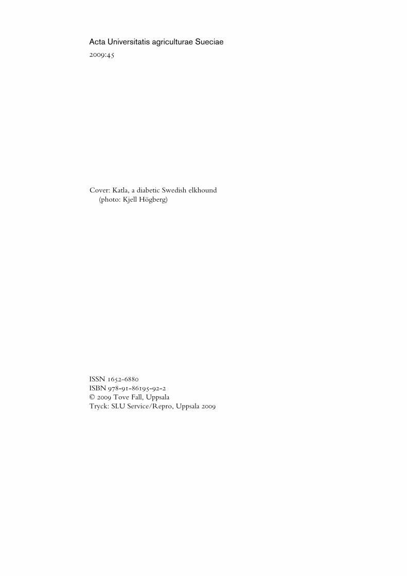
List of Publications
This thesis is based on the work reported in the following papers, which are referred to by Roman numerals in the text:
I Fall T, Hamlin HH, Hedhammar A, Kämpe O, Egenvall A. Diabetes mellitus in a population of 180,000 insured dogs: incidence, survival, and breed distribution. J Vet Intern Med. 2007 Nov-Dec; 21(6):1209-16
II Fall T, Holm B, Karlsson A, Ahlgren KM, Kämpe O, von Euler H. Glucagon stimulation test for estimating endogenous insulin secretion in dogs. Vet Rec. 2008 Aug 30;163(9):266-70.
III Fall T, Johansson Kreuger S, Juberget A, Bergström A, Hedhammar Å. Gestational diabetes mellitus in 13 dogs. J Vet Intern Med. 2008 Nov-Dec; 22(6):1296-300
IV Fall T, Hedhammar Å, Fall N, Hamlin HH, Wallberg A, Andersson G, Kämpe, O. Spontaneous diabetes mellitus in Elkhounds – a suitable animal model for gestational diabetes mellitus (manuscript)
Papers I-III are reproduced with the permission of the publishers.

8
Abbreviations
dla dog leucocyte antigen gad65 glutamic acid decarboxylase-65 gh growth hormone glut glucose transporter hla human leucocyte antigen ica islet cell antibodies iddm insulin-dependent diabetes mellitus igf insulin-like growth factor lada latent autoimmune diabetes mellitus of the adult mody maturity-onset diabetes mellitus of the young mrna messenger ribonucleic acid niddm non-insulin-dependent diabetes mellitus scdmp Swedish canine diabetes mellitus project who World Health Organisation

9
1 Introduction
Diabetes mellitus consists of a group of metabolic diseases that are characterised by a chronic excess of blood glucose, resulting from defects in insulin secretion, insulin action, or both (ADA, 2003). Diabetes mellitus constitutes a major global public health problem in humans, and is also of concern in dogs. Although diabetes mellitus is considered to be a common endocrine disorder in dogs, its underlying disease mechanisms are poorly understood. The overall aim of this doctoral research further characterise the different subtypes of canine diabetes mellitus.
Common concepts in the field of diabetes mellitus are summarised in chapter 2. Sections 2.1-2.3 and 2.5 are based on studies in humans and laboratory animals, while section 2.4 cover dog-specific features. The following chapters (3-7) describe and discuss the four studies included in the doctoral work. Furthermore, a new classification system for canine diabetes mellitus is proposed based on earlier and newly gained knowledge.

10

11
2 Diabetes mellitus
2.1 Physiology
2.1.1 Glucose uptake and transport
Glucose, blood sugar, represents a major source of energy for mammalian cells. Glucose is obtained directly from the diet, and by synthesis from substrates in organs such as the liver (Wood & Trayhurn, 2003). Brain cells and red blood cells are entirely dependent on glucose as their energy source. Glucose is stored in the liver and muscle as glycogen. Because cell membranes are impermeable to polar molecules, the transport of glucose into cells requires specific transport proteins. Two classes of glucose transport proteins have been described in mammalian cells: the sodium-dependent glucose co-transporter and the facilitative glucose transporters (glut)(Mueckler, 1994).
The sodium-dependent glucose co-transporter is expressed in the kidney and small intestine (Wright, 2001). Facilitative glucose carriers are expressed by most, if not all cells. They have different isoforms, which have distinct tissue distributions. The isoform glut-4 transports glucose into muscle and adipose tissue and is activated by the hormone insulin, which is secreted by the endocrine parts of the pancreas (Zhao & Keating, 2007).
2.1.2 Insulin synthesis and secretion
Insulin is rapidly secreted in response to elevations of blood glucose. Insulin is secreted endocrine cell clusters in the pancreas, which are named the islets of Langerhans after their discoverer. These cell clusters contain different types of cells, which produces hormones such as insulin, glucagon, somatostatin, ghrelin, and pancreatic polypeptide (Huang et al., 2009; Wierup et al., 2002). Insulin is produced and secreted solely by beta cells in the islets of Langerhans (Rutter, 2001).

12
Glucose sensing and insulin secretion
Glucose is constantly transported across the beta cell membrane through glut-2/glut-1, which keeps extracellular and intracellular glucose concentrations similar (Richardson et al., 2007; Rutter, 2001). Glucose must be metabolised through glycolysis, to exert any significant effect on the beta cell. The intracellular enzyme glucokinase, which is responsible for the first step of glycolysis, has some specific characteristics which make it the rate-limiting step of the beta cell ‘‘glucose sensor system’’ (Matschinsky et al., 1986). Glucose metabolites are further processed within the beta cell mitochondria which results in an increased atp/adp ratio and ultimately membrane depolarisation by impeding potassium efflux. This depolarisation will lead to an influx of extracellular calcium, which will trigger the exocytosis of insulin (Ammala et al., 1993). (See Figure 1).
The insulin secretory response is greater after oral than after intravenous administration of glucose, as an effect of gut hormones and other mechanisms that sensitise the beta cell to glucose metabolites (Cavaghan & Polonsky, 2005).
Figure 1. Simplified schematic representation of signalling pathways in pancreatic beta cells. Drawn by the author.

13
Biosynthesis
The synthesis of insulin includes transcription of the preproinsulin gene (INS) to mrna, translation of mrna into preproinsulin, and processing of preproinsulin via proinsulin into mature insulin, which is stored in two pools of granulæ, one of them docked to the cell membrane and the other one waiting in the cytoplasm (Daniel et al., 1999). During the processing of proinsulin, a peptide called C-peptide is cleaved off to form insulin. Mature insulin is composed of two peptide chains, the A and B chains. The amino acid sequence of proinsulin differs among species. The amino acid sequence of proinsulin in the dog is shown in Figure 2, with indications of differences with the human sequence.
The metabolites of glucose stimulate biosynthesis of insulin. About one hour after an increase in serum glucose, the production of insulin will reach a maximum, with a concentration as much as 10- to 20-fold higher than basal levels (Guest et al., 1989).
Figure 2. Amino acid sequence of dog proinsulin. The amino acids marked in red differ between dogs and humans. Straight lines indicate cleavage points. Drawn by the author on the basis of information in (Kwok et al., 1983; Bell et al., 1979).

14
2.1.3 Insulin effect
Insulin is an anabolic hormone with major systemic effects. It increases glucose uptake in muscle and fat, and inhibits hepatic glucose production, thus serving as the primary regulator of the blood glucose concentration. Insulin also stimulates cell growth and differentiation, and promotes the storage of substrates in fat, liver and muscle by stimulating lipogenesis, glycogen and protein synthesis, and inhibiting lipolysis, glycogenolysis and protein breakdown (Saltiel & Kahn, 2001). See Figure 3 for a schematic representation of the effect of insulin.
Figure 3. Schematic representation of the metabolic effects of the activated insulin receptor inmuscle and fat cells. Drawn by the author from information in Saltiel & Kahn (2001). FFA;free fatty acids.

15
Insulin acts through the insulin receptor, which is a so called tyrosine kinase receptor (reviewed in Patti & Kahn, 1998). When insulin binds to the receptor, the receptor itself becomes phosphorylated and activates a number of signalling cascades within the cell. Vesicles containing glut-4 are transported from intracellular storage to the cell membrane, allowing glucose influx. These pathways also influence protein synthesis, enzyme activation and inactivation, and gene expression, resulting in the regulation of glucose, lipid and protein metabolism (Kahn & Saltiel, 2005).
2.2 Summary of important disease mechanisms
When an insufficient amount of insulin is secreted and/or the tissue sensitivity to insulin is low, diabetes mellitus will develop. The symptoms of diabetes mellitus are mainly attributable either to the abundance of glucose in the blood or to the shortage of nutrients within cells. The classical symptoms of diabetes mellitus include increased thirst, increased production of urine, increased appetite and weight loss. The name of the syndrome relates to the glycosuria seen in diabetic patients."Diabetes" is derived from a Greek word meaning 'siphon' or ‘flow’. "Mellitus" is a Latin adjective meaning 'honey-like'. Diabetes mellitus is often referred to simply as "diabetes", and in this thesis, both expressions will be used.
At the beginning of the last century, insulin deficiency due to beta cell destruction was considered to be the main cause leading to diabetes mellitus in humans. Mechanisms such as autoimmune events and chemical stress due to toxins or pancreatitis may cause destruction of islets. The ability of beta cells to proliferate is limited, especially in adults. Some degree of regeneration can occur after physiological stimulation such as pregnancy (Butler et al., 2007) or injury (Dor et al., 2004) but the “new” beta cells are thought to derive from already existing insulin-expressing cells in the pancreatic duct epithelium. Other causes of defective insulin secretion may include malfunctioning glucose metabolism in the beta cell, due, for example, to defects in beta cell glucokinase, as is the case in a rare form of diabetes with dominant inheritance, namely mature onset diabetes mellitus of the young type 2 (mody2) (Froguel et al., 1993).
After the development of a radioimmunoassay for measuring insulin by Yalow and Berson (1960), it became obvious that the majority of human diabetic patients had residual insulin secretion at diagnosis. Their diabetes mellitus seemed to be attributable to reduced peripheral tissue sensitivity to insulin.1 Many factors are now known to reduce the tissue sensitivity to 1 Mainly patients with type 2 diabetes mellitus
16
insulin, including visceral adiposity, inflammation, infections, glucocorticoids, pregnancy, and genetic defects in the insulin receptor (Reaven, 1998). In a person with insulin resistance (decreased insulin sensitivity), beta cells will normally meet the augmented demands by increasing their production of insulin. If the beta cells cannot meet these high demands, hyperglycaemia will develop. This phenomenon is called relative insulin deficiency.
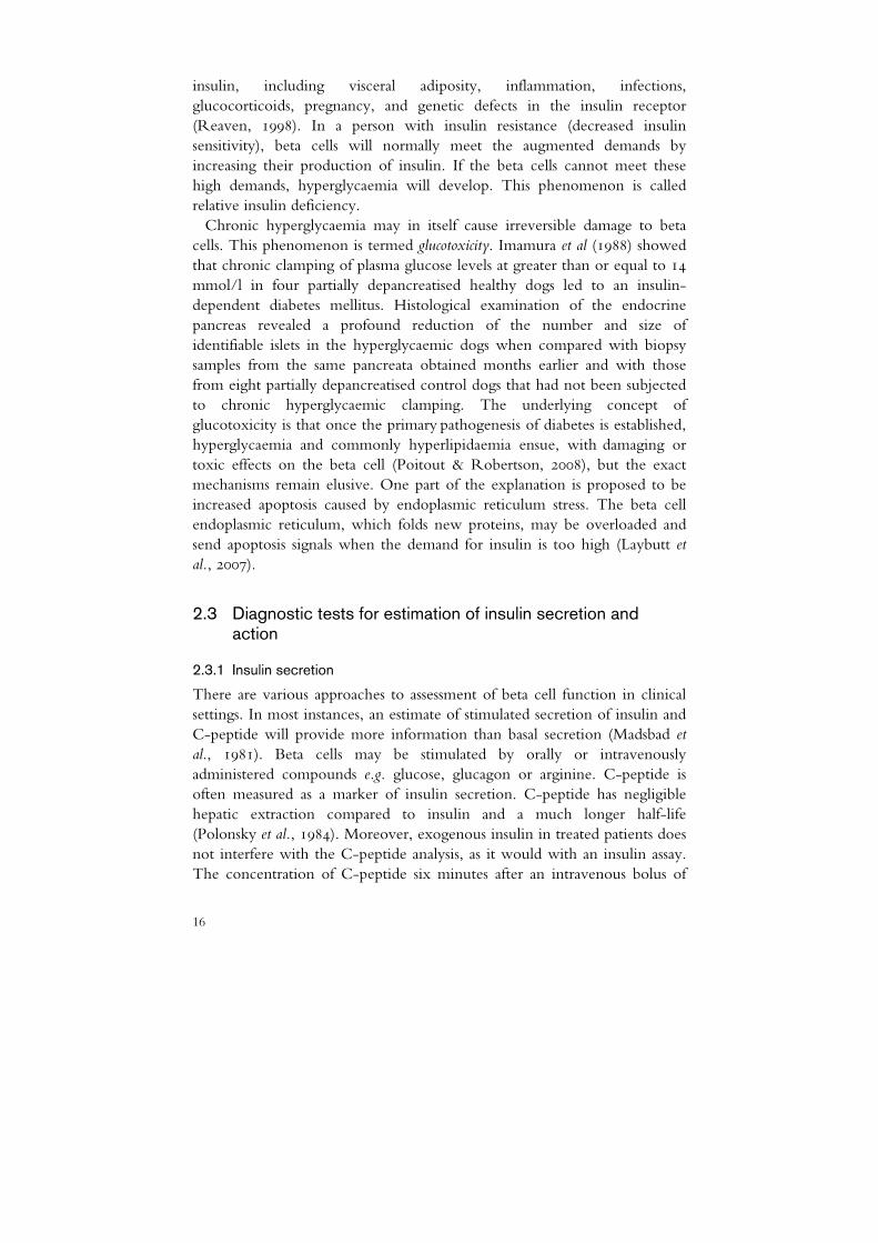
Chronic hyperglycaemia may in itself cause irreversible damage to beta cells. This phenomenon is termed glucotoxicity. Imamura et al (1988) showed that chronic clamping of plasma glucose levels at greater than or equal to 14 mmol/l in four partially depancreatised healthy dogs led to an insulin-dependent diabetes mellitus. Histological examination of the endocrine pancreas revealed a profound reduction of the number and size of identifiable islets in the hyperglycaemic dogs when compared with biopsy samples from the same pancreata obtained months earlier and with those from eight partially depancreatised control dogs that had not been subjected to chronic hyperglycaemic clamping. The underlying concept of glucotoxicity is that once the primary pathogenesis of diabetes is established, hyperglycaemia and commonly hyperlipidaemia ensue, with damaging or toxic effects on the beta cell (Poitout & Robertson, 2008), but the exact mechanisms remain elusive. One part of the explanation is proposed to be increased apoptosis caused by endoplasmic reticulum stress. The beta cell endoplasmic reticulum, which folds new proteins, may be overloaded and send apoptosis signals when the demand for insulin is too high (Laybutt et al., 2007).
2.3 Diagnostic tests for estimation of insulin secretion and action
2.3.1 Insulin secretion
There are various approaches to assessment of beta cell function in clinical settings. In most instances, an estimate of stimulated secretion of insulin and C-peptide will provide more information than basal secretion (Madsbad et al., 1981). Beta cells may be stimulated by orally or intravenously administered compounds e.g. glucose, glucagon or arginine. C-peptide is often measured as a marker of insulin secretion. C-peptide has negligible hepatic extraction compared to insulin and a much longer half-life (Polonsky et al., 1984). Moreover, exogenous insulin in treated patients does not interfere with the C-peptide analysis, as it would with an insulin assay. The concentration of C-peptide six minutes after an intravenous bolus of

17
glucagon is often used to assess residual beta cell function in patients with diabetes (Scheen et al., 1996; Faber & Binder, 1977).
2.3.2 Insulin sensitivity
Numerous different techniques for estimating the effect of insulin in peripheral tissue have been reviewed by Monzillo and Hamdy (2003). One of the two gold standard methods approved by the American Diabetes Association (1998) is the hyperinsulinaemic euglycaemic clamp, in which the patient is exposed to a predetermined amount of insulin while the plasma glucose is maintained within the euglycaemic range by infusion of glucose. The amounts of insulin and glucose administered are used to calculate the insulin sensitivity. The other gold standard method is the “frequently sampled intravenous glucose tolerance test”, where glucose is administered and samples for glucose and insulin are analysed at numerous time points. The test results from this method are more variable than those from the hyperinsulinaemic euglycaemic clamp (Steil et al., 1994). Both methods, however, are labour-intensive.
2.4 Canine diabetes mellitus
2.4.1 Historical perspective
Spontaneous diabetes mellitus in dogs was described as early as in 1861 in two case reports (Leblanc, 1861; Thiernesse, 1861). One of the cases was reported in a 15-year old petit griffon, and in the other one in a 6-year old sighthound. At that time, diabetes mellitus was diagnosed by the presence of glycosuria, but the analysis was not readily available. In the sighthound case, the diagnostic suspicion was first investigated by the attending veterinarian, by tasting the urine and finding it sweet (Leblanc, 1861). Fröhner (1892) reported on the first case series of five diabetic dogs and estimated the frequency of diabetes mellitus to be 1:10,000 dogs in his veterinary practice.
Dogs were used extensively as experimental animals during the “insulin discovery era” and were crucial both for the discovery of “pancreatic diabetes mellitus” in 1893 by Minkowski and Mering and in the discovery and isolation of insulin in 1921 by Banting and Best (Minkowski, 1929 reprinted in 1989).
Reports on larger case series of spontaneous diabetes in dogs did not appear until the 1960s and originated from the United Kingdom and Sweden (Krook et al., 1960; Wilkinson, 1960). Ricketts et al (1953) found that diabetes mellitus occurs mainly in older dogs. He also found that the ratio of affected female to male dogs was 3 to 1. This finding was confirmed

18
in a number of studies, but with somewhat different ratios (Foster, 1975). Campbell (1958) and Wilkinson (1960) reported that many of the female cases developed shortly after œstrus. Wilkinson also wrote that ovariohysterectomy may seem a drastic therapeutic measure for diabetic female dogs, but that he would nevertheless recommend it.
Few authors committed themselves to the issue of breed predominance in diabetes prior to the doctoral research of Wilkinson (1960). He identified five breeds besides mongrels that he considered to have a higher incidence than the general population, namely the dachshund, spaniel, poodle, fox terrier and cairn terrier. Krook (1960), reported that the rottweiler, dachshund, spaniel, Swedish hound (now called Hamilton hound) and mongrel were overrepresented in his Swedish pathology material.
A milestone in canine diabetology was the discovery of progesterone-induced mammary growth hormone (gh) and its relationship to diabetes mellitus (Selman et al., 1994b; Eigenmann et al., 1983). During the last decade, the interest for the ætiology of canine diabetes mellitus has increased with a number of interesting studies being published, mainly focusing on autoantibodies, dla association, and candidate genes (Davison et al., 2008; Short et al., 2007; Kennedy et al., 2006).
2.4.2 Clinical manifestations
Dogs with diabetes mellitus show all the classical symptoms seen in humans. The major complaint at first consultation is usually an increased thirst and increased urination. The owner might also have noticed weight loss, in spite of a good appetite. Other symptoms may be a dull hair-coat, muscle wasting, tiredness, loss of vision, and infections (Foster, 1975). If the dog is not correctly treated for its disease, it may pass into a ketoacidotic state, which is a severe condition that requires intensive care.
Most dogs are older than five years at onset of diabetes mellitus (Davison et al., 2005; Guptill et al., 2003). Several studies have shown that female dogs have an increased risk for diabetes (Guptill et al., 2003; Marmor et al., 1982; Foster, 1975; Krook et al., 1960), but in one recent large British study this association was not confirmed (Davison et al., 2005).
2.4.3 Diagnosis
The diagnosis is based on measurement of glucose in the blood and urine. Healthy dogs do not have detectable amounts of glucose in the urine and have fasting blood glucose concentration between 3.5 and 6.0 mmol/L.2 In 2Reference range, Clinical Pathology Laboratory, University Animal Hospital at the Swedish
University of Agricultural Sciences

19
general veterinary practice, tests for insulin secretion are not performed. Dogs are sometimes tested for concurrent and potentially diabetogenic disorders, such as hyperadrenocorticism or pancreatitis.
2.4.4 Treatment
Dogs with diabetes mellitus are most often dependent on insulin for survival (Ling et al., 1977). Dog owners are instructed how to give subcutaneous injections of insulin and how to treat a potential hypoglycaemia. One year survival is reported to be 64% among dogs that survive stabilisation with insulin (Doxey et al., 1985). Intact female dogs are usually spayed shortly after diagnosis, because of the insulin-antagonistic features of the sex hormone progesterone (Eigenmann et al., 1983).
2.4.5 Occurrence
Diabetes mellitus is considered to be a common endocrinopathy in dogs. The incidence is unknown, since all published epidemiological studies have been cross-sectional or hospital-based. The reported prevalence has varied from 0.3 % to 1.3 % (Davison et al., 2005; Fracassi et al., 2004; Guptill et al., 2003). Some studies have shown a winter peak in the onset of canine diabetes (Davison et al., 2005; Atkins & MacDonald, 1987) whereas in other studies no seasonal predisposition has been found (Guptill et al., 2003; Marmor et al., 1982).
Certain breeds have been shown to have either an increased or a decreased risk of developing the diabetes mellitus, implying the existence of important genetic factors in the aetiology. Breeds that are consistently reported to have a high frequency of diabetes are the samoyed, cairn terrier and Australian terrier, and those that are consistently reported to have a low frequency are the golden retriever, boxer and the German shepherd (Davison et al., 2005; Fracassi et al., 2004; Guptill et al., 2003; Hess et al., 2000a; Doxey et al., 1985; Marmor et al., 1982; Krook et al., 1960).
2.4.6 Assessment of beta cell function
Previous studies of insulin secretion in diabetic dogs have yielded contradictory results, probably because of the different populations tested and the different diagnostic criteria for diabetes mellitus applied. Mattheuuws et al (1984) studied 71 diabetic dogs and found that 32 of them had no insulin secretion, 15 had high basal insulin but no response to an intravenous glucose load and 24 showed a significant increase in insulin after an intravenous glucose load. The inclusion criterion for the dogs in that study was a blood glucose concentration of >6.7 mmol/L. Many of the dogs

20
in the study were asymptomatic. Montgomery et al (1996) used an inclusion criterion of a fasting blood glucose >13.9 mmol/L combined with clinical signs of diabetes mellitus and reported that 35/42 diabetic dogs had low or absent C-peptide concentrations even after intravenous glucagon stimulation, indicating an absolute insulin deficiency. However, 7 of the 42 dogs had a basal insulin secretion higher than in healthy dogs, but with a lower increment after stimulation, which could correspond to insulin resistance and beta cell exhaustion. Eigenmann et al (1983) tested 21 dogs with either acromegaly or diabetes mellitus secondary to progestin (synthetic progesterone) treatment or diœstrus. Only ten of these animals had glucose concentrations of >10 mmol/L. All dogs in that study showed a marked hyperinsulinemia that responded little or not at all to an intravenous glucose load.
2.4.7 Histopathology
Findings in pancreatic biopsy samples from diabetic dogs have been conflicting. Two fairly large studies of diabetic dogs (Ling et al., 1977; Gepts & Toussaint, 1967) (n=33, n=30) have shown a reduced number or total absence of islets, together with degeneration, hyalinisation or vacuolisation. Pancreatic biopsy samples from dogs with diabetes mellitus secondary to progestin treatment or diœstrus also showed degeneration and vacuolisation of the beta cells (Eigenmann et al., 1983). Insulitis was not seen in any of these three studies.
On the other hand, Alejandro et al (1988) reported that 6/13 diabetic dogs in their study had lymphocytic infiltration associated with islets (insulitis) and that 5/18 displayed extensive exocrine pancreatic damage. There were no control group in any of these studies. The latter report is often referred to by textbooks and review articles, indicating that a third of diabetes mellitus cases are attributable to pancreatitis (Catchpole et al., 2005; Rand et al., 2004). However, a recent study showed that chronic pancreatitis is a common finding at autopsies of dogs in general practice. In that particular study 34% of 200 dogs with various diseases were considered as having chronic pancreatitis (Watson et al., 2007).
Histopathological investigation in 12 juvenile diabetic Greyhounds revealed extensive exocrine and endocrine atrophy of the pancreas (Brenner et al., 2009).
2.4.8 Immune markers
Autoantibodies directed against glutamic acid decarboxylase 65 (gad65) and ia-2, a tyrosine phosphatase-like protein, is common in immune-mediated
21
human diabetes (type 1 diabetes). Alejandro et al (1988) did not find any evidence of autoreactive components in the sera of 18 diabetic dogs. Hoenig and Dawe (1992) observed an unspecific anti-islet reactivity in sera from nine out of 23 newly diagnosed dogs with diabetes representing various breeds. One recent study showed that five out of 30 investigated diabetic dogs had gad65 and ia-2 antibodies. It is unclear whether these autoantibodies indicate a autoimmunity process, or are secondary to an immune response against antigens that are released as a consequence of beta cell destruction by some other disease process (Davison et al., 2008).
2.4.9 Genetic markers
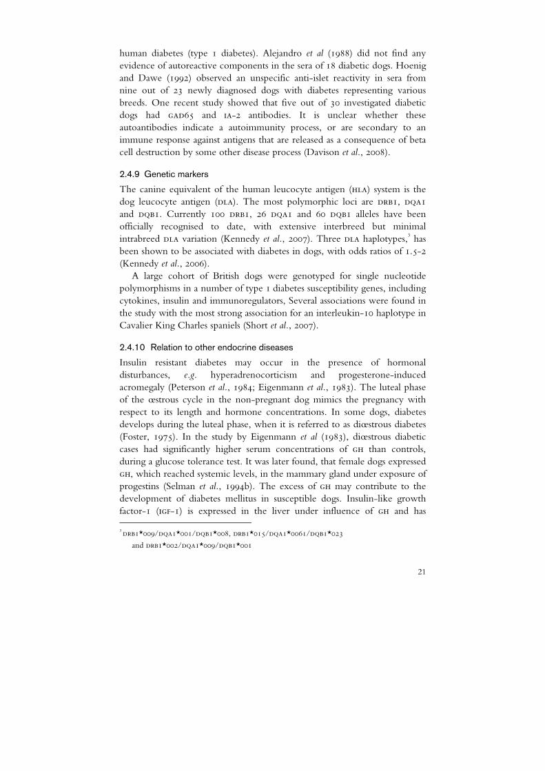
The canine equivalent of the human leucocyte antigen (hla) system is the dog leucocyte antigen (dla). The most polymorphic loci are drb1, dqa1 and dqb1. Currently 100 drb1, 26 dqa1 and 60 dqb1 alleles have been officially recognised to date, with extensive interbreed but minimal intrabreed dla variation (Kennedy et al., 2007). Three dla haplotypes,3 has been shown to be associated with diabetes in dogs, with odds ratios of 1.5-2 (Kennedy et al., 2006).
A large cohort of British dogs were genotyped for single nucleotide polymorphisms in a number of type 1 diabetes susceptibility genes, including cytokines, insulin and immunoregulators, Several associations were found in the study with the most strong association for an interleukin-10 haplotype in Cavalier King Charles spaniels (Short et al., 2007).
2.4.10 Relation to other endocrine diseases
Insulin resistant diabetes may occur in the presence of hormonal disturbances, e.g. hyperadrenocorticism and progesterone-induced acromegaly (Peterson et al., 1984; Eigenmann et al., 1983). The luteal phase of the œstrous cycle in the non-pregnant dog mimics the pregnancy with respect to its length and hormone concentrations. In some dogs, diabetes develops during the luteal phase, when it is referred to as diœstrous diabetes (Foster, 1975). In the study by Eigenmann et al (1983), diœstrous diabetic cases had significantly higher serum concentrations of gh than controls, during a glucose tolerance test. It was later found, that female dogs expressed gh, which reached systemic levels, in the mammary gland under exposure of progestins (Selman et al., 1994b). The excess of gh may contribute to the development of diabetes mellitus in susceptible dogs. Insulin-like growth factor-1 (igf-1) is expressed in the liver under influence of gh and has 3 drb1*009/dqa1*001/dqb1*008, drb1*015/dqa1*0061/dqb1*023
and drb1*002/dqa1*009/dqb1*001

22
shown to be increased in female dogs treated with progestins (Bhatti et al., 2007; Selman et al., 1994a). In dogs with diabetes due to insulin resistance, diabetes may or may not be transient after treatment of the cause of insulin resistance. Untreated insulin resistant diabetes can lead to insulin deficiency as a consequence of chronic hyperglycaemia, which in itself can produce permanent beta cell dysfunction, probably due to glucotoxicity (Imamura et al., 1988). Diœstrous diabetes is generally considered common in populations, where spaying of young female dogs is rare, but there no further investigations have been published on this subject since 1983.
In a study by Hess et al (2000b), who investigated concurrent disorders in 221 dogs with diabetes mellitus, 23% were found to have concurrent hyperadrenocorticism (Cushing’s syndrome). Peterson and co-workers performed glucose and insulin tolerance tests in 60 dogs with untreated hyperadrenocorticism. Most dogs showed hyperinsulinaemia, except for a few dogs with overt diabetes mellitus and hypoinsulinaemia (Peterson et al., 1984).
2.5 Human diabetes mellitus
The aetiology and epidemiological features of diabetes in humans are heterogeneous. The terminology and classification of human diabetes mellitus have changed over the years towards an aetiology-based system. The World Health Organisation (who) and an expert committee from the American Diabetes Association (ada) have recently each published a document with essentially the same classification system of diabetes mellitus (Table 1) (ADA, 2003; WHO, 1999). The capacity for insulin secretion and the magnitude of insulin resistance vary with the type of diabetes mellitus and also within the type, depending on the stage of the disease.

23
Table 1. Aetiological classification of diabetes mellitus in humans I. Type 1 diabetes mellitus (beta-cell destruction, usually leading to absolute insulin deficiency4)
A. Immune mediated
B. Idiopathic
II. Type 2 diabetes mellitus (may range from predominantly insulin resistance with relative insulin deficiency to a predominantly secretory defect with insulin resistance)
III. Other specific types
A. Genetic defects of beta cell function
1. Chromosome 12, HNF-1-alpha (mody3)
2. Chromosome 7, glucokinase (mody2)
3. Chromosome 20, HNF-4-alpha (mody1)
4. Mitochondrial DNA
5. Others
B. Genetic defects in insulin action
1. Type A insulin resistance
2. Leprechaunism
3. Rabson-Mendenhall syndrome
4. Lipoatrophic diabetes mellitus
5. Others
C. Diseases of the exocrine pancreas
1. Pancreatitis
2. Trauma/pancreatectomy
3. Neoplasia
4. Cystic fibrosis
5. Haemochromatosis
6. Fibrocalculous pancreatopathy
7. Others
D. Endocrinopathies
1. Acromegaly
2. Cushing’s syndrome
3. Glucagonoma
4. Phaeochromocytoma
5. Hyperthyroidism
6. Somatostatinoma
7. Aldosteronoma
8. Others
4 Patients with any form of diabetes mellitus may require insulin treatment at some stage of their disease. Such use of insulin does not, in itself, classify the patient.

24
E. Drug- or chemical-induced
1. Vacor
2. Pentamidine
3. Nicotinic acid
4. Glucocorticoids
5. Thyroid hormone
6. Diazoxide
7. Beta-adrenergic agonists
8. Thiazides
9. Dilantin
10. alpha-Interferon
11. Others
F. Infections
1. Congenital rubella
2. Cytomegalovirus
3. Others
G. Uncommon forms of immune-mediated diabetes mellitus
1. “Stiff-man” syndrome
2. Anti-insulin receptor antibodies
3. Others
H. Other genetic syndromes sometimes associated with diabetes mellitus
1. Down syndrome
2. Klinefelter’s syndrome
3. Turner’s syndrome
4. Wolfram’s syndrome
5. Friedreich’s ataxia
6. Huntington’s chorea
7. Laurence-Moon-Biedl syndrome
8. Myotonic dystrophy
9. Porphyria
10. Prader-Willi syndrome
11. Others
IV. Gestational diabetes mellitus

25
2.5.1 Type 1 diabetes mellitus
This form of diabetes was previously encompassed by the terms insulin-dependent diabetes mellitus (iddm) or juvenile-onset diabetes mellitus. It is most often observed in children, but may also occur in adults (World Health Organisation, 1999). Destruction of beta cells is the crucial event leading to type 1 diabetes with ensuing cessation of insulin secretion (Bennet & Knowler, 2005). Individuals with type 1 diabetes are dependent on insulin for survival and are prone to develop ketosis if not treated properly.
The presence of certain autoantibodies against islet cells (ica) is commonly reported in type 1 diabetes. These autoantibodies are mainly directed against glutamic acid decarboxylase 65 (gad65) and ia-2, a tyrosine phosphatase-like protein. Individuals possessing one or more of these autoantibodies can be subclassified as having autoimmune type 1 or type 1a diabetes mellitus (World Health Organisation, 1999). These autoantibodies may be detected years before the onset of clinical disease (Knip et al., 1998). This subtype, type 1a, shows a strong genetic association with the human leucocyte antigen (hla). The HLA-DR/DQ alleles with association with type 1 diabetes can be either predisposing or protective (Nejentsev et al., 2007).
There are, especially in the non-white population, patients with insulin deficiency at diagnosis and ketoacidosis without the presence of autoimmunity. These patients are classified as having idiopathic type 1 or sometimes type 1b (World Health Organisation, 1999). There is an ongoing discussion on reclassification of this group as ketosis-prone diabetes instead of type 1 diabetes, as these patients may have a transient need for insulin treatment (Balasubramanyam et al., 2008).
In Sweden, the incidence of type 1 diabetes is 3.4 cases per 10,000 years at risk in children younger than 15 years (Patterson et al., 2009). The incidence of type 1 diabetes is increasing by about 4% per year in Europe and it is estimated that the incidence will be nearly doubled by the year 2020 compared to year 2005 (Patterson et al., 2009). Studies have shown different risk estimates for type 1 diabetes in different countries, in different seasons of the year, and in rural compared to urban areas (Karvonen et al., 2000; Patterson et al., 1996; Levy-Marchal et al., 1995).
A variant of autoimmune diabetes mellitus is the latent autoimmune diabetes in adults (lada). This form of diabetes differs from type 1 diabetes in clinical characteristics by showing a more preserved beta cell function at diagnosis (Stenstrom et al., 2005). Although patients diagnosed with lada do not by definition require insulin early after diagnosis, within six years, the

26
beta cell function is severely impaired, leading to dependency on insulin medication (Littorin et al., 1999).
2.5.2 Type 2 diabetes mellitus
Type 2 diabetes was previously covered by the terms non-insulin-dependent diabetes mellitus (niddm) and adult-onset diabetes mellitus (World Health Organisation, 1999).
Type 2 diabetes is often associated with obesity and develops when chronic overnutrition occurs concomitantly with genetic susceptibility to insulin resistance as well as in the presence of a relative insulin deficiency of non-autoimmune aetiology. Insulin resistance develops as a consequence of inflammatory and hormonal factors, endoplasmic reticulum stress, and accumulation of by-products of nutritional ‘overload’ in insulin-sensing tissues. Overnutrition is thought to increase the burden of the adipocyte endoplasmic reticulum, which may lead to a disruption of the normal folding of proteins and an activation of the unfolded protein response, which is known to induce stress response pathways (Ozcan et al., 2004).
Beta cell failure triggers the transition from an obese, insulin-resistant state to full blown type 2 diabetes. The beta cell failure involves both a partial loss of beta cell mass and a deterioration of cell function. (Muoio & Newgard, 2008). In type 2 diabetes patients, residual insulin secretion is common during an extended time after diagnosis. The incidence of type 2 diabetes is estimated at 38 cases per 10,000 years at risk in people older than 30 years in the county of Uppsala, Sweden (Ringborg et al., 2008). Several gene variations (alleles) associated with an increased risk of type 2 diabetes are described, most of them common in the populations studied and conferring disorders of beta cell development and function (McCarthy & Zeggini, 2009).
2.5.3 Other specific types of diabetes mellitus
This group contains variants of diabetes that develop in association with disorders other than those defined as type 1, type 2 or gestational diabetes. The group comprises a variety of types of diabetes, in which the underlying disease or disease process can be identified in a relatively specific manner (e.g., diabetes secondary to pancreatic disease, endocrine disease, or administration of certain drugs). The extent of residual insulin secretion varies widely with the cause of the diabetes (World Health Organisation, 1999).
27
Monogenic defects of beta cell function
Several forms of diabetes are associated with monogenic defects in beta cell function, characterised by onset of mild hyperglycaemia before the age of 25 years. These are usually inherited in an autosomal dominant pattern and are usually referred to as maturity-onset diabetes mellitus of the young (mody). To date, the underlying point-mutations of nine different mody types have been identified (Nyunt et al., 2009).
Diseases of the exocrine pancreas
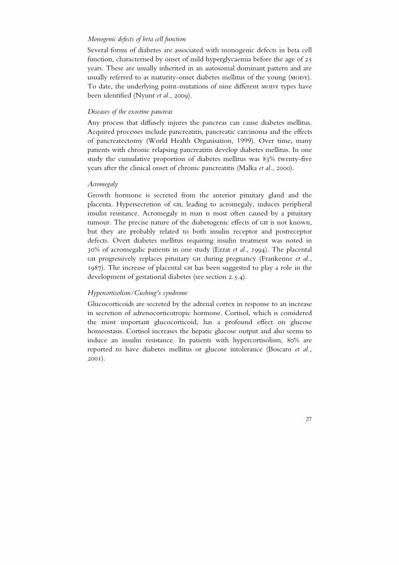
Any process that diffusely injures the pancreas can cause diabetes mellitus. Acquired processes include pancreatitis, pancreatic carcinoma and the effects of pancreatectomy (World Health Organisation, 1999). Over time, many patients with chronic relapsing pancreatitis develop diabetes mellitus. In one study the cumulative proportion of diabetes mellitus was 83% twenty-five years after the clinical onset of chronic pancreatitis (Malka et al., 2000).
Acromegaly
Growth hormone is secreted from the anterior pituitary gland and the placenta. Hypersecretion of gh, leading to acromegaly, induces peripheral insulin resistance. Acromegaly in man is most often caused by a pituitary tumour. The precise nature of the diabetogenic effects of gh is not known, but they are probably related to both insulin receptor and postreceptor defects. Overt diabetes mellitus requiring insulin treatment was noted in 30% of acromegalic patients in one study (Ezzat et al., 1994). The placental gh progressively replaces pituitary gh during pregnancy (Frankenne et al., 1987). The increase of placental gh has been suggested to play a role in the development of gestational diabetes (see section 2.5.4).
Hypercortisolism/Cushing’s syndrome
Glucocorticoids are secreted by the adrenal cortex in response to an increase in secretion of adrenocorticotropic hormone. Cortisol, which is considered the most important glucocorticoid, has a profound effect on glucose homeostasis. Cortisol increases the hepatic glucose output and also seems to induce an insulin resistance. In patients with hypercortisolism, 80% are reported to have diabetes mellitus or glucose intolerance (Boscaro et al., 2001).

28
Other types
Other types of diabetes mellitus, including drug- or chemical-induced diabetes, specific infections, and rare genetic defects of insulin action, are not further described in this thesis.
2.5.4 Gestational diabetes mellitus
Gestational diabetes is defined as a carbohydrate intolerance that results in hyperglycaemia of varying degrees of severity with onset or first recognition during pregnancy. Individuals at high risk for gestational diabetes include older women and those with a history of glucose intolerance. About 2-12% of all human pregnancies (depending on the population and diagnostic criteria) are associated with gestational diabetes and even though the disease usually resolves after parturition, the risk for type 2 diabetes is increased later in life. The relative risk for type 2 diabetes after a diabetic pregnancy is about seven times higher than after a non-diabetic pregnancy (Bellamy et al., 2009). Although human gestational diabetes is usually asymptomatic at diagnosis, the consequences may be substantial, with complications such as macrosomia, dystocia and neonatal hypoglycaemia (Hunt & Schuller, 2007; Brown & Goldfine, 2005).
The insulin resistance normally observed during pregnancy plays an important role for the growth of the fœtus (Catalano et al., 1998). Several hormones such as progesterone, cortisol, and placental gh have been proposed as responsible for the insulin resistance seen in human pregnancies, but their individual roles and possible interactions are unclear (Kirwan et al., 2002; Ryan & Enns, 1988). This insulin resistance plays an important part in the development of gestational diabetes.
2.6 The dog as a genetic model for complex diseases
The domestic dog has emerged as an ideal model for gene mapping of human complex diseases, as it has a spectrum of diseases similar to that in man, and also an advantageous population structure (Karlsson & Lindblad-Toh, 2008). When dog breeds were created less than 200 years ago, the genetic variation within each breed became limited. The process had unintended consequences for the health of pure-bred dogs, with high rates of specific diseases in certain breeds, reflecting enrichment of risk alleles owing to random fixation at genetic bottlenecks and hitch-hiking of mutations near desirable traits.
Dogs often share their environment with their owner, which is not the case for the most common model organism, the mouse. If something in the

29
environment triggers disease in humans, it is possible that it will also affect the dog. In order to use dogs for finding gene variations associated with diabetes mellitus, it is important to clearly define different types of the syndrome in the dog. Thus, the research presented here should provide essential clinical information allowing successful identification of genetic risk factors for different subtypes of canine diabetes.

30

31
3 Aims
The overall aim of this research was to further characterise different subtypes of canine diabetes mellitus. The specific aims were:
to describe relevant epidemiological features of diabetes mellitus in the Swedish dog population with respect to breed, age and gender distribution;
to evaluate the glucagon stimulation test for the assessment of beta cell function in diabetic dogs and potential differentiation of diabetes mellitus subtypes in dogs;
to investigate whether subtypes of diabetes mellitus differs between breeds;
to describe one specific subtype of canine diabetes mellitus concerning its clinical characteristics and prognosis.

32

33
4 Material and methods
Overall issues regarding the dogs and research methods used in studies (papers) I-IV are outlined in this section. For detailed descriptions, the reader is referred to each individual paper.
4.1 The Agria Insurance database
The Swedish dog population is unique in that a large proportion of the animals are covered by an insurance plan and that most dogs are not neutered. The Agria insurance company5 has been shown to cover approximately 30-40 % of the entire Swedish dog population (Egenvall et al., 1999). The insured dog population in Sweden reflects the general Swedish dog population, with regard to gender distribution and breed structure. However, the mean age of the insured population (5.2 years) is somewhat lower than the general Swedish population (5.7 years) but the median ages are equal (5 years). This is explained by the different age limits that have been applied for the different insurance forms. In general, during the study period of paper I, life insurance ended at 10 years of age and the veterinary care insurance at 12 years of age. Mixed breeds and some breeds such as elkhounds and border collies are underrepresented in the insured population, with about 50 % of dogs insured for veterinary care compared to about 68 % in the general population (Egenvall et al., 1999). One of the advantages of using an insurance database like Agria for research is that it is possible to use for longitudinal cohort studies, starting with a large number of healthy dogs.
5 Agria Insurance, PO 70306, SE-107 23, Stockholm, Sweden

34
4.2 The Swedish Kennel Club
The Swedish Kennel Club has kept fully computerised records of pedigrees and ownership of all pure-bred dogs in Sweden since the year 1976. In study IV, their register was accessed in order to find Swedish elkhounds of a susceptible age for diabetes mellitus. These dogs served as healthy control dogs. The Swedish Kennel Club registry was also accessed for pedigree information used for the study presented in study IV.
4.3 The Swedish canine diabetes mellitus project
A large number of participating diabetic and healthy dogs were required for the work of this doctoral research. The Swedish canine diabetes mellitus project (scdmp) was started as a basis for this work in December 2005. A broad network of veterinarians, veterinary nurses, breed clubs and dog owners was established. The network was recruited through articles, television interviews, lectures and newsletters. A special web-site (www.hunddiabetes.se) was created and letters directed to owners and veterinarians were sent out on a regular basis. Three major dog insurance companies (Agria Djurförsäkringar, Svealand and Folksam) made it possible, for the project, to contact owners of affected dogs.
The samples and clinical information were collected mainly from Sweden and in a few instances from Norway. All treatments and sampling of dogs were approved by the owners and conformed with the decision of the Uppsala Animal Ethical Committee (no. C267/5) and the Swedish Animal Welfare Agency (no. 2005-2038).
In study II, five veterinarians with a special interest in diabetes from different parts of the country were invited to participate in the study.
Study III required medical records for dogs with gestational diabetes, and clinicians from 250 clinics were asked to share such records with the scdmp. The medical records for all diabetic dogs at three large animal hospitals were also reviewed with the aim of finding all dogs with onset of diabetes at pregnancy.
Study IV was based on the medical records from 63 diabetic and 26 healthy Swedish and Norwegian elkhounds.
In total, 250 serum and EDTA samples from dogs affected by diabetes mellitus and 300 such samples from healthy dogs were collected within the project. The diagnosis of diabetes in each dog was based on clinical signs, including polyuria and polydipsia and fasting hyperglycaemia (>7 mmol/L). Blood samples were collected from each dog and were stored in -80°C until analysed. A questionnaire was completed by the owner of every dog that

35
participated in the project and the veterinarian in charge supplied the medical records. The survey included questions on the date of onset of symptoms, concurrent disease, insulin regimen, castration date and feeding regimen.
To evaluate the potential side effects of glucagon, an adverse effect protocol was attached to the questionnaires to the dog owner and attending veterinarian. The reported side effects are described in paper II.
4.4 Clinical pathology
All analyses were performed at the University Animal Hospital Laboratory, Swedish University of Agricultural Sciences. The analyses performed included:
Glucose (papers II and IV) Fructosamine (paper II) Insulin (paper II) C-peptide (papers II and IV) Progesterone (paper IV)
The methods are described in detail in each paper.
4.5 Methodological considerations concerning study II
4.5.1 Dose of glucagon
As mentioned above, the glucagon stimulation test was evaluated for use in dogs in study II. In the study by Montgomery et al (1996), it was shown that an intravenous bolus dose of 1 mg glucagon was sufficient to obtain a significant insulin release in dogs. Contact with other researchers using the test revealed, however, that in some dogs of miniature breeds this dose had adverse gastrointestinal effects. In a study of patients with type 2 diabetes and healthy controls, the C-peptide response to bolus doses of 0.5 mg and 1 mg glucagon were equivalent (Ahren et al., 1987). It was decided to give dogs <10 kg bodyweight a bolus dose of 0.5 mg and larger dogs a bolus dose of 1 mg. To determine whether the different doses gave different C-peptide responses, the Wilcoxon rank sum test was performed to assess the difference between the six control dogs <10 kg and the 14 control dogs >10 kg. The results and individual C-peptide concentrations are shown in Figure 4. Since the population was small, the power for detecting differences was, however, low. The proportions of small dogs in the diabetes and control

36
dogs were similar in study II (9/31 and 6/20 dogs respectively), which would reduce the risk of selection bias.
In humans, C-peptide concentrations peaks 6 minutes after intravenous glucagon administration, but as the 10-minute C-peptide had been found to be higher than the 5-minute sample in dogs in the study by Montgomery et al (1996), it was decided to not sample dogs after 5 minutes.
Figure 4. C-peptide concentrations in a group of six healthy dogs weighing <10 kg receiving 0.5 mg glucagon and in 14 healthy dogs >10 kg receiving 1 mg of glucagon. Samples are taken before and 10 and 20 minutes after intravenous administration of glucagon.
4.5.2 Sample management
Glucagon stimulation tests for the study II were performed at five centres in Sweden from January to October 2006, generating three serum tubes from each dog. After centrifugation, each serum sample was divided in two aliquots. One aliquot from each sample was sent in ambient temperature to the laboratory and analysed upon arrival, and the other one stored at -20 ºC awaiting transport on dry ice to the laboratory in October 2006. The study was based on the samples stored at -20 ºC. The analytical results from the aliquots sent at ambient temperature were used by the attending veterinarians for clinical management of the diabetic dogs. The concentrations of C-peptide were significantly lower in serum samples
0.5 m
g1 m
g
0.5 m
g1 m
g
0.5 m
g1 m
g0.0
0.5
1.0
1.5p=0.16
p=0.59
p=0.68
0 min 10 min 20 min
nmol
/L

37
stored at -20 ºC than in those that had been sent to the laboratory at an ambient temperature and analysed upon arrival. The mean difference was 0·05 nmol/l, with a range from 0 to 0·16 mmol/l. C-peptide in human samples is known to be sensitive to storage at -20 ºC if stored without the protease inhibitor aprotinin (Myrick et al., 1989). Hence, the C-peptide results in paper II should not serve as a reference range for C-peptide measured in fresh serum. Table 2 shows a small-scale stability study for samples stored in room temperature for 48 hours with and without aprotinin. From the experience of this study, it can be concluded that C-peptide stability must be taken in consideration, when designing of similar studies. One opportunity may be to use samples sent at ambient temperature with aprotinin added to the sample. The sera for the C-peptide analysis in paper IV were stored at -80 ºC, after arrival to the laboratory.
Table 2. Concentrations of C-peptide (nmol/L) in three dog serum samples after storage with and without the protease inhibitor aprotinin at room temperature for 48 h
0h 24h 48h
sample 1
stored without aprotinin 0.13 0.13 0.10
stored with aprotinin 0.13 0.13 0.13
sample 2
stored without aprotinin 0.17 0.13 0.20
stored with aprotinin 0.17 0.17 0.17
sample 3
stored without aprotinin 0.27 0.27 0.20
stored with aprotinin 0.33 0.30 0.27

38

39
5 Results and discussion
A detailed discussion of each specific study is given in the respective papers. In the general discussion below, the principal features of the investigations are summarised and a new classification system for diabetes mellitus in dogs is proposed.
5.1 Epidemiology
5.1.1 Frequency of diabetes mellitus in Swedish insured dogs
There are different ways of expressing disease frequency, but the two of the most common are prevalence and incidence rates. Prevalence relates to the number of affected animals existing at a specific point in time. The incidence rate refers to the number of new cases per animal-time unit at risk. Prevalence is dependent on the duration of disease, and on the length of time for which an animal survives with the disease, and is therefore generally not appropriate for use in research on risk factors for disease.
The study population in study I comprised more than 180,000 dogs (51% females), aged 5-12 years, of 294 breeds, contributing a total of 670,000 years at risk. Claims for diabetes mellitus had been submitted for 860 of the dogs. Incidence rates for all dogs and by gender are shown in Table 3.
Table 3. Incidence rates of diabetes mellitus in a population of insured dogs
n, number of new cases
Animals N Years at risk Incidence rate, cases per 10,000 years at risk (95% confidence interval)
All 860 652,898 13 (12-14)
Male 242 318,406 8 (7-9)
Female 618 334,491 19 (17-20)

40
The incidence varied by breed, with the Australian terrier, samoyed, Swedish Lapphund, Swedish elkhound and border collie having the highest rates (45-183 cases/10,000 years at risk) and the papillon and boxer having the lowest (0 cases/10,000 years at risk). For all breeds with more than 40 cases, a cumulative percentage of affected dogs was calculated and these figures are shown in Table 4 at 8, 10 and 12 years.
The risk of misclassification was deemed small in this study. Most veterinarians can both readily recognise the symptoms and diagnose the dogs. Furthermore, diabetes mellitus lead to death if left untreated. Hence, most insured diabetic dogs are assumed to eventually be diagnosed by a clinician.
The finding that certain breeds are at high or low risk for diabetes mellitus confirms the results of previous studies (Davison et al., 2005; Guptill et al., 2003; Hess et al., 2000a) with the exception of a number of Scandinavian breeds, such as the Swedish elkhound, Swedish Lapphund, the drever and the Hamilton hound, which we hound to be at high risk in our study. This is probably because these breeds are uncommon outside Scandinavia.
It is possible to extrapolate most of the results from paper I to the general Swedish dog population in the age group studied. The results are generally not valid for extrapolation to non-Scandinavian countries mainly due to the different breed distribution and elective neuter routines.
Table 4. Cumulative percentages of dogs that had developed diabetes at 8, 10 and 12 years of age. Data based on 860 cases of diabetes in a population of 180,000 insured dogs.
5.1.2 Age at onset
The mean age at the first insurance claim for diabetes mellitus was found to be 8.6 years in study I. For gestational diabetes (study III), the median age at diagnosis was 5.9 years. In study IV, the mean age at diagnosis of non-
Percentage (95% CI)
8 years 10 years 12 years
All 0.3 (0.2-0.3) 0.7 (0.7-0.8) 1.2 (1.1-1.2)
Females 0.4 (0.3-0.4) 1.0 (0.9-1.1) 1.6 (1.5-1.7)
Males 0.1 (0.1-0.2) 0.4 (0.3-0.5) 0.6 (0.5-0.7)
Border collie 0.4 (0.1-0.6) 2.2 (1.5-2.8) 3.4 (2.3-4.4)
Sw. Elkhound 1.3 (0.8-1.8) 2.5 (1.7-3.3) 3.6 (2.3-4.9)
Samoyed 1.9 (0.9-2.8) 5.3 (3.4-7.1) 9.0 (5.9-12.0)
Labrador 0.2 (0.1-0.3) 0.6 (0.4-0.9) 1.3 (0.9-1.7)
Drever 0.7 (0.4-1.0) 2.4 (1.7-3.1) 3.0 (2.2-3.9)
CI, confidence interval

41
pregnant dogs was 8.0 years. In study I, the mean age at the first insurance claim varied by breed. The Swedish elkhound had the youngest age at first claim (7.8 years) and the cairn terrier and west highland white terrier had the oldest (9.3 years). There is a time lag from the first symptoms until diagnosis, the mean of which was estimated in study IV to be 21 days. However, some owners do not consult a veterinarian until months later in the disease process.
5.1.3 Gender
Of the 860 diabetes cases in study I, 618 (72%) were of female gender and 242 dogs were males (28%). The proportion of female cases varied significantly by breed, with Swedish elkhounds, Norwegian elkhounds and border collies having the highest proportion (98-100%) and the rottweiler and Labrador retriever the lowest (33-51%, but with CIs overlapping 0.5). Within the scdmp, however, a small breed, the Polish Lowland Sheepdog, seemed to have a male predilection. Of 14 confirmed cases, only two were females (unpublished data). The finding of a high proportion of female elkhounds was confirmed in study IV, which is further discussed in section 4.8.
In populations where juvenile spay of dogs is common, the increased risk of diabetes in females seems to have been eradicated (Davison et al., 2005). Hence, the increased risk of diabetes in females is probably explained mainly by diœstrous diabetes and also the more rare form gestational diabetes. If we simply assume that the risk of other types of diabetes is equal between genders, the total number of male cases subtracted from the total number of females would equal the sum of diœstrous diabetes. That calculation crudely estimates the proportion of diœstrus/gestational diabetes mellitus in the population studied in study 1, to 44% (376/860), and the proportion among female dogs 60% (376⁄618). See Figure 5.

42
Figure 5. Estimation of diabetic cases related to female gender (probable dioestrus diabetes)
versus other causes unrelated to gender. Based on 860 cases of diabetes mellitus in the Agria
database 1995-2004.
5.1.4 Survival after diagnosis
In study I, the median survival time (n=686, deaths=413) after the first diabetes mellitus claim was 57 days among all dogs and 2.0 years among dogs surviving the first day after diagnosis. The risk of dying is clearly highest early after diagnosis, probably on account of unwillingness to treat the dog, but also because of complications such as ketoacidosis. Breed was shown to influence the survival after diagnosis. The hunting dog breeds, in particular, have a high mortality at day of diagnosis. In study IV, the median survival time was much longer, but it should be noted that the recruitment of dogs for that study was biased towards dogs with longer survival and less euthanasia.
5.1.5 Other diseases as risk factors for diabetes mellitus
In study I, the hazard ratio for development of diabetes in dogs with hyperadrenocorticism was 9.3 (95% ci, 5.4-15.9) compared to dogs without hyperadrenocorticism. Pancreatitis, pancreatic insufficiency, adrenocortical insufficiency and hypothyroidism were evaluated as risk factors for diabetes, but were not found to contribute to the models, neither increasing nor decreasing the risk of diabetes. Those diseases (except for hypothyroidism) may have been under-diagnosed in the population, and should not be excluded as potential risk factors for diabetes mellitus. Further studies are therefore required.
female male0
200
400
600
800
other causes
probable dioestrus diabetesN
o of
cas
es other causes
probable dioestrous diabetes N
o. o
f cas
es

43
5.2 Glucagon stimulation test
In paper II, the glucagon stimulation test was evaluated for dogs and found effective in increasing blood glucose, insulin and C-peptide concentrations in healthy dogs. In diabetic dogs, most dogs had low concentrations of C-peptide and insulin and high concentrations of glucose. A few newly diagnosed female dogs had normal to high C-peptide concentrations, indicative of residual beta cell mass. The test is of potential value for classifying the type of diabetes in newly diagnosed dogs (see section 6.10).
5.3 Diabetes mellitus in elkhounds
In the studies summarised in this thesis several important conclusions were drawn regarding diabetes in Swedish and Norwegian elkhounds:
Diabetes predominantly affects female dogs (I, IV) Although gestational diabetes is uncommonly diagnosed in dogs in general, elkhounds seem to be predisposed to the syndrome (III)
In non-pregnant elkhounds, the first symptoms of diabetes occur shortly after œstrus (IV)
Dogs may recover from diabetes after pregnancy or at the end of diœstrus (III, IV)
The probability of remission is associated with the glucose concentrations at diagnosis and the length of the interval from the first symptoms to ovariohysterectomy (IV). Hence, in dogs with dioestrus diabetes, treatment directed against the source of the insulin resistance should be initiated as soon as possible after diagnosis to avoid glucotoxic effects on beta cells
On the basis of the findings in this doctoral research, it is concluded that elkhounds must be genetically predisposed to diœstrous and gestational diabetes. The elkhounds could be a helpful link in explaining the genetic background to human gestational diabetes in the future. Other breeds with a high female incidence, such as the border collie and the beagle, may have similar clinical features. This may be important in experimental animal research, as beagle is usually the breed studied. Many studies concerning the impact of progestin upon glucose metabolism are carried out in beagles, and it would be interesting to repeat them in breeds not commonly affected by diœstrous diabetes.

44
5.4 Review of existing classification systems
A major requirement for epidemiological, genetic and clinical research on diabetes mellitus and for its clinical management is an appropriate system of classification that provides a framework within which its various forms and stages can be to identified and differentiated (American diabetes organisation, 2003).
Several attempts to classify canine diabetes have been made over the years. It has been proposed that canine diabetes is comparable to lada (Catchpole et al., 2005; Rand et al., 2004). This comparison was motivated by the fact that canine diabetes commonly affects middle-aged to older dogs and that newly diagnosed animals have higher stimulated C-peptide levels than those that have had the diagnosis for more than a year (Montgomery et al., 1996). I would like to argue against this comparison, as patients with lada do not by definition (Fourlanos et al., 2005) require insulin for the first six months, in contrast to most diabetic dogs. Moreover, the decline in the C-peptide response to glucagon during the years after diagnosis is seen in many forms of diabetes, including type 1 (Palmer, 2009).
The iddm⁄niddm classification system was used in humans until 1999 and is still often used for dogs. It is based on the question whether a diabetic patient needs insulin (iddm) or not (niddm). Since most diabetic dogs are treated with insulin from the onset of the disease, they would all be classified as iddm. Such a classification system does not help the clinician at all regarding the prognosis and treatment options. Thus, this is not a good classification system for use in dogs.
Catchpole et al. (2005) suggested a system whereby dogs are divided into two groups depending on the cause of the disease, namely insulin deficiency or insulin resistant diabetes. The system is attractive in many ways, as it addresses the aetiology of the disease. However, some problems remain. The first is that the majority of the cases are still idiopathic, and the system does not providing a good classification for such cases. Another problem is the difficulty in communication with human diabetologists, as they tend to interpret the two categories directly as type 1 and type 2 diabetes. Furthermore, since dogs seem to pass from an insulin resistant state into insulin deficiency through glucotoxicity within weeks after diagnosis, it may be confusing to place them in either class.
For the above reasons, a modified system for classifying diabetes mellitus in dogs constructed in a fashion similar to that in human medicine (see Table 1) is proposed, as discussed below.

45
5.5 A new classification system for canine diabetes mellitus
A new system for classification of diabetes mellitus in dogs is proposed and presented in Table 5. The system is based on current knowledge and is an attempt to categorise diabetes mellitus in dogs according to its cause. When more is learned about the pathogenesis of diabetes in dogs, this system will need to be reviewed and revised accordingly.
Table 5. Aetiological classification of diabetes mellitus in dogs
Classification in dogs Corresponding class in human system (Table 1)
Juvenile diabetes mellitus
A. Beta cell hypoplasia
B. Combined beta cell deficiency and pancreatic acinar atrophy
None
None
Progesterone-related
A. Gestational
B. Diœstrous
Class IV
Secondary to pancreatitis Class III:C:1
Endocrine tumours
A. Hyperadrenocorticism
B. Acromegaly
C. Glucagonoma
Class III:D:2
Class III:D:1
Class III:D:3
Iatrogenic
A. Glucocorticoids
B. Progestin
C. Secondary to insulinoma treatment
Class III:E:4
Class III:E:11
Class III:C:2/Class III:E:11
Immune-mediated diabetes mellitus6 Class IA
Idiopathic diabetes mellitus None
5.5.1 Juvenile diabetes mellitus
Beta cell hypoplasia
This form of diabetes is diagnosed before the age of six months. Biopsies of the pancreas reveal a small number of islets, without signs of insulitis or exocrine pancreatic inflammation (Minkus et al., 1997; Atkins et al., 1988). Dogs are insulinopenic at diagnosis. This form of diabetes is mainly seen in keeshonds but a few cases have also been reported in golden retrievers, poodles and Labrador retrievers (Minkus et al., 1997; Atkins et al., 1988; Kramer et al., 1980).
6The importance of autoimmunity in canine diabetes mellitus is not yet clear

46
Combined beta cell deficiency and pancreatic acinar atrophy
This form of diabetes is diagnosed before the age of six months. Aside from symptoms of diabetes, the dog may show diarrhoea and weight loss. Blood analysis reveals concurrent exocrine pancreatic insufficiency and insulinopenic diabetes mellitus. Reported histopathological findings include lesions in both the endocrine and exocrine pancreas, with acinar cell apoptosis, zymogen granule loss, cytoplasmic clearing or vacuolar changes, lobular atrophy and islet loss. In greyhounds, a lymphocytic or lymphoplasmacytic pancreatitis is also reported (Brenner et al., 2009). This form of diabetes mellitus is seen in greyhounds and German shepherds (Brenner et al., 2009; Neiger et al., 1996; Atkins et al., 1988; Ling et al., 1977). Many of the reported cases are euthanised shortly after diagnosis.
5.5.2 Progesterone-related
A. Gestational
Gestational diabetes, which is described in detail in paper III, is defined as diabetes with onset during pregnancy. It is considered uncommon in dogs (Johnson, 2008) and there are only two case reports in the literature (Norman et al., 2006). Dogs may be treated with abortion, caesarean section, or insulin. The diabetes may resolve after the end of pregnancy. This form of diabetes has been reported mainly in Nordic Spitz breeds, but also in a German shepherd and a Labrador retriever (Norman et al., 2006; paper III).
B. Diœstrous
Here the onset is during the luteal phase of the œstrous cycle in an intact female dog. Insulin/C-peptide may be high initially, and the diabetes can resolve after ovariohysterectomy (Eigenmann et al., 1983; paper IV). Within the scdmp, diœstrous diabetes mellitus has been seen in several breeds, including the elkhounds, border collie, Australian kelpie, Irish setter and beagle. Pancreatic biopsies reveal vacuolisation of islets and a decreased number of islets (Eigenmann et al., 1983). In section 5.1.3, a rough estimation of the proportion of diœstrous diabetes in the Swedish dog population is 60% of all female diabetes cases.
5.5.3 Secondary to pancreatitis
In this form of diabetes, a diagnosis of pancreatitis precedes the onset of diabetes. There have been several studies on dogs suffering from diabetes concurrently with acute or chronic pancreatitis (Hess et al., 2000b; Alejandro et al., 1988) but none in which the causal association has been established. In

47
cases of combined pancreatitis and diabetes, exocrine pancreatic insufficiency may also develop (Watson, 2003). Dogs with both chronic pancreatitis and diabetes may be challenging to treat, because of the lack of effective treatments for the former condition. Chronic pancreatitis in combination with diabetes has been reported to occur in English cocker spaniels (Watson et al., 2006). A relation between previous pancreatitis and diabetes was not confirmed in the present study I, but since the chronic pancreatitis may be strongly under-diagnosed the power of the analysis was low.
5.5.4 Endocrine tumours
A. Hyperadrenocorticism/Cushing’s disease
In this form of diabetes, a diagnosis of hyperadrenocorticism precedes the onset of diabetes. High concentrations of insulin/C-peptide may be found at early stages, and if the primary disease is correctly treated, the diabetes may resolve (Campbell & Latimer, 1984; Peterson et al., 1984). If no beta cells remain in a dog with hyperadrenocorticism, the prognosis is worse, because of the clinical challenge of treating this double endocrine disorder (Peterson et al., 1981). The relation between previous hyperadrenocorticism and diabetes was confirmed in study I.
B. Acromegaly
Pituitary-dependent acromegaly is uncommon in dogs. There are two case reports of dogs with pituitary tumours and insulin resistance, which proceeded to overt diabetes in one dog (Fracassi et al., 2007; van Keulen et al., 1996). Both dogs were euthanised.
C. Glucagonoma
Dogs with pancreatic glucagon-producing tumours may develop diabetes and superficial necrolytic dermatitis. It has been reported that dogs develop diabetes in the later stages of the illness, months after the skin disease is first observed (Gross et al., 1990). In reported cases, dogs have been euthanised because of post-surgical complications.
5.5.5 Iatrogenic
A. Glucocorticoid treatment
The iatrogenic form of diabetes due to hypercortisolism has been reported to occur in dogs treated with oral and parenteral glucocorticoid therapy.

48
The diabetes may resolve after the medication has been discontinued (Jeffers et al., 1991; Campbell & Latimer, 1984).
B. Progestin
This form of diabetes occurs in female dogs after administration of progestins, e.g. medroxyprogesterone (Bhatti et al., 2006; Selman et al., 1997; Eigenmann et al., 1983; Eigenmann & Rijnberk, 1981). The diabetes may occur transiently if progestin administration is discontinued or if the dog is treated with a progesterone antagonist such as aglépristone (Bhatti et al., 2006). Within the scdmp, a case of transient diabetes in a male dog was noted. This dog, a 6-year-old German pointer was treated for prostatic hyperplasia with medroxyprogesterone (Promon®)(unpublished data).
C. Secondary to insulinoma treatment
Insulinoma, a rare beta cell tumour, may be treated with surgery or medically. Medical treatment may consist of dietary changes, administration of glucocorticoids, and in some cases streptozocin. Diabetes is a well known complication of treatment with streptozocin and insulinoma surgery (Bell et al., 2005; Moore et al., 2002; Tobin et al., 1999). The prognosis in this type of diabetes depends on whether the insulin-secreting tumour has metastasised or not.
5.5.6 Immune-mediated diabetes
For allocation to the class immune-mediated diabetes in dogs, the diagnostic criteria and tools need to be refined. Hoenig and Dawe (1992) found nonspecific anti-islet reactivity in nine out of 23 dogs of various breeds newly diagnosed with diabetes. Davison et al (2008) found increased gad65 and IA-2 antibody reactivity in five out of 30 newly diagnosed dogs compared to controls. The increases were much smaller than what is seen in human type 1 diabetes. The auto-antibody positive dogs were mainly crossbred. It is not clear whether these antibodies are part of the cause of diabetes in these cases or a consequence of beta cell destruction due to other causes. More studies on this subject are needed. Preliminary results from our own work did not confirm the presence of autoantibodies to gad65 in three of the sera reported positive from the Davison study. Further, we have screened 120 diabetic and 120 healthy control sera from Swedish dogs for autoantibodies with immunofluorescence techniques including dog pancreata and a certified islet cell antibody assay (ica) using human pancreata. All samples tested negative for pancreatic autoantibodies (personal communication, Kerstin Ahlgren, Dept. of Medical Sciences, Uppsala). If autoimmunity occurs at all in the dog, it might be T-cell mediated, and

49
there could be a need for research on reactive T cells, similar to that performed in Japanese type 1b human patients (Shimada et al., 2003).
5.5.7 Idiopathic diabetes
Idiopathic diabetes is the most frequently reported form of diabetes in the dog. This class will hopefully be reduced as more information is acquired about canine diabetes. It is important that clinicians try to exclude the other types of diabetes before subtyping it as idiopathic.

50

51
6 Conclusions
Canine diabetes mellitus comprises a group of heterogeneous disorders resulting in a hyperglycaemic state which gives rise to similar symptoms among affected dogs.
The distribution of the different subtypes of canine diabetes mellitus differs between breeds, indicating different genetic predispositions for the different subtypes.
Survival after diagnosis varies by breed, probably depending on a combination of the owner’s willingness to treat and potentially also the subtype of diabetes mellitus.
The glucagon stimulation test is effective and safe for assessing beta cell function in dogs.
Elkhounds are predisposed to gestational and diœstrous diabetes and may serve as a useful animal model for human gestational diabetes in ongoing genetic studies of the disease.
The probability of remission from diœstrous diabetes mellitus is associated with the time from onset of symptoms to surgery, and with the glucose concentrations at diagnosis.
Progesterone concentrations are similar in dogs with diœstrous diabetes and healthy dogs.
Dogs with gestational diabetes can be difficult to control with insulin. In some cases, usually when the pregnancy is quickly terminated, dogs recover from diabetes.

52

53
7 Future research
New insights have been gained regarding the characteristics of canine diabetes mellitus. However, several areas have been indentified in which studies are ongoing or further research is needed:
A genome-wide association study with 60 cases and 100 controls is under way for mapping genetic variants associated with diabetes in elkhounds.
Epidemiological studies addressing effects of environmental risk factors such as exercise and nutrition upon the risk of developing various types of canine diabetes have been initiated.
Further investigations of breed-specific characteristics of diabetes mellitus are required.
Studies addressing the hypothesis that abnormalities in the gh/igf-1 axis is a component of the development of diabetes in elkhounds.
The role of autoimmunity in the development of canine diabetes needs to be analysed further.

54

55
8 Populärvetenskaplig sammanfattning
8.1.1 Diabetes mellitus hos hund
Diabetes mellitus (sockersjuka) är ett stort folkhälsoproblem som även drabbar hundar. Diabetes mellitus är inte en enskild sjukdom utan en gemensam benämning för flera olika sjukdomar som alla karaktäriseras av höga blodsockernivåer. Det höga blodsockret orsakas antingen av för låg produktion eller otillräcklig effekt av det blodsockersänkande hormonet insulin. För såväl hund som människa är ökad törst, ökad urinvolym, ökad aptit och avmagring klassiska symptom på diabetes mellitus. De flesta hundar som drabbas är äldre än fem år. Fler tikar än hanhundar insjuknar och vissa raser har visat sig vara mer drabbade än andra.
Diagnosen ställs genom att mäta sockerhalten (glukos) i blod och urin. Hundar med diabetes behöver oftast livslång behandling med insulininjektioner. Många hundar avlivas direkt när de fått diagnosen. En tik som fått diabetes bör kastreras eftersom det är välkänt att tikens långa efterlöpsperioder påverkar blodsockernivåerna negativt.
Hundar är idealiska för studier av sjukdomars genetiska bakgrund eftersom individerna inom varje hundras är genetiskt mycket lika men olika jämfört med individer av andra raser. För att bestämma den genetiska bakgrunden till diabetes hos hund behövs mer kunskap om huruvida det finns olika former av hundens diabetes. Sådan kunskap kan också vara till stor nytta i det kliniska arbetet med diabetessjuka hundar då det troligtvis är olika prognos och behandling som krävs vid de olika formerna av diabetes mellitus.
8.1.2 Typer av diabetes mellitus hos människa
Hos människa finns ett stort antal olika typer av diabetes mellitus beskrivna. De tre vanligaste är typ 1-diabetes, typ 2-diabetes och graviditetsdiabetes. Typ 1-diabetes drabbar oftast barn och ungdomar och beror på en

56
autoimmun nedbrytning av de insulinproducerande cellerna i bukspottkörteln. Patienter med typ 1–diabetes behöver insulininjektioner dagligen hela livet. Typ-2 diabetes drabbar oftast vuxna och beror på en kombination av nedsatt känslighet för insulin i vävnaderna och en nedsatt förmåga att tillverka tillräckliga mängder insulin. Den minskade känsligheten kan bland annat relateras till övervikt och brist på motion. Patienter med denna typ av diabetes kan behandlas med särskild kost, motion, blodsockersänkande tabletter men kan ibland även behöva insulininjektioner. Graviditetsdiabetes är en ganska vanlig komplikation vid graviditet. Under en normal graviditet måste kroppen tillverka mer insulin eftersom insulinkänsligheten minskar. Om inte kroppen förmår tillverka tillräckligt mycket insulin utvecklas diabetes.
8.1.3 Avhandlingsarbetet
Syftet med avhandlingsarbetet var att öka kunskapen om diabetes mellitus hos hund och då framförallt definiera olika former av syndromet. För att uppnå dessa mål utfördes fyra studier.
I den första studien användes Agria djurförsäkringars databas för att studera förekomsten av diagnosen diabetes mellitus under åren 1995-2004. Databasen innehöll mer än 180.000 hundar varav 860 fått diagnosen diabetes. I studien konstaterades att förekomsten diabetes mellitus varierade kraftigt mellan olika raser. Australisk terrier, samojed, svensk lapphund och jämthund var de raser som löpte högst risk att utveckla diabetes medan papillon och boxer hade lägst risk. Risken för en försäkrad tolvårig svensk hanhund att ha insjuknat i diabetes uppskattades till 0,6%, och för en försäkrad tolvårig svensk tik 1,6%. I tabell 6 visas skattningar för några av högriskraserna. Medelåldern för att insjukna i diabetes var 8,6 år. Många hundar avlivades vid diagnos vilket gjorde att medelöverlevnaden var kort, bara 56 dagar. För hundar som levde mer än en dag efter diagnos, det vill säga hundar som inte avlivades direkt eller var akut döende, var medelöverlevnaden två år efter diagnos.

57
Table 2. Risken att ha insjuknat i diabetes mellitus vid 8, 10 och 12 års ålder hos några av riskraserna för diabetes. Tabellen baseras på försäkringsdata från Agria djurförsäkringar.
Ett anmärkningsvärt fynd i denna studie var att i stort sett bara tikar drabbades i vissa raser (jämthund, gråhund och border collie). i I övriga raser var ungefär 60% av de drabbade hundarna tikar.
I följande studie testades insulinproduktionen hos både friska hundar och diabetessjuka hundar med ett test; ”glukagonstimuleringstest”. Detta test används ofta på människor för att bedöma förmågan till insulinproduktion. Blodprover analyserades från hundarna före samt tio och tjugo minuter efter en injektion av det blodsockerförhöjande läkemedlet glukagon. Vi såg att hundar med diabetes mellitus generellt hade lägre insulinproduktion jämfört med friska hundar. Det fanns några diabetessjuka hundar som hade en normal insulinproduktion, vilket troligtvis innebar att de hade en nedsatt känslighet för insulin.
I den tredje studien sammanställdes information om hundar som drabbats av diabetes mellitus under dräktighet. Denna form av diabetes mellitus är ovanlig och de vetenskapliga rapporterna i litteraturen är få. Genom den här studien finns nu en samlad information om när under dräktigheten hundar drabbas, vilka blodanalysvärden hundarna har samt vilken prognosen är. Studien visade att det är svårt att medicinskt behandla dräktiga hundar med diabetes mellitus och att sjukdomen ofta blir permanent hos dräktiga hundar som insulinbehandlats. Många tikar tillfrisknar dock helt om dräktigheten avbryts via abort eller kejsarsnitt. Tiken bör sedan kastreras för att förhindra återfall i samband med nästa löp.
I den fjärde studien undersökte vi två raser där nästan enbart tikar drabbas av diabetes mellitus. Information och blodprover insamlades från 63 diabetessjuka jämt- och gråhundar med och från 26 friska hundar av dessa raser. I studien såg vi att samtliga fall av diabetes mellitus drabbade okastrerade tikar och att de hade fått sina första symptom kort efter avslutat löp. Efter varje löp är koncentrationen av det dräktighetsbevarande hormonet progesteron hög och även icke-dräktiga hundar genomgår en period av så kallad skendräktighet. Det är under denna period som hundar i
8 år 10 år 12 år
Samtliga raser 0.3 % 0.7 % 1.2 %
Tikar 0.4 % 1.0 % 1.6 %
Hanhundar 0.1 % 0.4 % 0.6 %
Border collie 0.4 % 2.2 % 3.4 %
Jämthund 1.3 % 2.5 % 3.6 %
Samojed 1.9 % 5.3 % 9.0 %
Drever 0.7 % 2.4 % 3.0 %

58
dessa raser drabbas av diabetes. Vi mätte nivåerna av progesteron och jämförde med de friska hundarna som också var provtagna efter avslutad löpperiod. Det gick inte att påvisa någon skillnad mellan friska och sjuka hundar avseende progesteronvärden. Fyrtiosex procent av tikarna tillfrisknade helt från sin sjukdom efter kastration. Vi studerade riskfaktorer för att inte bli frisk efter kastration och såg att ett högt blodsockervärde vid diagnos och en lång väntetid mellan diagnos och kastration ökade risken för permanent diabetes.
Avhandlingen avslutas med att författaren föreslår ett nytt system att klassificera diabetes mellitus hos hund. Klassificeringen är baserad på de nya kunskaperna från studierna i avhandlingen samt på kunskaper sammanställda från befintlig litteratur. Följande klassindelning föreslås:
Tabell 7. Klassindelning av diabetes mellitus hos hund
Juvenil diabetes mellitus
A. Betacellhypoplasi
B. Kombinerad betacellsbrist och bukspottkörtelatrofi
Progesteronrelaterad
A. Dräktighetsdiabetes
B. Diöstrusdiabetes
Sekundärt till bukspottkörtelsjukdom
Endokrina tumörer
A. Cushings sjukdom
B. Akromegali
C. Glukagonom
Iatrogen (Läkemedelsorsakad)
A. Glukokortikoider
B. Progesteronpreparat
C. Sekundärt till insulinom-behandling
Immunmedierad diabetes mellitus7
Idiopatisk diabetes mellitus
7Denna grupp är inte färdigutredd

59
References
ADA (1998). Consensus Development Conference on Insulin Resistance. 5-6 November 1997. American Diabetes Association. Diabetes Care 21(2), 310-4.
ADA (2003). Report of the expert committee on the diagnosis and classification of diabetes mellitus. Diabetes Care 26 Suppl 1, S5-20.
Ahren, B., Nobin, A. & Schersten, B. (1987). Insulin and C-peptide secretory responses to glucagon in man: studies on the dose-response relationships. Acta Med Scand 221(2), 185-90.
Alejandro, R., Feldman, E.C., Shienvold, F.L. & Mintz, D.H. (1988). Advances in canine diabetes mellitus research: etiopathology and results of islet transplantation. J Am Vet Med Assoc 193(9), 1050-5.
Ammala, C., Eliasson, L., Bokvist, K., Larsson, O., Ashcroft, F.M. & Rorsman, P. (1993). Exocytosis elicited by action potentials and voltage-clamp calcium currents in individual mouse pancreatic B-cells. J Physiol 472, 665-88.
Atkins, C.E., LeCompte, P.M., Chin, H.P., Hill, J.R., Ownby, C.L. & Brownfield, M.S. (1988). Morphologic and immunocytochemical study of young dogs with diabetes mellitus associated with pancreatic islet hypoplasia. Am J Vet Res 49(9), 1577-81.
Atkins, C.E. & MacDonald, M.J. (1987). Canine diabetes mellitus has a seasonal incidence: implications relevant to human diabetes. Diabetes Res 5(2), 83-7.
Balasubramanyam, A., Nalini, R., Hampe, C.S. & Maldonado, M. (2008). Syndromes of ketosis-prone diabetes mellitus. Endocr Rev 29(3), 292-302.
Bell, G.I., Swain, W.F., Pictet, R., Cordell, B., Goodman, H.M. & Rutter, W.J. (1979). Nucleotide sequence of a cDNA clone encoding human preproinsulin. Nature 282(5738), 525-7.
Bell, R., Mooney, C.T., Mansfield, C.S. & Jones, B.R. (2005). Treatment of insulinoma in a springer spaniel with streptozotocin. J Small Anim Pract 46(5), 247-50.
Bellamy, L., Casas, J.P., Hingorani, A.D. & Williams, D. (2009). Type 2 diabetes mellitus after gestational diabetes: a systematic review and meta-analysis. Lancet 373(9677), 1773-9.
Bennet, P.H. & Knowler, W.C. (2005). Definition, Diagnosis, and Classification of Diabetes Mellitus and Glucose Homeostasis In: Kahn, C.R. (Ed.) Joslin's Diabetes Mellitus. Joslin Diabetes Center. pp. 331-340.

60
Bhatti, S.F., Duchateau, L., Okkens, A.C., Van Ham, L.M., Mol, J.A. & Kooistra, H.S. (2006). Treatment of growth hormone excess in dogs with the progesterone receptor antagonist aglepristone. Theriogenology 66(4), 797-803.
Bhatti, S.F., Rao, N.A., Okkens, A.C., Mol, J.A., Duchateau, L., Ducatelle, R., van den Ingh, T.S., Tshamala, M., Van Ham, L.M., Coryn, M., Rijnberk, A. & Kooistra, H.S. (2007). Role of progestin-induced mammary-derived growth hormone in the pathogenesis of cystic endometrial hyperplasia in the bitch. Domest Anim Endocrinol 33(3), 294-312.
Boscaro, M., Barzon, L., Fallo, F. & Sonino, N. (2001). Cushing's syndrome. Lancet 357(9258), 783-91.
Brenner, K., Harkin, K.R., Andrews, G.A. & Kennedy, G. (2009). Juvenile pancreatic atrophy in Greyhounds: 12 cases (1995-2000). J Vet Intern Med 23(1), 67-71.
Brown, F. & Goldfine, A. (2005). Diabetes and Pregnancy in "Joslin's Diabetes Mellitus". 14 edition.
Butler, P.C., Meier, J.J., Butler, A.E. & Bhushan, A. (2007). The replication of beta cells in normal physiology, in disease and for therapy. Nat Clin Pract Endocrinol Metab 3(11), 758-68.
Campbell, E.A. (1958). The treatment and control of diabetes in the dog. The Australian Veterinary Journal 34(July), 222-225.
Campbell, K.L. & Latimer, K.S. (1984). Transient diabetes mellitus associated with prednisone therapy in a dog. J Am Vet Med Assoc 185(3), 299-301.
Catalano, P.M., Thomas, A.J., Huston, L.P. & Fung, C.M. (1998). Effect of maternal metabolism on fetal growth and body composition. Diabetes Care 21 Suppl 2, B85-90.
Catchpole, B., Ristic, J.M., Fleeman, L.M. & Davison, L.J. (2005). Canine diabetes mellitus: can old dogs teach us new tricks? Diabetologia 48(10), 1948-56.
Cavaghan, M.K. & Polonsky, K.S. (2005). Insulin secretion in vivo. In: Kahn, C.R. (Ed.) Joslin's Diabetes Mellitus. Boston: Lippincott, Williams and Wilkins.
Daniel, S., Noda, M., Straub, S.G. & Sharp, G.W. (1999). Identification of the docked granule pool responsible for the first phase of glucose-stimulated insulin secretion. Diabetes 48(9), 1686-90.
Davison, L.J., Herrtage, M.E. & Catchpole, B. (2005). Study of 253 dogs in the United Kingdom with diabetes mellitus. Vet Rec 156(15), 467-71.
Davison, L.J., Weenink, S.M., Christie, M.R., Herrtage, M.E. & Catchpole, B. (2008). Autoantibodies to GAD65 and IA-2 in canine diabetes mellitus. Vet Immunol Immunopathol 126(1-2), 83-90.
Dor, Y., Brown, J., Martinez, O.I. & Melton, D.A. (2004). Adult pancreatic beta-cells are formed by self-duplication rather than stem-cell differentiation. Nature 429(6987), 41-6.
Doxey, D.L., Milne, E.M. & Mackenzie, C.P. (1985). Canine diabetes mellitus: a retrospective survey. J Small Anim Pract 26, 555-561.
Egenvall, A., Hedhammar, A., Bonnett, B.N. & Olson, P. (1999). Survey of the Swedish dog population: age, gender, breed, location and enrollment in animal insurance. Acta Vet Scand 40(3), 231-40.

61
Eigenmann, J.E., Eigenmann, R.Y., Rijnberk, A., van der Gaag, I., Zapf, J. & Froesch, E.R. (1983). Progesterone-controlled growth hormone overproduction and naturally occurring canine diabetes and acromegaly. Acta Endocrinol (Copenh) 104(2), 167-76.
Eigenmann, J.E. & Rijnberk, A. (1981). Influence of medroxyprogesterone acetate (Provera) on plasma growth hormone levels and on carbohydrate metabolism. I. Studies in the ovariohysterectomized bitch. Acta Endocrinol (Copenh) 98(4), 599-602.
Ezzat, S., Forster, M.J., Berchtold, P., Redelmeier, D.A., Boerlin, V. & Harris, A.G. (1994). Acromegaly. Clinical and biochemical features in 500 patients. Medicine (Baltimore) 73(5), 233-40.
Faber, O.K. & Binder, C. (1977). C-peptide response to glucagon. A test for the residual beta-cell function in diabetes mellitus. Diabetes 26(7), 605-10.
Foster, S.J. (1975). Diabetes mellitus--a study of the disease in the dog and cat in Kent. J Small Anim Pract 16(5), 295-315.
Fourlanos, S., Dotta, F., Greenbaum, C.J., Palmer, J.P., Rolandsson, O., Colman, P.G. & Harrison, L.C. (2005). Latent autoimmune diabetes in adults (LADA) should be less latent. Diabetologia 48(11), 2206-12.
Fracassi, F., Gandini, G., Diana, A., Preziosi, R., Ingh, T.S., Famigli-Bergamini, P. & Kooistra, H.S. (2007). Acromegaly due to a somatroph adenoma in a dog. Domest Anim Endocrinol 32(1), 43-54.
Fracassi, F., Pietra, M., Boari, A., Aste, G., Giunti, M. & Famigli-Bergamini, P. (2004). Breed distribution of canine diabetes mellitus in Italy. Vet Res Commun 28 Suppl 1, 339-42.
Frankenne, F., Pirens, G., Gomez, F. & Hennen, G. (1987). [Discovery of a placental variant of human growth hormone: biochemistry, physiology and implication in the secretion of hypophyseal forms]. Reprod Nutr Dev 27(2B), 523-4.
Froguel, P., Zouali, H., Vionnet, N., Velho, G., Vaxillaire, M., Sun, F., Lesage, S., Stoffel, M., Takeda, J., Passa, P. & et al. (1993). Familial hyperglycemia due to mutations in glucokinase. Definition of a subtype of diabetes mellitus. N Engl J Med 328(10), 697-702.
Fröhner, E. (1892). Ueber Zuckerharnruhr beim Hunde. Monatshefte fur prakt. Thierheilkunde 3, 149-163.
Gepts, W. & Toussaint, D. (1967). Spontaneous diabetes in dogs and cats. A pathological study. Diabetologia 3(2), 249-65.
Gross, T.L., O'Brien, T.D., Davies, A.P. & Long, R.E. (1990). Glucagon-producing pancreatic endocrine tumors in two dogs with superficial necrolytic dermatitis. J Am Vet Med Assoc 197(12), 1619-22.
Guest, P.C., Rhodes, C.J. & Hutton, J.C. (1989). Regulation of the biosynthesis of insulin-secretory-granule proteins. Co-ordinate translational control is exerted on some, but not all, granule matrix constituents. Biochem J 257(2), 431-7.
Guptill, L., Glickman, L. & Glickman, N. (2003). Time trends and risk factors for diabetes mellitus in dogs: analysis of veterinary medical data base records (1970-1999). Vet J 165(3), 240-7.
Hess, R.S., Kass, P.H. & Ward, C.R. (2000a). Breed distribution of dogs with diabetes mellitus admitted to a tertiary care facility. J Am Vet Med Assoc 216(9), 1414-7.

62
Hess, R.S., Saunders, H.M., Van Winkle, T.J. & Ward, C.R. (2000b). Concurrent disorders in dogs with diabetes mellitus: 221 cases (1993-1998). J Am Vet Med Assoc 217(8), 1166-73.
Hoenig, M. & Dawe, D.L. (1992). A qualitative assay for beta cell antibodies. Preliminary results in dogs with diabetes mellitus. Vet Immunol Immunopathol 32(3-4), 195-203.
Huang, Y.H., Sun, M.J., Jiang, M. & Fu, B.Y. (2009). Immunohistochemical localization of glucagon and pancreatic polypeptide on rat endocrine pancreas: coexistence in rat islet cells. Eur J Histochem 53(2), 81-5.
Hunt, K.J. & Schuller, K.L. (2007). The increasing prevalence of diabetes in pregnancy. Obstet Gynecol Clin North Am 34(2), 173-99, vii.
Imamura, T., Koffler, M., Helderman, J.H., Prince, D., Thirlby, R., Inman, L. & Unger, R.H. (1988). Severe diabetes induced in subtotally depancreatized dogs by sustained hyperglycemia. Diabetes 37(5), 600-9.
Jeffers, J.G., Shanley, K.J. & Schick, R.O. (1991). Diabetes mellitus induced in a dog after administration of corticosteroids and methylprednisolone pulse therapy. J Am Vet Med Assoc 199(1), 77-80.
Johnson, C.A. (2008). Glucose homeostasis during canine pregnancy: Insulin resistance, ketosis, and hypoglycemia. Theriogenology 70(9), 1418-23.
Kahn, C.R. & Saltiel, A.R. (2005). The Molecular Mechanism of Insulin Action and the Regulation of Glucose and Lipid metabolism. In: Kahn, C.R. (Ed.) Joslin's Diabetes Mellitus. Boston: Lippincott, Williams and Wilkins.
Karlsson, E.K. & Lindblad-Toh, K. (2008). Leader of the pack: gene mapping in dogs and other model organisms. Nat Rev Genet 9(9), 713-25.
Karvonen, M., Viik-Kajander, M., Moltchanova, E., Libman, I., LaPorte, R. & Tuomilehto, J. (2000). Incidence of childhood type 1 diabetes worldwide. Diabetes Mondiale (DiaMond) Project Group. Diabetes Care 23(10), 1516-26.
Kennedy, L.J., Barnes, A., Short, A., Brown, J.J., Lester, S., Seddon, J., Fleeman, L., Francino, O., Brkljacic, M., Knyazev, S., Happ, G.M. & Ollier, W.E. (2007). Canine DLA diversity: 1. New alleles and haplotypes. Tissue Antigens 69 Suppl 1, 272-88.
Kennedy, L.J., Davison, L.J., Barnes, A., Short, A.D., Fretwell, N., Jones, C.A., Lee, A.C., Ollier, W.E. & Catchpole, B. (2006). Identification of susceptibility and protective major histocompatibility complex haplotypes in canine diabetes mellitus. Tissue Antigens 68(6), 467-76.
Kirwan, J.P., Hauguel-De Mouzon, S., Lepercq, J., Challier, J.C., Huston-Presley, L., Friedman, J.E., Kalhan, S.C. & Catalano, P.M. (2002). TNF-alpha is a predictor of insulin resistance in human pregnancy. Diabetes 51(7), 2207-13.
Knip, M., Karjalainen, J. & Akerblom, H.K. (1998). Islet cell antibodies are less predictive of IDDM among unaffected children in the general population than in sibs of children with diabetes. The Childhood Diabetes in Finland Study Group. Diabetes Care 21(10), 1670-3.
Kramer, J.W., Nottingham, S., Robinette, J., Lenz, G., Sylvester, S. & Dessouky, M.I. (1980). Inherited, early onset, insulin-requiring diabetes mellitus of Keeshond dogs. Diabetes 29(7), 558-65.

63
Krook, L., Larsson, S. & Rooney, J.R. (1960). The interrelationship of diabetes mellitus, obesity, and pyometra in the dog. Am J Vet Res 21, 120-7.
Kwok, S.C., Chan, S.J. & Steiner, D.F. (1983). Cloning and nucleotide sequence analysis of the dog insulin gene. Coded amino acid sequence of canine preproinsulin predicts an additional C-peptide fragment. J Biol Chem 258(4), 2357-63.
Laybutt, D.R., Preston, A.M., Akerfeldt, M.C., Kench, J.G., Busch, A.K., Biankin, A.V. & Biden, T.J. (2007). Endoplasmic reticulum stress contributes to beta cell apoptosis in type 2 diabetes. Diabetologia 50(4), 752-63.
Leblanc, U. (1861). Du diabète chez les animaux. Clinique vétérinaire(April/May), 225-235. Levy-Marchal, C., Patterson, C. & Green, A. (1995). Variation by age group and seasonality
at diagnosis of childhood IDDM in Europe. The EURODIAB ACE Study Group. Diabetologia 38(7), 823-30.
Ling, G.V., Lowenstine, L.J., Pulley, L.T. & Kaneko, J.J. (1977). Diabetes mellitus in dogs: a review of initial evaluation, immediate and long-term management, and outcome. J Am Vet Med Assoc 170(5), 521-30.
Littorin, B., Sundkvist, G., Hagopian, W., Landin-Olsson, M., Lernmark, A., Ostman, J., Arnqvist, H.J., Blohme, G., Bolinder, J., Eriksson, J.W., Lithner, F., Schersten, B. & Wibell, L. (1999). Islet cell and glutamic acid decarboxylase antibodies present at diagnosis of diabetes predict the need for insulin treatment. A cohort study in young adults whose disease was initially labeled as type 2 or unclassifiable diabetes. Diabetes Care 22(3), 409-12.
Madsbad, S., Krarup, T., McNair, P., Christiansen, C., Faber, O.K., Transbol, I. & Binder, C. (1981). Practical clinical value of the C-peptide response to glucagon stimulation in the choice of treatment in diabetes mellitus. Acta Med Scand 210(3), 153-6.
Malka, D., Hammel, P., Sauvanet, A., Rufat, P., O'Toole, D., Bardet, P., Belghiti, J., Bernades, P., Ruszniewski, P. & Levy, P. (2000). Risk factors for diabetes mellitus in chronic pancreatitis. Gastroenterology 119(5), 1324-32.
Marmor, M., Willeberg, P., Glickman, L.T., Priester, W.A., Cypess, R.H. & Hurvitz, A.I. (1982). Epizootiologic patterns of diabetes mellitus in dogs. Am J Vet Res 43(3), 465-70.
Matschinsky, F.M., Meglasson, M., Ghosh, A., Appel, M., Bedoya, F., Prentki, M., Corkey, B., Shimizu, T., Berner, D., Najafi, H. & et al. (1986). Biochemical design features of the pancreatic islet cell glucose-sensory system. Adv Exp Med Biol 211, 459-69.
Mattheeuws, D., Rottiers, R., Kaneko, J.J. & Vermeulen, A. (1984). Diabetes mellitus in dogs: relationship of obesity to glucose tolerance and insulin response. Am J Vet Res 45(1), 98-103.
McCarthy, M.I. & Zeggini, E. (2009). Genome-wide association studies in type 2 diabetes. Curr Diab Rep 9(2), 164-71.
Minkowski, O. (1989). Historical development of the theory of pancreatic diabetes by Oscar Minkowski, 1929: introduction and translation by Rachmiel Levine. Diabetes 38(1), 1-6.
Minkus, G., Breuer, W., Arun, S., Kirsch, M., Muller, D., Mueller, J. & Hermanns, W. (1997). Ductuloendocrine cell proliferation in the pancreas of two young dogs with diabetes mellitus. Vet Pathol 34(2), 164-7.

64
Montgomery, T.M., Nelson, R.W., Feldman, E.C., Robertson, K. & Polonsky, K.S. (1996). Basal and glucagon-stimulated plasma C-peptide concentrations in healthy dogs, dogs with diabetes mellitus, and dogs with hyperadrenocorticism. J Vet Intern Med 10(3), 116-22.
Monzillo, L.U. & Hamdy, O. (2003). Evaluation of insulin sensitivity in clinical practice and in research settings. Nutr Rev 61(12), 397-412.
Moore, A.S., Nelson, R.W., Henry, C.J., Rassnick, K.M., Kristal, O., Ogilvie, G.K. & Kintzer, P. (2002). Streptozocin for treatment of pancreatic islet cell tumors in dogs: 17 cases (1989-1999). J Am Vet Med Assoc 221(6), 811-8.
Mueckler, M. (1994). Facilitative glucose transporters. Eur J Biochem 219(3), 713-25. Muoio, D.M. & Newgard, C.B. (2008). Mechanisms of disease: molecular and metabolic
mechanisms of insulin resistance and beta-cell failure in type 2 diabetes. Nat Rev Mol Cell Biol 9(3), 193-205.
Myrick, J.E., Gunter, E.W., Maggio, V.L., Miller, D.T. & Hannon, W.H. (1989). An improved radioimmunoassay of C-peptide and its application in a multiyear study. Clin Chem 35(1), 37-42.
Neiger, R., Jaunin, V.B. & Boujon, C.E. (1996). Exocrine pancreatic insufficiency combined with insulin-dependent diabetes mellitus in a juvenile German shepherd dog. J Small Anim Pract 37(7), 344-9.
Nejentsev, S., Howson, J.M., Walker, N.M., Szeszko, J., Field, S.F., Stevens, H.E., Reynolds, P., Hardy, M., King, E., Masters, J., Hulme, J., Maier, L.M., Smyth, D., Bailey, R., Cooper, J.D., Ribas, G., Campbell, R.D., Clayton, D.G. & Todd, J.A. (2007). Localization of type 1 diabetes susceptibility to the MHC class I genes HLA-B and HLA-A. Nature 450(7171), 887-92.
Norman, E.J., Wolsky, K.J. & MacKay, G.A. (2006). Pregnancy-related diabetes mellitus in two dogs. N Z Vet J 54(6), 360-4.
Nyunt, O., Wu, J.Y., McGown, I.N., Harris, M., Huynh, T., Leong, G.M., Cowley, D.M. & Cotterill, A.M. (2009). Investigating maturity onset diabetes of the young. Clin Biochem Rev 30(2), 67-74.
Ozcan, U., Cao, Q., Yilmaz, E., Lee, A.H., Iwakoshi, N.N., Ozdelen, E., Tuncman, G., Gorgun, C., Glimcher, L.H. & Hotamisligil, G.S. (2004). Endoplasmic reticulum stress links obesity, insulin action, and type 2 diabetes. Science 306(5695), 457-61.
Palmer, J.P. (2009). C-peptide in the natural history of type 1 diabetes. Diabetes Metab Res Rev 25(4), 325-8.
Patterson, C.C., Carson, D.J. & Hadden, D.R. (1996). Epidemiology of childhood IDDM in Northern Ireland 1989-1994: low incidence in areas with highest population density and most household crowding. Northern Ireland Diabetes Study Group. Diabetologia 39(9), 1063-9.
Patterson, C.C., Dahlquist, G.G., Gyurus, E., Green, A. & Soltesz, G. (2009). Incidence trends for childhood type 1 diabetes in Europe during 1989-2003 and predicted new cases 2005-20: a multicentre prospective registration study. Lancet 373(9680), 2027-33.
Patti, M.E. & Kahn, C.R. (1998). The insulin receptor--a critical link in glucose homeostasis and insulin action. J Basic Clin Physiol Pharmacol 9(2-4), 89-109.

65
Peterson, M.E., Altszuler, N. & Nichols, C.E. (1984). Decreased insulin sensitivity and glucose tolerance in spontaneous canine hyperadrenocorticism. Res Vet Sci 36(2), 177-82.
Peterson, M.E., Nesbitt, G.H. & Schaer, M. (1981). Diagnosis and management of concurrent diabetes mellitus and hyperadrenocorticism in thirty dogs. J Am Vet Med Assoc 178(1), 66-9.
Poitout, V. & Robertson, R.P. (2008). Glucolipotoxicity: fuel excess and beta-cell dysfunction. Endocr Rev 29(3), 351-66.
Polonsky, K.S., Pugh, W., Jaspan, J.B., Cohen, D.M., Karrison, T., Tager, H.S. & Rubenstein, A.H. (1984). C-peptide and insulin secretion. Relationship between peripheral concentrations of C-peptide and insulin and their secretion rates in the dog. J Clin Invest 74(5), 1821-9.
Rand, J.S., Fleeman, L.M., Farrow, H.A., Appleton, D.J. & Lederer, R. (2004). Canine and feline diabetes mellitus: nature or nurture? J Nutr 134(8 Suppl), 2072S-2080S.
Reaven, G.M. (1998). Insulin resistance and human disease: a short history. J Basic Clin Physiol Pharmacol 9(2-4), 387-406.
Richardson, C.C., Hussain, K., Jones, P.M., Persaud, S., Lobner, K., Boehm, A., Clark, A. & Christie, M.R. (2007). Low levels of glucose transporters and K+ATP channels in human pancreatic beta cells early in development. Diabetologia 50(5), 1000-5.
Ricketts, H.T., Petersen, E.S., Steiner, P.E. & Tupikova, N. (1953). Spontaneous diabetes mellitus in the dog: An account of eight cases. Diabetes 2(4), 288-294.
Ringborg, A., Lindgren, P., Martinell, M., Yin, D.D., Schon, S. & Stålhammar, J. (2008). Prevalence and incidence of Type 2 diabetes and its complications 1996-2003--estimates from a Swedish population-based study. Diabet Med 25(10), 1178-86.
Rutter, G.A. (2001). Nutrient-secretion coupling in the pancreatic islet beta-cell: recent advances. Mol Aspects Med 22(6), 247-84.
Ryan, E.A. & Enns, L. (1988). Role of gestational hormones in the induction of insulin resistance. J Clin Endocrinol Metab 67(2), 341-7.
Saltiel, A.R. & Kahn, C.R. (2001). Insulin signalling and the regulation of glucose and lipid metabolism. Nature 414(6865), 799-806.
Scheen, A.J., Castillo, M.J. & Lefebvre, P.J. (1996). Assessment of residual insulin secretion in diabetic patients using the intravenous glucagon stimulatory test: methodological aspects and clinical applications. Diabetes Metab 22(6), 397-406.
Selman, P.J., Mol, J.A., Rutteman, G.R. & Rijnberk, A. (1994a). Progestin treatment in the dog. I. Effects on growth hormone, insulin-like growth factor I and glucose homeostasis. Eur J Endocrinol 131(4), 413-21.
Selman, P.J., Mol, J.A., Rutteman, G.R., van Garderen, E. & Rijnberk, A. (1994b). Progestin-induced growth hormone excess in the dog originates in the mammary gland. Endocrinology 134(1), 287-92.
Selman, P.J., Mol, J.A., Rutteman, G.R., van Garderen, E., van den Ingh, T.S. & Rijnberk, A. (1997). Effects of progestin administration on the hypothalamic-pituitary-adrenal axis and glucose homeostasis in dogs. J Reprod Fertil Suppl 51, 345-54.
Shimada, A., Kodama, K., Morimoto, J., Oikawa, Y., Irie, J., Nakagawa, Y., Matsubara, K., Maruyama, T. & Saruta, T. (2003). Detection of GAD-reactive CD4+ cells in so-called "type 1B" diabetes. Ann N Y Acad Sci 1005, 378-86.

66
Short, A.D., Catchpole, B., Kennedy, L.J., Barnes, A., Fretwell, N., Jones, C., Thomson, W. & Ollier, W.E. (2007). Analysis of candidate susceptibility genes in canine diabetes. J Hered 98(5), 518-25.
Steil, G.M., Murray, J., Bergman, R.N. & Buchanan, T.A. (1994). Repeatability of insulin sensitivity and glucose effectiveness from the minimal model. Implications for study design. Diabetes 43(11), 1365-71.
Stenström, G., Gottsater, A., Bakhtadze, E., Berger, B. & Sundkvist, G. (2005). Latent autoimmune diabetes in adults: definition, prevalence, beta-cell function, and treatment. Diabetes 54 Suppl 2, S68-72.
Thiernesse (1861). Lecture du diabète sucré chez les animaux. Bulletin de l´Academie Royale de Medecine de Belgique 2 série(séance du 6 juillet 1861)
Tobin, R.L., Nelson, R.W., Lucroy, M.D., Wooldridge, J.D. & Feldman, E.C. (1999). Outcome of surgical versus medical treatment of dogs with beta cell neoplasia: 39 cases (1990-1997). J Am Vet Med Assoc 215(2), 226-30.
van Keulen, L.J., Wesdorp, J.L. & Kooistra, H.S. (1996). Diabetes mellitus in a dog with a growth hormone-producing acidophilic adenoma of the adenohypophysis. Vet Pathol 33(4), 451-3.
Watson, P.J. (2003). Exocrine pancreatic insufficiency as an end stage of pancreatitis in four dogs. J Small Anim Pract 44(7), 306-12.
Watson, P.J., Roulois, A.J., Holloway, A., Posch, B. & Herrtage, M.E. (2006). Chronic Pancreatitis in Cocker Spaniels shows features of Human Autoimmune Pancreatitis. In: Proceedings of ECVIM, Amsterdam.
Watson, P.J., Roulois, A.J., Scase, T., Johnston, P.E., Thompson, H. & Herrtage, M.E. (2007). Prevalence and breed distribution of chronic pancreatitis at post-mortem examination in first-opinion dogs. J Small Anim Pract 48(11), 609-18.
Wierup, N., Svensson, H., Mulder, H. & Sundler, F. (2002). The ghrelin cell: a novel developmentally regulated islet cell in the human pancreas. Regul Pept 107(1-3), 63-9.
Wilkinson, J.S. (1960). Spontaneous Diabetes Mellitus. The Veterinary Record 72(28), 548-555.
Wood, I.S. & Trayhurn, P. (2003). Glucose transporters (GLUT and SGLT): expanded families of sugar transport proteins. Br J Nutr 89(1), 3-9.
World Health Organization Geneva (1999). Definition, Diagnosis and Classification of Diabetes Mellitus and its Complications: Report of a WHO Consultation. Part 1: Diagnosis and Classification of Diabetes Mellitus. World Health Org.
Wright, E.M. (2001). Renal Na(+)-glucose cotransporters. Am J Physiol Renal Physiol 280(1), F10-8.
Yalow, R.S. & Berson, S.A. (1960). Immunoassay of endogenous plasma insulin in man. J Clin Invest 39, 1157-75.
Zhao, F.Q. & Keating, A.F. (2007). Functional properties and genomics of glucose transporters. Curr Genomics 8(2), 113-28.

67
Acknowledgements
This work was carried out at the Department of Clinical Sciences, Division of Small Animal Clinical Sciences, SLU, Uppsala. Financial support was kindly provided by NCoEDG, LUPA, SKK, Agria Djurförsäkringar, FORMAS, Mikael Forsgren’s foundation and Amanda Personne’s Foundation. I would like to express my gratitude to everybody who contributed to my doctoral research, and especially: My fantastic main supervisor: Åke Hedhammar Thank you for leading me gently in the right direction, but at the same time giving me freedom to try my own wings. Moreover, all your national and international contacts and friends have made this project so unique. I have truly enjoyed being supervised by you! My enthusiastic co-supervisors (in alphabetical order). Thank you for doing my years as a PhD-student so superb. I want to thank you especially, Göran Andersson for sharing all your enthusiasm and scientific knowledge. I really appreciate your hawk-eyed reading of my manuscripts. Leif Andersson for your outstanding competence and financial support ☺ I look forward to the search for the diabetes genes. Agneta Egenvall for our fantastic work together with Paper I, which I think really made the core of this thesis! Thank you for good advices on my manuscripts. Henrik von Euler for cheering up life in the office. Thanks for all scientific discussions and the caffe lattes! You always boost my self-esteem. Helene Hansson Hamlin for seeing me and not just the science! You are really someone to trust. Olle Kämpe for having you as a perfect link to human medicine. I wish that I had a fraction of your scientific and medical knowledge! Thank you for all encouragement.

68
My warmest thanks also go to… Collaborating veterinarians, technicians and dog-owners for all inspiration, for your time and knowledge and of course for all the valuable blood samples and information And thanks to all the dogs as well of course! Kerstin Lindblad-Toh and all the Broadies. for believing in me and for inviting me to experience the Broad. Kerstin, you are such an extraordinary role model in the scientific community! Ann-Sofie Lagerstedt and Björn Ekesten, for your support and belief in me! It has been a pleasure to work at the department under your leadership. Jens Häggström, for your everyday help with my research. Your presence at the department is fundamental! Inger Lilliehöök and Bernt Jones for all your kind advice, especially on Paper II, and for your collaboration concerning the collection of control samples. Ulf Emanuelsson for your helpful discussions on statistical matters and for sending Nils to that NOVA course in Finland in 2006… Åsa Juberget, for all help with papers III and IV, they wouldn’t be there without you! The elkhounds must be so happy to have their own vet! Kerstin Ahlgren, for excellent collaboration on the autoimmunity studies and for letting me into the laboratory! Bitten Andrén, for sharing your knowledge on canine DM with me and for helping me out in the clinic. Thank you also for your friendship and for sharing your life experience. Älghundklubben and PON-unionen – for excellent collaboration!

69
Agria Djurförsäkringar, Sveland and Folksam for helping me to find the dogs Renata Nilsson for all help with the Australian Terriers. Ingrid and Berndt Lindgren for your expertise in the clinical management of diabetes in dogs Helena Skarp at the SKK for you invaluable help with the dog registry Maud Marsden for excellent advice on grammatics and spelling the SLU library staff and in particular Michael Eklund, for quickly providing all the articles I needed, even from 1861. Kjell-Åke Ahlenius for keeping my computer in shape in spite of numerous wine accidents. The Staff at Cornell University (Small Animal Internal Medicine) for showing me American way to educate veterinary students and for teaching me a lot about internal medicine. Katarina Tengvall, Nina Sundberg, Mia Ohlson and Izabella Baranowska for being such excellent co-workers helping me out in the genetics lab and for cheering me up by taking me for sushi when I was a bored house wife. Patricio Rivera, you are one of the kindest and most warm-hearted persons I know. I’m honoured that you included me in your doctoral work. Good luck with your thesis! To my favourite clinical pathology team, Josefine Öberg and Emma Strage, it has been perfect to have two people so happy to chat about insulin assays so close! Anna Hillström It has been a pleasure to have been side-by-side with you in studies and work for almost 10 years now! Sanna Johansson Kreuger, Annelie Wallberg and Malin Mared, Veterinary students who worked on canine diabetes their last semester – you really pushed the project forward with your hard work.

70
All colleagues and friends at the Dept of Clinical Sciences and the University Animal Hospital, for friendship, support and interesting discussions. A special thanks to Lena & Lisa for keeping me informed and in a good mood. The reception staff led by Karin for your compliance, Sara K-L for your interest in diabetes, Mikaela for being you, Niklas Wikström for all your enthusiasm – we miss you! The staff at the internal medicine ward for helping me with the diabetes patients. Ragnvi and Pia for interesting discussions, Susanne P for baby talks, Kristina E for taking care of my bills, Kerstin Bergvall, Karin H-J and Ann Petterson for showing me how to practise evidence-based medicine, Astrid Hoppe and Gunilla T-W for being such nice role models and for letting me grow and take resposability for the lectures in endocrinology! Annika Bergström - I’m impressed with your energy. Thank you for an excellent collaboration with Paper III and for putting me up on horse-back again. My fellow junior colleagues, former and present PhD students, Lena Pelander, Mia Norell, Ingrid Ljungvall, Malin Öhlund, Lena Stengärde, Charlotte Silverlås, Emma Löf, Odd & Katja Höglund, Niklas Bergknut, Lotta Sindhöj, Ane Nödtvedt, Cecilia Wolff, Maria Dimopoulou, Johanna Penell, Marie Mörk, Ann Nyman, Sara Larsdotter, Anna Kendall, Miia Rihimääki- Thank you for cheering up the everyday life at the university with lunch dates, dog walks and so much more. Anna Ohlson, Best friends forever! You always make me happy and say the right things! Let’s always work at the same place! Birgitta and Per-Arne, for welcoming me to the family and for all support during my and Nils’ dissertation year! Katja and Linn, for all your everyday support with everything from baby/dog-sitting and chasing us in the Boston half-marathon to advice on where to submit papers – “if you’re never rejected, you aim too low…” Welcome to Uppsala! Steinar and Cecilie for entertaining climbing stories, baby-sitting and wine evenings. Please move closer! Mamma och Pappa, for all love, support and belief in me during my never ending studies. And sorry for saying that academic people are boring and that I never ever would do a PhD… Nils, Ebba and Ikka for being the most fantastic little family. There are no words for how much you mean to me, I love you!

71