Embed Size (px)
Citation preview

Characterisation of a C1qtnf5 Ser163Arg Knock-In Mouse Model ofLate-Onset Retinal Macular Degeneration
Shu, X. H., Luhmann, U. F. O., Aleman, T. S., Barker, S. E., Lennon, A., Tulloch, B., Chen, M., Xu, H.,Jacobson, S. G., Ali, A. S., & Wright, A. (2011). Characterisation of a C1qtnf5 Ser163Arg Knock-In Mouse Modelof Late-Onset Retinal Macular Degeneration. PLoS ONE, 6(11), [e27433].https://doi.org/10.1371/journal.pone.0027433
Published in:PLoS ONE
Document Version:Publisher's PDF, also known as Version of record
Queen's University Belfast - Research Portal:Link to publication record in Queen's University Belfast Research Portal
General rightsCopyright for the publications made accessible via the Queen's University Belfast Research Portal is retained by the author(s) and / or othercopyright owners and it is a condition of accessing these publications that users recognise and abide by the legal requirements associatedwith these rights.
Take down policyThe Research Portal is Queen's institutional repository that provides access to Queen's research output. Every effort has been made toensure that content in the Research Portal does not infringe any person's rights, or applicable UK laws. If you discover content in theResearch Portal that you believe breaches copyright or violates any law, please contact [email protected].
Download date:22. Feb. 2022
Characterisation of a C1qtnf5 Ser163Arg Knock-In MouseModel of Late-Onset Retinal Macular DegenerationXinhua Shu1,3,4, Ulrich F. O. Luhmann2, Tomas S. Aleman5, Susan E. Barker2, Alan Lennon1, Brian
Tulloch1, Mei Chen6, Heping Xu6, Samuel G. Jacobson5, Robin Ali2, Alan F. Wright1*
1 MRC Human Genetics Unit, Institute of Genetics and Molecular Medicine, Edinburgh, United Kingdom, 2 UCL Institute of Ophthalmology, London, United Kingdom,
3 Department of Biological and Biomedical Sciences, Glasgow Caledonian University, Glasgow, United Kingdom, 4 Department of Vision Sciences, Glasgow Caledonian
University, Glasgow, United Kingdom, 5 Scheie Eye Institute, University of Pennsylvania School of Medicine, Philadelphia, Pennsylvania, United States of America, 6 Centre
for Vision and Vascular Science, Queen’s University Belfast, Belfast, United Kingdom
Abstract
A single founder mutation resulting in a Ser163Arg substitution in the C1QTNF5 gene product causes autosomal dominantlate-onset retinal macular degeneration (L-ORMD) in humans, which has clinical and pathological features resembling age-related macular degeneration. We generated and characterised a mouse ‘‘knock-in’’ model carrying the Ser163Arg mutationin the orthologous murine C1qtnf5 gene by site-directed mutagenesis and homologous recombination into mouseembryonic stem cells. Biochemical, immunological, electron microscopic, fundus autofluorescence, electroretinography andlaser photocoagulation analyses were used to characterise the mouse model. Heterozygous and homozygous knock-in miceshowed no significant abnormality in any of the above measures at time points up to 2 years. This result contrasts withanother C1qtnf5 Ser163Arg knock-in mouse which showed most of the features of L-ORMD but differed in geneticbackground and targeting construct.
Citation: Shu X, Luhmann UFO, Aleman TS, Barker SE, Lennon A, et al. (2011) Characterisation of a C1qtnf5 Ser163Arg Knock-In Mouse Model of Late-OnsetRetinal Macular Degeneration. PLoS ONE 6(11): e27433. doi:10.1371/journal.pone.0027433
Editor: Thomas Claudepierre, Faculty of Medicine University of Leipzig, Germany
Received April 8, 2011; Accepted October 17, 2011; Published November 16, 2011
Copyright: � 2011 Shu et al. This is an open-access article distributed under the terms of the Creative Commons Attribution License, which permits unrestricteduse, distribution, and reproduction in any medium, provided the original author and source are credited.
Funding: This work was supported by The European Commission (FP7), Integrated project ‘‘EVI GENORET’’ (LSHG-CT-2005-512036), Medical Research Council(UK), Macula Vision Research Foundation, RP Fighting Blindness, TENOVUS Scotland, Vision Research Trust, W.H. Ross Foundation, National Eye Research Centre,and Carnegie Trust for the Universities of Scotland. The funders had no role in study design, data collection and analysis, decision to publish, or preparation of themanuscript.
Competing Interests: The authors have declared that no competing interests exist.
* E-mail: [email protected]
Introduction
Late-onset retinal macular degeneration (L-ORMD) is a fully
penetrant autosomal dominant disorder associated with a late-
onset macular degeneration resembling age-related macular
degeneration (AMD) [1,2,3]. L-ORMD shows disease onset in
the fifth to sixth decade with impaired dark adaptation
associated with both punctate and diffuse sub-retinal pigment
epithelium (RPE) deposits, leading to central and later
peripheral visual loss and, at late stages, a pan-retinal atrophy
often with choroidal neovascularisation (CNV) and disciform
scarring. The most striking and consistent pathological feature is
a thick (#50 mm) extracellular sub-RPE deposit, worst in the
macula but extending to the extreme retinal periphery [2,3].
The deposits resemble basal laminar deposits that can be seen
both in aged and in AMD eyes, with wide-spaced collagen, RPE
basal processes penetrating the deposits and appearances
consistent with exocytosis of packets of fibrillar material into
the deposits [3]. An unusual phenotypic feature is the presence
of long ciliary zonules, which extend from the ciliary epithelium
to the anterior lens [4,5].
L-ORMD is caused by a single founder Ser163Arg mutation in
the Complement 1q Tumour Necrosis Factor 5 (C1QTNF5) gene
[1]. C1QTNF5 (formerly called CTRP5) encodes a short-chain
collagen which is strongly expressed in RPE, ciliary epithelium
and adipose tissue [1,4,6]. The protein is predicted to contain an
N-terminal secretory signal, a short helical collagen repeat and a
C-terminal globular complement 1q (gC1q) domain which
facilitates trimerisation [1,7,8]. The functional consequences of
a Ser163Arg mutation in the gC1q domain has been reported to
involve destabilisation of the protein as a result of an abnormal
surface charge [7,8]. Similar to the closely-related collagens VIII
and X, C1QTNF5 appears to be secreted into the adjacent
extracellular matrix but its function is currently unknown.
C1QTNF5 protein interacts with the CUB domains of the
Membrane type Frizzled Related Protein (MFRP) [8]. MFRP is
expressed as a dicistronic transcript with C1QTNF5 [1] and is
mutated in human autosomal recessive nanophthalmos and also
in the mouse, in the Mfrp rd6 mutant, which is associated with
retinal degeneration [9,10]. Recently, Park et al. reported that the
expression of C1QTNF5 is increased in mtDNA-depleted
myocytes and that it stimulates the phosphorylation of AMP-
activated protein kinase [11]. These authors also showed that
serum C1QTNF5 has significantly higher expression in obese/
diabetic mice than in controls.
In order to investigate the pathogenic role of the C1QTNF5
Ser163Arg mutation in vivo, we generated a C1qtnf5 Ser163Arg
knock-in mouse model of L-ORMD by homologous recombi-
nation into mouse embryonic stem cells and analysed the
consequences of the mutation on retinal function and mor-
phology.
PLoS ONE | www.plosone.org 1 November 2011 | Volume 6 | Issue 11 | e27433

Results
Generation of C1qtnf5 Ser163Arg miceBoth human C1QTNF5 and mouse C1qtnf5 proteins contain
243 amino acids with 94% identity. In humans, the Ser163Arg
mutation is caused by a single point mutation in codon 163
(AGC.AGG) changing the encoded serine to an arginine residue.
In the mouse, serine is also encoded by an AGC codon, therefore
the same point mutation (AGC.AGG) in mouse C1qtnf5
introduces the mutation found in L-ORMD patients. The
targeting strategy and C1qtnf5 Ser163Arg targeting construct are
described in detail in Materials and Methods and summarised in
Figure 1. The targeting vector contained long (6.8 kb) and short
(1.4 kb) genomic fragments from the Mfrp/C1qtnf5 locus together
with a neomycin resistance (neo) cassette flanked by flippase (Flp)
recombination target (FRT) sites in order to remove the neo
cassette following successful targeting (Figure 1B). LoxP sites were
also introduced, which can be used in the future for deleting the
second and final exon of C1qtnf5, creating a C1qtnf5 null mouse.
The linearized construct was electroporated into mouse 129SV
embryonic stem (ES) cells and 271 G418 (neo) resistant clones
were isolated. These were initially screened by polymerase chain
reaction (PCR) amplification, which identified 10 potentially
targeted clones, which were further characterised by PCR,
Southern blotting and sequencing (data not shown). Four ES cell
clones with the C1qtnf5 Ser163Arg neo allele present were fully
validated and 3 of these were injected into C57BL/6J mouse
blastocysts to generate chimaeric mice. Two highly chimaeric
males (with 85% and 98% chimaerism) were each mated with two
Flp recombinase deleter C57BL/6J females to remove the neo
cassette. Two mice (one male and one female) were found to be
mosaic for the Ser163Arg mutation in the F1 progeny. The two
mosaic mice were each mated with wild-type mice, which gave rise
to 15 pups. The 15 animals were screened by PCR to determine
whether complete excision of the neomycin cassette had occurred
at the targeted C1qtnf5 locus. Five animals were heterozygous for
the C1qtnf5 Ser163Arg mutation and were validated by Southern
blotting and sequencing (Figure 1C, D). Intercrossing of
heterozygous C1qtnf5+/Ser163Arg mice on a 129SV background
generated wild-type, heterozygous C1qtnf5+/Ser163Arg and homo-
zygous C1qtnf5Ser163Arg/Ser163Arg mice. Both heterozygous
(C1qtnf5+/Ser163Arg) and homozygous (C1qtnf5Ser163Arg/Ser163Arg)
mice were fertile, with normal weight and lifespan, and did not
show evidence of systemic disease.
The mRNA from the mutant C1qtnf5 Ser163Arg allele was
found by reverse transcriptase PCR (RT-PCR) to be expressed at
similar levels to the wild-type allele, showing that the introduction
of the mutant allele did not affect its expression (Figure 2A). The
expression of Mfrp, which is co-ordinately expressed as a
dicistronic partner with C1qtnf5, was also unaltered (Figure 2A).
C1qtnf5 protein levels in the eyes of both heterozygous and
homozygous C1qtnf5 Ser163Arg knock-in (KI) mice were also
similar to wild-type (Figure 2B).
Phenotypic analysis of the C1qtnf5 Ser163Arg miceA series of experiments were carried out to characterise the
ocular phenotype of the C1qtnf5 Ser163Arg mice. Histological
evaluation by light microscopy and electron microscopy
showed the neural retina of C1qtnf5 heterozygous and
homozygous KI mice to be essentially normal from age 6 to
24 months of age, compared with those of age-matched wild-
type mice (summarised in Figure 3A). We evaluated the
integrity of the RPE and the thickness of Bruch’s membrane
(BM) in young (6 months) and old (20 months) wild-type and
Ser163Arg KI mice and focused in particular on age-related
changes in the retinal pigment epithelium (RPE) and
underlying BM. Although aged animals showed amorphous
sub-RPE deposits, there was no obvious difference in these
deposits between wild-type and Ser163Arg KI mice (Figure 3B).
The BM thickness increased with age, so there was a significant
correlation between age and BM thickness in each genotype
respectively. The values of mean BM thickness of old mice (20–
24 months) were approximately twice those from young
animals (6 months), but we did not identify a significant
difference between age-matched wild-type and Ser163Arg KI
mice (Figure 3B, C), indicating that the major cause of the
increase in BM thickness was normal ageing.
To assess any possible differences in age-related RPE pathology
between wild-type, heterozygous or homozygous mutant C1qtnf5
KI littermates with age, we applied a previously described method
for RPE pathology grading in animals between 6 and 24 months of
age [12,13]. In Figure 4, the mean sum of the observed RPE
damage per section and per animal is shown, estimated from the
analysis of three sections per animal. All three genotype groups
showed a similar range of RPE damage, that was not significantly
different between the three groups (Pearson correlation,
p = 0.0026, N = 35 r2 = 0.244). This suggests that the observed
RPE damage increases with age in all three groups and so is most
likely to be due to normal ageing and not to an effect of the
Ser163Arg KI allele. These findings are very similar to those in a
previous study, in which we also showed a significant increase of
RPE damage with age in wild-type mice over a period of 2 to 24
months [13].
Analysis of changes in the normal distribution of fluorophores
inside the retina and RPE was performed using autofluorescence-
scanning laser ophthalmoscopy (AF-SLO) in three wild-type,
C1qtnf5+/Ser163Arg and C1qtnf5Ser163Arg/Ser163Arg KI mice at 15–16
months of age respectively. There was no significant difference in
autofluorescence in inner and outer retina between wild-type and
C1qtnf5 Ser163Arg KI mice, although the number of autofluores-
cence spots in the inner and outer retina of C1qtnf5Ser163Arg/Ser163Arg
mice were slightly greater than those of wild-type mice but this was
not statistically significant (Figure 5).
The electroretinogram (ERG) was used to assess retinal
function in C1qtnf5 Ser163Arg KI mice at 10–12 and 15–18
months of age (Figure 6). Representative ERG responses from
18-month-old mutant mice are compared to responses from a
wild-type control in Figure 6A. Maximal rod photoreceptor
function was elicited with bright light stimuli in the dark-adapted
state (Figure 6A, black traces). Cone function was obtained in
dark-adapted mice by recording ERGs within a short interval
following rod-suppressing bleaching lights (Figure 6A, bottom
traces). Delayed dark adaptation following a photobleach, a
prominent feature in human L-ORMD [1,2], was studied in the
mice by determining the recovery of the rod ERG photoresponse
(a-wave) following a light exposure that completely suppressed it
(Figure 6A, gray traces overlapping the DA rod response). ERG
responses in mutant animals are similar to those in the wild-type
control. ERG a-waves and b-waves were measured convention-
ally for each of the ERG responses and summary statistics for
each group shown (Figure 6B). Rod a-wave amplitudes in
homozygous (mean 6 SD = 250653 mV) and heterozygous
(238635 mV) C1qtnf5 Ser163Arg KI mice were similar to each
other and to wild-type (276656 mV) mice (P.0.05). Cone b-wave
amplitudes in homozygous (122623 mV) and heterozygous
(109620 mV) C1qtnf5 Ser163Arg KI animals were also similar to
each other and to wild-type (115623 mV) mice (P.0.05). Recovery
C1qtnf5 Knock-In Mouse
PLoS ONE | www.plosone.org 2 November 2011 | Volume 6 | Issue 11 | e27433

of the a-wave (expressed as a fraction of dark-adapted results) in
both homozygotes (0.3860.08) and heterozygotes (0.4560.10)
reached levels comparable to those of wild-type (0.4360.09) mice
(P.0.05).
Effects of photocoagulation/laser induced choroidalneovascularisation
Wild-type and heterozygous or homozygous KI mice at 15–16
month of age were exposed to photocoagulation/laser-induced
Figure 1. Introduction of the Ser163Arg mutation into the mouse C1qtnf5 gene. A) Schematic representation of the murine Mfrp/C1qtnf5genes. Boxes represent exons, the solid line represents intronic sequence (not drawn to scale). B) The targeting construct showing the long (6.8 kb)homology arm (LA), short (1.4 kb) homology arm (SA) and the central fragment with the Ser163Arg mutation labelled with a *. FRT: FlippaseRecognition Target sites, Neo: the neomycin selection cassette. LoxP: sites flanking the introduced mutation and Neo gene, allowing subsequent Cre-recombinase-mediated deletion to generate a knockout mouse. C) Southern blot performed using genomic DNA from two heterozygous mice with a39 C1qtnf5 probe showing wild-type genomic DNA digested by AvrII, resulting in a 11.3-kb band, while genomic DNA containing the targetedSer163Arg mutant showed the expected 6.6-kb band following Flp-mediated excision of the neo cassette. D) Validation of the Ser163Arg pointmutation in heterozygous mice by DNA sequencing. The wild-type codon is AGC (serine), the mutant is AGG (arginine), the heterozygous mice showboth alleles, highlighted in blue. E) Genotyping of tail biopsy DNA from wild-type and mutant (C1qtnf5 Ser163Arg) mice by PCR amplification of thenative wild-type and mutant fragments. The primers anneal close to the FRT-sites flanking the neo cassette (see Figure 1 and Materials and Methods).The wild-type allele corresponds to the lower 432 bp band and the mutant allele to the upper 548 bp band. The gel shows genotypes in a mixture ofC1qtnf5 Ser163Arg homozygous mutant (n = 9), heterozygous mutant (n = 4) and wild-type mice (n = 2). A DNA molecular weight marker V (8–587 bp;HaeIII digested pBR322 (Roche)) is shown in the right hand lane.doi:10.1371/journal.pone.0027433.g001
C1qtnf5 Knock-In Mouse
PLoS ONE | www.plosone.org 3 November 2011 | Volume 6 | Issue 11 | e27433

CNV. The results were analysed by in vivo fluorescein angiography
and immunohistochemistry. There was no obvious difference
between the CNV laser lesions at 1 and 2 weeks post-laser in wild-
type mice compared to those in C1qtnf5 Ser163Arg KI mice
(Figure 7A). To evaluate the response of microglial cells to laser-
induced CNV, retinal and RPE/choroidal flat mounts from laser
injured wild-type and C1qtnf5 KI mice were stained with anti-Iba1
antibody for labelling of microglia. There was no significant
difference in microglial cells in the laser lesion area between wild-
type and C1qtnf5 Ser163Arg KI mice (Figure 7B).
Effects of human mutant C1QTNF5 over-expression inmouse retina
We generated lentivirus to over-express human C1QTNF5 wild-
type and Ser163Arg mutant proteins. Initial in vitro infection of
HeLa cells showed that the wild-type protein was diffusely located
in the cytoplasm but most of the mutant protein was retained in
the endoplasm reticulum (Figure 8A), consistent with previous
observations when C1qtnf5 is over-expressed [8]. Over-expression
of wild-type and mutant C1QTNF5 in wild-type mouse retina did
not cause any pathological effects on either RPE or photoreceptors
(Figure 8B, C).
Discussion
Mouse models of human inherited retinal diseases have proved
to be invaluable tools for the analysis of disease mechanisms and,
more recently, for evaluating therapy [14,15]. The situation with
late-onset or genetically complex disorders, such as age-related
macular degeneration, has been less clear-cut due to the presence
of anatomical differences (e.g. the mouse has no macula) and the
requirement for ageing in addition to inherited and environmental
influences, such as diet, genetic background or light exposure. All
of these factors may influence both ageing and inflammatory
processes, making these disorders intrinsically more difficult to
model [16,17]. Some mouse models of inherited macular
dystrophies, including the Abca4 model of Stargardt disease,
reproduce the biochemical features of the human disease but only
manifest a very slow photoreceptor degeneration, in contrast to the
human disease [18,19]. A transgenic mouse model of Stargardt-
like macular degeneration resulting from ELOVL4 mutation on the
other hand showed both biochemical and functional features of
the human disease [20]. Other macular disease models show few
features of the corresponding human disease. For example, a
mouse model of Sorsby fundus dystrophy, with a Timp3 Ser156Cys
knock-in mutation, showed premature age-related changes in
Bruch’s membrane and RPE, which were evident at 8 months
compared with 30 months in wild-type littermates but lacked the
most striking features of the human disease, namely massive sub-
RPE deposits (resembling those found in L-ORMD), choroidal
neovascularization or retinal atrophy [21].
The lack of overt phenotypic abnormality in C1qtnf5 Ser163Arg
knock-in mice up to 24 months is therefore disappointing but by
no means unprecedented in view of the mixed success with mouse
models of late-onset macular degeneration. The lack of a macula
in the mouse seems unlikely to be a problem since L-ORMD
becomes a pan-retinal disease in the later stages. Another possible
factor is the short lifespan of the mouse compared with humans, in
whom it may take 60–70 years to develop the full disease [22].
However, very recently, Chavali et al. [23] reported a very similar
knock-in mouse model of L-ORMD in which the same Ser163Arg
mutation had been introduced. In contrast to the present study,
these mice showed most of the key features of L-ORMD - ERG
changes consistent with early dark adaptation abnormalities,
accumulation of hyperautofluorescent spots, RPE and Bruch’s
membrane abnormalities, drusen, loss of photoreceptors and
retinal vascular leakage. The Chavali et al. [23] mouse was made
using a similar targeting construct containing introduced LoxP
sites (for subsequently making a C1qtnf5 deletion knockout) and
FRT sites flanking the neomycin selection cassette but these,
together with the S163R mutation, were introduced into a
bacterial artificial chromosome (BAC) containing the Mfrp and
C1qtnf5 genes rather than our approach in which right and left
arms were PCR amplified and cloned separately into a plasmid
construct and the mutation introduced by standard site directed
mutagenesis. In our study, expression of both Mfrp and C1qtnf5
were found to be unaffected in the resultant knock-in mice
compared with wildtype (Figure 2). The expression of C1qtnf5 was
unaffected in the Chavali et al. [23] mouse whereas the expression
of Mfrp was not checked. One important difference was that
Chavali et al. targeted C57BL/6J embryonic stem (ES) cell and
maintained the resultant knock-in mice on a 100% C57BL/6J
genetic background whereas the present mouse model was
constructed using a targeted 129SV ES cell and analysed on a
mixed 129SV and C57BL/6J background.
Phenotypic differences in mouse models of human disease are
commonplace, as are effects of genetic backgrounds (http://
phenome.jax.org/) [24]. Many mutations causing human disease
show no clinical phenotype in the mouse, even if the targeted
protein is completely absent and an appropriate biochemical
phenotype is present(e.g. Lesch-Nyhan syndrome, Lowe syn-
drome, X-linked adrenoleukodystrophy, Fabry disease, galacto-
semia, Tay-Sachs disease) [25]. Others show mild or partial
phenotypes which vary according to the genetic background,
including cystic fibrosis, cardiomyopathy, Huntington disease,
adenomatous polyposis, glomerulonephritis, Marfan syndrome
and hypertension [25,26,27,28,29,30,31,32,33]. In some cases, the
genetic background differences led to the direct identification of
modifier genes [34,35] while in others, multiple genes appeared to
be involved [36]. Phenotypic differences in ocular mutants on
Figure 2. C1qtnf5 and Mfrp expression in C1qtnf5 Ser163Argknock-in (KI) mice. A) Similar expression of C1qtnf5 and Mfrp byreverse transcriptase-PCR in RPE/choroid from wild-type (wt) andheterozygous (wt/ki) or homozygous (ki/ki) C1qtnf5 Ser163Arg KI mice.B) Western blot of eye cups from wild-type and C1qtnf5 heterozygousand homozygous knock-in mice stained with anti-C1QTNF5 antibody(top) and anti-b tubulin antibody (bottom).doi:10.1371/journal.pone.0027433.g002
C1qtnf5 Knock-In Mouse
PLoS ONE | www.plosone.org 4 November 2011 | Volume 6 | Issue 11 | e27433

Figure 3. Retinal structure and ultrastructure in C1qtnf5 knock-in (KI) and wild-type (Wt) mice. A) Retinal sections from mice of theC1qtnf5 wild-type, Ser163 Arg heterozygous (Het) and homozygous (Mut) genotypes at the ages indicated, stained and examined by lightmicroscopy. B) Ultrastructures of the basal site of the RPE and Bruch’s membrane of retinas from wild-type (Wt) and C1qtnf5 Ser163Arg KI (Het, Mut)mice at the ages of 6 months and 20 months, respectively. C) The thickness of Bruch’s membrane increased with age (Pearson correlation Wt: n = 6,p = 0.103, r2 = 0.526; Het: n = 22, p = 0.0005, r2 = 0.459; Mut: n = 21, p = 0.0039, r2 = 0.362) but no significant difference between wild-type and C1qtnf5Ser163Arg KI mice was found.doi:10.1371/journal.pone.0027433.g003
C1qtnf5 Knock-In Mouse
PLoS ONE | www.plosone.org 5 November 2011 | Volume 6 | Issue 11 | e27433

different genetic backgrounds are also common, including
abnormities of ocular development, early and late onset forms of
retinal degeneration and cataract [37,38][39,40][41]. Again,
direct identification of disease modifiers such as variation at the
Rpe65 locus, has proved useful [42]. It remains possible that the
phenotypic differences between the C1qtnf5 knockout mice
reported here and by Chavali et al. [23] result from differences
in the targeting construct, which might for example affect the
expression level of the mutant (or wild-type) allele, but an equally
likely interpretation is the difference in genetic background.
Further work is therefore required to clarify this difference.
Finally, over-expression of the wild-type and mutated forms of
C1QTNF5 did not appear to result in any greater RPE or outer
nuclear layer pathology than was observed following injection with
the control lentiviral construct LNT-GFP or injection with PBS.
This suggests, at least over the time points studied, that the
mutated protein does not exert a gain-of-function pathogenic
effect on either the RPE or ONL, although it is possible that if
much longer time points post-injection were studied, a subtle
phenotype might become apparent. It remains unclear whether L-
ORMD results from a gain-of-function or loss-of-function
although over-expression of C1QTNF5 in vitro does lead to ER
retention [8], as observed with lentivirus over-expression of the
mutant protein in HeLa cells (Figure 8).
Resolving phenotypic differences between our C1qtnf5 KI
mouse and that reported by Chavali et al. [23] will be difficult.
Firstly, it will require back-crossing both KI models onto C57BL/
6J or 129SV strains respectively and then repeating the phenotypic
analyses in parallel. If the difference is associated with genetic
background, it may have therapeutic implications but resolving the
mechanism will not be easy since it may be due to polygenic
factors influencing penetrance. In vitro over-expression of the
mutant protein in mouse embryonic fibroblasts from potentially
susceptible compared with non-susceptible strains could help to
clarify whether genetic background effects are due to differences in
the cellular handling of misfolded proteins, but this may vary
between tissues, and many alternative explanations are possible.
Materials and Methods
Ethics statementAll the animal experiments were performed in accordance with
the ARVO Statement for the Use of Animals in Ophthalmic and
Vision Research. Animal work was carried out with the approval
of the Home Office under project licence number PL 6003868.
Generation of C1qtnf5 Ser163Arg knock-in miceThe C1QTNF5 and MFRP genes are located immediately
adjacent to one another in both human and mouse genomes and
are expressed as a dicistronic transcript [1,4]. The mouse Mfrp/
C1qtnf5 gene is located on chromosome 9 (position 44204272-
44211776) and extends for 7.5 kb. The Mfrp/C1qtnf5 gene is
composed of 14 exons which are expressed in a 4.22 kb bicistronic
mRNA. The mouse Mfrp protein is encoded by exons 1 to 13,
while the C1qtnf5 protein is encoded by exons 13 and 14
(Figure 1A). We employed a classical approach of homologous
recombination to replace wild-type C1qtnf5 with a modified
C1qtnf5 allele. In order to make the knock-in targeting construct,
three genomic fragments were PCR amplified using 129SV
genomic DNA as template: the long (59) homology arm which
contained exons 7 to 13 and part of exon 14; the short (39)
homology arm containing the 39 end of exon 14 and the region
located downstream of the Mfrp/C1qtnf5 gene; and a central
fragment, containing a portion of exon 14. The Ser163Arg
mutation (base change AGC to AGG) in C1qtnf5 exon 14 was then
introduced by site-directed mutagenesis into the central fragment
Figure 4. Retinal pigment epithelium (RPE) damage scores in C1qtnf5 wild-type and Ser163 Arg mutant mice. RPE damage was scoredin wild-type and C1qtnf5 Ser163Arg knock-in (KI) mice at ages between 6 and 24 months old and no signficant difference was found between any ofthe genotypes (n (wt) = 6, n(het) = 15, n(mut) = 14). Overall, there is a signficant correlation of RPE damage with age (Pearson correlationn(wt,het,mut) = 35, p = 0.0026, r2 = 0.244).doi:10.1371/journal.pone.0027433.g004
C1qtnf5 Knock-In Mouse
PLoS ONE | www.plosone.org 6 November 2011 | Volume 6 | Issue 11 | e27433

(Figure 1). The above three PCR fragments were cloned into the
targeting vector G139 (provided by genOway, Lyon, France) and
the linearized construct (Figure 1) was transfected into mouse
129SV ES cells according to genOway’s standard electroporation
procedures (i.e. 56106 ES cells in presence of 40 mg of linearized
plasmid, 260volt, 500 mF). Positive selection was started 48 hours
after electroporation by addition of 200 mg/ml of G418. G418-
resistant clones were screened for the correct homologous
recombination event by PCR and southern blotting. Three ES
clones were used to inject into C57BL/6J blastocysts and a total of
19 chimeric mice were produced from the three injection sessions.
Two highly chimeric males (displaying 85% and 98% chimerism)
were each mated with two Flp-deleter C57BL/6J females to get
heterozygous mice carrying the mutated C1qtnf5 allele devoid of
the neo selection cassette. Agouti F1 progeny were screened for
assessing the Flp-mediated excision event of the neo cassette on the
C1qtnf5 targeted allele by PCR and southern blotting, using
genomic DNA isolated from tail biopsies (Figure 1C, E). As there
was mosaicism for the Flp C1qtnf5 allele in the F1 mice, the mosaic
animals were then crossed with wild-type C57BL/6J mice to
generate a pure line of Flp excised heterozygous C1qtnf5 knock-in
mice. The resultant germline C1qtnf5 Ser163Arg mutant mice
were then inter-crossed to produce stocks of heterozygous and
homozygous mice on a mixed 129Sv and C57BL/6J genetic
background. Genotyping was performed by PCR amplification of
a 432 nucleotide base pair (bp) fragment corresponding to the
wild-type allele and a 548 bp fragment for the mutant (Ser163Arg)
allele using the following primers:-
Forward primer WRI1-F; GAGATGAGTAGTTCCTGA-
GATGACCACAAGG
Reverse primer WRI1-R; TTGGCAAACCTGGGTGTGT-
TATCG
The annealing temperature was 65uC (30 secs) and amplifica-
tion was continued for 35 cycles.
For in vivo procedures, the mice were anesthetized by a single
intraperitoneal (i.p.) injection of a mixture of medetomidine
Figure 5. Fundus autofluorescence (AF) in C1qtnf5 wild-type and Ser163Arg knock-in mice. A) Representative fundus images of inner andouter retina obtained by AF-SLO imaging from wild-type, heterozygous and homozygous mutant C1qtnf5 S163 KI mice at 16–18 months of age. Noobvious difference in clearly demarcated autofluorescent areas or punctuated pattterns in the fundus images were observed in any of the threegentotypes. Wt: wild-type mice, Het: heterozygous KI mice, Mut: homozygous KI mice. B,C) Semiquantitative analysis of the inner (B) and outer (C)retinal background autofluorescence. The mean background autofluorescence (AF-pixel above threshold) for the right and left eye per animal isshown for each genotype (n = 3 animals per genotype). No significant difference in background autofluorescence in the inner retina (B, Kruskal-Walliswith Dunn’s multiple comparison test, p = 0.393) or in the outer retina (C, Kruskal-Wallis with Dunn’s multiple comparison test, p = 0.067) wasobserved between any of the three gentoytpes.doi:10.1371/journal.pone.0027433.g005
C1qtnf5 Knock-In Mouse
PLoS ONE | www.plosone.org 7 November 2011 | Volume 6 | Issue 11 | e27433

hydrochloride (1 mg/kg body weight; Domitor; Pfizer Animal
Health, New York, NY), and ketamine (60 mg/kg body weight) in
water. Whenever necessary, the pupils were dilated with 1 drop of
1% tropicamide.
Autofluorescence Scanning Laser Ophthalmoscopy (AF-SLO) and semi-quantitative assessment of backgroundautofluorescence
Autofluorescence imaging was performed using a HRA2
scanning laser ophthalmoscope with a 55u angle lense as described
previously [13]. We used the autofluorescent mode of the HRA2
to scan the retina with a 488 nm laser that provides the excitation
light to stimulate emission of autofluorescence from any possible
fluorophores in the retina, including for example lipofuscin in the
RPE, which will then be detected by a camera and result in the
autofluorescent photographs shown [8]. These photographs are
taken systematically withe the optic disc positioned at the centre of
the image and the image is focussed either on the inner retinal
vasculature or the outer retina respectively. Projection images of
30 frames per fundus are shown and were visually evaluated for
brighter spots or areas in the image which would indicate the
appearance of autofluorescence. Additionally, in an attempt to
semi-quantitatively evaluate the diffuse background autofluores-
cence of the AF-SLO fundus photographs, all images were
converted into 16-bit greyscale images and Image J software was
used to analyse the area of pixels above a set threshold for all
images. The mean of AF-pixel per fundus photograph above the
threshold was calculated for each animal (n = 3 per genotype)
using the values obtained for the right and the left eye.
Figure 6. Electroretinography in wild-type and C1qtnf5 Ser163Arg knock-in mice. A, Dark-adapted (DA) ERGs evoked by 2.2 log scot-cd.s.m22 flashes (black traces) in 19-month-old heterozygote and homozygote, C1qtnf5 Ser163Arg knock-in mice compared to responses from arepresentative, wild-type (WT) control. DA cone function (bottom grey traces) was isolated in dark-adapted mice by recording ERGs within a shortinterval following a rod-suppressing bleaching light; recovery of the rod-mediated ERG response (grey traces overlapping DA rod traces) wasassessed by recording ERGs 10 minutes following this bleach. B, Summary statistics of ERG parameters, showing a-wave (rods, left panel) and b-wave(cones, middle panel) in wild-type (WT), C1qtnf5 Ser163Arg heterozygous (Heter) and homozygous (Homoz) knock-in mice. Recovery of the a-wave(photoresponse) after the bleaching exposure is shown (right panel) and expressed as a fraction of the dark-adapted amplitude. The results for botheyes (right eye symbols are slightly displaced to the left) are plotted; circles are the 10–12 month old mice and hexagons are 15–18 month old mice.The grey line in each panel represents 2SD below the mean for WT mice.doi:10.1371/journal.pone.0027433.g006
C1qtnf5 Knock-In Mouse
PLoS ONE | www.plosone.org 8 November 2011 | Volume 6 | Issue 11 | e27433

Figure 7. Laser-induced choroidal neovascularization in wild-type and C1qtnf5 Ser163 Arg knock-in mice. A, Fundus images from theearly and late phase of in vivo fundus fluorescein angiography at 1 and 2 weeks after photocoagulation/laser induced choroidal neovascularization(CNV) in 15–16 month old wild-type and C1qtnf5 KI mice. Wt: wild-type mice, Het: heterozygous KI mice, Mut: homozygous KI mice. B, The microgliain laser lesions on RPE flat mounts resulting from CNV laser treated C1qtnf5 KI mice after 2 weeks were labelled with anti-Iba1 (green) and co-labelledwith anti-lectin (blood vessels) antibodies. Scale bar, 250 mm. C,D, Quantitative analysis of area of hyperfluorescence in the fundus images from 1week old animals (C) and 2 week old animals (D) as a measure for CNV lesion size. No significant difference between the hyperfluorescent areas wereobserved at 1 week (Kruskal-Wallis with Dunn’s mutliple comparison test, p = 0.911) or 2 weeks (Kruskal-Wallis with Dunn’s mutliple comparison test,p = 0.638). The number of animals for both time points were n (wt) = 4, n (het) = 12, n(mut) = 10.doi:10.1371/journal.pone.0027433.g007
C1qtnf5 Knock-In Mouse
PLoS ONE | www.plosone.org 9 November 2011 | Volume 6 | Issue 11 | e27433

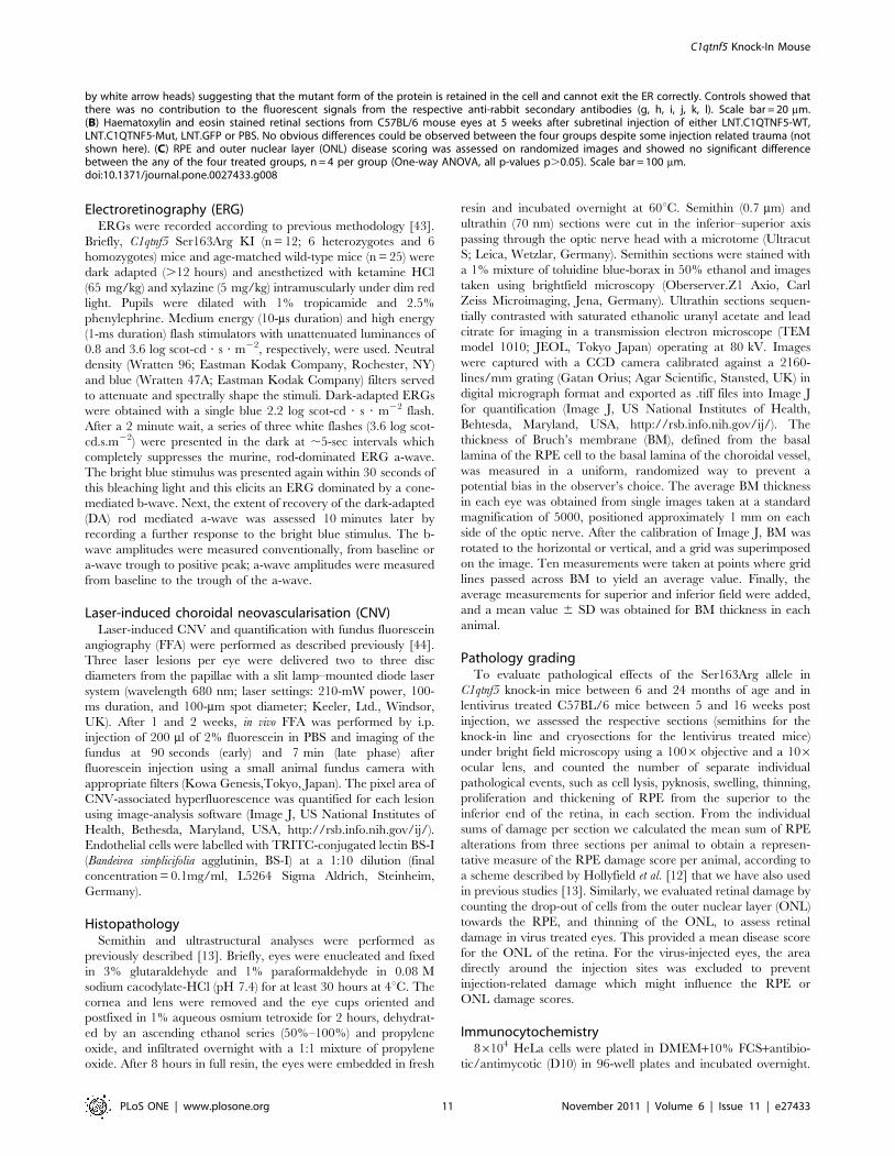
Figure 8. Effects of Lentiviral expression of wild-type or Ser163Arg mutant C1qtnf5 in mouse retinas. (A) HeLa cells were infected withlentivirus contain wild-type C1QTNF5 (LNT.C1QTNF5-WT) or Ser163Arg mutant C1QTNF5 (LNT.C1QTNF5-Mut). At 72 hr post-infection, cells werestained with anti-CTRP5 (green) and the endoplasmic reticulum (ER) marker anti-BiP (red) antibodies. Cells infected with LNT.CTRP5 or LNT. CTRP5-Mut both showed intracellular staining surrounding the nucleus (a, d) suggesting that both proteins enter the secretory pathway. While the stainingon LNT-CTRP5 infected cells is rather diffuse (a), the LNT-CTRP5-Mut showed punctate staining (d) that co-localised with the ER marker BiP (e, f, shown
C1qtnf5 Knock-In Mouse
PLoS ONE | www.plosone.org 10 November 2011 | Volume 6 | Issue 11 | e27433
Electroretinography (ERG)ERGs were recorded according to previous methodology [43].
Briefly, C1qtnf5 Ser163Arg KI (n = 12; 6 heterozygotes and 6
homozygotes) mice and age-matched wild-type mice (n = 25) were
dark adapted (.12 hours) and anesthetized with ketamine HCl
(65 mg/kg) and xylazine (5 mg/kg) intramuscularly under dim red
light. Pupils were dilated with 1% tropicamide and 2.5%
phenylephrine. Medium energy (10-ms duration) and high energy
(1-ms duration) flash stimulators with unattenuated luminances of
0.8 and 3.6 log scot-cd ? s ? m22, respectively, were used. Neutral
density (Wratten 96; Eastman Kodak Company, Rochester, NY)
and blue (Wratten 47A; Eastman Kodak Company) filters served
to attenuate and spectrally shape the stimuli. Dark-adapted ERGs
were obtained with a single blue 2.2 log scot-cd ? s ? m22 flash.
After a 2 minute wait, a series of three white flashes (3.6 log scot-
cd.s.m22) were presented in the dark at ,5-sec intervals which
completely suppresses the murine, rod-dominated ERG a-wave.
The bright blue stimulus was presented again within 30 seconds of
this bleaching light and this elicits an ERG dominated by a cone-
mediated b-wave. Next, the extent of recovery of the dark-adapted
(DA) rod mediated a-wave was assessed 10 minutes later by
recording a further response to the bright blue stimulus. The b-
wave amplitudes were measured conventionally, from baseline or
a-wave trough to positive peak; a-wave amplitudes were measured
from baseline to the trough of the a-wave.
Laser-induced choroidal neovascularisation (CNV)Laser-induced CNV and quantification with fundus fluorescein
angiography (FFA) were performed as described previously [44].
Three laser lesions per eye were delivered two to three disc
diameters from the papillae with a slit lamp–mounted diode laser
system (wavelength 680 nm; laser settings: 210-mW power, 100-
ms duration, and 100-mm spot diameter; Keeler, Ltd., Windsor,
UK). After 1 and 2 weeks, in vivo FFA was performed by i.p.
injection of 200 ml of 2% fluorescein in PBS and imaging of the
fundus at 90 seconds (early) and 7 min (late phase) after
fluorescein injection using a small animal fundus camera with
appropriate filters (Kowa Genesis,Tokyo, Japan). The pixel area of
CNV-associated hyperfluorescence was quantified for each lesion
using image-analysis software (Image J, US National Institutes of
Health, Bethesda, Maryland, USA, http://rsb.info.nih.gov/ij/).
Endothelial cells were labelled with TRITC-conjugated lectin BS-I
(Bandeirea simplicifolia agglutinin, BS-I) at a 1:10 dilution (final
concentration = 0.1mg/ml, L5264 Sigma Aldrich, Steinheim,
Germany).
HistopathologySemithin and ultrastructural analyses were performed as
previously described [13]. Briefly, eyes were enucleated and fixed
in 3% glutaraldehyde and 1% paraformaldehyde in 0.08 M
sodium cacodylate-HCl (pH 7.4) for at least 30 hours at 4uC. The
cornea and lens were removed and the eye cups oriented and
postfixed in 1% aqueous osmium tetroxide for 2 hours, dehydrat-
ed by an ascending ethanol series (50%–100%) and propylene
oxide, and infiltrated overnight with a 1:1 mixture of propylene
oxide. After 8 hours in full resin, the eyes were embedded in fresh
resin and incubated overnight at 60uC. Semithin (0.7 mm) and
ultrathin (70 nm) sections were cut in the inferior–superior axis
passing through the optic nerve head with a microtome (Ultracut
S; Leica, Wetzlar, Germany). Semithin sections were stained with
a 1% mixture of toluidine blue-borax in 50% ethanol and images
taken using brightfield microscopy (Oberserver.Z1 Axio, Carl
Zeiss Microimaging, Jena, Germany). Ultrathin sections sequen-
tially contrasted with saturated ethanolic uranyl acetate and lead
citrate for imaging in a transmission electron microscope (TEM
model 1010; JEOL, Tokyo Japan) operating at 80 kV. Images
were captured with a CCD camera calibrated against a 2160-
lines/mm grating (Gatan Orius; Agar Scientific, Stansted, UK) in
digital micrograph format and exported as .tiff files into Image J
for quantification (Image J, US National Institutes of Health,
Behtesda, Maryland, USA, http://rsb.info.nih.gov/ij/). The
thickness of Bruch’s membrane (BM), defined from the basal
lamina of the RPE cell to the basal lamina of the choroidal vessel,
was measured in a uniform, randomized way to prevent a
potential bias in the observer’s choice. The average BM thickness
in each eye was obtained from single images taken at a standard
magnification of 5000, positioned approximately 1 mm on each
side of the optic nerve. After the calibration of Image J, BM was
rotated to the horizontal or vertical, and a grid was superimposed
on the image. Ten measurements were taken at points where grid
lines passed across BM to yield an average value. Finally, the
average measurements for superior and inferior field were added,
and a mean value 6 SD was obtained for BM thickness in each
animal.
Pathology gradingTo evaluate pathological effects of the Ser163Arg allele in
C1qtnf5 knock-in mice between 6 and 24 months of age and in
lentivirus treated C57BL/6 mice between 5 and 16 weeks post
injection, we assessed the respective sections (semithins for the
knock-in line and cryosections for the lentivirus treated mice)
under bright field microscopy using a 1006 objective and a 106ocular lens, and counted the number of separate individual
pathological events, such as cell lysis, pyknosis, swelling, thinning,
proliferation and thickening of RPE from the superior to the
inferior end of the retina, in each section. From the individual
sums of damage per section we calculated the mean sum of RPE
alterations from three sections per animal to obtain a represen-
tative measure of the RPE damage score per animal, according to
a scheme described by Hollyfield et al. [12] that we have also used
in previous studies [13]. Similarly, we evaluated retinal damage by
counting the drop-out of cells from the outer nuclear layer (ONL)
towards the RPE, and thinning of the ONL, to assess retinal
damage in virus treated eyes. This provided a mean disease score
for the ONL of the retina. For the virus-injected eyes, the area
directly around the injection sites was excluded to prevent
injection-related damage which might influence the RPE or
ONL damage scores.
Immunocytochemistry86104 HeLa cells were plated in DMEM+10% FCS+antibio-
tic/antimycotic (D10) in 96-well plates and incubated overnight.
by white arrow heads) suggesting that the mutant form of the protein is retained in the cell and cannot exit the ER correctly. Controls showed thatthere was no contribution to the fluorescent signals from the respective anti-rabbit secondary antibodies (g, h, i, j, k, l). Scale bar = 20 mm.(B) Haematoxylin and eosin stained retinal sections from C57BL/6 mouse eyes at 5 weeks after subretinal injection of either LNT.C1QTNF5-WT,LNT.C1QTNF5-Mut, LNT.GFP or PBS. No obvious differences could be observed between the four groups despite some injection related trauma (notshown here). (C) RPE and outer nuclear layer (ONL) disease scoring was assessed on randomized images and showed no significant differencebetween the any of the four treated groups, n = 4 per group (One-way ANOVA, all p-values p.0.05). Scale bar = 100 mm.doi:10.1371/journal.pone.0027433.g008
C1qtnf5 Knock-In Mouse
PLoS ONE | www.plosone.org 11 November 2011 | Volume 6 | Issue 11 | e27433

On the following day, cells were infected with either
LNT.CTRP5-WT or LNT.CTRP5-Mut in D10. At 72 h post-
infection, cells well fixed with ice-cold methanol, washed with PBS
containing 0.05% Tween-20 (PBS-T). Cells were blocked with 3%
BSA in PBS-T for 1 hr at room temperature (RT) before they
were incubated with rabbit-anti-CTRP5 (1:250 in block solution,
antibody provided by A.F. Wright [8]) for 1 hr at RTuC. Cells
were washed 4 times in PBS-T and then incubated with goat anti-
rabbit-Alexa488 (1:400 in block, Invitrogen, Paisley, UK) for
45 minutes at RT. For double labelling with the ER-marker BiP,
the cells were then washed 4 times in PBS and re-blocked for 1 hr
before incubation with the rabbit anti-BiP (1:100 in block, G8918,
Sigma, Gillingham, UK) antibody for 1 hr at RT. After 4
additional wash steps, the goat anti-rabbit-Alexa546 antibody
(1:400 in block, Invitrogen, Paisley, UK) was applied for 45 mins,
the cells washed again 4 times before they were counterstained
with Hoechst (1:1000 in PBS-T) and washed once more. For
imaging, the wells were kept hydrated with blocking solution in the
96-well plate and imaged with Q-Capture software (Q Imaging,
Maidenhead, UK) on an inverted Axio Oberver.Z1 microscope
(Carl Zeiss, Jena, Germany).
Reverse transcriptase-polymerase chain reaction (RT-PCR)Total RNAs from mouse eye cup tissues were extracted using
RNAeasy Mini kit (QIAGEN) according to the manufacturer’s
instructions. Two micrograms of the resulting RNA was reverse
transcribed with random primers using a Transcriptor High Fidelity
cDNA Synthesis Kit (Roche). The cDNAs of C1qtnf5 and Mfrp were
then amplified using platinum Taq DNA polymerase (Invitrogen) as
previous described [45]. PCR primers were manufactured by Sigma
Aldrich. Primer sequences are available on request.
Western blottingEyecups from wild-type, heterozygous and homozygous C1qtnf5
Ser163Arg knock-in mice were lysed in a buffer containing
50 mM Tris-Cl pH 7.5, 150 mM NaCl, 1% NP-40, 0.5%
deoxycholate, 0.05% SDS, 2 mM EDTA, 1 mM sodium-vana-
date, 5 mM sodium-fluoride and 10 mM iodoacetamide with a
protease inhibitor cocktail (Roche). The lysates were centrifuged
for 10 min at 13,000 rpm and the post-nuclear supernatants were
collected. Equal amounts of protein were electrophoresed on 10%
SDS-polyacrylamide gels and transferred to a nitrocellulose
membrane. The primary antibodies were used at 1:1,000 dilution
for anti-C1QTNF5 and 1:2,000 dilution for antib-tubulin
antibodies. HRP-conjugated secondary antibodies were used at
1:5,000 dilution. The membrane-bound antibodies were detected
by ECL (Amersham Biosciences).
Lentivirus productionSecond generation HIV-1-based self-inactivating lentiviral vec-
tors pseudotyped with the vesicular stomatitis virus glycoprotein
envelope contained either the murine C1qtnf5 wild type or the
murine C1qtnf5 Ser163Arg mutant cDNA or the GFP reporter gene
(Lenti.GFP). All vectors contain deletions in the 39 long-terminal
repeats, making them self-inactivating, and include the central
polypurine tract (cPPT), which is necessary for second-strand DNA
synthesis, and may enhance the nuclear transport of the provirus.
All cDNAs are driven by the spleen focus-forming virus (SFFV)
promoter and each vector contains the woodchuck hepatitis virus
post-transcriptional regulatory element (WPRE) to stabilize mRNA
and increase expression. Vectors were produced using transient
triple transfection of 293T cells as described previously [46]. At 28–
72 hr post-transfection, supernatant was harvested and filtered
through a 0.45 mm filter. The virus particles were concentrated by
ultracentrifugation at 90,000 g for 2 hr and the pelleted virus was
resuspended in OptiMEM and stored at 280uC. Virus was titred
using a Reverse Transcriptase activity ELISA kit (Roche Diagnos-
tics, Burgess Hill, UK) according to the manufacturer’s instructions.
Transgene expression was detected by immunocytochemistry on
infected HeLa cells. Cells were infected with 1 ml of virus in a 24 well
plate. Three days post-infection, cells were washed and fixed with
ice-cold methanol and stained with a polyclonal rabbit anti-human
C1QTNF5 antibody [8] and goat anti-rabbit AlexaFluor546
antibody. Cells were counterstained with Hoechst, imaged using a
Zeiss Axioplan II fluorescent microscope and analysed using
QCapture Pro software (QIMaging, Maidenhead,UK).
Subretinal injectionsSubretinal administration of vectors was performed in anesthe-
tized 12 to 16-week-old C57BL/6 mice (Harlan UK Ltd.), under
direct retinoscopy through an operating microscope as previously
described [47]. A 1.5-cm 34-gauge hypodermic needle (Hamilton,
UK) was used to inject 2 microlitres of virus suspension (titer of 108
particles/ml) into the subretinal space to produce a bullous retinal
detachment in the superior hemisphere and a second in the
inferior hemisphere. Mice received C1qtnf5-wild type virus in one
eye and C1qtnf5-Ser163Arg mutant virus in the other eye. Control
mice received PBS or titre-matched Lenti.GFP. Retinal vessels
remained patent during and following the injections. All animals
received chloramphenicol 1% eye ointment to the cornea.
Statistical analysisStatistical analyses were performed using GraphPad Prism 5 for
Windows (GraphPad Software Inc, La Jolla, USA). Changes in the
thickness of Bruch’s membrane or in RPE damage with age were
assessed using Pearson correlation statistics, a parametric measure
of association for two continuous random variables. Differences in
autofluorsecence were assessed using a Kruskal-Wallis with Dunn’s
multiple comparison test. RPE and outer nuclear layer disease
scoring was assessed on randomized images using a one-way
analysis of variance.
Acknowledgments
We thank genOway (Lyons, France) for help with vector design and
targeting strategy, mouse ES cell injections and targeting experiments. We
also thank Craig Nicol for help with the artwork.
Author Contributions
Conceived and designed the experiments: AFW RA SGJ. Performed the
experiments: XS UL TSA SEB AL BT MC HX. Analyzed the data: XS
UL TSA SEB AL BT MC HX. Wrote the paper: AFW XS UL SGJ.
References
1. Hayward C, Shu X, Cideciyan AV, Lennon A, Barran P, et al. (2003) Mutation
in a short-chain collagen gene, CTRP5, results in extracellular deposit formationin late-onset retinal degeneration: a genetic model for age-related macular
degeneration. Hum Mol Genet 12: 2657–2667.2. Kuntz CA, Jacobson SG, Cideciyan AV, Li ZY, Stone EM, et al. (1996) Sub-
retinal pigment epithelial deposits in a dominant late-onset retinal degeneration.
Invest Ophthalmol Vis Sci 37: 1772–1782.
3. Milam AH, Curcio CA, Cideciyan AV, Saxena S, John SK, et al. (2000)
Dominant late-onset retinal degeneration with regional variation of sub-retinalpigment epithelium deposits, retinal function, and photoreceptor degeneration.
Ophthalmology 107: 2256–2266.4. Ayyagari R, Mandal MN, Karoukis AJ, Chen L, McLaren NC, et al. (2005)
Late-onset macular degeneration and long anterior lens zonules result from a
CTRP5 gene mutation. Invest Ophthalmol Vis Sci 46: 3363–3371.
C1qtnf5 Knock-In Mouse
PLoS ONE | www.plosone.org 12 November 2011 | Volume 6 | Issue 11 | e27433

5. Subrayan V, Morris B, Armbrecht AM, Wright AF, Dhillon B (2005) Long
anterior lens zonules in late-onset retinal degeneration (L-ORD).Am J Ophthalmol 140: 1127–1129.
6. Wong GW, Krawczyk SA, Kitidis-Mitrokostas C, Revett T, Gimeno R, et al.
(2008) Molecular, biochemical and functional characterizations of C1q/TNFfamily members: adipose-tissue-selective expression patterns, regulation by
PPAR-gamma agonist, cysteine-mediated oligomerizations, combinatorialassociations and metabolic functions. Biochem J 416: 161–177.
7. Shu X, Tulloch B, Lennon A, Hayward C, O’Connell M, et al. (2006)
Biochemical characterisation of the C1QTNF5 gene associated with late-onsetretinal degeneration. A genetic model of age-related macular degeneration. Adv
Exp Med Biol 572: 41–48.8. Shu X, Tulloch B, Lennon A, Vlachantoni D, Zhou X, et al. (2006) Disease
mechanisms in late-onset retinal macular degeneration associated with mutationin C1QTNF5. Hum Mol Genet 15: 1680–1689.
9. Sundin OH, Leppert GS, Silva ED, Yang JM, Dharmaraj S, et al. (2005)
Extreme hyperopia is the result of null mutations in MFRP, which encodes aFrizzled-related protein. Proc Natl Acad Sci U S A 102: 9553–9558.
10. Kameya S, Hawes NL, Chang B, Heckenlively JR, Naggert JK, et al. (2002)Mfrp, a gene encoding a frizzled related protein, is mutated in the mouse retinal
degeneration 6. Hum Mol Genet 11: 1879–1886.
11. Park SY, Choi JH, Ryu HS, Pak YK, Park KS, et al. (2009) C1q tumor necrosisfactor alpha-related protein isoform 5 is increased in mitochondrial DNA-
depleted myocytes and activates AMP-activated protein kinase. J Biol Chem284: 27780–27789.
12. Hollyfield JG, Bonilha VL, Rayborn ME, Yang X, Shadrach KG, et al. (2008)Oxidative damage-induced inflammation initiates age-related macular degener-
ation. Nat Med 14: 194–198.
13. Luhmann UF, Robbie S, Munro PM, Barker SE, Duran Y, et al. (2009) Thedrusenlike phenotype in aging Ccl2-knockout mice is caused by an accelerated
accumulation of swollen autofluorescent subretinal macrophages. InvestOphthalmol Vis Sci 50: 5934–5943.
14. Pierce EA (2001) Pathways to photoreceptor cell death in inherited retinal
degenerations. Bioessays 23: 605–618.15. Stone EM (2009) Progress toward effective treatments for human photoreceptor
degenerations. Curr Opin Genet Dev 19: 283–289.16. Zeiss CJ (2010) Animals as models of age-related macular degeneration: an
imperfect measure of the truth. Vet Pathol 47: 396–413.17. Rakoczy EP, Yu MJ, Nusinowitz S, Chang B, Heckenlively JR (2006) Mouse
models of age-related macular degeneration. Exp Eye Res 82: 741–752.
18. Mata NL, Tzekov RT, Liu X, Weng J, Birch DG, et al. (2001) Delayed dark-adaptation and lipofuscin accumulation in abcr+/2 mice: implications for
involvement of ABCR in age-related macular degeneration. Invest OphthalmolVis Sci 42: 1685–1690.
19. Radu RA, Mata NL, Bagla A, Travis GH (2004) Light exposure stimulates
formation of A2E oxiranes in a mouse model of Stargardt’s maculardegeneration. Proc Natl Acad Sci U S A 101: 5928–5933.
20. Karan G, Lillo C, Yang Z, Cameron DJ, Locke KG, et al. (2005) Lipofuscinaccumulation, abnormal electrophysiology, and photoreceptor degeneration in
mutant ELOVL4 transgenic mice: a model for macular degeneration. Proc NatlAcad Sci U S A 102: 4164–4169.
21. Weber BH, Lin B, White K, Kohler K, Soboleva G, et al. (2002) A mouse model
for Sorsby fundus dystrophy. Invest Ophthalmol Vis Sci 43: 2732–2740.22. Wright AF, Jacobson SG, Cideciyan AV, Roman AJ, Shu X, et al. (2004)
Lifespan and mitochondrial control of neurodegeneration. Nat Genet 36:1153–1158.
23. Chavali VR, Khan NW, Cuckras CA, Bartsch DU, Jablonski MM, et al. (2011)
A CTRP5 Gene S163R mutation Knock-In Mouse Model for Late-OnsetRetinal Degeneration. Hum Mol Genet.
24. Montagutelli X (2000) Effect of the genetic background on the phenotype ofmouse mutations. J Am Soc Nephrol 11 Suppl 16: S101–105.
25. Elsea SH, Lucas RE (2002) The mousetrap: what we can learn when the mouse
model does not mimic the human disease. Ilar J 43: 66–79.26. Robson MG, Cook HT, Pusey CD, Walport MJ, Davies KA (2003) Antibody-
mediated glomerulonephritis in mice: the role of endotoxin, complement andgenetic background. Clin Exp Immunol 133: 326–333.
27. Dickinson P, Dorin JR, Porteous DJ (1995) Modelling cystic fibrosis in the
mouse. Mol Med Today 1: 140–148.
28. Van Raamsdonk JM, Metzler M, Slow E, Pearson J, Schwab C, et al. (2007)
Phenotypic abnormalities in the YAC128 mouse model of Huntington disease
are penetrant on multiple genetic backgrounds and modulated by strain.
Neurobiol Dis 26: 189–200.
29. Wheeler FC, Fernandez L, Carlson KM, Wolf MJ, Rockman HA, et al. (2005)
QTL mapping in a mouse model of cardiomyopathy reveals an ancestral
modifier allele affecting heart function and survival. Mamm Genome 16:
414–423.
30. Dietrich WF, Lander ES, Smith JS, Moser AR, Gould KA, et al. (1993) Genetic
identification of Mom-1, a major modifier locus affecting Min-induced intestinal
neoplasia in the mouse. Cell 75: 631–639.
31. Yang T, Huang YG, Ye W, Hansen P, Schnermann JB, et al. (2005) Influence of
genetic background and gender on hypertension and renal failure in COX-2-
deficient mice. Am J Physiol Renal Physiol 288: F1125–1132.
32. Rozmahel R, Wilschanski M, Matin A, Plyte S, Oliver M, et al. (1996)
Modulation of disease severity in cystic fibrosis transmembrane conductance
regulator deficient mice by a secondary genetic factor. Nat Genet 12: 280–287.
33. Lima BL, Santos EJ, Fernandes GR, Merkel C, Mello MR, et al. A new mouse
model for marfan syndrome presents phenotypic variability associated with the
genetic background and overall levels of Fbn1 expression. PLoS One 5: e14136.
34. Wheeler FC, Tang H, Marks OA, Hadnott TN, Chu PL, et al. (2009) Tnni3k
modifies disease progression in murine models of cardiomyopathy. PLoS Genet
5: e1000647.
35. Silverman KA, Koratkar R, Siracusa LD, Buchberg AM (2002) Identification of
the modifier of Min 2 (Mom2) locus, a new mutation that influences Apc-
induced intestinal neoplasia. Genome Res 12: 88–97.
36. Haston CK, McKerlie C, Newbigging S, Corey M, Rozmahel R, et al. (2002)
Detection of modifier loci influencing the lung phenotype of cystic fibrosis
knockout mice. Mamm Genome 13: 605–613.
37. Kiernan AE, Li R, Hawes NL, Churchill GA, Gridley T (2007) Genetic
background modifies inner ear and eye phenotypes of jag1 heterozygous mice.
Genetics 177: 307–311.
38. Mehalow AK, Kameya S, Smith RS, Hawes NL, Denegre JM, et al. (2003)
CRB1 is essential for external limiting membrane integrity and photoreceptor
morphogenesis in the mammalian retina. Hum Mol Genet 12: 2179–2189.
39. Danciger M, Yang H, Ralston R, Liu Y, Matthes MT, et al. (2007) Quantitative
genetics of age-related retinal degeneration: a second F1 intercross between the
A/J and C57BL/6 strains. Mol Vis 13: 79–85.
40. Gerido DA, Sellitto C, Li L, White TW (2003) Genetic background influences
cataractogenesis, but not lens growth deficiency, in Cx50-knockout mice. Invest
Ophthalmol Vis Sci 44: 2669–2674.
41. Brunner S, Skosyrski S, Kirschner-Schwabe R, Knobeloch KP, Neidhardt J,
et al. Cone versus rod disease in a mutant Rpgr mouse caused by different
genetic backgrounds. Invest Ophthalmol Vis Sci 51: 1106–1115.
42. Samardzija M, Wenzel A, Naash M, Reme CE, Grimm C (2006) Rpe65 as a
modifier gene for inherited retinal degeneration. Eur J Neurosci 23: 1028–1034.
43. Williams DS, Aleman TS, Lillo C, Lopes VS, Hughes LC, et al. (2009)
Harmonin in the murine retina and the retinal phenotypes of Ush1c-mutant
mice and human USH1C. Invest Ophthalmol Vis Sci 50: 3881–3889.
44. Balaggan KS, Binley K, Esapa M, MacLaren RE, Iqball S, et al. (2006) EIAV
vector-mediated delivery of endostatin or angiostatin inhibits angiogenesis and
vascular hyperpermeability in experimental CNV. Gene Ther 13: 1153–1165.
45. Shu X, Zeng Z, Eckmiller MS, Gautier P, Vlachantoni D, et al. (2006)
Developmental and tissue expression of Xenopus laevis RPGR. Invest
Ophthalmol Vis Sci 47: 348–356.
46. Demaison C, Brouns G, Blundell MP, Goldman JP, Levinsky RJ, et al. (2000) A
defined window for efficient gene marking of severe combined immunodeficient-
repopulating cells using a gibbon ape leukemia virus-pseudotyped retroviral
vector. Hum Gene Ther 11: 91–100.
47. Smith AJ, Schlichtenbrede FC, Tschernutter M, Bainbridge JW, Thrasher AJ,
et al. (2003) AAV-Mediated gene transfer slows photoreceptor loss in the RCS
rat model of retinitis pigmentosa. Mol Ther 8: 188–195.
C1qtnf5 Knock-In Mouse
PLoS ONE | www.plosone.org 13 November 2011 | Volume 6 | Issue 11 | e27433