Embed Size (px)
Citation preview
Chapter VIII
Promising biomolecules
Isabel Oliveira 1,2, Ana L. Carvalho 1,2, Hajer Radhouani 1,2, Cristiana
Gonçalves1,2, Joaquim M. Oliveira1,2,3, Rui L. Reis1,2,3
13B’s Research Group – Biomolecules, Biodegradables and Biomimetics, University of
Minho, Headquarters of the European Institute of Excellence on Tissue Engineering and
Regenerative Medicine, Avepark, Parque da Ciência e Tecnologia, Zona Industrial da
Gandra. 4805-017 Barco, GMR, Portugal
2ICVS/3B’s – PT Government Associate Laboratory, Braga/Guimarães, Portugal
3The Discoveries Centre for Regenerative and Precision Medicine, Headquarters at
University of Minho, Avepark 4805-017 Barco, Guimarães, Portugal
Abstract The osteochondral defect (OD) comprises the articular cartilage and its
subchondral bone. The treatment of these lesions remains as one of the most prob-
lematic clinical issues, since these defects include different tissues, requiring dis-
tinct healing approaches. Among the growing applications of regenerative medi-
cine, clinical articular cartilage repair has been used for two decades and it is an
effective example of translational medicine; one of the most used cell based repair
strategy include implantation of autologous cells in degradable scaffolds such as
alginate, agarose, collagen, chitosan, chondroitin sulfate, cellulose, silk fibroin, hy-
aluronic acid, gelatin, among others. Concerning the repair of osteochondral de-
fects, tissue engineering and regenerative medicine started to design single- or bi-
phased scaffold constructs, often containing hydroxyapatite-collagen composites,
usually used as a bone substitute. Biomolecules such as natural and synthetic have
been explored to recreate the cartilage-bone interface through multilayered biomi-
metic scaffolds. In this chapter, is given a succinct description about the most rele-
vant natural and synthetic biomolecules used on cartilage and bone repair, describ-
ing the procedures to obtain these biomolecules, their chemical structure, common
modifications to improve its characteristics and also their application in the biomed-
ical fields.

2
8.1 Introduction
Tissue engineering field has been responsible for promising applications develop-
ment to regenerate and repair osteochondral defects. The biomolecules studied and
applied for cartilage and bone repair, are selected considering the knowledge of the
anatomical complexity of both structures. The increase of knowledge in the bio-
molecular area, with progress in new technology such as cellular, molecular biology
and biochemistry, offer a good opportunity to create biomolecules with stimulating
and precise properties [1, 2].
The main properties sought in a biomolecule to be applied for cartilage and bone
regeneration are: biocompatibility, bioactivity, biomimetic skill, biodegradability,
being bio-responsive, highly porous, suitable for cell attachment, proliferation and
differentiation, osteoconductive, non-cytotoxic, flexible and elastic, and non-anti-
genic [3, 4]. Currently, several natural and synthetic polymers, with most of these
characteristics, have been studied as therapy for cartilage repair. The natural poly-
mers, more used to osteochondral defects, are collagen, fibrin, alginate, silk, and
chitosan. These biopolymers have been investigated as bioactive scaffolds for bone
engineering such as alginate, agarose, fibrin, hyaluronic acid (HA), collagen, gela-
tin, chitosan, chondroitin sulphate, and cellulose [5, 6], Regarding synthetic poly-
mers, they are often considered to be used for proteins and growth factors delivery
with or without cells locally to enhance tissue repair and regeneration such as
Poly(ethylene glycol) (PEG), Poly(lactic-co-glycolic acid) (PLGA), Poly-L-lactic
acid(PLLA),Polycaprolactone(PCL),Poly(N-isopropylacrylamide)(PolyNiPAAm),
and polycarbonate [7, 8]. At this point, it is important to clear up that both natural
and synthetic materials can be considered biomolecules, since that they can be
equally used to medical and surgical purposes.
In the following section, promising trends in the development of biomolecules for
cartilage and bone repair are described.
8.2 Biomolecules
Bone and cartilage are two different tissues with specific structural and mechanical
properties belonging to osteochondral interface [9].
The biodegradable scaffolds, with or without cells and/or growth factors, have been
widely used for cartilage and bone repair. Cartilage tissue shows single conjunction
of nonlinear tensile and compressive properties due to arranged collagen fibrils,

3
proteoglycans, and proteins. The structural and mechanical properties of natural tis-
sues can be mimicked by engineered bio-nanocomposites through the use of poly-
mers and nanoparticles [10].
It is important to highlight that classical scaffolds cannot be together for both chon-
drogenesis and osteogenesis. Scaffolds with two layers have more potential, being
organized by different space structures and mechanical forces. One layer is respon-
sible by the repair of the cartilage and the other layer supports the regeneration of
the subchondral bone [11].
8.2.1 Natural materials
Nature has been using natural polymers long before the creation of plastics and other
synthetic materials. For that reason, the natural biomolecules development is not
truly a scientific area. Nonetheless this not turns it less important, in fact the use of
this products is currently resurging in the biomedical field. Natural polymers remain
attractive compared with synthetic polymers since they can avoid waste-disposal
problems that are associated with traditional synthetic polymers. Moreover, these
polymers present more advantages related to the possibility of several chemical
modifications, potential to be degradable and biocompatible due to their natural
origin [12].
Over the past few years, this area have gained special attention and a large number
of biomolecules have been reported as a great option to apply on osteochondral de-
fects repair and regeneration [13-20].
Silk fibroin
Silk fibroin (SF) is a natural polymeric biomolecule composed of two proteins: hy-
drophobic fibroin and hydrophilic sericin. This biopolymer shows several interest-
ing characteristics for tissue engineering, such as mammalian cell compatibility,
remarkable oxygen and water vapor permeability, biodegradability and suitable me-
chanical properties, with an unique combination of elasticity and strength [21]. Re-
cently, the attention for silk biomolecules has increasing, since this biomolecule has
shown to be a great option as a therapeutic material like biomimetic scaffolds for
tissue engineering [22, 23].
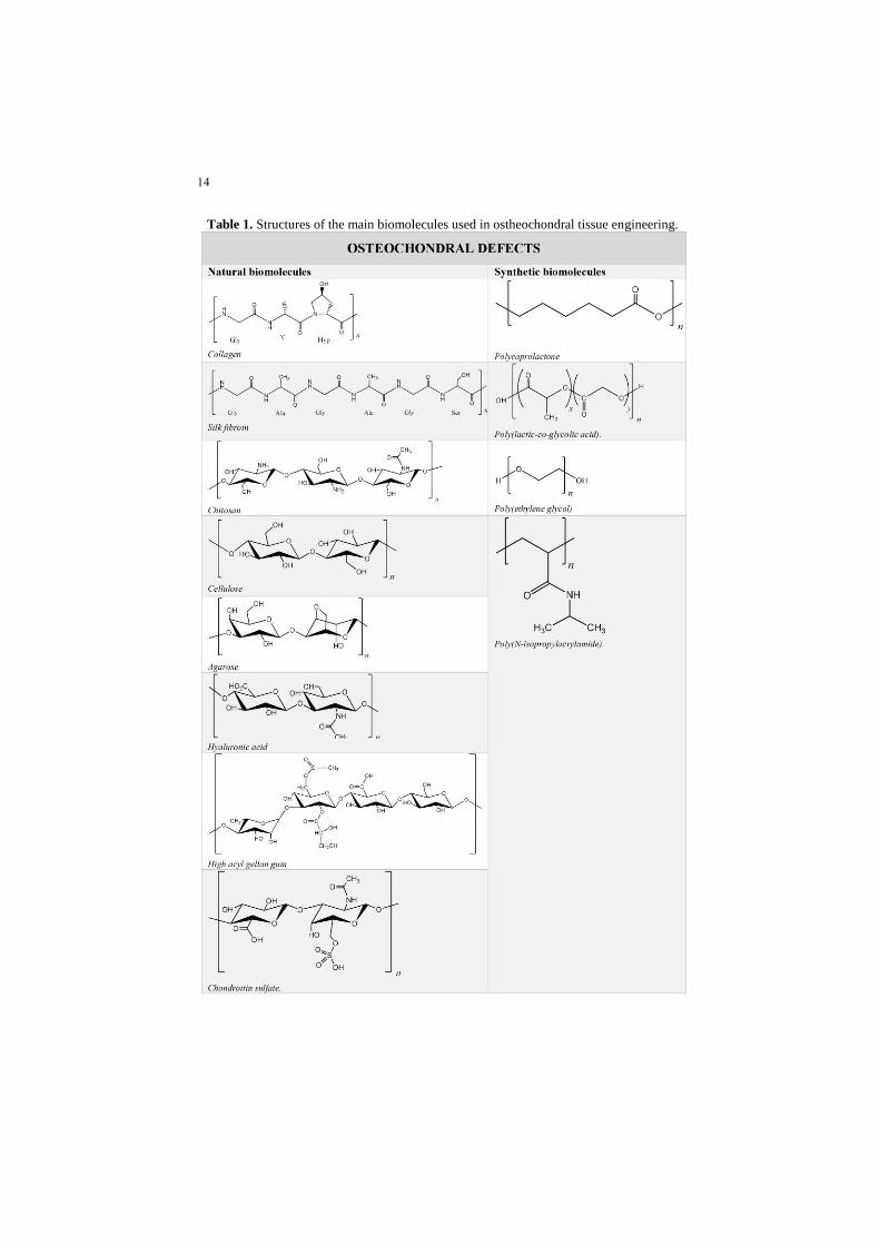
Silk fibroin presents crystalline regions, with an amino acid sequence (Table 1) due
to their heavy chains (350 kDa) and non-repetitive amorphous regions. Moreover,
SF also presents three crystal structures denoted as silk I, silk II and silk III [24, 25].

4
Silk proteins, present in glands of silk, can be obtained from silkworms, spiders,
scorpions, mites and bees and spun into fibers during their metamorphosis [26]. The
cannibalistic nature of spiders represents a difficulty on the commercial production
of spider silk. For that reason, silkworm's silk, an established fiber, has been the
option for textile but also for medical research area. In fact, the yield of fiber from
a single silk cocoon is 600–1500 m, compared to only about 137 m from the am-
pullate gland of a spider and about 12 m from the spider web [27]. Silk based bio-
materials can be easily prepared from Bombyx mori silkworm silk. The raw silk
fibers (cocoons) are degummed Na2CO3 solution at 100 ºC and then rinsed with
distilled water in order to extract the highly immunogenic sericin protein (other pro-
tein secreted by silk worm) and then air-dried. SF thus extracted is dissolved in a
ternary solvent containing CaCl2, CH3CH2OH and H2O, at 70 ºC for 1 h, with con-
tinuous stirring. The final regenerated SF solution is obtained following dialysis
against distilled water and filtration at room temperature [28].
As refereed above, SF fibres have been extensively used in biomedical field, namely
as surgical sutures, not presenting side effects for humans [24]. In fact, due to its
excellent properties, SF is considered one of the best biomolecule for tissue engi-
neering, more specifically for skeletal and ostheocontral applications [26, 29]. An-
other add-value of SF is the fact that besides the ways it is processed (films, sponges
or hydrogels), it is able to sustain cell adhesion, proliferation, even shown capable
of ECF production in vivo and in vitro with a low inflammatory potential [30].
Several research studies have shown the potential of SF for osteochondral defect
regeneration. In fact, silk fibroin scaffolds showed to be osteoinductive with good
integration of stem cell-silk biomolecule, which is elementary in the bone or carti-
lage area [31].
Collagen
Collagen is the primary structural material of vertebrates and is the most abundant
protein in mammalian, accounting for about 20–30% of the whole-body protein
content [32]. In the human body, collagen is mostly found in fibrous tissues such as
tendons, skin, ligaments and also in tissues (as cartilage, bone and intervertebral
disc) and mostly of collagen in the body is of type I [33]. In fact, it is in the skin
that collagen is more abundant, and about 70% of the material, besides water that
are present in dermis of skin and tendon, is collagen [34]. Collagen is synthesized
by fibroblasts, which usually originate from pluripotential adventitial cells or retic-
5
ulum cells [35]. There are different collagen types, differentiated by their complex-
ity and diversity in their structure, their splice variants, the presence of additional,
non-helical domains, their assembly and their function. Types I and V collagen fi-
brils are the main contribution for the structural backbone of bone while types II
and XI collagens predominantly contribute to the fibrillar matrix of articular carti-
lage [36].
Collagen is gaining popularity not only for the easier way of fabrication since it can
be processed, through chemical and biochemical modifications, into a variety of
forms including cross-linked films, steps, sheets, beads, meshes, fibres, and
sponges. Furthermore, it presents a unique structural, physical, chemical and immu-
nological properties, being biodegradable, biocompatible and non-cytotoxic, sup-
porting cellular growth [37, 38].
Collagen can be extracted from bovine skin, pig skin and chicken waste, but these
kinds of sources could have religious and ethnic constraints. Taking this in account
many reports have been showed to extract collagen from marine sources as alterna-
tive source and have used to screen their potential industrial applications [39, 40].
Collagen molecules are structural macromolecule of the extracellular matrix (ECM)
comprising three polypeptide chains. These chains, aligned in a parallel manner and
coiled in triple-helix, wrap around each other to form that is stabilized by inter-
strand hydrogen bonds [38]. Collagen production is quite simple and a diver-
sity of matrix systems, such as meshes, hydrogels, scaffolds, injectable so-
lutions and dispersions, among others can be achieved [41]. Several studies
showed that collagen-based scaffolds provide a suitable scaffold for carti-
lage and bone regeneration, as it supports the adhesion, migration and pro-
liferation of cells in vitro [18, 42].
Chitosan
Chitosan polysaccharide is the only pseudo-natural cationic polymer and an im-
portant derivative of chitin. Chitin is a homopolymer comprised of 2-acetamido-2-
deoxy-b-D-glucopyranose units [43], Table 1. It has application on the biomedical
field and many others, due to its unique cationic character (protonation of the amino
group) [44]. Extensive research has been conducted in order to improve chitosan
characteristics for specific applications. This polysaccharide mostly finds its appli-
cation in wound dressings, scaffolds (on physical cross-linking) and as antimicro-
bial agent [45, 46]. Moreover, although it has several interesting properties (such as

6
biodegradability and non-toxicity) [47] this biopolymer has poor mechanical prop-
erties and high swelling ability, getting easily deformed, which is generally im-
proved by blending with other polymers [48].
Alginate
Alginate is a linear and anionic natural polysaccharide derived from brown seaweed
such as Laminaria hyperborea, Macrocystis pyrifera and Ascophyllum nodosum.
This biopolymer is composed of alternating blocks of α-1, 4-l-guluronic acid (G)
and β-1,4-d-manurunic acid (M) units [49], Table 1. The monomer sequence can
diverge depending on algal species and different tissues of the same species. The
ratio between monomers and the block structure have a significant effect on the
physicochemical and rheological properties of alginates. These biopolymers with
more industrial significance usually show high G content [50, 51]. Although it can
be produced by bacterial sources, it is commercially available from algae in form of
salt, e.g. sodium alginate. This biomolecule presents excellent properties, namely
their biodegradability, low toxicity and chemical versatility, but also its unique
property to form stable gel in aqueous media and mild condition by addition of
multivalent cations makes this biopolymer very useful for drug delivery and cell
immobilization [52].
The main industrial applications of alginates are linked to their ability to crosslink
with ions, retain water, gelling, viscosity booster and stabilizing properties [53].
Alginate’s extraction procedure from brown algae have suffered an optimization to
obtain more industrially and economically sustainable products, with controlled
properties as to envisage different therapeutic applications [50, 54].
Alginate is widely used on biotechnological industry, due to its ability to efficiently
bind divalent cations, leading to hydrogel formation, being also a perfect candidate
for chemical functionalization. By forming alginate derivatives through chemical
functionalization, the various properties such as solubility, hydrophobicity, and
physicochemical and biological characteristics may be improved. Several studies
reported the alginate chemical modification of hydroxyl groups through different
techniques such as oxidation [55], reductive-amination of oxidized alginate [56],
sulphation [57], copolymerization or cyclodextrin-linked alginate [58], and also
chemical modification of carboxyl groups using other techniques as esterification
[59], Ugi reaction [60] or amidation [61, 62]. Alginate has been extensively used
for many biomedical applications. It could form scaffolds through the use of ionic
cross-linking, allowing for encapsulation of cells.

7
Cellulose
Cellulose is the most widely spread natural raw material with total production of
1011–1012 tons/year. It is a cheap, biodegradable and renewable polymer, being fi-
brous and the main constituent of the cell wall of green plants. Cellulose has a ver-
satile production, and exist in a wide range of forms and shapes, e.g. as membrane
sponges, microspheres and non-woven, woven or knitted textiles [33]. Commercial
sources of cellulose include mainly wood or cotton. However, cellulose can also be
extracted from different parts of plants and other sources [33]. Usually, purification
and isolation processes almost engender the degradation of cellulose and also permit
the cellulose to undergo oxidation by reaction with both acids and bases [63].
Cellulose is one of the many beta-glucan compounds, it is a polycarbohydrate com-
posed of a series of cellubiose units, formed by two anhydroglucose subunits, Table
1. This polysaccharide presents unique properties and cannot be synthesized or hy-
drolyzed due to its intricate hydrogen bond network. This complex is also respon-
sible for the good mechanical properties of cellulose, important for its function in
nature [63]. Purification and isolation of cellulose comprises several steps including
a pulping process, partial hydrolysis, dissolution, re-precipitation, and extraction
with organic solvents. Cellulose polymer allows the proliferation of chondrocytes
and also have shown an interesting biocompatibility [64]. Moreover, it was found
that a product based on hydroxypropylmethylcellulose hydrogel may be used for
articular cartilage repair [16]. This versatile compound could also be modified for
bone defects, due to its good mechanical properties.
Agarose
Agarose is a neutral linear polysaccharide extracted from red algae (Rhodophyceae),
and is the major component of agar, being the other component agaropectin. It is
composed of alternating β-d-galactopyranose and anhydro-α-l-galactopyranose.
[16], Table 1. Agarose gel characteristics are determined by two parameters: tem-
perature and concentration, being obtained through changing temperatures. Cross-
linked alginate matrix can be formed via ionic bonding in the presence of Ca2+ [65].
Agarose hydrogels are at present well-characterized and have been studied in dif-
ferent biomedical fields. The hydrogels may be polymerized in situ, reducing inva-
siveness of the surgery and allowing the hydrogel to acquire the required shape [66].
These kind of hydrogels have been extensively studied for chondrocyte culture and
cartilage tissue engineering, by its mechanical and cell-seeding density proper-
ties [67-69]. Moreover, agarose also demonstrated to be a great option for cell ther-

8
apy with chondrocytes or mesenchymal stem cells (chondrocyte- or MSC-laden hy-
drogels) in osteochondral defects. This approach ensure physiologically relevant
mechanical properties and allows the formation of a repaired tissue containing col-
lagens and proteoglycans [70].
Hyaluronic acid
Hyaluronic acid (HA) is a polysaccharide that can be found in all tissues and body
fluids of vertebrates. It can also be found in some bacteria such as Streptococcus
genus. This polysaccharide, a linear polymer composed of N-acetylglucosamine
and glucuronic acid (Table 1), is especially abundant in loose connective tissue.
HA is a polyanion with a pKa around 2.9. It is a pseudoplastic material, and in
aqueous solutions it has shear-thinning properties, behaving like gelatin [71]. HA
has a remarkable hydrodynamic characteristics, especially in terms of its viscosity
and its ability to retain water; thus having an important role in tissue homeostasis
and biomechanical integrity [72]. HA is produced through bacterial fermentation of
streptococcus species or extracted from rooster combs, umbilical cords, synovial
fluids, skin or vitreous humour for commercial purposes. The usual sources for its
industrial production are bovine vitreous humour, bovine synovial liquid, and
rooster crest, with an increasing interest to bacterial cultivations [73].
Although the HA extraction protocols have been developed and optimized over the
years, but these protocols are still limited to low yields. The HA products are most
from animal origins, which have several risks of proteins and viruses’ contamina-
tions. But the risks can be reduced if there is a special care in choosing tissues from
healthy animals [74].
Alternatively, HA biomolecule could be functionalized in order to obtain a more
rigid and stable, hydrophobic, and less susceptible by enzyme material. These func-
tionalizations include sulphation, carbodiimude-mediated modification, esterifica-
tion, hydrazide modification, cross-linking with polyfunctional epoxides, divinyl
sulphone and glutaraldehyde, among others [71].
HA presents several advantages namely for adhesion of cells, extracellular matrix
deposition, transport of gases and nutrients, metabolic product release. It offers a
good interface of material-cell function mainly due to the HA-3D structure with a
significant porosity, surface and space area. This polymer has been reported as an
important role in joint lubrication, nutrition and preserving cartilage properties since
it can help in the control of water balance [71, 75, 76]. All these evidences turn HA
a good option for articular cartilage, besides the fact that it is also able to maintain

9
normal growth of cartilage cells and promote the integration of transplanted chon-
drocytes and damaged cartilage [77, 78].
Gellan Gum
Gellan gum, a bacterial polysaccharide, is a linear and anionic exopolysaccharide,
with the repeating unit consisting of α-l-rhamnose, β-d-glucose, and β-d-glucu-
ronate, in molar ratios of 1:2:1 (Table 1). Gellan is produced by the bacteria Sphin-
gomonas elodea and the process efficiency is dependent on many factors such as
media composition, pH, temperature, agitation rate and available oxygen. Native
form of gellan contains two types of acyl substituents, namely l-glyceryl and acetyl.
Usually, alkaline hydrolysis is used to remove both of the residues and gives
deacetylated gellan, also called low-acetyl or low-acyl [79].
Native as low-acyl gellan form hydrogels in the presence of mono-, di- and trivalent
cations [80]. With native gellan, it is possible to obtained a soft, easily deformable
gels; while with deacetylated one, it results to rigid and brittle gels. The production
of gellan gum is temperature dependent, at least 70 ºC is needed, with subsequent
cooling (to room temperature) to change the conformation of polymer chains [80-
82]. Commonly, gellan gum is used as agar substitute since it can be clarified by
filtration of hot deacetylated gellan gum [79]. In fact, due to the advantages of GG,
and it has received both US FDA and EU approval, being widely used in many food,
cosmetics, pharmacy and medicine applications [80, 83]. It has also being applied
in tissue engineering, mostly as a material for cartilage reconstruction [80, 83].
Moreover, some researchers have been modified gellan gum with methacrilated
groups to improve its physical and mechanical properties, without affecting its bio-
compatibility, being relevant in a widespread of tissue engineering applications
[84]. Moreover, gellan gum gel also showed to be a promising material for cartilage
tissue engineering [85].
Chondroitin sulfate
Chondroitin sulfate, member of the glycosaminoglycan family, is a complex sul-
fated polysaccharide containing repetitive units of glucuronic acid and galactosa-
mine extensively distributed in human, other mammals and invertebrates, as well as
some bacteria [86, 87] (Table 1). It is negatively charged and responsible for the
water retention of the cartilage, which is essential for pressure resistance [88].

10
Moreover, chondroitin sulfate has a molecular mass of 20–80 kDa [89] depending
with source, being always heterogeneous with respect to size. The sources for ex-
traction can be bovine, chicken and porcine and also marine species such as bony
fishes, whale, shark, squid and salmon [87, 90, 91]. This polysacchairde is largely
used as a biomacromolecule in the treatment of osteoarthritis via oral administration
and alone, or in combination with other active ingredients [91]. The isolation pro-
cess of from cartilage has been defined for many years and generally include four
steps: (1) chemical hydrolysis of cartilage; (2) breakdown of proteoglycan core; (3)
elimination of proteins and recovery; (4) purification [87]. Recently, chondroitin
sulfate was shown to enhance resistance to apoptosis in vascular cells [92].
8.2.2 Synthetic materials
Synthetic polymers are very attractive candidates as their material properties are typically
more flexible than those of natural materials, being also possible to control the mechani-
cal and chemical properties of synthetic polymers. Since it is possible to have a superior
control on the production, synthetic materials can be non-toxic, available, inexpensive to
produce and can be compatible with cells. For these reasons, synthetic polymers are being
used for tissue engineering and regenerative medicine, however they do not have the in-
herent qualities that can promote desirable cell responses. The most common synthetic
polymers are poly-lactic acid (PLA, which is present in both L and D forms), poly-
glycolic acid (PGA), and their copolymer poly-lactic-co-glycolic acid (PLGA) [87,
91]. Some examples of synthetic biomolecules will be discussed in the following
topics.
Polycaprolactone
Polycaprolactone (Table 1) is a hydrophobic and semi-crystalline polymer. Its crys-
tallinity tends to decrease with increasing molecular weight. Extensive research on
its application in the biomedical field has been performed due to its amazing prop-
erties, for instance good solubility, low melting point and exceptional blend-com-
patibility. This polymer can be prepared by either ring-opening polymerization of
ɛ-caprolactone using a variety of anionic, cationic and co-ordination catalysts or
also via free radical ring-opening polymerization of 2-methylene-1-3-dioxepane.
Polycaprolactone has many advantages over other polymers such as tailorable deg-
radation kinetics and mechanical properties (ease molding and manufacturing ena-
bling suitable pore sizes conducive to tissue in-growth, and controlled delivery of

11
drugs). Furthermore, functional groups can be added to turn the polymer more hy-
drophilic, adhesive, or biocompatible that enabled favorable cell responses [91, 92].
Recent research demonstrated that the PCL-HA scaffolds loaded with bone marrow
cells improved chondrogenesis and implantation of these scaffolds for osteochon-
dral repair enhanced integration with host bone [93].
Poly(lactic-co-glycolic acid)
Poly(lactic-co-glycolic acid) (Table 1) is a copolymer of poly lactic acid and poly
glycolic acid. This compound contains an asymmetric α-carbon which is typi-
cally described as the D or L form in classical stereochemical terms and some-
times as R and S form, respectively. The enantiomeric forms of the polymer poly
lactic acid are poly D-lactic acid and poly L-lactic acid. This polymer is the best-
defined material available for drug delivery regarding to shape and performance.
In fact, poly(lactic-co-glycolic acid) can have any shape and size, and can encap-
sulate molecules of any size. It is soluble in wide range of common solvents
including chlorinated solvents, tetrahydofuran, acetone, ethyl acetate and water
(by hydrolysis of its ester linkages). Due to the hydrolysis of this polymer, pa-
rameters of a solid formulation can change, such as the glass transition tempera-
ture, molecular weight and moisture content. The change in poly(lactic-co-gly-
colic acid) properties during polymer biodegradation influences the release and
degradation rates of incorporated drug molecules. Mechanical strength of the
poly(lactic-co-glycolic acid) is related to physical properties such as molecular
weight, polydispersity index that can be affect the ability of drug delivery device
and may control the device degradation rate and hydrolysis. Mechanical strength,
swelling behavior, capacity to suffer hydrolysis and biodegradation rate of the
polymer are influenced by the degree of crystallinity of poly(lactic-co-glycolic
acid) [87, 94]. Several researches showed the use of this biomolecules to treat
osteochondral defects [95, 96].
Poly(ethylene glycol)
Poly(ethylene glycol) (Table 1) is a linear or branched neutral polyether that has
molecular weights less than 1000. This biomolecule is viscous and soluble in water
and also in most organic solvents. The melting point of the solid is proportional to
molecular weight, approaching 67 ºC. Poly(ethylene glycol) is normally prepared
by an anionic initiation process with few chain-transfer and terminal steps. This
polymer has much interest in biomedical community because is nontoxic, soluble,

12
high mobile, and does not harm active proteins or cells. This biomolecule is weakly
immunogenic and FDA approved for internal consumption. Furthermore, it
interacts with cells membranes and partitioning controlled by making derivatives.
If covalently linked, poly(ethylene glycol) will solubilize other molecules, render
proteins nonimmunogenic, and toleragenic, change electroosmotic flow, change
render surfaces protein-rejecting, move molecules across cell membranes and
change pharmacokinetics [97, 98]. Hui and co-workers showed that the rehydrated
freeze-dried oligo[poly(ethylene glycol)fumarate] hydrogel can enhance formation
of hyaline-fibrocartilaginous mixed repair tissue of osteochondral defects in a small
model [99].
Poly(N-isopropylacrylamide)
Poly(N-isopropylacrylamide) (Table 1) is a biocompatible and stimuli-responsive
polymer with potential pharmaceutical applications, including controlled drug de-
livery, artificial muscles, cell adhesion mediators and precipitation of proteins. This
polymer can be synthesized from N-isopropylacrylamide, which is commercially
available and can be polymerized through different methods. This polymer, water-
soluble at room temperature, is able to suffer transition above 32 °C (the low critical
solution temperature, LCST). This temperature of such thermally sensitive poly(N-
isopropylacrylamide) can be adjusted to a desired temperature range by copolymer-
ization with a more hydrophilic comonomer, which raises the LCST, or a more hy-
drophobic comonomer, which lowers the LCST. When temperature is increased
above its LCST (hydrophobic) the hydrophobic isopropyl groups are exposed to the
water interface and so insoluble, suffering a collapse into gel form. Therefore re-
searches reported the modifications of poly(N-isopropylacrylamide) which showed
a great interest to tailor the LCST of poly(N-isopropylacrylamide) systems for drug
delivery [100, 101]. Moreover, it has been shown that hydrogel obtained by cova-
lently grafting poly(N-isopropylacrylamide) to hyaluronan is biocompatible and
does not interfere with the intrinsic healing response of osteochondral defects in a
rabbit model [102].
8.3 Final Remarks
The biomolecules discussed in this chapter covered some of the most current mate-
rials that can be used in osteochondral defects treatment. The biomolecule produc-
tion procedures, their chemical structures, and also their modifications as well as
their applications on bone and cartilage, was described for each one of them, as well
13
as its characteristic favorable and the unfavorable properties. To overcome these
unfavorable characteristics, several methodologies have been employed and are un-
der research nowadays. In fact, physical and chemical modifications could be per-
formed to overcome these problems.
The field of cartilage tissue engineering has developed novel biological solutions;
there is still a paucity of clinical options for treatment. Although the field has con-
centrated on finding therapies for local lesions, it has now developed sufficiently to
begin considering the challenge of finding novel solutions for the extensive joint
damage osteochondral defects. In fact, the development of novel biomolecules to
apply on osteochondral defects is an extremely active and challenging area of re-
search, due to the complexity of treating two different tissues. The best biomole-
cules should possess several properties and characteristics, which actively partici-
pate both on cartilage and bone repair and regeneration.
14
Table 1. Structures of the main biomolecules used in ostheochondral tissue engineering.

15
References
1. Mano, J. and R. Reis, Osteochondral defects: present situation and tissue
engineering approaches. Journal of tissue engineering and regenerative
medicine, 2007. 1(4): p. 261-273.
2. Mellor, L.F., et al., Extracellular calcium modulates chondrogenic and
osteogenic differentiation of human adipose-derived stem cells: a novel
approach for osteochondral tissue engineering using a single stem cell
source. Tissue Engineering Part A, 2015. 21(17-18): p. 2323-2333.
3. James, H.P., et al., Smart polymers for the controlled delivery of drugs–a
concise overview. Acta Pharmaceutica Sinica B, 2014. 4(2): p. 120-127.
4. Liechty, W.B., et al., Polymers for drug delivery systems. Annual review
of chemical and biomolecular engineering, 2010. 1: p. 149-173.
5. Singh, J., Natural polymers based drug delivery systems. World Journal
Of Pharmacy And Pharmaceutical Sciences, 2016. 5(4): p. 805-816.
6. Malafaya, P.B., G.A. Silva, and R.L. Reis, Natural–origin polymers as
carriers and scaffolds for biomolecules and cell delivery in tissue
engineering applications. Advanced drug delivery reviews, 2007. 59(4):
p. 207-233.
7. Sokolsky-Papkov, M., et al., Polymer carriers for drug delivery in tissue
engineering. Advanced drug delivery reviews, 2007. 59(4): p. 187-206.
8. Rodriguez, F., et al., Principles of polymer systems. 2014: CRC Press.
9. Martin, I., et al., Osteochondral tissue engineering. Journal of
biomechanics, 2007. 40(4): p. 750-765.
10. Slotkin, J.R., et al., Biodegradable scaffolds promote tissue remodeling
and functional improvement in non-human primates with acute spinal cord
injury. Biomaterials, 2017. 123: p. 63-76.
11. Neto, B.A., P.H. Carvalho, and J.R. Correa, Benzothiadiazole derivatives
as fluorescence imaging probes: Beyond classical scaffolds. Accounts of
chemical research, 2015. 48(6): p. 1560-1569.
12. Francis, R., N. Joy, and A. Sivadas, Relevance of Natural Degradable
Polymers in the Biomedical Field, in Biomedical Applications of
Polymeric Materials and Composites. 2016, Wiley-VCH Verlag GmbH &
Co. KGaA. p. 303-360.
13. Temenoff, J.S. and A.G. Mikos, Review: tissue engineering for
regeneration of articular cartilage. Biomaterials, 2000. 21(5): p. 431-40.
14. Bonzani, I.C., J.H. George, and M.M. Stevens, Novel materials for bone
and cartilage regeneration. Current opinion in chemical biology, 2006.
10(6): p. 568-75.
15. Doulabi , A.H., K. Mequanint, and H. Mohammadi, Blends and
Nanocomposite Biomaterials for Articular Cartilage Tissue Engineering.
Materials, 2014. 7: p. 5327-5355.

16
16. Vinatier, C., et al., Cartilage tissue engineering: towards a biomaterial-
assisted mesenchymal stem cell therapy. Current stem cell research &
therapy, 2009. 4(4): p. 318-29.
17. Johnstone, B., et al., Tissue engineering for articular cartilage repair--the
state of the art. European cells & materials, 2013. 25: p. 248-67.
18. Cao, Z., C. Dou, and S. Dong, Scaffolding biomaterials for cartilage
regeneration. Journal of nanomaterials, 2014. 2014.
19. Lee, E.J., F.K. Kasper, and A.G. Mikos, Biomaterials for tissue
engineering. Ann Biomed Eng, 2014. 42(2): p. 323-37.
20. Chajra, H., et al., Collagen-based biomaterials and cartilage engineering.
Application to osteochondral defects. Biomed Mater Eng, 2008. 18(1
Suppl): p. S33-45.
21. Koh, L.-D., et al., Structures, mechanical properties and applications of
silk fibroin materials. Progress in Polymer Science, 2015. 46: p. 86-110.
22. Kambe, Y., et al., Silk fibroin sponges with cell growth-promoting activity
induced by genetically fused basic fibroblast growth factor. Journal of
Biomedical Materials Research Part A, 2016. 104(1): p. 82-93.
23. Liu, H., et al., Composite scaffolds of nano-hydroxyapatite and silk fibroin
enhance mesenchymal stem cell-based bone regeneration via the
interleukin 1 alpha autocrine/paracrine signaling loop. Biomaterials,
2015. 49: p. 103-12.
24. Hashimoto, T., et al., Changes in the properties and protein structure of
silk fibroin molecules in autoclaved fabrics. Polymer Degradation and
Stability, 2015. 112: p. 20-26.
25. Lai, G.J., et al., Composite chitosan/silk fibroin nanofibers for modulation
of osteogenic differentiation and proliferation of human mesenchymal
stem cells. Carbohydr Polym, 2014. 111: p. 288-97.
26. Kundu, B., et al., Silk fibroin biomaterials for tissue regenerations. Adv
Drug Deliv Rev, 2013. 65(4): p. 457-70.
27. Lewis, R., Unraveling the weave of spider silkOne of nature's wondrous
chemical structures is being dissected so that it can be used in human
inventions. BioScience, 1996. 46(9): p. 636-638.
28. Rockwood, D.N., et al., Materials fabrication from Bombyx mori silk
fibroin. Nat Protoc, 2011. 6(10): p. 1612-31.
29. Jin, S.H., et al., The effects of tetracycline-loaded silk fibroin membrane
on proliferation and osteogenic potential of mesenchymal stem cells. J
Surg Res, 2014. 192(2): p. e1-9.
30. Foss, C., et al., Silk fibroin/hyaluronic acid 3D matrices for cartilage tissue
engineering. Biomacromolecules, 2013. 14(1): p. 38-47.
31. Saha, S., et al., Osteochondral tissue engineering in vivo A comparative
study using layered silk fibroin scaffolds from mulberry and PLoS ONE
2013. 8(11): p. e80004.
32. Miranda-Nieves, D. and E.L. Chaikof, Collagen and Elastin Biomaterials
for the Fabrication of Engineered Living Tissues. ACS Biomaterials
Science and Engineering, 2017. 3(5): p. 694-711.
17
33. Ong, K.L., S. Lovald, and J. Black, Orthopaedic Biomaterials in Research
and Practice. Second Edition ed, ed. L. Taylor and Francis Group. 2015,
Boca Raton: CRC Press. 476.
34. Gelse, K., E. Pöschl, and T. Aigner, Collagens—structure, function, and
biosynthesis. Advanced Drug Delivery Reviews, 2003. 55(12): p. 1531-
1546.
35. Gaharwar, A.K., et al., Nanocomposite Polymer: Biomaterials for Tissue
Repair of Bone and Cartilage: A Material Science Perspective, in
Nanomaterials in Tissue Engineering: Fabrication and Applications, A.K.
Gaharwar, et al., Editors. 2013, Woodhead Publishing: Cambridge. p. 468.
36. Lee, J.C. and E.J. Volpicelli, Bioinspired Collagen Scaffolds in Cranial
Bone Regeneration: From Bedside to Bench. Adv Healthc Mater, 2017.
37. Chaudhari, A.A., et al., Future Prospects for Scaffolding Methods and
Biomaterials in Skin Tissue Engineering: A Review. Int J Mol Sci, 2016.
17(12).
38. Chattopadhyay, S. and R.T. Raines, Review collagen-based biomaterials
for wound healing. Biopolymers, 2014. 101(8): p. 821-33.
39. Abedin, M.Z., et al., Isolation and characterization of pepsin-solubilized
collagen from the integument of sea cucumber (Stichopus vastus). Journal
of the science of food and agriculture, 2013. 93(5): p. 1083-8.
40. Potaros, T., et al., Characteristics of Collagen from Nile Tilapia
(Oreochromis niloticus) Skin Isolated by Two Different Methods. Kasetsart
Journal, 2009. 43: p. 584-593.
41. Gorgieva, S. and V. Kokol Collagen- vs. Gelatine-Based Biomaterials and
Their Biocompatibility: Review and Perspectives, in Biomaterials
Applications for Nanomedicine, R. Pignatello, Editor. 2011, INTECH
Open Access Publisher.
42. Zhang, L., J. Hu, and K.A. Athanasiou, The role of tissue engineering in
articular cartilage repair and regeneration. Crit Rev Biomed Eng, 2009.
37(1-2): p. 1-57.
43. Sionkowska, A., et al., The influence of UV-irradiation on thermal and
mechanical properties of chitosan and silk fibroin mixtures. J Photochem
Photobiol B, 2014. 140: p. 301-5.
44. Bhardwaj, N., et al., Potential of 3-D tissue constructs engineered from
bovine chondrocytes/silk fibroin-chitosan for in vitro cartilage tissue
engineering. Biomaterials, 2011. 32(25): p. 5773-81.
45. Je, J.Y. and S.K. Kim, Chitosan as potential marine nutraceutical. Adv
Food Nutr Res, 2012. 65: p. 121-35.
46. Zhang, K., et al., Repair of an articular cartilage defect using adipose-
derived stem cells loaded on a polyelectrolyte complex scaffold based on
poly(l-glutamic acid) and chitosan. Acta Biomater, 2013. 9(7): p. 7276-
88.
47. Bano, I., et al., Chitosan: A potential biopolymer for wound management.
Int J Biol Macromol, 2017. 102: p. 380-383.

18
48. Bhardwaj, N. and S.C. Kundu, Silk fibroin protein and chitosan
polyelectrolyte complex porous scaffolds for tissue engineering
applications. Carbohydrate Polymers, 2011. 85(2): p. 325-333.
49. Dragan, E.S., Design and applications of interpenetrating polymer
network hydrogels. A review. Chemical Engineering Journal, 2014. 243: p.
572-590.
50. Draget, K.I., O. Smidsrød, and G. Skjåk-Bræk, Alginates from Algae, in
Biopolymers Online. 2005, Wiley Online Libray.
51. Fertah, M., et al., Extraction and characterization of sodium alginate from
Moroccan Laminaria digitata brown seaweed Arabian Journal of
Chemistry, 2015. 8(1): p. 1-142.
52. Cardoso, M.J., R.R. Costa, and J.F. Mano, Marine Origin Polysaccharides
in Drug Delivery Systems. Mar Drugs, 2016. 14(2).
53. Nalamothu, N., A. Potluri, and M.B. Muppalla, Review on marine
alginates and its applications. Indo American Journal of Pharmaceutical
Research, 2014. 4(10): p. 4006-4015.
54. Silva, T.H., et al., Materials of marine origin: a review on polymers and
ceramics of biomedical interest. International Materials Reviews 2012,
2012.
55. Boontheekul, T., H.J. Kong, and D.J. Mooney, Controlling alginate gel
degradation utilizing partial oxidation and bimodal molecular weight
distribution. Biomaterials, 2005. 26(15): p. 2455-65.
56. Li, C., et al., Preparation and drug release of hydrophobically modified
alginate. Chemistry, 2009. 1: p. 93-96.
57. Alban, S., A. Schauerte, and G. Franz, Anticoagulant sulfated
polysaccharides: Part I. Synthesis and structure-activity relationships of
new pullulan sulfates. Carbohydrate Polymers, 2002. 47(3): p. 267-276.
58. Pluemsab, W., N. Sakairi, and T. Furuike, Synthesis and inclusion property
of alpha-cyclodextrin-linked alginate. Polymer, 2005. 46(23): p. 9778-
9783.
59. Pelletier, S., et al., Amphiphilic derivatives of sodium alginate and
hyaluronate for cartilage repair: rheological properties. Journal of
biomedical materials research, 2001. 54(1): p. 102-8.
60. Bu, H.T., et al., Interaction of unmodified and hydrophobically modified
alginate with sodium dodecyl sulfate in dilute aqueous solution -
Calorimetric, rheological, and turbidity studies. Colloids and Surfaces a-
Physicochemical and Engineering Aspects, 2006. 278(1-3): p. 166-174.
61. Yang, J.-S., Y.-J. Xie, and W. He, Research progress on chemical
modification of alginate: A review. Carbohydrate Polymers, 2010. 84(1):
p. 33-39.
62. Galant, C., et al., Altering associations in aqueous solutions of a
hydrophobically modified alginate in the presence of beta-cyclodextrin
monomers. The journal of physical chemistry. B, 2006. 110(1): p. 190-5.
63. Suhas, et al., Cellulose: A review as natural, modified and activated
carbon adsorbent. Bioresour Technol, 2016. 216: p. 1066-76.

19
64. Muller, F.A., et al., Cellulose-based scaffold materials for cartilage tissue
engineering. Biomaterials, 2006. 27(21): p. 3955-63.
65. Zhang, L., J. Hu, and K.A. Athanasiou, The role of tissue engineering in
articular cartilage repair and regeneration. Critical reviews in biomedical
engineering, 2009. 37(1-2): p. 1-57.
66. Varoni, E., et al., Agarose gel as biomaterial or scaffold for implantation
surgery: characterization, histological and histomorphometric study on
soft tissue response. Connect Tissue Res, 2012. 53(6): p. 548-54.
67. Yodmuang, S., et al., Silk microfiber-reinforced silk hydrogel composites
for functional cartilage tissue repair. Acta Biomater, 2015. 11: p. 27-36.
68. Khanarian, N.T., et al., A functional agarose-hydroxyapatite scaffold for
osteochondral interface regeneration. Biomaterials, 2012. 33(21): p.
5247-58.
69. Zignego, D.L., et al., The mechanical microenvironment of high
concentration agarose for applying deformation to primary chondrocytes.
J Biomech, 2014. 47(9): p. 2143-8.
70. Rackwitz, L., et al., Functional cartilage repair capacity of de-
differentiated, chondrocyte-and mesenchymal stem cell-laden hydrogels in
vitro. Osteoarthritis and cartilage, 2014. 22(8): p. 1148-1157.
71. Collins, M.N. and C. Birkinshaw, Hyaluronic acid based scaffolds for
tissue engineering--a review. Carbohydrate Polymers, 2013. 92(2): p.
1262-79.
72. Zhao, F., et al., The Application of Polysaccharide Biocomposites to
Repair Cartilage Defects. International Journal of Polymer Science, 2014.
2014: p. 9.
73. Murado, M.A., et al., Optimization of extraction and purification process
of hyaluronic acid from fish eyeball. Food and Bioproducts Processing,
2012. 90(C3): p. 491-498.
74. Liu, L., et al., Microbial production of hyaluronic acid: current state,
challenges, and perspectives. Microbial cell factories, 2011. 10: p. 99.
75. Kang, J.Y., et al., Novel porous matrix of hyaluronic acid for the three-
dimensional culture of chondrocytes. International journal of
pharmaceutics, 2009. 369(1-2): p. 114-20.
76. Kim, I.L., R.L. Mauck, and J.A. Burdick, Hydrogel design for cartilage
tissue engineering: a case study with hyaluronic acid. Biomaterials, 2011.
32(34): p. 8771-82.
77. Unterman, S.A., et al., Hyaluronic acid-binding scaffold for articular
cartilage repair. Tissue engineering. Part A, 2012. 18(23-24): p. 2497-
506.
78. Park, Y.B., et al., Single-stage cell-based cartilage repair in a rabbit
model: cell tracking and in vivo chondrogenesis of human umbilical cord
blood-derived mesenchymal stem cells and hyaluronic acid hydrogel
composite. Osteoarthritis Cartilage, 2017. 25(4): p. 570-580.
79. Prajapati, V.D., et al., An insight into the emerging exopolysaccharide
gellan gum as a novel polymer. Carbohydr Polym, 2013. 93(2): p. 670-8.
20
80. Osmalek, T., A. Froelich, and S. Tasarek, Application of gellan gum in
pharmacy and medicine. Int J Pharm, 2014. 466(1-2): p. 328-40.
81. da Silva, R.M.P., et al., Poly(N-Isopropylacrylamide) Surface-Grafted
Chitosan Membranes as a New Substrate for Cell Sheet Engineering and
Manipulation. Biotechnology and Bioengineering, 2008. 101(6): p. 1321-
1331.
82. da Silva, L.P., et al., Engineering cell-adhesive gellan gum spongy-like
hydrogels for regenerative medicine purposes. Acta Biomater, 2014.
10(11): p. 4787-97.
83. Kang, D., F. Zhang, and H. Zhang, Fabrication of stable aqueous
dispersions of graphene using gellan gum as a reducing and stabilizing
agent and its nanohybrids. Materials Chemistry and Physics, 2015. 149-
150: p. 129-139.
84. Coutinho, D.F., et al., Modified Gellan Gum hydrogels with tunable
physical and mechanical properties. Biomaterials, 2010. 31(29): p. 7494-
502.
85. Tang, Y., et al., An improved complex gel of modified gellan gum and
carboxymethyl chitosan for chondrocytes encapsulation. Carbohydrate
Polymers, 2012. 88(1): p. 46-53.
86. Thalla, P.K., et al., Chondroitin sulfate coatings display low platelet but
high endothelial cell adhesive properties favorable for vascular implants.
Biomacromolecules, 2014. 15(7): p. 2512-20.
87. Shi, Y.G., et al., Chondroitin sulfate: extraction, purification, microbial
and chemical synthesis. Journal of Chemical Technology and
Biotechnology, 2014. 89(10): p. 1445-1465.
88. Jerosch, J., Effects of Glucosamine and Chondroitin Sulfate on Cartilage
Metabolism in OA: Outlook on Other Nutrient Partners Especially
Omega-3 Fatty Acids. International Journal of Rheumatology, 2011. 2011:
p. 17.
89. Lai, J.Y., et al., Nanoscale modification of porous gelatin scaffolds with
chondroitin sulfate for corneal stromal tissue engineering. International
journal of nanomedicine, 2012. 7: p. 1101-14.
90. Fardellone, P., et al., Comparative efficacy and safety study of two
chondroitin sulfate preparations from different origin (avian and bovine)
in symptomatic osteoarthritis of the knee. Open Rheumatol J, 2013. 7: p.
1-12.
91. Vazquez, J.A., et al., Chondroitin sulfate, hyaluronic acid and
chitin/chitosan production using marine waste sources: characteristics,
applications and eco-friendly processes: a review. Marine drugs, 2013.
11(3): p. 747-74.
92. Charbonneau, C., et al., Stimulation of cell growth and resistance to
apoptosis in vascular smooth muscle cells on a chondroitin
sulfate/epidermal growth factor coating. Biomaterials, 2011. 32(6): p.
1591-600.

21
93. Wei, B., et al., Three-dimensional polycaprolactone-hydroxyapatite
scaffolds combined with bone marrow cells for cartilage tissue
engineering. J Biomater Appl, 2015. 30(2): p. 160-70.
94. Place, E.S., et al., Synthetic polymer scaffolds for tissue engineering.
Chemical Society Reviews, 2009. 38(4): p. 1139-1151.
95. Solchaga, L.A., et al., Repair of osteochondral defects with hyaluronan-
and polyester-based scaffolds. Osteoarthritis Cartilage, 2005. 13(4): p.
297-309.
96. Kang, S.W., et al., The use of poly(lactic-co-glycolic acid) microspheres
as injectable cell carriers for cartilage regeneration in rabbit knees. J
Biomater Sci Polym Ed, 2006. 17(8): p. 925-39.
97. Schmaljohann, D., Thermo-and pH-responsive polymers in drug delivery.
Advanced drug delivery reviews, 2006. 58(15): p. 1655-1670.
98. Woodruff, M.A. and D.W. Hutmacher, The return of a forgotten
polymer—Polycaprolactone in the 21st century. Progress in Polymer
Science, 2010. 35(10): p. 1217-1256.
99. Hui, J.H., et al., Oligo[poly(ethylene glycol)fumarate] hydrogel enhances
osteochondral repair in porcine femoral condyle defects. Clin Orthop
Relat Res, 2013. 471(4): p. 1174-85.
100. Emami, J., et al., Formulation and optimization of celecoxib-loaded PLGA
nanoparticles by the Taguchi design and their in vitro cytotoxicity for lung
cancer therapy. Pharmaceutical development and technology, 2015. 20(7):
p. 791-800.
101. Harris, J.M., Poly (ethylene glycol) chemistry: biotechnical and
biomedical applications. 2013: Springer Science & Business Media.
102. D'Este, M., et al., Evaluation of an injectable thermoresponsive
hyaluronan hydrogel in a rabbit osteochondral defect model. J Biomed
Mater Res A, 2016. 104(6): p. 1469-78.