Embed Size (px)
Citation preview

Chapter 7 Notes

7.1 History of Cell Theory• 1665- Robert Hooke made a simple
microscope and looked at a piece of cork and called the structures he saw cells.
• Late 1600s- Anton van Leeuwenhoek designed his own microscope.
• 1838- German scientist, Matthias Schleiden studied plant tissues and concluded that plants are made of cells
• 1839- Theodor Schwann reported that animal tissues were also made of cells.

7.1 History of the Cell Theory
Robert Hooke made a simple microscope and looked at dead cork cells. He called these box-shaped structures cells meaning “small rooms.”
A cell is the basic structural and functional unit of all living organisms.


Anton van Leeuwenhoek designed his own microscope in the late 1600s.
He used this simple microscope to look at pond water which led to the discovery of animal-like organisms called protozoans.


•Matthias Schleiden – All plants are made of cells.• Theodor Schwann – All animals are
made of cells.•Rudolph Virchow – Cells come only
from preexisting cells.
Cell Theory contributors

The Cell Theory- mid 1800s
1. All living things are composed of one or more cells.
2. Cells are the basic unit of structure and organization of all living organisms.
3. Cells arise only from previously existing cells, with cells passing copies of their genetic material on to their daughter cells.

• Magnification is the enlargement of an object.
• Resolution is the ability of the microscope to make an image visible.
• Detail increases as magnification and resolution increases.
Microscope Technology

Compound light microscopes usually use 2 lenses to enlarge an object. To determine the total magnification, multiply the power of the two lenses. Maximum magnification is limited due to the limiting of visible area.

1.
2.
3.
4.
5.
6.
7.
8.
9.
10.


1. Ocular lens- the lens at the top that you look through, 10X or 15X power2. Tube- connects the eyepiece to the objective lenses3. Arm- supports eye piece, used to carry microscope4. Turret(nose piece)- revolves to change objective lenses5. Objective lens- magnifies the specimen, multiply the power with the ocular lens
a. scanning- 4xb. Low power- 10xc. High power- 40x-100xd. Oil immersion- 100x, uses oil to eliminate two refractive surfaces
6. Stage- platform where the slide or specimen is placed7. Stage clips- holds the slide in place8. Stage adjuster- moves stage front to back and side to side9. Condenser lens- focuses the light onto the specimen10.Iris diaphragm- adjusts the amount of light that passes through the specimen11.Iris diaphragm lever- structure used to adjust the iris diaphragm12.Light source- provides light for viewing the specimen13.Coarse adjustment- large knob used for focusing the image, large movement,
used with lower power objectives14.Fine adjustment- smaller knob used for focusing the image, smaller movement,
used with higher power objectives15.Base- supports the microscope, use to carry the microscope

• Electron microscope- uses magnets, instead of lenses, that aim a beam of electrons at cells, specimens cannot be living.

Other types of Electron Microscopes:TEM – transmission electron microscope – produces black and white shaded image of specimen SEM – scanning electron microscope – scans surface; gives 3-D imageSTM – scanning tunneling electron microscope – 3-D computer images

Basic Cell Types
• All cells have a structure called a plasma membrane. The plasma membrane is a special boundary that helps control what enters and leaves the cell.
• Two types of cells– Prokaryotic– Eukaryotic

Eukaryotic Cells
• Cells that have specialized structures that carry out specific cell functions– Ex. golgi apparatus, ribosomes
• Central nucleus that contains cells genetic information in the form of DNA
• Examples: skin cells, liver cells

Prokaryotic Cells
• Cells without specialized internal structures• No bound nucleus
• Example: Bacteria

Description Prokaryote Eukaryote
9. Organisms that break down molecules to generate energy
10. Organisms that have cells lacking internal membrane-bound organelles
11. Organisms whose cells do not have nuclei (ex. __________)
12. Organisms that have cells containing a nucleus and membrane bound organelles
13. Organisms are generally unicellular
14. Organisms are either unicellular or multicellular
15. Organisms that have plasma membranes
X
X
X
X
X
X
X X
Bacteria

7.2 Plasma Membrane• The plasma membrane helps to a maintain
a cell’s homeostasis.• All cells have a plasma membrane, a thin,
flexible layer to separate them from the watery environment in which they exist.
• Selective permeabilility of the membrane allows some substances (nutrients) to enter the cell while keeping others (wastes) out. This is analogous to a fishing net.

• Most of the molecules in the plasma membrane are lipids.
• The membrane is composed of a phospholipid bilayer, in which the two phospholipids are arranged tail to tail.
• The phosphate head of the molecule is attracted to water, a polar molecule. The fatty acid tail are nonpolar and repelled by water.

The plasma membrane is composed of the phospholipid bilayer.
- Polar head- outer layer -attracted to water hydrophilic
- Non-polar tail- inner layer - repels water hydrophobic

• The two layers of phospholipids molecules make a sandwich with the heads being the bread and the tails the peanut butter and jelly.
• This arrangement means water-soluable substances will NOT move easily through the membrane.
• They are stopped by the nonpolar middle. • This allows the membrane to separate the
environment inside the cell from the environment outside the cell.

Transport Proteins• Transmits signals inside the cell• Support structure• Provides pathways for substances(wastes
and nutrients) to enter or leave the cell

Cholesterol • Nonpolar • Prevents fatty acid tails from sticking together• Provides stability and fluidity

Carbohydrates• Identifies chemical signals• Defines the characteristics of the cell

Fluid Mosaic Model• Phospholipid bilayer-
two layers arranged tail to tail
• Fluid= other molecules able to “float” or move about the membrane
• Mosaic= other molecules create this effect

Selective Permeability
• The cell membrane has some control over what can cross it, so that only certain molecules either enter or leave the cell

7.4 Cellular TransportDiffusion is the net movement of particles from an area of high concentration to an area of lower concentration.• The amount of substance in a
particular area is called concentration.
• Additional energy is NOT required since the particles are in motion.

Dynamic equilibrium occurs when there is equal distribution of the molecules in the solution.

• Three factors affect the rate of diffusion. These are concentration, temperature, and pressure.
• As each of these increases, the rate of diffusion increases.
• The size and charge of a substance also affect the rate of diffusion.

Facilitated DiffusionWater can diffuse across the plasma membrane, but most substances cannot. These substances use transport proteins to move through the membrane in a process called facilitated diffusion. This also requires no additional energy.

Both diffusion and facilitated diffusion are forms of passive transport because they require no additional energy input by the cell to move substances.

OsmosisOsmosis is the diffusion of water across a selectively permeable membrane.

Cells in Solutions• Isotonic solution- when the
cell is in a solution that has the same concentration of water and solute(sugars, proteins, ions…) as its cytoplasm
• Cells placed in an isotonic solution do NOT change shape because the movement of molecules in and out of the cell is equal. Most cells in organisms are in isotonic solutions (ex. blood).

• Hypotonic solution- the cell is placed in a solution with a lower concentration on the outside of the cell, causes the cell to swell
• Cells placed in a hypotonic solution swell because there is more water outside the cell than inside the cell. Therefore, water moves inside the cell and the cell swells. (egg in water).

Animal cells in a hypotonic solution may burst, but plant cells do not because they have a cell wall.

• Hypertonic solution- the cell is placed in a solution with a higher concentration of solute on the outside of the cell, causes the cell to shrivel under pressure
• Cells placed in a hypertonic solution shrivel because there is a greater concentration of water inside the cell than outside the cell and water moves out of the cell.

Water loss in plants is called wilting.

Active TransportThe movement of substances across the plasma membrane against the concentration gradient(moving from low to higher concentration) requires energy and is called active transport. • This helps the cell to maintain
homeostasis.

An example of an active transport pump in animals is the Na+/K+ ATPase pump.• Maintains sodium ion and
potassium ion levels inside and outside the cell
Transport of large particles is by endocytosis or exocytosis.

Endocytosis is the process by which a cell surrounds a substance too large in the outside environment and takes the substance inside the cell by pinching off creating a vacuole.

The opposite process, exocytosis, is the secretion of materials at the plasma membrane. These substances might be wastes or hormones.• Both endocytosis and exocytosis
require energy

7.3 Cell Structure and Organelles
• Cell membrane- surrounds the cell and helps maintain homeostasis
• Cytoplasm- the semifluid environment inside the plasma membrane; the inside of a cell
• Cytoskeleton- supporting structure anchoring all the organelles in place and function in cell movement

Animal Cells vs. Plant Cells
Plant cells contain two structures that animal cells do not:
Cell wall- outer cellulose structure used for support
Chloroplasts- organelle that is the site of photosynthesis

Cell StructuresEndoplasmic reticulum
Chemical reaction take place hereRough ER- ribosomes present, makes proteinsSmooth ER- ribosomes not present, makes lipids
ribosomes Site of protein synthesis
Golgi apparatus
Modifies, sorts, and packages proteins
Lysosomes Breaks down dead organelles and wastes

Microtubule hollow structures that are part of cytoskeleton; gives cell shape
Mitochondria “powerhouse” of the cell; converts sugars into energy
Nucleus control center of the cell; contains DNA
Nucleolus located inside nucleus; makes ribosomes
Vacuole Place of storage inside of cell, much larger in plant cells
Chloroplasts Capture light and convert it to energy (photosynthesis), only found in plant cells

Cell Wall• The cell wall is a thick, rigid protective
layer around the plasma membrane.• It is used for support and protection
allowing the plant to stand up to various heights– Ex. Blades of grass, daisy stem, oak tree
• Made of cellulose which gives it flexibility
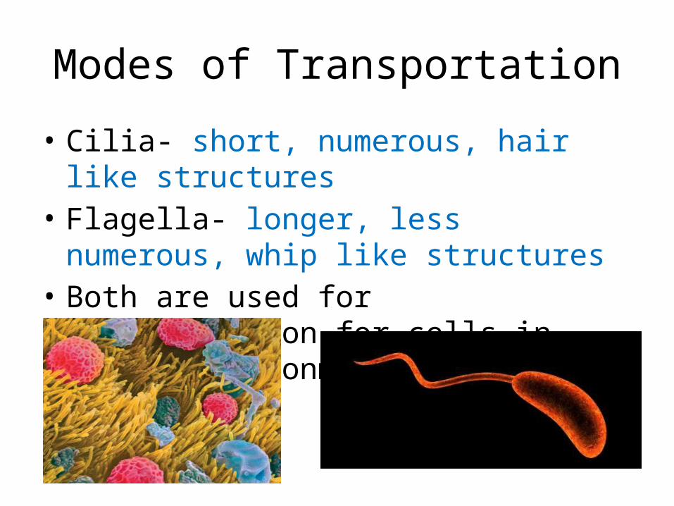
Modes of Transportation
• Cilia- short, numerous, hair like structures• Flagella- longer, less numerous, whip like
structures• Both are used for transportation for cells in
watery environments
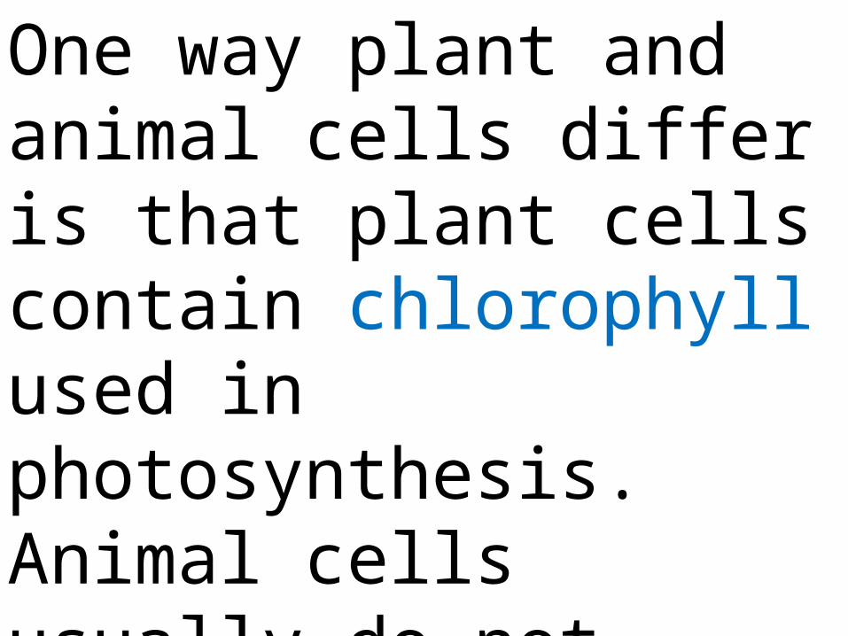
One way plant and animal cells differ is that plant cells contain chlorophyll used in photosynthesis. Animal cells usually do not contain chloroplasts and lack cell walls.

The cell can be compared to a factory because each organelle has a job to do. One of the major jobs is the protein synthesis which begins in the nucleus with information contained in DNA.


![CELL THEORY A Brief History. Robert Hooke named the cell [1665] based on observations of the cell walls of cork tissue](https://img.dokumen.tips/doc/110x75/56649f265503460f94c3dff5/cell-theory-a-brief-history-robert-hooke-named-the-cell-1665-based-on-observations.jpg)


