Embed Size (px)
Citation preview

Chapter 6 Human Heredity by Michael Cummings ©2006 Brooks/Cole-Thomson Learning
Chapter 6Cytogenetics: Karyotypes and
Chromosomal Aberrations
Chapter 6 Human Heredity by Michael Cummings ©2006 Brooks/Cole-Thomson Learning
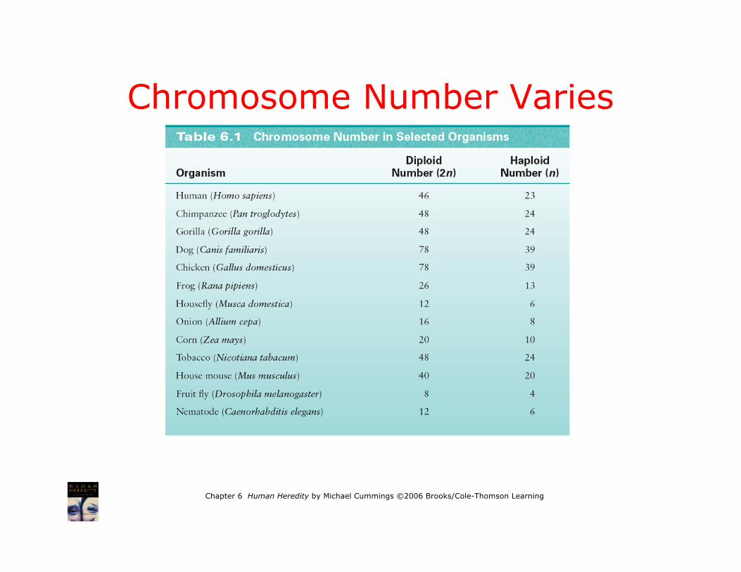
Chromosome Number Varies

Chapter 6 Human Heredity by Michael Cummings ©2006 Brooks/Cole-Thomson Learning
Human Chromosomes
• Diploid number(2N) = 46• 23 pairs
–22 pairs of autosomes–XX in females and XY in
males• Gametes (eggs and
sperm) are haploid andhave 23 chromosomes
Fig. 6.4

Chapter 6 Human Heredity by Michael Cummings ©2006 Brooks/Cole-Thomson Learning
The Centromere Divides theChromosome into Two Arms
Fig. 6.2
Fig. 6-2, p.123
17
ACROCENTRICMETACENTRIC SUBMETACENTRIC
Stalk
CentromereSatellite
qLongArm(q)
ShortArm(p) p
p
q
213

Chapter 6 Human Heredity by Michael Cummings ©2006 Brooks/Cole-Thomson Learning
At metaphase of mitosis

Chapter 6 Human Heredity by Michael Cummings ©2006 Brooks/Cole-Thomson LearningCredit: © Dr. K.G. Murti/Visuals Unlimited 203283
Telomeres(yellow)

Chapter 6 Human Heredity by Michael Cummings ©2006 Brooks/Cole-Thomson Learning
Cells Used for ChromosomalAnalysis
• Any cell with a nucleus• Lymphocytes• Skin cells• Tumor cells• Amniotic cells• Chorionic villi• Rare fetal cells from maternal blood

Chapter 6 Human Heredity by Michael Cummings ©2006 Brooks/Cole-Thomson Learning
Karyotype
• Chromosomesphotographedduringmetaphaseand arrangedin a standardsequence
Fig. 6.3

Chapter 6 Human Heredity by Michael Cummings ©2006 Brooks/Cole-Thomson Learning
Creating a Karyotype
Fig. 6.6

Chapter 6 Human Heredity by Michael Cummings ©2006 Brooks/Cole-Thomson Learning
Stains and Dyes
• Used to produce a pattern of bandsspecific to each type of chromosome
• One common method is G-banding–Treated with trypsin–Stained with Giemsa stain–Metaphase chromosomes approximately
550 bands–More bands can be produced in early
metaphase and late prophasechromosomes

Chapter 6 Human Heredity by Michael Cummings ©2006 Brooks/Cole-Thomson Learning
BandingTechniques
Stains anddyes are usedto identify thechromosomes
Fig. 6.8

Chapter 6 Human Heredity by Michael Cummings ©2006 Brooks/Cole-Thomson Learning
Fig. 6-5, p.124
Banding patterns allow individual chromosomes to be identified
Provide location of genes
Information about structural aberrations