Embed Size (px)
Citation preview

CIRCULATORY ANDRESPIRATORY SYSTEMS
FOCUS CONCEPT: Structure and FunctionAs you read, note how the structural features of the organs in thecirculatory and respiratory systems are related to the transport andexchange of materials.
This photograph shows the air sacs of a human lung. (SEM 780!)
47-1 The Circulatory System
47-2 Blood
47-3 The Respiratory System
930
CHAPTER 47
Copyright © by Holt, Rinehart and Winston. All rights reserved.

Copyright © by Holt, Rinehart and Winston. All rights reserved.
T H E C I RC U L ATO RY S YS T E MMost of the cells in the human body are not in direct contact
with the external environment. The circulatory system acts as
a transport service for these cells. Two fluids move through
the circulatory system: blood and lymph. The blood, heart,
and blood vessels form the cardiovascular system. The
lymph, lymph nodes, and lymph vessels form the lymphatic
system. The cardiovascular system and lymphatic system
collectively make up the circulatory system.
THE HEARTThe central organ of the cardiovascular system is the heart, themuscular organ that pumps blood through an intricate network ofblood vessels. The heart beats more than 2.5 billion times in anaverage life span. Yet this organ that does so much work is slightlylarger than a fist. The heart lies within the thoracic (chest) cavity,behind the sternum (breastbone) and between the two lungs. Atough, saclike membrane called the pericardium surrounds theheart and secretes a fluid that reduces friction as the heart beats.
Notice in Figure 47-1 that a septum vertically divides the heart intotwo sides. The right side pumps blood to the lungs, and the left sidepumps blood to the other parts of the body. Each side of the heart isdivided into an upper and lower chamber. Each upper chamber iscalled an atrium, and each lower chamber is called a ventricle.
931C I R C U L A T O R Y A N D R E S P I R A T O R Y S Y S T E M S
!
Describe the structure and function of the human heart.
"
Trace the flow of blood through the heart and body.
#
Distinguish between arteries,veins, and capillaries in terms of their structure and function.
$
Distinguish between pulmonarycirculation and systemic circulation.
!
Describe the functions of thelymphatic system.
O B J E C T I V E S
SECTION
47-1
The septum prevents mixing of bloodfrom the two sides of the heart, andthe valves ensure that blood flows inonly one direction.
FIGURE 47-1
Pulmonary (rightsemilunar) valve
Aortic (leftsemilunar) valve
Rightatrium
Tricuspid (right atrioventricular) valve
Right ventricle
Leftatrium
Mitral (left atrioventricular)valve
Left ventricle
Septum
RIGHT LEFT

A one-way valve separates each atrium from the ventriclebeneath it. These valves are called atrioventricular (AY-tree-oh-ven-TRIH-kyuh-luhr) (AV) valves. They consist of flaps of tissue that openin only one direction. The AV valve on the right side is called thetricuspid valve. The mitral valve, also called the bicuspid valve, ison the left. As the ventricles pump, blood pressure closes the AVvalves, preventing blood from flowing backward from the ventri-cles to the atria.
From the ventricles, blood flows out of the heart into large ves-sels. A semilunar (semee-LOON-uhr) (SL) valve separates the ventriclesfrom these large vessels on each side of the heart. The SL valve onthe right side is known as the pulmonary valve, and the SL valve onthe left side is known as the aortic valve. The SL valves preventblood from flowing back into the ventricles when the heart relaxes.
Circulation in the Heart Refer to Figure 47-2 to trace the path of the blood as it circulatesthrough the heart. Blood returning to the heart from parts of thebody other than the lungs has a high concentration of carbon diox-ide and a low concentration of oxygen. This deoxygenated bloodenters the right atrium. Notice in Figure 47-2 that the flow of bloodon the right side of the heart is illustrated with a blue arrow repre-senting deoxygenated blood, which has a deep bluish-red color.
The right atrium pumps the deoxygenated blood into the rightventricle. The muscles of the right ventricle contract and force theblood into the pulmonary arteries, which lead to the lungs. In thelungs, the carbon dioxide diffuses out of the blood and oxygen dif-fuses into the blood. The oxygenated blood returns to the left
C H A P T E R 4 7932Copyright © by Holt, Rinehart and Winston. All rights reserved.
Trace the path of blood through theheart. Which side of the heart containsdeoxygenated blood? Notice that illus-trations of a heart are drawn as if theheart were in a person facing you, thatis, the left side of the heart is shownon the right as you face it, and theright side of the heart is on the left asyou face it.
FIGURE 47-2
Right atrium
Tricuspid valve
Right ventricle
Left atrium
Mitral valve
Left ventricle
Superiorvena cava
Inferior vena cava
Descending aorta
Aorta
Pulmonaryarteries
Pulmonaryveins
Determining Heart RateMaterials stopwatch or clockwith second handProcedure1. Have your partner find the pulse
in your wrist and count yourheartbeats for 15 seconds whileyou are seated. Calculate yourresting heart rate in beats perminute.
2. Have your partner count yourheartbeats for 15 seconds whileyou are standing. Calculate yourheart rate in beats per minute.
3. Have your partner count yourheartbeats for 15 seconds whileyou are lying down. Calculateyour heart rate in beats perminute.
Analysis What causes yourpulse? What causes the change inyour heart rate in each position?
Quick Lab

atrium of the heart. Notice in Figure 47-2 that the flow of blood onthe left side of the heart is illustrated with a red arrow represent-ing oxygenated blood, which has a bright red color.
The oxygenated blood is then pumped into the left ventricle.Contraction of the muscular walls of the left ventricle force theblood into a large blood vessel called the aorta. From the aorta,blood is transported to all parts of the body except the lungs. Theleft ventricle is the thickest chamber of the heart because it has todo the most work to pump blood to all parts of the body.
Control of the HeartbeatThe heart consists of muscle cells that contract in waves. When thefirst group of cells are stimulated, they in turn stimulate neighbor-ing cells. Those cells then stimulate more cells. This chain reactioncontinues until all the cells contract. The wave of activity spreadsin such a way that the atria and the ventricles contract in a steadyrhythm. The first group of heart-muscle cells that get stimulated liein an area of the heart known as the sinoatrial node, shown inFigure 47-3.
The sinoatrial (SIEN-oh-AY-tree-uhl) (SA) node is a group of specializedheart-muscle cells located in the right atrium. These muscle cellsspontaneously initiate their own electrical impulse and contract.The SA node is often called the pacemaker because it regulates therate of contraction of the entire heart. The electrical impulse initi-ated by the SA node subsequently reaches another special area ofthe heart, known as the atrioventricular (AV) node. The AV node islocated in the septum between the atria, as shown in Figure 47-3. TheAV node relays the electrical impulse to the muscle cells that makeup the ventricles. As a result, the ventricles contract a fraction of asecond after the atria, completing one full heartbeat. In an averageadult at rest, the heart beats about 70 times each minute.
A heartbeat has two phases. Phase one, called systole (SIS-tohl),occurs when the ventricles contract, closing the AV valves andopening the SL valves to pump blood into the two major vesselsthat exit the heart. Phase two, called diastole (DIE-a-stohl), occurswhen the ventricles relax, allowing the back pressure of the bloodto close the SL valves and opening the AV valves. The closing ofthese two heart valves results in the characteristic lubb dup soundwe call a heartbeat. If one of the valves fails to close properly, someblood may flow backward, creating a different sound, which isknown as a heart murmur.
Blood VesselsThe circulatory system is known as a closed system because theblood is contained within either the heart or the blood vessels atall times. This type of system differs from an open system, in whichblood leaves the vessels and circulates within tissues throughoutthe organism’s body. The blood vessels that are part of the closedcirculatory system of humans form a vast network to help keep theblood flowing in one direction.
933C I R C U L A T O R Y A N D R E S P I R A T O R Y S Y S T E M S
Copyright © by Holt, Rinehart and Winston. All rights reserved.
Two areas of specialized tissue, knownas nodes, control the heartbeat. A per-son whose SA node is defective canhave an operation to implant an artifi-cial pacemaker. An artificial pacemakercan also help a defective AV node.Why is the term artificial pacemakerappropriate?
FIGURE 47-3
Sinoatrial node
Atrioventricular node

Arteries and Blood PressureEach beat of the heart forces blood through blood vessels. Thelarge, muscular vessels that carry blood away from the heart arecalled arteries. As shown in Figure 47-4, the thick walls of the ar-teries have three layers: an inner endothelial layer, a middle layer ofsmooth muscle, and an outer layer of connective tissue. This struc-ture gives arteries a combination of strength and elasticity, whichallows them to stretch as pressurized blood enters from the heart.You can feel this stretching of arteries—it is your pulse.
Contraction of the heart propels the blood through the arterieswith considerable force. The force that blood exerts against thewalls of a blood vessel is known as blood pressure. Blood pressureis always highest in the two main arteries that leave the heart. It isusually measured in the artery that supplies blood to the arm. Tomeasure blood pressure, a trained person inflates a cuff that isplaced around a person’s arm, temporarily stopping the flow ofblood through the artery. Connected to the cuff is a gauge contain-ing a column of mercury (Hg) that rises as the pressure in the cuffincreases. The trained person then releases the air in the cuffslowly while listening to the artery with a stethoscope and watch-ing the column of mercury. The first sounds of blood passingthrough the artery mean that the ventricles have pumped withenough force to momentarily overcome the pressure exerted bythe cuff. This is known as the systolic pressure, or the pressure ofthe blood when the ventricles contract. In a normal adult, the sys-tolic pressure is about 120 mm of Hg for males and 110 mm of Hgfor females. Continuing to release the air in the cuff, the trainedperson next listens for the disappearance of sound, which indi-cates a steady flow of blood through the artery in the arm. At thispoint, the pressure of the blood is sufficient to keep the arteriesopen constantly even with the ventricles relaxed. This is known asdiastolic pressure. In a normal adult, the diastolic pressure isabout 80 mm of Hg for males and 70 mm of Hg for females.
High blood pressure, or hypertension, is a leading cause ofdeath in many countries. Blood pressure that is higher than normalplaces a strain on the walls of the arteries and increases thechance that a vessel will burst. Because people with chronic highblood pressure often display no external symptoms, hypertensionis sometimes referred to as the silent killer.
Capillaries and VeinsRecall that when the left ventricle contracts, it forces blood intothe aorta, the body’s largest artery. From the aorta, blood travelsthrough a network of smaller arteries, which in turn divide andform even smaller vessels, called arterioles. The arterioles branchinto a network of tiny vessels, called capillaries. A capillary isshown in Figure 47-5.
The network formed by capillaries is so extensive that all of theapproximately 100 trillion cells in the body lie within about 125 µm
C H A P T E R 4 7934Copyright © by Holt, Rinehart and Winston. All rights reserved.
The diameter of a capillary is so smallthat red blood cells must move singlefile through these vessels, as shown inthis photograph (1,200!). All exchangeof nutrients and waste between bloodand cells occurs across the thin walls ofthe capillaries.
FIGURE 47-5
Connectivetissue
Smooth muscle tissue
Endothelial tissue
ARTERY
Notice the thick muscular layer of anartery. The layers of the artery wall areseparated by elastic tissue. This tissueprovides strength, preventing systolicpressure from bursting the artery.
FIGURE 47-4

Copyright © by Holt, Rinehart and Winston. All rights reserved.
935C I R C U L A T O R Y A N D R E S P I R A T O R Y S Y S T E M S
Connectivetissue
Smooth muscle tissue
Endothelial tissue
VEIN
VEINConnectivetissue
Smoothmuscle
Endothelium
Valve
This figure shows the structure of thevalves in veins. Many veins have valvesto keep the blood flowing in one direc-tion. If these valves fail to close prop-erly, some blood can leak backwardand expand a weak area of the vein.This results in a condition known asvaricose veins.
FIGURE 47-7
Like an artery, a vein has three layers.Notice the outer layer of connective tis-sue, the middle layer of smooth muscle,and the inner layer of endothelial tissue. Compare this figure with Figure 47-4. How do the three layers ina vein compare with those in an artery?
FIGURE 47-6
of a capillary. This close association between capillaries and cellsallows for rapid exchange of materials. Capillary walls are only onecell thick; gases and nutrients can diffuse through these thin walls.Wherever the concentration of oxygen or nutrients is higher in theblood than in the surrounding cells, the substance diffuses fromthe blood into the cells. Wherever the concentrations of carbondioxide and wastes are higher in the cells than in the blood, thesesubstances diffuse from the cells into the blood.
Blood flows through capillaries that merge to form larger vesselscalled venules (VEN-yoolz). Several venules in turn unite to form avein, a large blood vessel that carries blood to the heart. Veinsreturning deoxygenated blood from the lower parts of the bodymerge to form the inferior vena cava. Veins returning deoxy-genated blood from the upper parts of the body merge to form thesuperior vena cava. Refer back to Figure 47-2 and locate the inferiorvena cava and the superior vena cava.
As you can see in Figure 47-6, although the walls of the veins arecomposed of three layers, like those of the arteries, they are thinnerand less muscular. By the time blood reaches the veins, it is undermuch less pressure than it was in the arteries. With less pressurebeing exerted in the veins, the blood could flow backward and dis-rupt the pattern of circulation. To prevent that, valves in the veinshelp keep the blood flowing in one direction. Many veins passthrough skeletal muscle. When these muscles contract, they are ableto squeeze the blood through the veins. When these muscles relax,the valves can close, thus preventing the blood from flowing back-ward. Figure 47-7 shows the structure of a valve in a vein.
PATTERNS OF CIRCULATIONThe English scientist William Harvey (1578–1657) first showed thatthe heart and the blood vessels form one continuous, closed systemof circulation. He also reasoned that this system consists of two pri-mary subsystems: pulmonary circulation, in which the blood trav-els between the heart and lungs, and systemic circulation, in whichthe blood travels between the heart and all other body tissues.
Pulmonary CirculationDeoxygenated blood returning from all parts of the body exceptthe lungs enters the right atrium, where it is then pumped into theright ventricle. When the right ventricle contracts, the deoxy-genated blood is sent through the pulmonary artery to the lungs.The pulmonary artery is the only artery that carries deoxygenatedblood. The pulmonary artery branches into two smaller arteries,with one artery going to each lung. These arteries branch into arte-rioles and then into capillaries in the lungs.
In the lungs, carbon dioxide diffuses out of the capillaries andoxygen diffuses into the capillaries. The oxygenated blood then

flows into venules, which merge into the pulmonary veins thatlead to the left atrium of the heart. The pulmonary veins are theonly veins that carry oxygenated blood. From the left atrium, bloodis pumped into the left ventricle and then to the body through theaorta, supplying the cells with oxygen. In Figure 47-8, trace thepath blood takes as it passes through pulmonary circulation.
Systemic CirculationSystemic circulation is the movement of blood between the heartand all parts of the body except the lungs. Trace the path blood fol-lows in systemic circulation in Figure 47-9. Notice that oxygenatedblood is pumped out of the left ventricle and into the aorta. From theaorta, blood flows into other subsystems of systemic circulation.
Coronary circulation is one subsystem of systemic circulationthat supplies blood to the heart itself. The heart muscle is thick,and oxygen and nutrients must be supplied to each cell. If theblood supply to the heart is reduced or cut off, muscle cells willdie. This can happen when an artery is blocked by a blood clot orby atherosclerosis (ATH-uhr-oh-skler-OH-sis), a disease characterized bythe buildup of fatty materials on the interior walls of the coronaryartery. If either type of blockage reduces the flow of blood to theheart muscle cells, a heart attack will result.
Renal circulation, another subsystem of systemic circulation,supplies blood to the kidneys. Nearly one-fourth of the blood thatis pumped into the aorta by the left ventricle flows to the kidneys.The kidneys filter waste from the blood.
Hepatic portal circulation is a subsystem of systemic circula-tion. Nutrients are picked up by capillaries in the small intestineand are transported by the blood to the liver. Excess nutrients arestored in the liver for future needs. The liver receives oxygenatedblood from a large artery that branches from the aorta.
C H A P T E R 4 7936Copyright © by Holt, Rinehart and Winston. All rights reserved.
The pulmonary circulation between theheart and the lungs involves the pul-monary arteries and the pulmonaryveins. Deoxygenated blood flows fromthe right side of the heart to the lungs.Oxygenated blood is returned to theleft side of the heart from the lungs.This is the opposite of systemic andcoronary blood flow, in which oxygen-rich blood flows from the heart andoxygen-poor blood is returned to theheart.
FIGURE 47-8
From headTo head
From body
To body
Pulmonary veins Pulmonary veins
Pulmonary arteries
Rightatrium
Superior vena cava
Left ventricle
Rightventricle
Leftatrium
Heart
Aorta
Capillariesof intestines
Capillariesof legs
Capillariesof liver
Coronarycirculation
Inferiorvena cava
Hepaticportalcirculation
Renalcirculation
Notice three subsystems of systemic cir-culation. Other subsystems transportblood between the heart and the head,arms, organs, and legs.
FIGURE 47-9

Copyright © by Holt, Rinehart and Winston. All rights reserved.
LYMPHATIC SYSTEMIn addition to the cardiovascular system, the circulatory system alsoincludes the lymphatic system. One function of the lymphatic sys-tem is to return fluids that have collected in the tissues to the blood-stream. Fluids diffuse through the capillary walls just as oxygen andnutrients do. Some of these fluids pass into cells, some return to thecapillaries, and some remain in the intercellular spaces.
Excess fluid in the tissues moves into the tiny vessels of the lym-phatic system; this fluid is called lymph. Lymph vessels merge toform larger vessels. The lymph vessels are similar in structure tocapillaries, and the larger lymph vessels are similar in structure toveins. However, an important difference exists between blood ves-sels and lymph vessels. As you learned earlier, blood vessels forma complete circuit so that blood passes from the heart to all partsof the body and then back again to the heart. In contrast, lymph ves-sels form a one-way system that returns fluids collected in the tis-sues back to the bloodstream. In addition, the lymphatic systemhas no pump like the heart. Like the blood in veins, lymph must bemoved through the vessels by the squeezing of skeletal muscles.Like veins, the larger lymph vessels have valves to prevent the fluidfrom moving backward.
Notice in Figure 47-10 that lymph vessels form a vast networkthat extends throughout the body. The lymph that travels in thesevessels is a transparent yellowish fluid, much like the liquid part ofthe blood. As the lymph travels through these vessels on its way tothe heart, it passes through small organs known as lymph nodes.Notice in Figure 47-10 that lymph nodes are like beads on a string.These nodes filter the lymph as it passes, trapping foreign parti-cles, microorganisms, and other tissue debris. Lymph nodes alsostore lymphocytes, white blood cells that are specialized to fightdisease. When a person has an infection, the nodes may becomeinflamed, swollen, and tender because of the increased number of lymphocytes.
937C I R C U L A T O R Y A N D R E S P I R A T O R Y S Y S T E M S
Like the cardiovascular system, the lym-phatic system forms a vast network.Concentrated in certain regions of thisnetwork are lymph nodes that containsome of the disease-fighting cells of theimmune system.
FIGURE 47-10
1. Identify the structure that prevents blood frommixing between the left and right sides of theheart. Explain what prevents blood from flowingfrom the ventricles backward into the atria.
2. Identify the structure that controls the heartbeat,and describe the process by which it regulatesthe heartbeat.
3. Outline the path that blood follows in pulmonarycirculation.
4. Compare the lymphatic system with the cardio-vascular system.
5. In which blood vessels would you expect to findthe lowest average blood pressure? Explain youranswer.
6. CRITICAL THINKING Some babies are bornwith a hole in the septum between the two atria.Based on what you know about blood flowthrough the heart, explain why this conditionwould be harmful to the baby.
SECTION 47-1 REVIEW
Lymphnodes

C H A P T E R 4 7938
Literature & Life
he study of medicine inHarvey’s time normally con-
sisted of studying the works ofancient Greek doctors and philoso-phers, not direct observation.Harvey’s work as a physician andanatomist was revolutionarybecause he actually observed theaction of the heart and the move-ment of blood in live animals.
When the chest of a living ani-mal is laid open and the capsulethat immediately surrounds theheart is slit up or removed, theorgan is seen now to move, now to be at rest; there is a time whenit moves, and a time when it ismotionless.
These things are more obvious inthe colder animals, such as toads,frogs, serpents, small fishes, crabs,shrimps, snails, and shell-fish. Theyalso become more distinct in warm-blooded animals, such as the dogand hog, if they be attentively notedwhen the heart begins to flag, tomove more slowly, and, as it were,to die: the movements then becomeslower and rarer, the pauses longer,by which it is made much moreeasy to perceive and unravel whatthe motions really are, and howthey are performed. In the pause, asin death, the heart is soft, flaccid,exhausted, lying, as it were, at rest.
In the motion, and interval inwhich this is accomplished, three
principal circumstances are to benoted:
1. That the heart is erected, andrises upwards to a point, so that atthis time it strikes against the breastand the pulse is felt externally.
2. That it is everywhere con-tracted, but more especially towardsthe sides so that it looks narrower,relatively longer, more drawntogether. The heart of an eel taken
out of the body of the animal andplaced upon the table or the hand,shows these particulars; but thesame things are manifest in thehearts of all small fishes and ofthose colder animals where theorgan is more conical or elongated.
3. The heart being grasped inthe hand, is felt to become harderduring its action. Now this hard-ness proceeds from tension, pre-cisely as when the forearm isgrasped, its tendons are perceivedto become tense and resilient whenthe fingers are moved.
4. It may further be observed in
fishes, and the colder blooded ani-mals, such as frogs, serpents, etc.,that the heart, when it moves,becomes of a paler color, when qui-escent of a deeper blood-red color.
From these particulars it appearsevident to me that the motion ofthe heart consists in a certain uni-versal tension—both contraction inthe line of its fibres, and constric-tion in every sense.
Reading for MeaningBecause On the Motion of the Heartand Blood in Animals was written inLatin in the seventeenth century andwas translated into English in 1910,the language is very old-fashioned.Rewrite the last paragraph of thispassage, paraphrasing it in simple,modern language.
Read FurtherIn Harvey’s time, knowledge aboutan animal’s internal anatomy camefrom direct observation. Observa-tions of human anatomy and physi-ology were rare because theobservation techniques were injur-ious and often fatal. What equip-ment and techniques are usedtoday in modern hospitals to makeobservations of human anatomyand physiology that are not inva-sive or harmful to the patient? Howare these techniques used in thediagnosis of illness or disease?
On the Motion of the HeartThis excerpt is from William Harvey’s anatomical essay On the Motion of the Heart and Blood in Animals. It was first presented in 1628.
T
Copyright © by Holt, Rinehart and Winston. All rights reserved.

Copyright © by Holt, Rinehart and Winston. All rights reserved.
939C I R C U L A T O R Y A N D R E S P I R A T O R Y S Y S T E M S
B L O O DBlood is a liquid connective tissue that constitutes the
transport medium of the cardiovascular system. The two main
functions of the blood are to transport nutrients and oxygen to
the cells and to carry carbon dioxide and other waste
materials away from the cells. Blood also transfers heat to the
body surface and plays a major role in defending the body
against disease.
COMPOSITION OF BLOODBlood is composed of a liquid medium and blood solids. Bloodsolids consist of red blood cells, white blood cells, and platelets.The liquid makes up about 55 percent of the blood, and bloodsolids make up the remaining 45 percent. A healthy adult has about4 to 5 L of blood in his or her body.
PlasmaPlasma, the liquid medium, is a sticky, straw-colored fluid that isabout 90 percent water. Cells receive nourishment from dissolvedsubstances carried in the plasma. These substances, which mayinclude vitamins, minerals, amino acids, and glucose, are absorbedfrom the digestive system and transported to the cells. Plasma alsocarries hormones and brings wastes from the cells to the kidneysor the lungs to be removed from the body.
Proteins are carried in the plasma and have various functions.Some of the proteins in the plasma are essential for the formationof blood clots. Another protein, called albumin, plays an importantrole in the regulation of osmotic pressure between plasma andblood cells and between plasma and tissues. Other proteins, calledantibodies, help the body fight disease.
Red Blood CellsRed blood cells, or erythrocytes (uh-RITH-ruh-siets), shown in Figure 47-11,transport oxygen to cells in all parts of the body. Red blood cells areformed in the red marrow of bones. Red blood cells synthesize largeamounts of an iron-containing protein called hemoglobin. Hemoglo-bin is the molecule that actually transports oxygen and, to a lesserdegree, carbon dioxide. During the formation of a red blood cell, itscell nucleus and organelles disintegrate. The mature red blood cellbecomes little more than a membrane sac containing hemoglobin.
!
List the components of blood.
"
Distinguish between red blood cells, white blood cells,
and platelets in terms of their structure and function.
#
Summarize the process of blood clotting.
$
Explain what determines the compatibility of blood types
for transfusion.
O B J E C T I V E S
SECTION
47-2
Notice that a mature red blood cell(RBC) is disk-shaped and is concave onboth sides (5,250!). A red blood cell islittle more than a cell membrane filledwith hemoglobin. How is this structurerelated to its function?
FIGURE 47-11

Because red blood cells lack nuclei, they cannot divide and theyhave a limited survival period, usually 120 to 130 days. Of the morethan 30 trillion red blood cells circulating throughout the body atone time, 2 million disintegrate every second. To replace them,new ones form at the same rate in the red marrow of bones. Someparts of the disintegrated red blood cells are recycled. For exam-ple, the iron portion of the hemoglobin molecule is carried in theblood to the marrow, where it is reused in new red blood cells.
White Blood CellsWhite blood cells, or leukocytes (LOO-kuh-siets), help defend the bodyagainst disease. They are formed in the red marrow, the lymphnodes, and the spleen. White blood cells are larger than red bloodcells and significantly less plentiful. Each cubic millimeter of bloodnormally contains about 4 million red blood cells and 7,000 whiteblood cells. White blood cells can squeeze their way through open-ings in the walls of blood vessels and into the intercellular fluid. Inthat way, white blood cells can reach the site of infection and helpdestroy invading microorganisms.
Notice in Figure 47-12 that a white blood cell has a very differentstructure from that of a red blood cell. For instance, a white bloodcell may be irregularly shaped and may have a rough outer surface.There are other differences between red blood cells and white bloodcells as well. In contrast with the short-lived red blood cells, whiteblood cells may function for years. And while there is only one typeof red blood cell, there are several types of white blood cells.
The white blood cell shown in Figure 47-12 is the type of whiteblood cell known as a phagocyte (FA-guh-siets). Phagocytes are cellsthat engulf invading microorganisms. Locate the microorganismsthat are being engulfed by the phagocyte in Figure 47-12.Another type of white blood cell produces antibodies. Antibodiesare proteins that help destroy substances, such as bacteria andviruses, that enter the body and can cause disease. When a personhas an infection, the number of white blood cells can double.
C H A P T E R 4 7940Copyright © by Holt, Rinehart and Winston. All rights reserved.
Some white blood cells, like the phago-cyte shown in blue, engulf and destroyinvading microorganisms.
FIGURE 47-12
Copyright © by Holt, Rinehart and Winston. All rights reserved.
941C I R C U L A T O R Y A N D R E S P I R A T O R Y S Y S T E M S
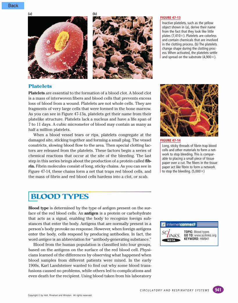
PlateletsPlatelets are essential to the formation of a blood clot. A blood clotis a mass of interwoven fibers and blood cells that prevents excessloss of blood from a wound. Platelets are not whole cells. They arefragments of very large cells that were formed in the bone marrow.As you can see in Figure 47-13a, platelets get their name from theirplatelike structure. Platelets lack a nucleus and have a life span of7 to 11 days. A cubic micrometer of blood may contain as many ashalf a million platelets.
When a blood vessel tears or rips, platelets congregate at thedamaged site, sticking together and forming a small plug. The vesselconstricts, slowing blood flow to the area. Then special clotting fac-tors are released from the platelets. These factors begin a series ofchemical reactions that occur at the site of the bleeding. The laststep in this series brings about the production of a protein called fib-rin. Fibrin molecules consist of long, sticky chains. As you can see inFigure 47-14, these chains form a net that traps red blood cells, andthe mass of fibrin and red blood cells hardens into a clot, or scab.
BLOOD TYPESBlood type is determined by the type of antigen present on the sur-face of the red blood cells. An antigen is a protein or carbohydratethat acts as a signal, enabling the body to recognize foreign sub-stances that enter the body. Antigens that are normally present in aperson’s body provoke no response. However, when foreign antigensenter the body, cells respond by producing antibodies. In fact, theword antigen is an abbreviation for “antibody-generating substance.”
Blood from the human population is classified into four groups,based on the antigens on the surface of the red blood cell. Physi-cians learned of the differences by observing what happened whenblood samples from different patients were mixed. In the early1900s, Karl Landsteiner wanted to find out why some blood trans-fusions caused no problems, while others led to complications andeven death for the recipient. Using blood taken from his laboratory
Inactive platelets, such as the yellowobject shown in (a), derive their namefrom the fact that they look like littleplates (7,410!). Platelets are colorlessand contain chemicals that are involvedin the clotting process. (b) The plateletschange shape during the clotting proc-ess. When activated, the platelets settleand spread on the substrate (4,900!).
FIGURE 47-13
Long, sticky threads of fibrin trap bloodcells and other materials to form a net-work to stop bleeding. This is compar-able to placing a small piece of tissuepaper over a cut. The fibers in the tissuepaper act like fibrin to form a networkto stop the bleeding. (5,000!)
FIGURE 47-14
(a) (b)
TOPIC: Blood typesGO TO: www.scilinks.orgKEYWORD: HM941

workers, Landsteiner made observations similar to those you seein Figure 47-15. He noticed that mixing blood samples from twopeople sometimes resulted in the cells clumping together, or agglu-tinating. However, at other times no clumping or agglutinationoccurred when blood samples were mixed. Landsteiner reasonedthat clumping occurred when blood samples of two different bloodtypes were mixed.
When samples of two different blood types are mixed together,reactions occur between the antigens on the red blood cells and theantibodies in the plasma, causing the cells to agglutinate. Whensamples of the same blood type are mixed, no reaction occurs andthe blood cells do not agglutinate. Landsteiner’s observations led tothe classification of human blood by blood types. Blood typinginvolves identifying the antigens in a sample. Three of the mostimportant human antigens are called A, B, and Rh. The A-B-O systemof blood typing, described below, is based on the A and B antigens.
A-B-O SystemThe A-B-O system is a means of classifying blood by the antigenslocated on the surface of the red blood cells and the antibodies cir-culating in the plasma. As shown in Table 47-1, an individual’s redblood cells may carry an A antigen, a B antigen, both A and B anti-gens, or no antigen at all. These antigen patterns are called bloodtypes A, B, AB, and O, respectively.
Notice in Table 47-1 that an individual with type A blood also hasanti-B antibodies against type B blood. If type B blood is given to arecipient with type A blood, the recipient’s anti-B antibodies will
C H A P T E R 4 7942Copyright © by Holt, Rinehart and Winston. All rights reserved.
Notice that there is no agglutination ofred blood cells in the slide in (a), whereblood samples from two people withthe same blood type were mixed(20!). Compare this with the slide in(b), where blood samples from twopeople with different blood types weremixed (20!).
FIGURE 47-15
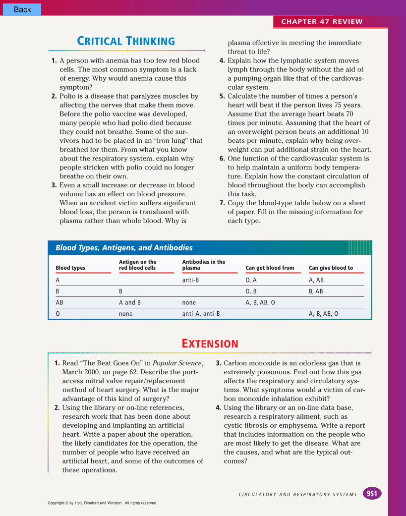
TABLE 47-1 Blood Types, Antigens, and Antibodies
Blood types
A
B
AB
O
Antigen on the red blood cells
A
B
A and B
none
Antibodies in theplasma
anti-B
anti-A
none
anti-A, anti-B
Can get blood from
O, A
O, B
A, B, AB, O
O
Can give blood to
A, AB
B, AB
AB
A, B, AB, O
(a) (b)

Copyright © by Holt, Rinehart and Winston. All rights reserved.
943C I R C U L A T O R Y A N D R E S P I R A T O R Y S Y S T E M S
react with the B antigens on the donated red blood cells and theblood will agglutinate. In addition, the donor’s type B blood has anti-A antibodies. Their presence will compound the antigen-antibody reaction in the recipient. The net result will be agglutinatedblood that will block the flow of blood through the vessels. For thisreason, transfusion recipients must receive blood that is compatiblewith their own. Based on the information in Table 47-1, why can aperson with type AB blood receive blood from any of the four types?
Rh SystemAn antigen that is sometimes present on the surface of red bloodcells is the Rh factor, named after the rhesus monkey in which it was first discovered. Eighty-five percent of the United States’ popu-lation is Rh-positive (Rh+), meaning that Rh antigens are present.People who do not have Rh antigens are called Rh-negative (Rh– ).
If an Rh– person receives a transfusion of blood that has Rh+ anti-gens, antibodies will react with the antigen and agglutination willoccur. The most serious problem with Rh incompatibility occursduring pregnancy. If the mother is Rh– and the father is Rh+, thechild may inherit the dominant Rh+ allele from the father. Theblood supplies of the mother and the fetus are separated duringpregnancy, but during delivery, a small amount of the fetus’s Rh+
blood may reach the mother’s bloodstream. If this happens, themother will develop antibodies to the Rh factor. If a second Rh+
child is conceived later, the mother’s antibodies can cross the pla-centa and attack the blood of the fetus. This condition is called erythroblastosis fetalis. The fetus may die as a result of this condi-tion, or if the child is born alive, he or she may need an immediatetransfusion of Rh+ blood.
To prevent this condition, an Rh– mother of an Rh+ child can begiven antibodies to destroy any Rh+ cells that have entered herbloodstream from the fetus. These antibodies must be adminis-tered to the mother within three days after the birth of her first Rh+
child. By destroying any Rh+ cells in her bloodstream, any dangerto a second child is prevented because the mother will not makeany antibodies against the blood cells of the Rh+ fetus.
1. What is plasma? Name at least one major func-tion of plasma.
2. Distinguish between the three solid componentsof the blood.
3. Identify the stages and structures involved in theclotting process.
4. Explain why a pregnant woman should know herblood type and the blood type of her baby’sfather.
5. Which blood type, in terms of the A-B-O and Rhantigens, can be donated to all others? Why?
6. CRITICAL THINKING Why do you think somepeople turn pale when they are frightened?
SECTION 47-2 REVIEW
Identifying OffspringMaterials pencil, paperProcedure Two babies arebelieved to have been swapped atbirth in error. Blood samples weretaken from each of the parents andbabies. The following results wereobtained from the blood samples:Family 1: mother, type B; father,
type O; baby, type AFamily 2: mother, type O; father,
type A; baby, type ODesign a chart or data table thatcorrectly pairs the biological parents with their baby.Analysis Are the babies with thecorrect biological parents? How doyou know?
Quick Lab

Copyright © by Holt, Rinehart and Winston. All rights reserved.
C H A P T E R 4 7944
T H E R E S P I R A T O R YS Y S T E MYou have read how the blood transports oxygen from the lungs
to cells and carries carbon dioxide from the cells to the lungs. It
is the function of the respiratory system to transport gases to
and from the cardiovascular system. The respiratory system
involves both external respiration and internal respiration.
External respiration is the exchange of gases between
the atmosphere and the blood. Internal respiration is the
exchange of gases between the blood and the cells of the body.
In Chapter 7, you learned how aerobic respiration involves
the use of oxygen to break down glucose in the cell. In this
section, you will examine the structures and mechanisms that
carry oxygen to the cells for use in aerobic respiration and
that eliminate the carbon dioxide that is produced by the
same process.
THE LUNGSThe lungs are the site of gas exchange between the atmosphere andthe blood. Notice in Figure 47-16 that the right lung has three divi-sions, or lobes. It is slightly heavier than the two-lobed left lung. Thelungs are located inside the thoracic cavity, bounded by the rib cageand the diaphragm. Lining the entire cavity and encasing the lungsare pleura, membranes that secrete a slippery fluid that decreasesfriction from the movement of the lungs during breathing.
The Passage of AirRefer to Figure 47-16 to trace the path air follows from the atmos-phere to the capillaries in the lungs. External respiration begins atthe mouth and at the nose. Air filters through the small hairs of thenose and passes into the nasal cavity, located above the roof of themouth. In the nasal cavity, mucous membranes warm and moistenthe air, which helps prevent damage to the delicate tissues thatform the respiratory system. The walls of the nasal cavity are alsolined with cilia. These cilia trap particles that are inhaled and areeventually swept into the throat, where they are swallowed.
SECTION
47-3
!
Trace the passage of air from theenvironment to the bloodstream.
"
Describe how gases are exchanged in the lungs.
#
Contrast the ways that oxygenand carbon dioxide are
transported in the bloodstream.
$
Summarize the skeletal and muscular changes that occur
during breathing.
!
Describe how the rate of breathing is controlled.
O B J E C T I V E S
TOPIC: Respiratory system
GO TO: www.scilinks.orgKEYWORD: HM944

The moistened, filtered air then moves into the pharynx (FER-inks), a tube at the back of the nasal cavities and the mouth. Thepharynx contains passageways for both food and air. When food isswallowed, a flap of cartilage, called the epiglottis, presses downand covers the opening to the air passage. When air is being takenin, the epiglottis is in an upright position, allowing air to pass intoa cartilaginous tube called the trachea (TRAY-kee-uh). The trachea isabout 10 to 12 cm long and has walls lined with ciliated cells thattrap inhaled particles. The cilia sweep the particles and mucusaway from the lungs toward the throat.
At the upper end of the trachea is the larynx (LER-inks). Soundsare produced when air is forced past two ligaments—the vocalcords—that stretch across the larynx. The pitch and volume of thesound produced varies with the amount of tension on the vocalcords and on the amount of air being forced past them.
The trachea then branches into two bronchi (BRAHN-kie), each ofwhich leads to a lung. The walls of the bronchi consist of smoothmuscle and cartilage and are lined with cilia and mucus. Within thelungs, the bronchi branch into smaller and smaller tubes. The small-est of these tubes are known as bronchioles, which are also linedwith cilia and mucus. Eventually the bronchioles end in clusters oftiny air sacs called alveoli (al-VEE-oh-LIE). A network of capillaries sur-round each alveolus, as you can see in the detailed view shown inFigure 47-16. All exchange of gases in the lungs occurs in the alveoli.To facilitate this exchange, the surface area of the lungs is enormous.Each lung contains nearly 300 million alveoli and has a total surfacearea of 70 m2—about 40 times the surface area of the skin.
945C I R C U L A T O R Y A N D R E S P I R A T O R Y S Y S T E M S
Copyright © by Holt, Rinehart and Winston. All rights reserved.
Trace the passage of air from theatmosphere to the lungs. Oxygen in the air finally reaches the alveoli, thefunctional units of the respiratory sys-tem. All exchange of gases betweenthe respiratory system and the circula-tory system occurs in the alveoli.
FIGURE 47-16
Pharynx
Alveoli
Capillaries
Epiglottis
Larynx
Trachea
Right lung
Left lung
Bronchus
Bronchiole
Heart
Alveoli
Diaphragm

Copyright © by Holt, Rinehart and Winston. All rights reserved.
GAS EXCHANGE ANDTRANSPORT
In the lungs, gases are exchanged between the alveoli and theblood in the capillaries. Oxygen to be transported throughout thebody moves into the bloodstream, and carbon dioxide to be elimi-nated from the body moves into the alveoli.
Gas Exchange in the LungsFigure 47-17 illustrates the direction in which oxygen and carbondioxide move in the alveoli. When air moves into the lungs, theoxygen in the air crosses the thin alveolar membranes as well asthe capillary walls and enters the blood. Carbon dioxide moves inthe opposite direction, crossing the capillary walls and thin alveo-lar membranes and entering the alveoli.
Air moving into the alveoli is rich in oxygen and contains littlecarbon dioxide. In contrast, blood in the capillaries surroundingthe alveoli is low in oxygen and contains high levels of carbon diox-ide. Thus, concentration gradients for both oxygen and carbondioxide exist. Remember from Chapter 5 that substances diffusefrom an area of higher concentration to an area of lower concen-tration. Consequently, oxygen diffuses from the alveoli into theblood, and carbon dioxide diffuses from the blood into the alveoli.The enormous surface area of the alveoli increases the rate of dif-fusion of these two gases.
Hemoglobin and Gas ExchangeWhen oxygen diffuses into the blood, only a small amount dis-solves in the plasma. Most of the oxygen—97 percent—moves intothe red blood cells, where it combines with hemoglobin. Eachhemoglobin molecule contains four iron atoms. Each iron atom can
Alveolus
CO2 molecule
O2 molecule
Capillary
C H A P T E R 4 7946
Because of concentration gradients,oxygen and carbon dioxide diffuseacross the alveoli and capillary walls.
FIGURE 47-17

bind to one oxygen molecule. Thus, one hemoglobin molecule cancarry up to four molecules of oxygen. When oxygenated bloodreaches cells, the oxygen concentration is higher in the blood thanin the cells. Thus, oxygen is released from the hemoglobin and dif-fuses out of the capillaries and into the surrounding cells.
Because the concentration of carbon dioxide is higher in the cells,it diffuses out of the cells and into the blood. Only about 8 percent ofthe carbon dioxide dissolves in the plasma. Approximately 25 per-cent binds to hemoglobin. The remaining 67 percent reacts withwater in the plasma to form carbonic acid. In turn, the carbonic aciddisassociates into bicarbonate ions and a proton. Thus, most of thecarbon dioxide travels in the blood as bicarbonate ions. When theblood reaches the lungs, the series of reactions is reversed. Bicar-bonate ions combine with a proton to form carbonic acid, which inturn forms carbon dioxide and water. The carbon dioxide diffuses outof the capillaries into the alveoli and is exhaled into the atmosphere.
MECHANISM OF BREATHINGBreathing is the process of moving air into and out of the lungs.Inspiration, shown in Figure 47-18a, is the process of taking air intothe lungs. When you take a deep breath, your chest expands asmuscles contract to move the ribs up and outward. At the sametime, your diaphragm, a large skeletal muscle, flattens and pushes
947C I R C U L A T O R Y A N D R E S P I R A T O R Y S Y S T E M S
Copyright © by Holt, Rinehart and Winston. All rights reserved.
The diaphragm, a large skeletal mus-cle that separates the thoracic cavityfrom the abdominal cavity, and themuscles between the ribs control themovement of the thoracic cavity dur-ing breathing. If these muscles wereparalyzed, then inspiration and expi-ration would not occur.
FIGURE 47-18
(a) INSPIRATION (b) EXPIRATION
Rib musclescontract(Rib cageexpands)
Rib musclesrelax(Rib cagecontracts)
Ribs
Diaphragmrelaxed(moves up)
Diaphragmcontracted(moves down)

down on the abdomen. Muscles in the abdominal wall in turn relax.This action provides room for the flattened diaphragm.
When the diaphragm flattens and the ribs are lifted up and out,the volume of the thoracic cavity increases. An increased volumereduces the air pressure within the cavity. At this point, the airpressure inside the thoracic cavity is lower than the air pressureoutside the body. As a result, air from the atmosphere moves intothe lungs.
During expiration, the reverse process takes place, as you cansee in Figure 47-18b. As the diaphragm and rib muscles relax, theelastic tissues of the lungs recoil, deflating the lungs. The size ofthe thoracic cavity decreases. Because the volume is smaller, theair pressure inside the cavity becomes greater than the air pres-sure outside the body. This pressure difference forces air out of thelungs until the pressures are again equal.
Regulation of BreathingThe rate at which oxygen is used depends on the activity of thecells. The greater their activity, the more oxygen they need and thefaster the body needs to breathe. The slower their activity, theslower the body needs to breathe.
The rate of breathing is controlled by the brain and brain stem,which monitors the concentration of carbon dioxide in the blood.As activity increases, high levels of carbon dioxide in the bloodstimulate nerve cells in the brain. The brain stem in turn stimulatesthe diaphragm to increase the breathing rate and depth. When thecarbon dioxide concentration in the blood returns to lower levels,the sensors in the brain send a message to the respiratory musclesto return to a slower breathing rate. All this is controlled subcon-sciously by control centers in the brain. However, a person cantemporarily override the respiratory control system at any time,holding his or her breath until losing consciousness. Then thebrain stem takes control, and normal breathing resumes. Thismechanism allows humans to swim underwater for short periodsand to sleep without concern for breathing.
C H A P T E R 4 7948Copyright © by Holt, Rinehart and Winston. All rights reserved.
1. What structures of the respiratory system prepareair for entry into the lungs?
2. Why does oxygen diffuse from the alveoli intocapillaries and then into cells in the body?
3. Describe the main way carbon dioxide is trans-ported in the blood.
4. How does an increased carbon dioxide concentra-tion in the blood affect expiration and inspiration?
5. What is the adaptive value of having the organ ofgas exchange—in this case, the alveolus—insidethe body?
6. CRITICAL THINKING Why does a single-celledorganism not need a respiratory system?
SECTION 47-3 REVIEW
CHAPTER 47 REVIEW
aorta (933)aortic valve (932)arteriole (934)artery (934)atherosclerosis (936)atrioventricular (AV) node
(933)atrioventricular valve (932)atrium (931)blood pressure (934)
capillary (934)cardiovascular system (931)circulatory system (931)coronary circulation (936)diastole (933)diastolic pressure (934)hepatic portal circulation (936)hypertension (934)inferior vena cava (935)lymph (937)
lymphatic system (931)lymphocyte (937)mitral valve (932)pacemaker (933)pericardium (931)pulmonary circulation (935)pulmonary valve (932)pulmonary vein (936)renal circulation (936)semilunar valve (932)
septum (931)sinoatrial (SA) node (933)superior vena cava (935)systemic circulation (935)systole (933)systolic pressure (934)tricuspid valve (932)vein (935)ventricle (931)venule (935)
! The circulatory system consists of the cardio-vascular system and the lymphatic system.
! The human heart is located in the thoraciccavity and has two atria and two ventricles.
! Heartbeat is initiated by the sinoatrial (SA)node, also known as the pacemaker. A heart-beat has two phases: systole and diastole.
! Blood vessels include arteries, capillaries,and veins. Arteries are thick, muscular ves-sels that transport blood away from theheart. Arteries branch into smaller vesselsknown as arterioles and capillaries.
! Capillaries merge to form venules, which thencollect into veins. Veins return blood to theheart.
! Pulmonary circulation involves blood flowbetween the heart and the lungs.
! Systemic circulation includes the heart, thekidneys, the liver, and all other organs,including skin and muscle.
! The lymphatic system returns intercellularfluid to the heart. Fluid in the lymphaticsystem is called lymph.
SUMMARY/VOCABULARY
Vocabulary
47-1
949C I R C U L A T O R Y A N D R E S P I R A T O R Y S Y S T E M S
antibody (940)antigen (941)blood type (941)
erythrocyte (939)fibrin (941)hemoglobin (939)
leukocyte (940)phagocyte (940)plasma (939)
platelet (941)Rh factor (943)
! Blood is composed of plasma, red bloodcells, white blood cells, and platelets. Redblood cells transport oxygen. White bloodcells help defend the body against disease.Platelets help form blood clots.
! Human blood can be grouped into fourtypes: A, B, AB, and O. In addition, blood iseither Rh-positive or Rh-negative.
Vocabulary
47-2
47-3
alveolus (945)bronchiole (945)bronchus (945)
epiglottis (945)expiration (948)external respiration (944)
inspiration (947)internal respiration (944)
larynx (945)trachea (945)
! Oxygen enters the bloodstream throughthe lungs.
! The epiglottis prevents food from enteringthe trachea during swallowing. The larynxcontains the vocal cords.
! Oxygen and carbon dioxide are exchangedbetween the alveoli and blood and betweenblood and the cells.
! Nearly all the oxygen in the body is trans-ported by hemoglobin. The level of carbondioxide in the blood determines the rate ofbreathing, which is controlled by the brain.
! During inspiration, the thoracic cavityexpands, pulling air into the lungs. Duringexpiration, the thoracic cavity gets smaller,forcing air out of the lungs.
Vocabulary
Copyright © by Holt, Rinehart and Winston. All rights reserved.

CHAPTER 47 REVIEW
REVIEW
Vocabulary1. Distinguish between systolic pressure and
diastolic pressure.2. What do the pulmonary arteries and the aorta
have in common? How are they different?3. What do the pulmonary veins and the in-
ferior vena cava have in common? How arethey different?
4. What do arteries, veins, and capillaries havein common?
5. Identify the solid components of the blood.
Multiple Choice6. The wall that divides the heart vertically is
the (a) ventricle (b) pericardium (c) septum(d) atrium.
7. During systole, blood moves from the (a) ventricles to the atria (b) atria to theveins (c) atria to the ventricles (d) ventriclesto the arteries.
8. Pulmonary circulation involves movement ofblood between the heart and the (a) lungs (b) brain (c) liver (d) kidneys.
9. One of the functions of the lymphatic systemis that it (a) interacts with the respiratorysystem (b) helps the body fight infection (c) consists of a series of two-way vessels (d) transports intercellular fluid away fromthe heart.
10. One function of plasma is to (a) carry sub-stances that nourish cells (b) aid in the forma-tion of blood clots (c) carry the majority of theoxygen supply of the blood (d) defend againstdisease.
11. The function of fibrin is to (a) transport oxy-gen (b) destroy invading microorganisms (c) stimulate the production of antibodies (d) help form a blood clot.
12. A person who has no antigens present on thered blood cells has blood type (a) AB Rh+ (b) O Rh– (c) A Rh+ (d) O Rh+.
13. During internal respiration, gases are (a) exchanged between the atmosphere andthe blood (b) exchanged between the bloodand the cells (c) produced by the heart (d) warmed and moistened.
14. Gases diffuse (a) from an area of high concen-tration to an area of low concentration (b) froman area of low concentration to an area of highconcentration (c) directly from the cells to theair passages (d) from the alveoli to the cells.
15. Cilia (a) move air molecules (b) moisten theair passages (c) sweep foreign particles intothe stomach (d) sweep particles out of theair passages.
Short Answer16. Describe the route of blood through the heart.
Include circulation through the lungs, andspecify whether the artery or the vein carriesoxygenated blood.
17. What are two major roles of the lymphaticsystem?
18. What structure do red blood cells lack that limits their life span?
19. A child is about to be born to parents whoare both Rh!. Given their Rh status, whatconcerns might the parents have about thehealth of their child ? Explain your answer.
20. How are human vocal sounds produced?21. What is one factor that stimulates the brain
stem to increase the breathing rate?22. Describe three differences between white
blood cells and red blood cells.23. Describe the movement of the diaphragm
and the rib muscles during inspiration andexpiration.
24. How are the structures of alveoli and capil-laries related to their function?
25. List the part of the heart denoted by eachletter in the diagram below.
C H A P T E R 4 7950
D
E
A
RIGHT LEFT
N
MB
C
F
G
LK
J
I
H
Copyright © by Holt, Rinehart and Winston. All rights reserved.
CRITICAL THINKING
1. A person with anemia has too few red bloodcells. The most common symptom is a lackof energy. Why would anemia cause thissymptom?
2. Polio is a disease that paralyzes muscles byaffecting the nerves that make them move.Before the polio vaccine was developed,many people who had polio died becausethey could not breathe. Some of the sur-vivors had to be placed in an “iron lung” thatbreathed for them. From what you knowabout the respiratory system, explain whypeople stricken with polio could no longerbreathe on their own.
3. Even a small increase or decrease in bloodvolume has an effect on blood pressure.When an accident victim suffers significantblood loss, the person is transfused withplasma rather than whole blood. Why is
plasma effective in meeting the immediatethreat to life?
4. Explain how the lymphatic system moveslymph through the body without the aid of a pumping organ like that of the cardiovas-cular system.
5. Calculate the number of times a person’sheart will beat if the person lives 75 years.Assume that the average heart beats 70times per minute. Assuming that the heart ofan overweight person beats an additional 10beats per minute, explain why being over-weight can put additional strain on the heart.
6. One function of the cardiovascular system isto help maintain a uniform body tempera-ture. Explain how the constant circulation ofblood throughout the body can accomplishthis task.
7. Copy the blood-type table below on a sheetof paper. Fill in the missing information foreach type.
CHAPTER 47 REVIEW
951C I R C U L A T O R Y A N D R E S P I R A T O R Y S Y S T E M S
Blood Types, Antigens, and Antibodies
Blood types
A
B
AB
O
Antigen on the red blood cells
B
A and B
none
Antibodies in theplasma
anti-B
none
anti-A, anti-B
Can get blood from
O, A
O, B
A, B, AB, O
Can give blood to
A, AB
B, AB
A, B, AB, O
1. Read “The Beat Goes On” in Popular Science,March 2000, on page 62. Describe the port-access mitral valve repair/replacementmethod of heart surgery. What is the majoradvantage of this kind of surgery?
2. Using the library or on-line references,research work that has been done aboutdeveloping and implanting an artificialheart. Write a paper about the operation,the likely candidates for the operation, thenumber of people who have received anartificial heart, and some of the outcomes ofthese operations.
3. Carbon monoxide is an odorless gas that isextremely poisonous. Find out how this gasaffects the respiratory and circulatory sys-tems. What symptoms would a victim of car-bon monoxide inhalation exhibit?
4. Using the library or an on-line data base,research a respiratory ailment, such as cystic fibrosis or emphysema. Write a reportthat includes information on the people whoare most likely to get the disease. What arethe causes, and what are the typical out-comes?
EXTENSION
Copyright © by Holt, Rinehart and Winston. All rights reserved.
Tidal Volume, Expiration Volume, and CO2 Production
CHAPTER 47 INVESTIGATION
OBJECTIVES
! Use indirect measurement to determine lung capacity.! Determine the effect of exercise on breathing rate and
CO2 production.
PROCESS SKILLS
! measuring! hypothesizing! collecting data! analyzing data! experimenting
MATERIALS
! safety goggles! 1 L bromothymol indicator solution! drinking straws! 100 mL Erlenmeyer flasks, 2 per group! 100 mL graduated cylinders! marker! plastic wrap! spirometer! stopwatch or clock with second hand
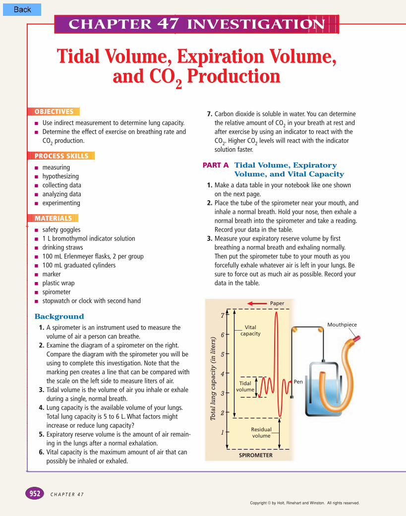
Background1. A spirometer is an instrument used to measure the
volume of air a person can breathe.2. Examine the diagram of a spirometer on the right.
Compare the diagram with the spirometer you will beusing to complete this investigation. Note that themarking pen creates a line that can be compared withthe scale on the left side to measure liters of air.
3. Tidal volume is the volume of air you inhale or exhaleduring a single, normal breath.
4. Lung capacity is the available volume of your lungs.Total lung capacity is 5 to 6 L. What factors mightincrease or reduce lung capacity?
5. Expiratory reserve volume is the amount of air remain-ing in the lungs after a normal exhalation.
6. Vital capacity is the maximum amount of air that canpossibly be inhaled or exhaled.
7. Carbon dioxide is soluble in water. You can determinethe relative amount of CO2 in your breath at rest andafter exercise by using an indicator to react with theCO2. Higher CO2 levels will react with the indicatorsolution faster.
PART A Tidal Volume, ExpiratoryVolume, and Vital Capacity
1. Make a data table in your notebook like one shownon the next page.
2. Place the tube of the spirometer near your mouth, andinhale a normal breath. Hold your nose, then exhale anormal breath into the spirometer and take a reading.Record your data in the table.
3. Measure your expiratory reserve volume by firstbreathing a normal breath and exhaling normally.Then put the spirometer tube to your mouth as youforcefully exhale whatever air is left in your lungs. Besure to force out as much air as possible. Record yourdata in the table.
C H A P T E R 4 7952
1
2
3
4
5
6
7
SPIROMETER
Tota
l lun
g c
apac
ity (
in li
ters
)
Tidalvolume
Vitalcapacity
Residualvolume
Paper
Pen
Mouthpiece
Copyright © by Holt, Rinehart and Winston. All rights reserved.

4. The table includes values for young adult males. Theaverage volume for young adult females is 20–25 per-cent lower than that of males. Calculate the averagevolumes for young adult females. Athletes can have vol-umes that are 30–40 percent greater than the averagefor their gender. Calculate the average volume for anathlete.
PART B Breathing Rate and CO2Production
5. Discuss the purpose of this part of the investigationwith your partners. You will use bromothymol blue as anindicator of the CO2 you bubble into each flask. Developa hypothesis that describes a relationship between airvolume exhaled during rest or exercise and the volumeof CO2 exhaled.
6. CAUTION Wear safety goggles at alltimes during this procedure. If you
get the indicator solution on your skin or cloth-ing, wash it off at the sink while calling to yourteacher. If you get the indicator solution in youreyes, immediately flush it out at the eyewashstation while calling to your teacher.
7. Label the two flasks as 1 and 2.8. Add 100 mL of indicator solution to each flask. Cover
the mouth of each flask with plastic wrap.9. Remove the plastic wrap from flask 1. Blow gently
through one straw into flask 1 until the solution turnsa yellowish color, exhaling slowly so that the solutiondoes not bubble up. Be careful not to inhale the solu-tion or get it in your mouth.
10. Record on your Part B Data Table the time in secondsthat it took to see a color change in flask 1.
11. Exercise by jogging in place or doing jumping jacksfor 2 min. Immediately blow gently through a newstraw into flask 2 until the solution becomes the sameyellowish color as the solution in flask 1.
12. In your Part B Data Table, record the amount of timein seconds that it took to get the same yellow color inflask 2 as you got in flask 1.
13. Calculate the difference in the amount of time it tookto see a color change in the two flasks. What can youinfer about the amount of CO2 you exhaled beforeand after exercise?
14. Clean up your materials. Pour the solu-tions down the sink, and rinse the sink
thoroughly with water. Wash your hands before leav-ing the lab.
Analysis and Conclusions1. How did your tidal volume compare with that of your
classmates?2. What are the independent and dependent variables in
Part B of the Investigation? How did you vary theindependent variable and measure changes in thedependent variable?
3. Why were the flasks covered with plastic wrap?4. Do your data support your hypothesis from Part B?
Explain your answers.5. How do you know whether you produced more car-
bon dioxide before or after you exercised? Supportyour answer with evidence from this lab.
6. What were some of the possible sources of error inyour experiment?
Further InquiryDesign an experiment to determine whether exerciseaffects heart rate in the same way it affects breathing rateand tidal volume.
953C I R C U L A T O R Y A N D R E S P I R A T O R Y S Y S T E M S
PART A TIDAL VOLUME, EXPIRATORY VOLUME, AND VITAL CAPACITY
Average for youngadult males
Average for youngadult females Average for athletes Your readings
Tidal volume 500 mL
Expiratory reserve volume 100 mL
Vital capacity 4,600 mL
Time for colorchange in flask 1
Time for colorchange in flask 2
Difference in time between flask 1 and flask 2
PART B DATA TABLE
Copyright © by Holt, Rinehart and Winston. All rights reserved.