Embed Size (px)
DESCRIPTION
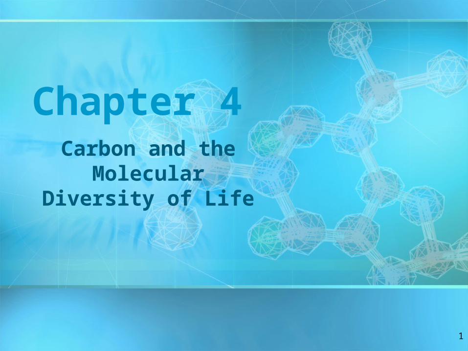
Chapter 4. Carbon and the Molecular Diversity of Life. Figure 4.1. Carbon Chemistry. Carbon is the Backbone of Biological Molecules (macromolecules) All living organisms Are made up of chemicals based mostly on the element carbon. Carbon Chemistry. - PowerPoint PPT Presentation
Citation preview
1
Chapter 4Carbon and the
Molecular Diversity of Life

2
Carbon Chemistry• Carbon is the Backbone of Biological
Molecules (macromolecules)• All living organisms Are made up of
chemicals based mostly on the element carbon
Figure 4.1

3
Carbon Chemistry
• Organic chemistry is the study of carbon compounds
• Carbon atoms can form diverse molecules by bonding to four other atoms or molecules
• Carbon compounds range from simple molecules to complex ones
• Carbon has four valence electrons and may form single, double, triple, or quadruple bonds

4
• The bonding versatility of carbon allows it to form many diverse molecules, including carbon skeletons
(a) Methane
(b) Ethane
(c) Ethene (ethylene)
Molecular Formula
Structural Formula
Ball-and-Stick Model
Space-Filling Model
H
H
H
H
H
H
H
H
H
H
H H
HH
C
C C
C C
CH4
C2H
6
C2H4
Name and Comments
Figure 4.3 A-C

5
• The electron configuration of carbon gives it covalent compatibility with many different elements
H O N C
Hydrogen
(valence = 1)
Oxygen
(valence = 2)
Nitrogen
(valence = 3)
Carbon
(valence = 4)
Figure 4.4

6
• Carbon may bond to itself forming carbon chains
• Carbon chains form the skeletons of most organic molecules
• Carbon chains vary in length and shape
HHH
HH
H H H
HH
H
H H H
H H HH H
H
H
H
H
H
H
HH
HH H H H
H HH H
H H H H
H H
H H
HHHH H
H
H
C C C C C
C C C C C C C
CCCCCCCC
C
CC
CC
C
C
CCC
CC
H
H
H
HHH
H
(a) Length
(b) Branching
(c) Double bonds
(d) Rings
Ethane Propane
Butane isobutane
1-Butene 2-Butene
Cyclohexane Benzene
H H H HH
Figure 4.5 A-D

7
Hydrocarbons
• Hydrocarbons are molecules consisting of only carbon and hydrogen
• Hydrocarbons Are found within many of a cell’s organic molecules
(a) A fat molecule (b) Mammalian adipose cells100 µm
Fat droplets (stained red)
Figure 4.6 A, B

8
Functional Groups• Functional groups
are the parts of molecules involved in chemical reactions
• They Are the chemically reactive groups of atoms within an organic molecule
• Give organic molecules distinctive chemical properties
CH3
OH
HO
O
CH3
CH3
OH
Estradiol
Testosterone
Female lion
Male lionFigure 4.9

9
• Six functional groups are important in the chemistry of life– Hydroxyl– Carbonyl– Carboxyl– Amino– Sulfhydryl– Phosphate

10
Some important functional groups of organic compounds
FUNCTIONALGROUP
STRUCTURE
(may be written HO )
HYDROXYL CARBONYL CARBOXYL
OH
In a hydroxyl group (—OH), a hydrogen atom is bonded to an oxygen atom, which in turn is bonded to the carbon skeleton of the organic molecule. (Do not confuse this functional group with the hydroxide ion, OH–.)
When an oxygen atom is double-bonded to a carbon atom that is also bonded to a hydroxyl group, the entire assembly of atoms is called a carboxyl group (—COOH).
C
O O
C
OH
Figure 4.10
The carbonyl group ( CO) consists of a carbon atom joined to an oxygen atom by a double bond.

11
Some important functional groups of organic compounds
Acetic acid, which gives
vinegar its sour tatste
NAME OF
COMPOUNDS
Alcohols (their
specific names usually
end in -ol)
Ketones if the carbonyl
group is within a carbon
skeleton
Aldehydes if the carbonyl
group is at the end of the
carbon skeleton
Carboxylic acids, or
organic acids
EXAMPLE
Propanal, an aldehyde
Acetone, the simplest ketone
Ethanol, the
alcohol present in
alcoholic
beverages
H
H
H
H H
C C OH
H
H
H
HH
H
HC C H
C
C C
C C C
O
H OH
O
H
H
H H
H O
H
Figure 4.10

12
• Some important functional groups of organic compounds
The amino group (—NH2) consists of a nitrogen atom bonded to two hydrogen atoms and to the carbon skeleton.
AMINO SULFHYDRYL PHOSPHATE
(may be written HS )
The sulfhydryl group consists of a sulfur atom bonded to an atom of hydrogen; resembles a hydroxyl group in shape.
In a phosphate group, a phosphorus atom is bonded to four oxygen atoms; one oxygen is bonded to the carbon skeleton; two oxygens carry negative charges; abbreviated P . The phosphate group (—OPO3
2–) is an ionized form of a phosphoric acid group (—OPO3H2; note the two hydrogens).
NH
H
SH
O P
O
OH
OH
Figure 4.10

13
Macromolecules– Are large molecules composed of smaller
molecules– Are complex in their structures
Figure 5.1

14
Macromolecules•Most macromolecules are polymers, built from monomers• Four classes of life’s organic molecules are polymers
– Carbohydrates (include sugars, starches etc)
– Proteins– Nucleic acids– Lipids (fats)
15
• A polymer– Is a long molecule consisting of many
similar building blocks called monomers– Specific monomers make up each
macromolecule– E.g. amino acids are the monomers for
proteins

16
The Synthesis and Breakdown of Polymers
• Monomers form larger molecules by condensation reactions called dehydration synthesis
(a) Dehydration reaction in the synthesis of a polymer
HO H1 2 3 HO
HO H1 2 3 4
H
H2O
Short polymer Unlinked monomer
Longer polymer
Dehydration removes a watermolecule, forming a new bond
Figure 5.2A
17
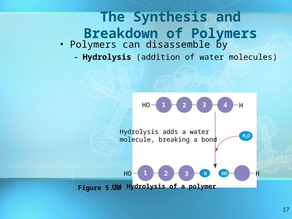
The Synthesis and Breakdown of Polymers
• Polymers can disassemble by– Hydrolysis (addition of water molecules)
(b) Hydrolysis of a polymer
HO 1 2 3 H
HO H1 2 3 4
H2O
HHO
Hydrolysis adds a watermolecule, breaking a bond
Figure 5.2B
18
• Although organisms share the same limited number of monomer types, each organism is unique based on the arrangement of monomers into polymers
• An immense variety of polymers can be built from a small set of monomers

19
Carbohydrates• Serve as fuel and building
material• Include both sugars and their
polymers (starch, cellulose, etc.)
20
Sugars
• Monosaccharides– Are the simplest sugars– Can be used for fuel– Can be converted into other organic
molecules– Can be combined into polymers

21
• Examples of monosaccharides
Triose sugars(C3H6O3)
Pentose sugars(C5H10O5)
Hexose sugars(C6H12O6)
H C OH
H C OH
H C OH
H C OH
H C OH
H C OH
HO C H
H C OH
H C OH
H C OH
H C OH
HO C H
HO C H
H C OH
H C OH
H C OH
H C OH
H C OH
H C OH
H C OH
H C OH
H C OH
C OC O
H C OH
H C OH
H C OH
HO C H
H C OH
C O
H
H
H
H H H
H
H H H H
H
H H
C C C COOOO
Ald
oses
Glyceraldehyde
RiboseGlucose Galactose
Dihydroxyacetone
Ribulose
Keto
ses
FructoseFigure 5.3

22
• Monosaccharides– May be linear– Can form rings
H
H C OH
HO C H
H C OH
H C OH
H C
O
C
H
1
2
3
4
5
6
H
OH
4C
6CH2OH 6CH2OH
5C
HOH
C
H OH
H
2 C
1C
H
O
H
OH
4C
5C
3 C
H
HOH
OH
H
2C
1 C
OH
H
CH2OH
H
H
OHHO
H
OH
OH
H5
3 2
4
(a) Linear and ring forms. Chemical equilibrium between the linear and ring structures greatly favors the formation of rings. To form the glucose ring, carbon 1 bonds to the oxygen attached to carbon 5.
OH3
O H OO
6
1
Figure 5.4

23
• Disaccharides– Consist of two monosaccharides– Are joined by a glycosidic linkage

24
Dehydration reaction in the synthesis of maltose. The bonding of two glucose units forms maltose. The glycosidic link joins the number 1 carbon of one glucose to the number 4 carbon of the second glucose. Joining the glucose monomers in a different way would result in a different disaccharide.
Dehydration reaction in the synthesis of sucrose. Sucrose is a disaccharide formed from glucose and fructose.Notice that fructose,though a hexose like glucose, forms a five-sided ring.
(a)
(b)
H
HO
H
HOH H
OH
O H
OH
CH2OH
H
HO
H
HOH
H
OH
O H
OH
CH2OH
H
O
H
HOH H
OH
O H
OH
CH2OH
H
H2O
H2O
H
H
O
H
HOH
OH
O H
CH2OH
CH2OH HO
OHH
CH2OH
HOH
H
H
HO
OHH
CH2OH
HOH H
O
O H
OHH
CH2OH
HOH H
O
HOH
CH2OH
H HO
O
CH2OH
H
H
OH
O
O
1 2
1 41– 4
glycosidiclinkage
1–2glycosidic
linkage
Glucose
Glucose Glucose
Fructose
Maltose
Sucrose
OH
H
H
Figure 5.5

25
Polysaccharides• Polysaccharides
– Are polymers of sugars– Serve many roles in organisms

26
Storage Polysaccharides• Starch
– Is a polymer consisting entirely of glucose monomers
– Is the major storage form of glucose in plants
Chloroplast Starch
Amylose Amylopectin
1 m
(a) Starch: a plant polysaccharideFigure 5.6

27
• Glycogen– Consists of glucose monomers– Is the major storage form of glucose in animals
Mitochondria Giycogen granules
0.5 m
(b) Glycogen: an animal polysaccharide
Glycogen
Figure 5.6
28
Structural Polysaccharides• Cellulose
– Is a polymer of glucose
29
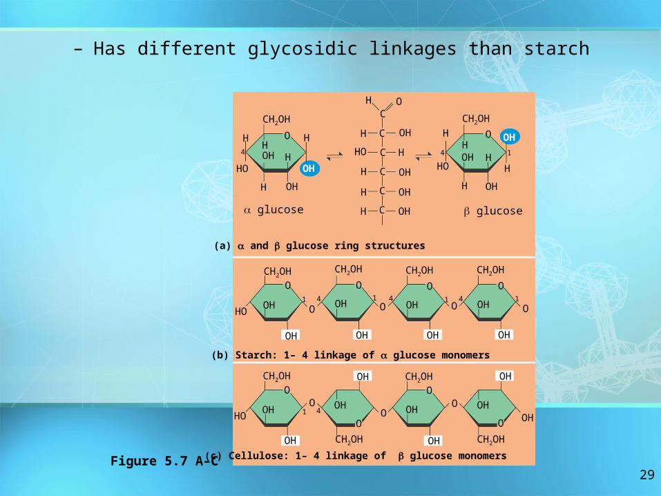
– Has different glycosidic linkages than starch
(c) Cellulose: 1– 4 linkage of glucose monomers
H O
O
CH2OH
HOH H
H
OH
OHH
H
HO
4
C
C
C
C
C
C
H
H
H
HO
OH
H
OHOHOH
H
O
CH2OH
HH
H
OH
OHH
H
HO
4 OH
CH2OH
OOH
OH
HO
41
O
CH2OH
OOH
OH
O
CH2OH
OOH
OH
CH2OH
O
OH
OH
O O
CH2OH
OOH
OH
HO
4O
1
OH
O
OH
OHO
CH2OH
O
OH
O OH
O
OH
OH
(a) and glucose ring structures
(b) Starch: 1– 4 linkage of glucose monomers
1
glucose glucose
CH2OH
CH2OH
1 4 41 1
Figure 5.7 A–C

30
Plant cells
0.5 m
Cell walls
Cellulose microfibrils in a plant cell wall
Microfibril
CH2OH
CH2OH
OH
OH
OO
OHOCH2OH
O
OOH
OCH2OH OH
OH OHO
O
CH2OH
OO
OH
CH2OH
OO
OH
O
O
CH2OHOH
CH2OHOHOOH OH OH OH
O
OH OH
CH2OH
CH2OH
OHO
OH CH2OH
OO
OH CH2OH
OH
Glucose monomer
O
O
O
O
O
O
Parallel cellulose molecules areheld together by hydrogenbonds between hydroxyl
groups attached to carbonatoms 3 and 6.
About 80 cellulosemolecules associate
to form a microfibril, themain architectural unitof the plant cell wall.
A cellulose moleculeis an unbranched glucose polymer.
OH
OH
O
OOH
Cellulosemolecules
Figure 5.8
– Is a major component of the tough walls that enclose plant cells
31
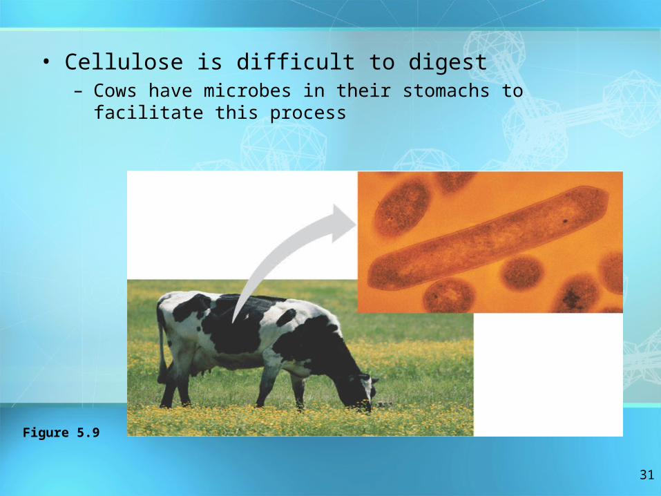
• Cellulose is difficult to digest– Cows have microbes in their stomachs to facilitate this
process
Figure 5.9
32
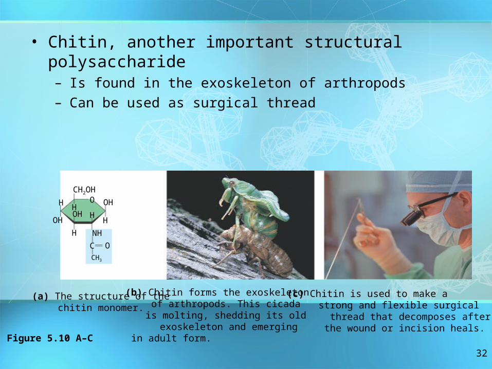
• Chitin, another important structural polysaccharide– Is found in the exoskeleton of arthropods– Can be used as surgical thread
(a) The structure of the chitin monomer.
O
CH2OH
OHHH OH
H
NH
CCH3
O
H
H
(b) Chitin forms the exoskeleton of arthropods. This cicada is molting, shedding its old exoskeleton and emergingin adult form.
(c) Chitin is used to make a strong and flexible surgical
thread that decomposes after the wound or incision heals.
OH
Figure 5.10 A–C

33
Lipids• Lipids are a diverse group of
hydrophobic molecules• Lipids
– Are the one class of large biological molecules that do not consist of polymers
– Share the common trait of being hydrophobic

34
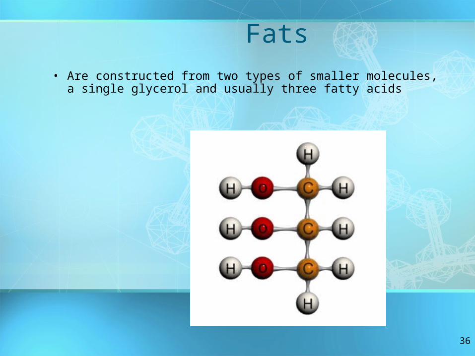
Fats– Are constructed from two types of smaller molecules, a single
glycerol and usually three fatty acids– Vary in the length and number and locations of double bonds
they contain

35
Fats– Are constructed from two types of smaller molecules, a single
glycerol and usually three fatty acids– Vary in the length and number and locations of double bonds
they contain
36
Fats• Are constructed from two types of smaller molecules, a single
glycerol and usually three fatty acids
37
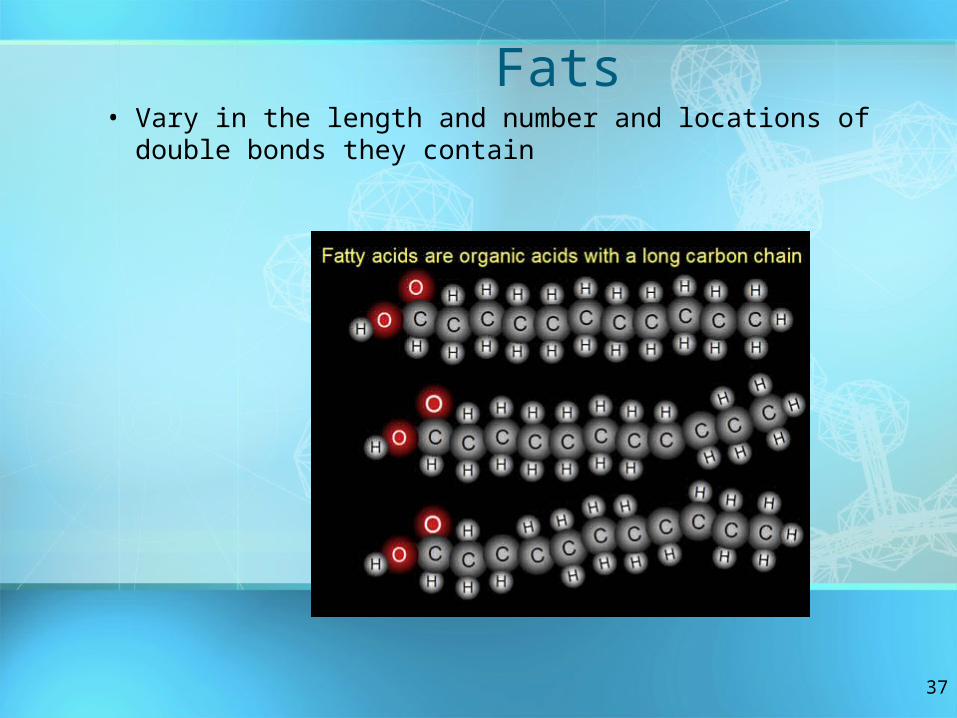
Fats• Vary in the length and number and locations of double
bonds they contain

38
• Saturated fatty acids– Have the maximum number of hydrogen atoms
possible– Have no double bonds
(a) Saturated fat and fatty acid
Stearic acid
Figure 5.12

39
• Unsaturated fatty acids– Have one or more double bonds
(b) Unsaturated fat and fatty acidcis double bondcauses bending
Oleic acid
Figure 5.12

40
• Phospholipids– Have only two fatty acids– Have a phosphate group instead of a third fatty
acid

41
• Phospholipid structure– Consists of a hydrophilic “head” and
hydrophobic “tails”
CH2
O
PO O
O
CH2CHCH2
OO
C O C O
Phosphate
Glycerol
(a) Structural formula (b) Space-filling model
Fatty acids
(c) Phospholipid symbol
Hyd
rop
hob
i c t
ails
Hydrophilichead
Hydrophobictails
–
Hyd
rop
hi li c
head
CH2 Choline+
Figure 5.13
N(CH3)3
42
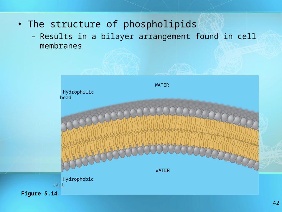
• The structure of phospholipids– Results in a bilayer arrangement found in cell
membranes
Hydrophilichead
WATER
WATER
Hydrophobictail
Figure 5.14
43
Steroids• Steroids
– Are lipids characterized by a carbon skeleton consisting of four fused rings

44
• One steroid, cholesterol– Is found in cell membranes– Is a precursor for some hormones
HO
CH3
CH3
H3C CH3
CH3
Figure 5.15

45
Proteins• Proteins have many structures,
resulting in a wide range of functions
• Proteins do most of the work in cells and act as enzymes
• Proteins are made of monomers called amino acids

46
• An overview of protein functions
Table 5.1

47
• Enzymes– Are a type of protein that acts as a catalyst, speeding up
chemical reactions
Substrate(sucrose)
Enzyme (sucrase)
Glucose
OH
H O
H2O
Fructose
3 Substrate is convertedto products.
1 Active site is available for a molecule of substrate, the
reactant on which the enzyme acts.
Substrate binds toenzyme.
22
4 Products are released.Figure 5.16
48
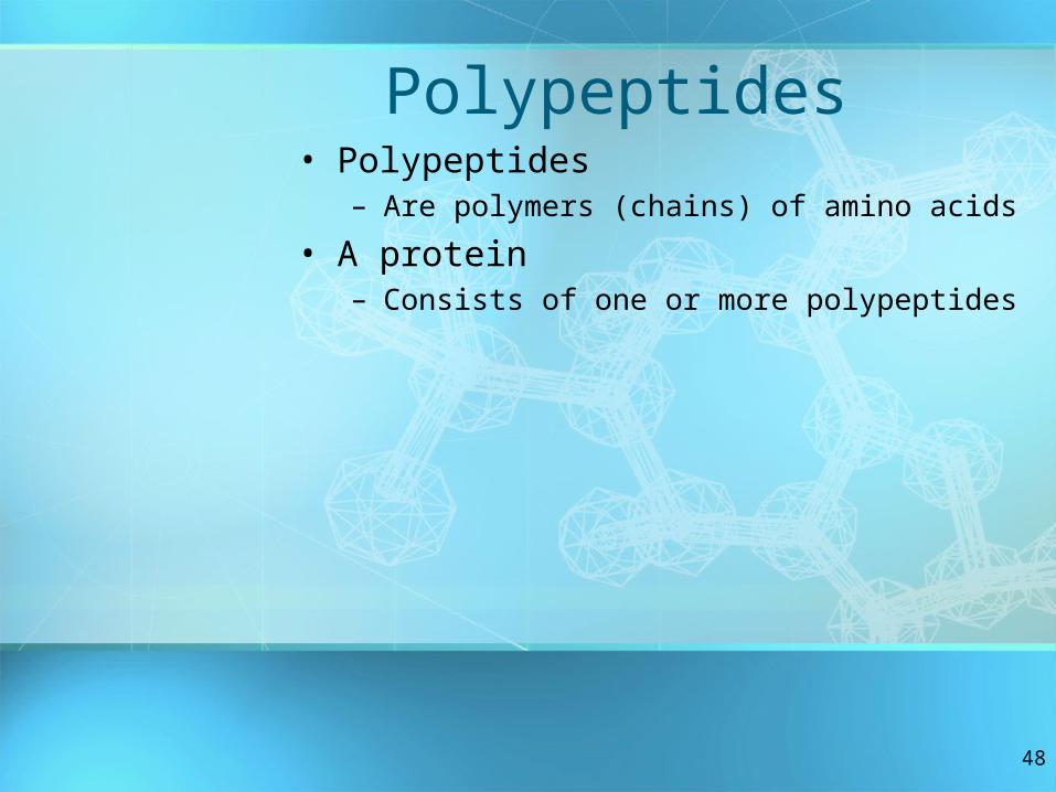
Polypeptides• Polypeptides
– Are polymers (chains) of amino acids
• A protein– Consists of one or more polypeptides

49
• Amino acids– Are organic molecules possessing both carboxyl
and amino groups– Differ in their properties due to differing side chains,
called R groups
50
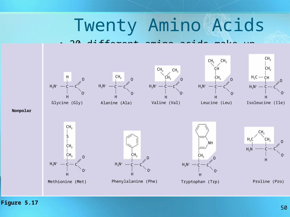
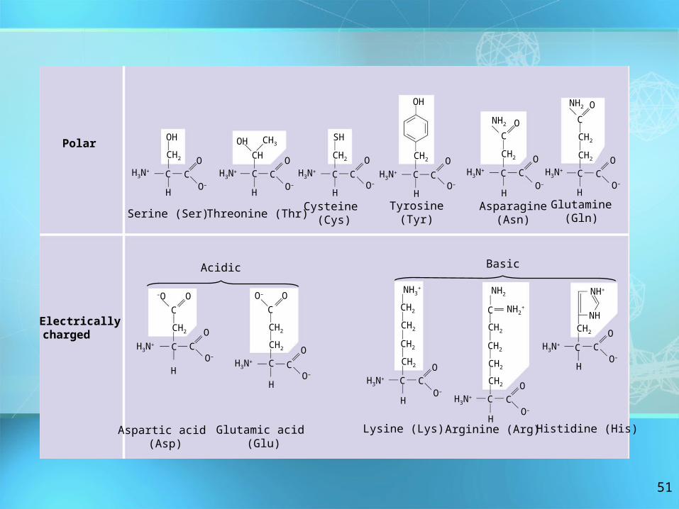
Twenty Amino Acids• 20 different amino acids make up proteins
O
O–
H
H3N+ C C
O
O–
H
CH3
H3N+ C
H
C
O
O–
CH3 CH3
CH3
C C
O
O–
H
H3N+
CH
CH3
CH2
C
H
H3N+
CH3CH3
CH2
CH
C
H
H3N+
C
CH3
CH2
CH2
CH3N+
H
C
O
O–
CH2
CH3N+
H
C
O
O–
CH2
NH
H
C
O
O–
H3N+ C
CH2
H2C
H2N C
CH2
H
C
Nonpolar
Glycine (Gly) Alanine (Ala) Valine (Val) Leucine (Leu) Isoleucine (Ile)
Methionine (Met) Phenylalanine (Phe)
C
O
O–
Tryptophan (Trp) Proline (Pro)
H3C
Figure 5.17
S
O
O–
51
O–
OH
CH2
C C
H
H3N+
O
O–
H3N+
OH CH3
CH
C C
HO–
O
SH
CH2
C
H
H3N+ C
O
O–
H3N+
C C
CH2
OH
H H H
H3N+
NH2
CH2
OC
C CO
O–
NH2 O
C
CH2
CH2
C CH3N
+
O
O–
O
Polar
Electricallycharged
–O O
C
CH2
C CH3N
+
H
O
O–
O– O
C
CH2
C CH3N
+
H
O
O–
CH2
CH2
CH2
CH2
NH3+
CH2
C CH3N
+
H
O
O–
NH2
C NH2+
CH2
CH2
CH2
C CH3N
+
H
O
O–
CH2
NH+
NHCH2
C CH3N
+
H
O
O–
Serine (Ser) Threonine (Thr)Cysteine
(Cys)Tyrosine
(Tyr)Asparagine
(Asn)Glutamine
(Gln)
Acidic Basic
Aspartic acid (Asp)
Glutamic acid (Glu)
Lysine (Lys) Arginine (Arg) Histidine (His)

52
Amino Acid Polymers
• Amino acids– Are linked by peptide bonds

53
Protein Conformation and Function• A protein’s specific conformation (shape)
determines how it functions

54
Four Levels of Protein Structure
• Primary structure– Is the unique sequence
of amino acids in a polypeptide
Figure 5.20–
Amino acid
subunits
+H3NAmino
end
oCarboxyl end
oc
GlyProThrGlyThr
Gly
GluSeuLysCysProLeu
MetVal
Lys
ValLeu
AspAlaValArgGly
SerPro
Ala
Gly
lle
SerProPheHisGluHis
Ala
GluValValPheThrAla
Asn
AspSer
GlyProArg
ArgTyrThr
lleAla
Ala
Leu
LeuSer
ProTyrSerTyrSerThr
Thr
Ala
ValVal
ThrAsnProLysGlu
ThrLys
SerTyrTrpLysAlaLeu
GluLleAsp

55
O C helix
pleated sheetAmino acid
subunitsNCH
C
O
C N
H
CO H
R
C NH
C
O H
C
R
N
HH
R C
O
R
C
H
NH
C
O H
NCO
R
C
H
NH
H
C
R
C
O
C
O
C
NH
H
R
C
C
ON
HH
C
R
C
O
NH
R
C
H C
ON
HH
C
R
C
O
NH
R
C
H C
ON
HH
C
R
C
O
N H
H C R
N HO
O C N
C
RC
H O
CHR
N HO C
RC
H
N H
O CH C R
N H
CC
N
R
H
O C
H C R
N H
O C
RC
H
H
C
RN
H
CO
C
NH
R
C
H C
O
N
H
C
• Secondary structure– Is the folding or coiling of the polypeptide into a
repeating configuration– Includes the helix and the pleated sheet
H H
Figure 5.20
56
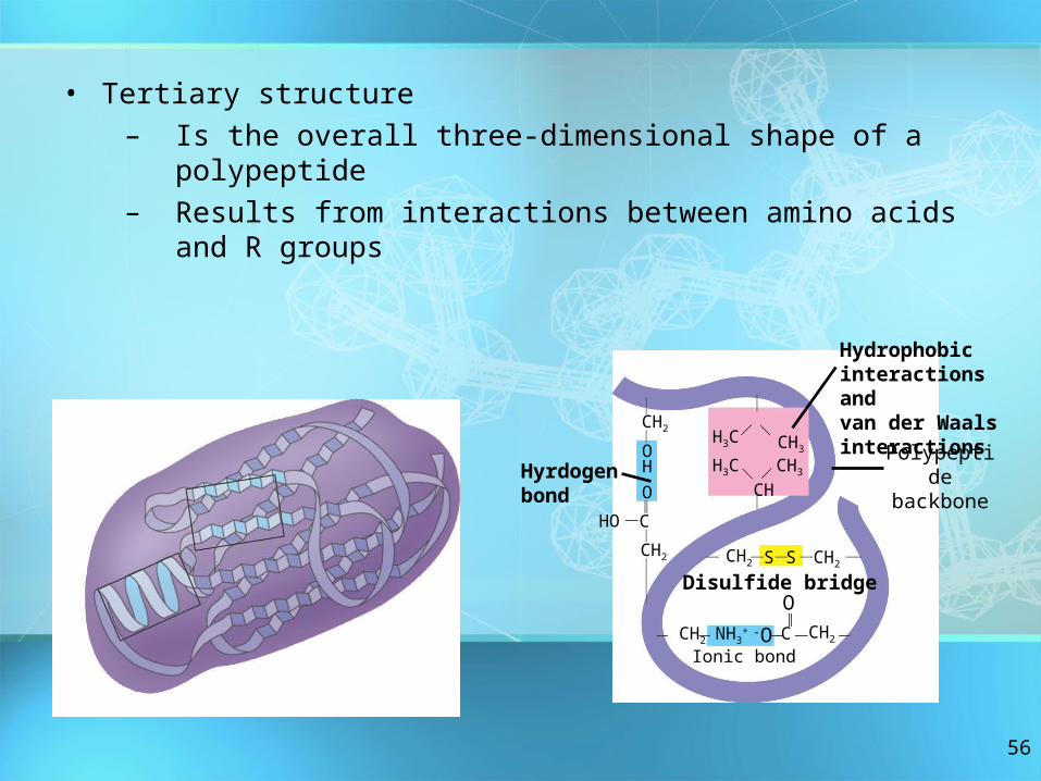
• Tertiary structure– Is the overall three-dimensional shape of a polypeptide– Results from interactions between amino acids and R
groups
CH2CH
OH
O
CHO
CH2
CH2 NH3+ C-O CH2
O
CH2SSCH2
CH
CH3
CH3
H3C
H3C
Hydrophobic interactions and van der Waalsinteractions Polypeptid
ebackbone
Hyrdogenbond
Ionic bond
CH2
Disulfide bridge

57
• Quaternary structure– Is the overall protein structure that results from the
aggregation of two or more polypeptide subunits
Polypeptidechain
Collagen
Chains
ChainsHemoglobin
IronHeme

58
Review of Protein Structure
+H3NAmino end
Amino acidsubunits
helix

59
Sickle-Cell Disease: A Simple Change in Primary Structure
• Sickle-cell disease– Results from a single amino acid
substitution in the protein hemoglobin

60
Fibers of abnormalhemoglobin deform cell into sickle shape.
Primary structure
Secondaryand tertiarystructures
Quaternary structure
Function
Red bloodcell shape
Hemoglobin A
Molecules donot associatewith oneanother, eachcarries oxygen.Normal cells arefull of individualhemoglobinmolecules, eachcarrying oxygen
10 m 10 m
Primary structure
Secondaryand tertiarystructures
Quaternary structure
Function
Red bloodcell shape
Hemoglobin SMolecules interact with one another tocrystallize into a fiber, capacity to carry oxygen is greatly reduced.
subunit subunit
1 2 3 4 5 6 7 3 4 5 6 721
Normal hemoglobin
Sickle-cell hemoglobin . . .. . .
Figure 5.21
Exposed hydrophobic
region
Val ThrHis Leu Pro Glul Glu Val His Leu Thr Pro Val Glu

61
What Determines Protein Conformation?
• Protein conformation Depends on the physical and chemical conditions of the protein’s environment
• Temperature, pH, etc. affect protein structure

62
•Denaturation is when a protein unravels and loses its native conformation(shape)
Denaturation
Renaturation
Denatured protein
Normal protein
Figure 5.22

63
The Protein-Folding Problem• Most proteins
– Probably go through several intermediate states on their way to a stable conformation
– Denaturated proteins no longer work in their unfolded condition
– Proteins may be denaturated by extreme changes in pH or temperature
64
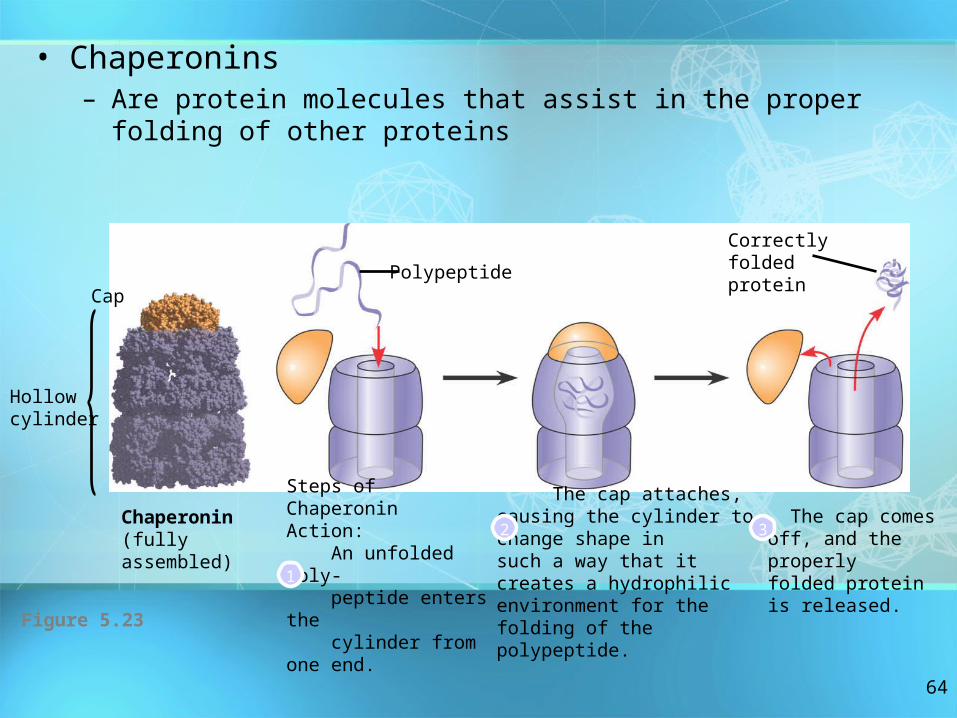
• Chaperonins– Are protein molecules that assist in the proper folding of
other proteins
Hollowcylinder
Cap
Chaperonin(fully assembled)
Steps of ChaperoninAction: An unfolded poly- peptide enters the cylinder from one end.
The cap attaches, causing the cylinder to change shape insuch a way that it creates a hydrophilic environment for the folding of the polypeptide.
The cap comesoff, and the properlyfolded protein is released.
Correctlyfoldedprotein
Polypeptide
2
1
3
Figure 5.23

65
• X-ray crystallography– Is used to determine a protein’s three-dimensional
structure
X-raydiffraction pattern
Photographic filmDiffracted X-
raysX-ray
source
X-ray
beam
CrystalNucleic acid Protein
(a) X-ray diffraction pattern(b) 3D computer modelFigure 5.24
66
Nucleic Acids• Nucleic acids store and transmit
hereditary information• Genes
– Are the units of inheritance– Program the amino acid sequence of
polypeptides– Are made of nucleotide sequences on
DNA

67
The Roles of Nucleic Acids• There are two types of nucleic acids
– Deoxyribonucleic acid (DNA)– Ribonucleic acid (RNA)

68
Deoxyribonucleic Acid• DNA
– Stores information for the synthesis of specific proteins
– Found in the nucleus of cells
69
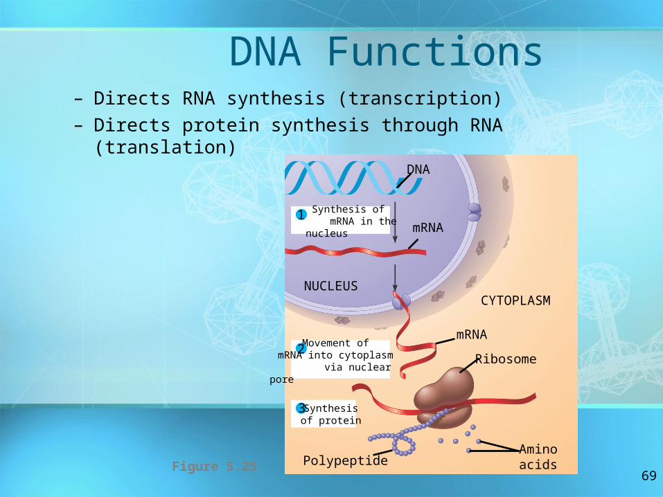
DNA Functions– Directs RNA synthesis (transcription)– Directs protein synthesis through RNA (translation)
1
2
3
Synthesis of mRNA in the nucleus
Movement of mRNA into cytoplasm
via nuclear pore
Synthesisof protein
NUCLEUSCYTOPLASM
DNA
mRNA
Ribosome
AminoacidsPolypeptide
mRNA
Figure 5.25
70
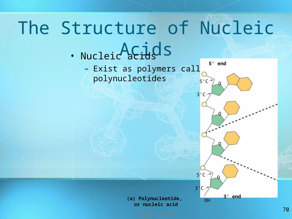
The Structure of Nucleic Acids• Nucleic acids
– Exist as polymers called polynucleotides
(a) Polynucleotide, or nucleic acid
3’C
5’ end
5’C
3’C
5’C
3’ endOH
Figure 5.26
O
O
O
O

71
• Each polynucleotide– Consists of monomers called nucleotides– Sugar + phosphate + nitrogen base
Nitrogenousbase
Nucleoside
O
O
O
O P CH2
5’C
3’CPhosphate
group Pentosesugar
(b) NucleotideFigure 5.26
O

72
Nucleotide Monomers• Nucleotide monomers
– Are made up of nucleosides (sugar + base) and phosphate groups
(c) Nucleoside componentsFigure 5.26
CHCH
Uracil (in RNA)U
Ribose (in RNA)
Nitrogenous bases Pyrimidines
CN
NC
OH
NH2
CHCH
OC
NH
CHHN
CO
CCH3
N
HNC
C
HO
O
CytosineC
Thymine (in DNA)T
NHC
N C
CN
C
CH
N
NH2 O
NHC
NHH
C C
N
NH
C NH2
AdenineA
GuanineG
Purines
OHOCH2
HH H
OH
H
OHOCH2
HH H
OH
H
Pentose sugars
Deoxyribose (in DNA)Ribose (in RNA)OHOH
CH
CH
Uracil (in RNA)U
4’
5”
3’OH H
2’
1’
5”
4’
3’ 2’
1’

73
Nucleotide Polymers• Nucleotide polymers
– Are made up of nucleotides linked by the–OH group on the 3´ carbon of one nucleotide and the phosphate on the 5´ carbon on the next

74
Gene• The sequence of bases along a nucleotide
polymer– Is unique for each gene

75
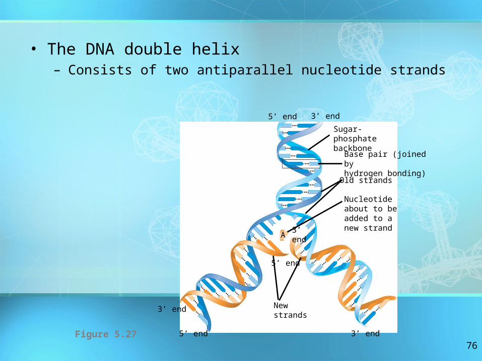
The DNA Double Helix• Cellular DNA molecules
– Have two polynucleotides that spiral around an imaginary axis
– Form a double helix
76
• The DNA double helix– Consists of two antiparallel nucleotide strands
3’ end
Sugar-phosphatebackbone
Base pair (joined byhydrogen bonding)
Old strands
Nucleotideabout to be added to a new strand
A
3’ end
3’ end
5’ end
Newstrands
3’ end
5’ end
5’ end
Figure 5.27

77
A,T,C,G• The nitrogenous bases in DNA
– Form hydrogen bonds in a complementary fashion (A with T only, and C with G only)
78
DNA and Proteins as Tape Measures of Evolution
• Molecular comparisons – Help biologists sort out the
evolutionary connections among species

79
The Theme of Emergent Properties in the Chemistry of
Life: A Review• Higher levels of organization
– Result in the emergence of new properties
• Organization– Is the key to the chemistry of life