Embed Size (px)
Citation preview

Ph. D. Thesis 2014
27
Chapter 3
Materials and Methods

Ph. D. Thesis 2014
28
Section A: Plant Description
Cynodon dactylon
Cynodon dactylon (L) Pers. is commonly known as ‘Doob’ grass in India
and is considered as toxic weeds that are very invasive (Fernandez, 2003) and fast
growing. It is also known as “world worst weed” (Holm et al., 1977) and is found
abundant along the roadsides, in lawns and uncultivable soil. It has no odour with
sweet mucilaginous taste. It is one of the important medicinal weed plants of
Chhattisgarh state. C. dactylon is the most sacred plant next to Ocimum sanctum.
Hindus worship the God Ganesha with the leaves of this plant.
The classification of this species is as under:
Kingdom Plantae
Sub-Kingdom Tracheobionta
Division Magnoliophyta
Class Liliopsida
Subclass Commelinidae
Order Cyperales
Family Poaceae
Genus Cynodon
Species C. dactylon (L.) Pers.
The local name of Cynodon dactylon in Indian language are Doorva or
Doob in Hindi, Arugu in Tamil, Bhargavi or Doorwa or Granthi, or Svetain in
Sanskrit, Gorikae in Kannada, Karuka-pulli in Malayalam, Doorva in Marathi,
Garika in Telgu, Ghass in Urdu, Dub in Punjabi and Durba in Bengali.
Cynodon dactylon is a hardy, drought resistant, and perennial herb with
rhizome. Its stem is creeping, rooting at nodes. Its leaves are flat or sometimes
folded or convolute. Inflorescence of this plant consisting of 2-12 spikes arranged
star-like at apex of stem; spikes with numerous spikelets, arranged in 2 rows on

Ph. D. Thesis 2014
29
one side of spike. Spikelets flat with one floret. Seeds are ovoid, yellow to reddish.
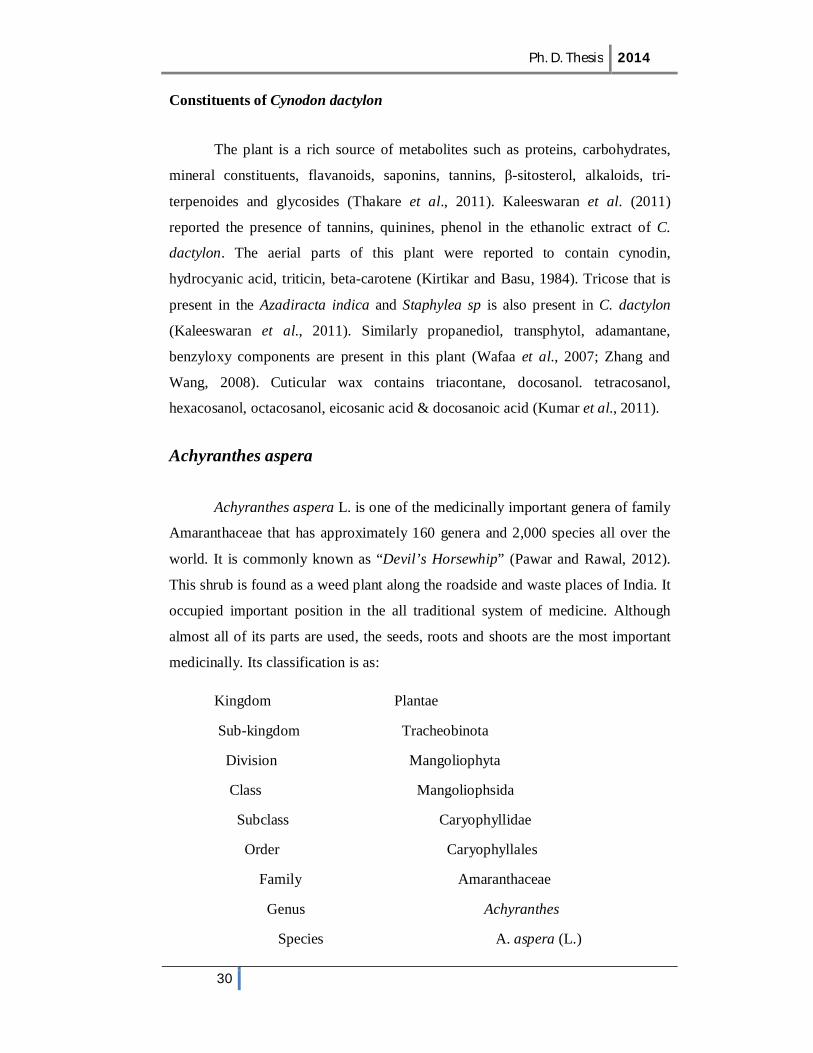
It is used as a control for soil erosion (Plate-I).
Plate I: Cynodon dactylon
Uses and biological activities
In India, this plant is used in the treatment of malaria, thirst, and anorexia,
burning sensations in the body (Karthik and Ravikumar, 2011). It is a good nerve
tonic, enhance memory power and very effective against anemia, asthma, bronchial
problem like cough, cold, sore throat, Influenza, fever, hypertension, snake bite,
kidney problem, swelling, skin diseases, leucoderma, stress and infection etc. Paul
et al. (2012) reviewed the therapeutic uses of this plant. Cynodon dactylon
possesses pharmacological properties such as antiviral, antifungal, antiseptic,
antibacterial (Devi et al., 2009; Chaudhari et al., 2011; Rao et al., 2011; Hema et
al., 2013), immunomodulatory (Santhi and Annapoorani, 2010), antihaemorrhagic,
antihelminthic, analgesic, antipyretic activity (Garg and Khosa, 2008, Kumar et al.,
2011), antioxidant (Pal et al., 2008; Sindhu et al., 2009). There are some
important papers showing the antiulcer (Patil et al., 2005), anti diabetic (Singh et
al., 2007), hepatoprotective (Surendra et al., 2008), heart protective (Garjani et al.,
2009), anti convulsive (Pal, 2009; Garg and Paliwal, 2011), wound healing
(Thakare et al., 2011) and hypolipedemic (Kumar and Kalaivani, 2011),
hypotensive (Bharti et al., 2012) effect on mammalian models.
Ph. D. Thesis 2014
30
Constituents of Cynodon dactylon
The plant is a rich source of metabolites such as proteins, carbohydrates,
mineral constituents, flavanoids, saponins, tannins, β-sitosterol, alkaloids, tri-
terpenoides and glycosides (Thakare et al., 2011). Kaleeswaran et al. (2011)
reported the presence of tannins, quinines, phenol in the ethanolic extract of C.
dactylon. The aerial parts of this plant were reported to contain cynodin,
hydrocyanic acid, triticin, beta-carotene (Kirtikar and Basu, 1984). Tricose that is
present in the Azadiracta indica and Staphylea sp is also present in C. dactylon
(Kaleeswaran et al., 2011). Similarly propanediol, transphytol, adamantane,
benzyloxy components are present in this plant (Wafaa et al., 2007; Zhang and
Wang, 2008). Cuticular wax contains triacontane, docosanol. tetracosanol,
hexacosanol, octacosanol, eicosanic acid & docosanoic acid (Kumar et al., 2011).
Achyranthes aspera
Achyranthes aspera L. is one of the medicinally important genera of family
Amaranthaceae that has approximately 160 genera and 2,000 species all over the
world. It is commonly known as “Devil’s Horsewhip” (Pawar and Rawal, 2012).
This shrub is found as a weed plant along the roadside and waste places of India. It
occupied important position in the all traditional system of medicine. Although
almost all of its parts are used, the seeds, roots and shoots are the most important
medicinally. Its classification is as:
Kingdom Plantae
Sub-kingdom Tracheobinota
Division Mangoliophyta
Class Mangoliophsida
Subclass Caryophyllidae
Order Caryophyllales
Family Amaranthaceae
Genus Achyranthes
Species A. aspera (L.)
Ph. D. Thesis 2014
31
The vernacular name of Achyranthes aspera in different Indian language
are Latjeera or Chirchita (Hindi); Apamarga (Sanskrit); Aghedi (Gujarati); Apang
(Bengali); Agadha (Marathi); Kadaladi (Malyalam); Kutri (Punjabi); Nayurivi
(Tamil); Uttaranee (Kannada).
Achyranthes aspera is annual or perennial herb. Its stem is yellow brownish
in color, erect, branched and hairy. The leaves are thick, opposite oval or rounded.
The flowers are in an auxiliary or terminal spikes and bisexual greenish white.
Fruits are easily disarticulated. The seeds are sub cylindrical truncate reddish
brown. The flowering time of this plant is from July to September and the seeds
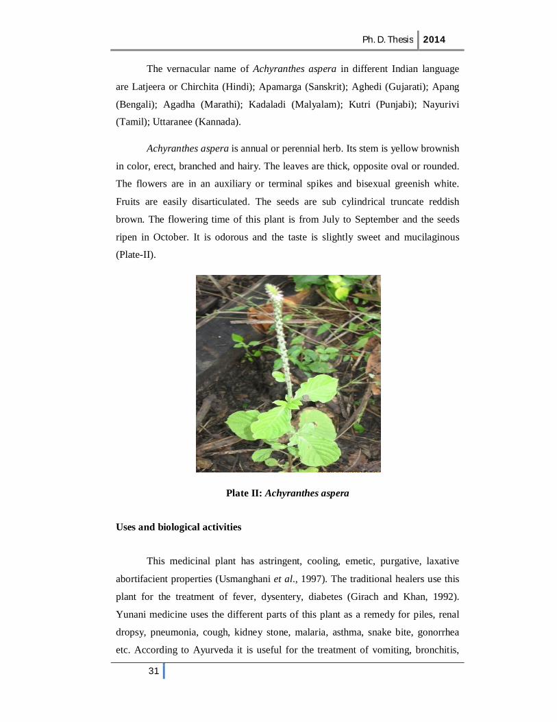
ripen in October. It is odorous and the taste is slightly sweet and mucilaginous
(Plate-II).
Plate II: Achyranthes aspera
Uses and biological activities
This medicinal plant has astringent, cooling, emetic, purgative, laxative
abortifacient properties (Usmanghani et al., 1997). The traditional healers use this
plant for the treatment of fever, dysentery, diabetes (Girach and Khan, 1992).
Yunani medicine uses the different parts of this plant as a remedy for piles, renal
dropsy, pneumonia, cough, kidney stone, malaria, asthma, snake bite, gonorrhea
etc. According to Ayurveda it is useful for the treatment of vomiting, bronchitis,
Ph. D. Thesis 2014
32
heart disease, piles, itching abdominal pains, ascites, dyspepsia, dysentery, blood
disease etc. (Dwivedi et al., 2008). The Maasai people of Kenya use this plant for
the treatment of malaria (Bussmann, 2006). Dey (2011) and Raji (2013) reviewed
the various pharmacological properties of Achyranthes aspera. It has many
pharmacological properties such as diuretic, hypoglycemic (Akhtar and Iqbal,
1991), prothyrodic (Tahiliani and Kar, 2000), cancer chemo preventive
(Chakraborty et al., 2002), Immunomodulatory (Rao et al., 2002), hepato-
protective (Bafna and Mishra, 2004), antifertility (Vasudeva and Sharma, 2006),
larvicidal (Bagavan et al., 2008), antibacterial & antifungal (Saravanan et al.,
2008; Elumalaii et al., 2009; Majula et al., 2009 Ashokkumar et al., 2010; Parmar
and Rawal, 2012), antioxidant activity (Edwin et al., 2008; Malarvilli and
Gomathi, 2009; Priya et al., 2010; Umamaheswari et al., 2012), analgesic &
antipyretic (Mehta et al., 2009), antiallergic (Datir et al., 2009), anti-helminthic
(Zehir et al., 2009), Nepheroprotective (Jayakumar et al., 2009; Aggarwal, et al.,
2012), anti-inflammatory (Vijaykumar et al., 2009; Bhosale et al., 2012),
spermicidal (Paul et al., 2010), antinociceptive (Barua et al., 2010), antimicrobial
(Khan et al., 2010; Ankad et al., 2013), antidandruff (Suresh kumar et al., 2010),
wound healing (Gupta and Jain, 2011; Ghosh et al., 2011), broncho-protective
(Srivastava et al., 2011), antimutagenic and thrombolytic (Rishikesh et al., 2013)
activities etc.
Constituents of Achyranthes aspera
Sutar et al. (2011) and Rishikesh et al. (2013) reported the presence of
tannins, saponins, flavonoids and alkaloids in the extract of leaves. Londonkar et
al. 2011 reported the presence of carbohydrates, glycoproteins, sterols, triterpenes,
flavanoids and coumarins in the methanolic extract of leaves and that of
chloroform and petroleum either extracts showed the presence of triterpenes,
sterols, azulene derivatives and absence of alkaloids, flavonoids, phenylpropanoid
and glycosides. Ecdysteron, an insect moulting hormone and a long chain alcohol
are also found in A. aspera (Indian Herbal Pharmacopeia). Rashmi and Dayal
(2003) reported that the seeds of Achyranthes aspera contain steroids, oleic acid,
linoleic acid, linolenic acid, hydrocarbons, saponins, alkaloids and amino acids.

Ph. D. Thesis 2014
33
Fatty acid composition of seeds showed the presence of lauric, myristic, palmitic,
stearic, arachidic, behenic, oleic and linoleic acids (Daulatabad and Ankalgi,
1985). Jabeen et al. (2010) detected and measured the concentration of trace
elements (Zn, Cu, Cr, Ni, Co, Pb, Mn, Fe, Na, Ca and Mg) in the plant. Similarly,
Hussain et al. (2006) reported the accumulation of heavy metals like Pb, Cu, Zn,
Cr, Fe and Ni by the plant.
Ph. D. Thesis 2014
34
Section B
3.1: Preparation of Plant extract:
3.1.1 Collection of material:
Fresh, mature and visibly uninfected plants of Cynodon dactylon and
Achyranthes aspera were collected from different location of Raipur district. The
plants were taxonomically identified in the Department of Botany, Govt. N. P. G.
College of Science, Raipur. The whole plant of C. dactylon and roots stem, leaves
and seeds of Achyranthes aspera were removed separately and shade dried at room
temperature for 15 days. The air-dried plant parts were powdered in an electric
blender and passed through 200-mesh sieve. The dried powder of whole plant of C.
dactylon and roots stem, leaves and seeds of Achyranthes aspera were packed in
plastic bags and stored at room temperature until further use.
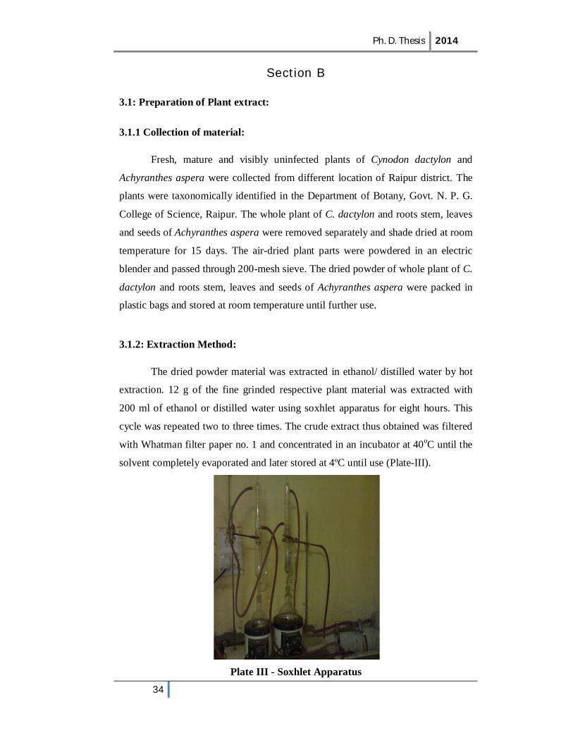
3.1.2: Extraction Method:
The dried powder material was extracted in ethanol/ distilled water by hot
extraction. 12 g of the fine grinded respective plant material was extracted with
200 ml of ethanol or distilled water using soxhlet apparatus for eight hours. This
cycle was repeated two to three times. The crude extract thus obtained was filtered
with Whatman filter paper no. 1 and concentrated in an incubator at 40oC until the
solvent completely evaporated and later stored at 4ºC until use (Plate-III).
Plate III - Soxhlet Apparatus

Ph. D. Thesis 2014
35
3.1.3: Preparation of stock and working solutions:
The crude extracts of the plant material was used for preparing stock
solutions by dissolving the known amount of the extract in a definite volume of
mother solvents. The concentration of alcoholic extract was kept high so that
minimum quantity of vehicle was required for treatment. Different concentration of
test solution viz., 50 and 100 ppm were prepared from stock solution by diluting
with mother solvent. Homogeneous solutions were obtained by gentle shaking or
stirring.
3.2: Phytochemical Screening:
Phytochemical tests were carried out both with aqueous and alcoholic
extracts of plants, using standard procedures to identify the major group of
following metabolites.
3.2.1: Alkaloids: A drop of Mayer’s reagent was added by the side of the test tube
to 1 ml of the extract treated with few drops of dilute hydrochloric acid. A creamy
or white precipitate indicated positive reaction.
Mayer’s reagent (Potassium mercuric iodide): 1.36 g of mercuric chloride in 60 ml
of distilled water and 5 g of potassium iodide in 10 ml of distilled water were
dissolved separately. The two solutions were mixed together and diluted to 100 ml
with distilled water.
3.2.2: Anthroquinone: 1 ml extract was heated with 1 ml of ferric chloride
solution and 1 ml of HCl in a test tube. The tube was cooled and the contents were
filtered. The filtrate was shaken with diethyl ether and ammonium hydroxide was
added to it. Appearance of a pink or deep red colour indicated presence of
anthroquinone.
3.2.3: Carbohydrates:
Molisch’s Test-1 ml of the plant extract was treated with 2 or 3 drops of 1%
alcoholic alpha naphthol. 2 ml of concentrated sulphuric acid was added along the
sides of the test tube. A brown ring appeared between the junctions of two liquid
that confirmed the carbohydrate in the sample.

Ph. D. Thesis 2014
36
Fehling’s test- 1 ml of extract was treated with 1 ml of Fehling’s solution A and B
and heated on a water bath. A reddish precipitate indicated the presence of
carbohydrates.
3.2.4: Cardio glycosides: 5 ml of the plant extract was mixed with 2 ml of glacial
acetic acid containing a drop of ferric chloride solution. 1 ml of concentrated
sulphuric acid was added to it. A brown ring at the interface indicates a deoxy-
sugar characteristic of cardio glycosides.
3.2.5: Flavonoids: 5 ml of the dilute ammonia solution was added to 1 ml of the
extract followed by addition of 5 ml concentrated sulphuric acid. Appearance of
yellow colouration indicated the presence of flavonoids (Ayoola et al., 2008).
3.2.6: Phenols: 1 to 2 drops of neutral ferric chloride was added to the dilute 1 ml
of extract in a test tube. The appearance of characteristic colour indicated (violet,
blue or green) the presence of phenols (Sawhney et al., 2011).
The extract was spotted on a filter paper. A drop of phosphomolybdic acid was
added to spot followed by exposure to ammonia vapor. Appearance of blue color
on spot indicated the presence of phenol (Kumar et al., 2007).
3.2.7: Quinones: A few drops of sodium hydroxide solution was mixed with the
plant extract and shaken vigorously; a blue or red color indicated the presence of
quinones.
3.2.8: Saponins: 5 ml extract was diluted to 20 ml with distilled water. The
suspension was shaken in a graduated cylinder for 15 minute. 2 cm layer of foam
indicates the presence of saponins (Evans, 1996).
3.2.9: Sterols: 1 ml plant extract was boiled with 2 ml of acetic anhydride. 1 or 2
drop of concentrated sulphuric acid was added to it slowly along the sides of the
test tubes. An array of color change showed presence of sterols.
3.2.10: Tannins: 20 mg of extract was boiled in 20 ml of distilled water in test
tube and filtered. 5 drops of 0.1% ferric chloride was added to it. Brownish green
or blue black coloration indicated the presence of tannins.

Ph. D. Thesis 2014
37
3.2.11: Terpenoids: 5 ml of extract was mixed with 2 ml of chloroform. 2 ml of
concentrated sulphuric acid was layered over it. A reddish brown colouration at the
interface showed the presence of terpenoids (Ayoola et al., 2008).
3.3: Experimental animal:
Clarias batrachus (Linnaeus, 1758), commonly known as magur is native
to Southeastern Asia was chosen for this study. It is a natural inhabitant of shallow,
derelict and swampy water bodies of tropical and subtropical countries, namely
India, Pakistan, Bangladesh, Nepal, Burma, Rangoon, Sri Lanka, Malaya,
Thailand, Indonesia and Philippines (Menon, 1979). It can also withstand slightly
brackish water and is more suited to live in poorly oxygenated water than other air
breathing fishes. In India it is mostly restricted to North Bihar, Bengal, Assam,
Orissa and certain parts of South India (Munshi and Ghosh, 1994). It received its
common name of the ‘Walking Catfish’ (Lagler, 1977) on its ability to walk
overland from pond to pond when their original habitat dries up or after a heavy
rainfall. They possess a much reduced air-bladder and their gills are stiffened to
prevent their collapse when out of water and in a special part of the gill chamber
are spongy tree like arborescent or dendritic organs growing from the upper ends
of the II and IV gill arches. Taxonomically it is placed in class Osteichthyes, order
Cypriniformes, suborder Siluroidei and family Clariidae (according to Berg, 1947).
It is known for its highly nutritive, restorative, therapeutic qualities and is
recommended as a diet during sickness and convalescence and for under nourished
and growing children. The flesh of this fish contains higher protein and low fat as
compared to carps. The protein of the fish contains many essential amino acids and
its fat is easily digestible and contains high value of physiologically available iron
and copper essential for haemoglobin synthesis (Thakur and Das, 1986). The
calorific value of this fish is higher (120cal/ 100g) than that of major carps (97 cal/
100g) (Roy, 1983). Therefore, it has high market value and preferred over major
carp. C. batrachus (Plate-IV) is a bottom dweller, nocturnal, slow growing
seasonal breeder (Munshi and Ghosh, 1994) and facultative air breather (Gupta and
Pati, 1998).
Ph. D. Thesis 2014
38
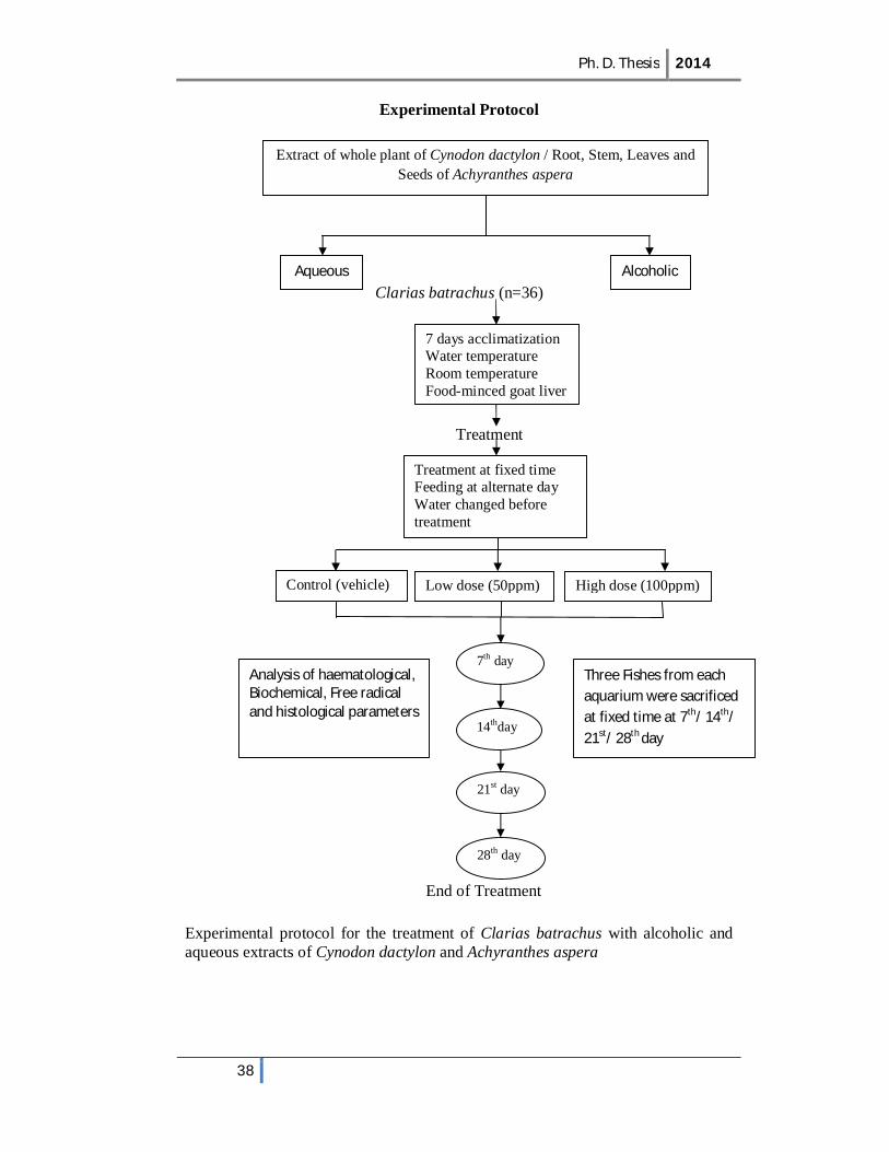
Experimental Protocol
Clarias batrachus (n=36)
Treatment
End of Treatment
Experimental protocol for the treatment of Clarias batrachus with alcoholic and aqueous extracts of Cynodon dactylon and Achyranthes aspera
Extract of whole plant of Cynodon dactylon / Root, Stem, Leaves and Seeds of Achyranthes aspera
Aqueous Alcoholic
Treatment at fixed time Feeding at alternate day Water changed before treatment
7 days acclimatization Water temperature Room temperature Food-minced goat liver
Control (vehicle) Low dose (50ppm) High dose (100ppm)
7th day
14thday
21st day
28th day
Three Fishes from each aquarium were sacrificed at fixed time at 7th/ 14th/ 21st/ 28th day
Analysis of haematological, Biochemical, Free radical and histological parameters
Ph. D. Thesis 2014
39
Experimental design
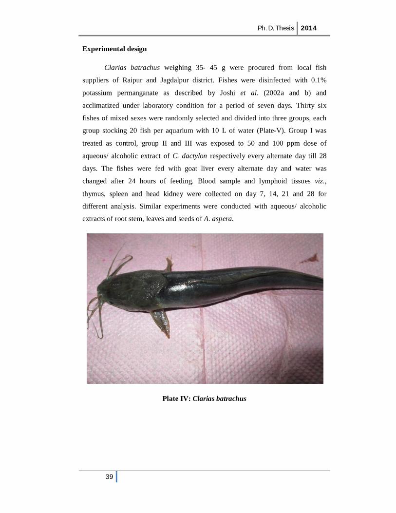
Clarias batrachus weighing 35- 45 g were procured from local fish
suppliers of Raipur and Jagdalpur district. Fishes were disinfected with 0.1%
potassium permanganate as described by Joshi et al. (2002a and b) and
acclimatized under laboratory condition for a period of seven days. Thirty six
fishes of mixed sexes were randomly selected and divided into three groups, each
group stocking 20 fish per aquarium with 10 L of water (Plate-V). Group I was
treated as control, group II and III was exposed to 50 and 100 ppm dose of
aqueous/ alcoholic extract of C. dactylon respectively every alternate day till 28
days. The fishes were fed with goat liver every alternate day and water was
changed after 24 hours of feeding. Blood sample and lymphoid tissues viz.,
thymus, spleen and head kidney were collected on day 7, 14, 21 and 28 for
different analysis. Similar experiments were conducted with aqueous/ alcoholic
extracts of root stem, leaves and seeds of A. aspera.
Plate IV: Clarias batrachus

Ph. D. Thesis 2014
40
Plate V: Experimental aquarium setup in the laboratory
Hematological Analysis:
Blood was collected in EDTA coated tubes for heamatological parameters
by cutting the caudal peduncle of Clarias batrachus. The haematological studies
were carried out immediately after collection of blood samples. Total erythrocytes
and leukocytes were counted in improved Neubaeur heamocytometer using
Hayem’s diluting fluid and Turk’s diluting fluid respectively in a binocular
microscope. For the study of differential leukocyte count, a drop of fresh blood
was smeared on a clean glass slide, air dried and stained with Leishman stain.
Biochemical analysis:
Total protein:
The second blood sample was taken without anticoagulant in a tube and
was left for 30 minutes at room temperature, centrifuged for 10 minutes at 3000
rpm. The serum was separated, collected and stored at 4⁰C until use. The samples
were analyzed for total protein following Lowry et al. (1951).
Ph. D. Thesis 2014
41
Reagents used:
1. 2% Sodium carbonate solution: 2 g sodium carbonate was dissolved in 100 ml
of 0.1 N sodium hydroxide solution.
2. 1% of Sodium potassium tartarate: 1g of sodium potassium tartarate was
dissolved in 100 ml of distilled water.
3. 0.5% Copper sulphate in sodium potassium tartarate: 50 mg of copper sulphate
was dissolved in 10 ml of sodium potassium tartarate solution. The solution
was freshly prepared.
4. Alkaline copper sulphate solution: 50 ml of solution I (2% of sodium
carbonate solution) and 1 ml of solution II (sodium potassium tartarate) were
mixed before use.
5. Folin- Ciocalteu phenol reagent: 1 ml of Folin Ciocalteu reagent was diluted
with 1 ml of distilled water before use.
6. Standard protein solution: 20 mg of bovine serum albumin was dissolved in
100 ml of 0.9% sodium chloride solution.
Procedure
5 ml of freshly prepared alkaline copper sulphate solution was taken into
different test tubes. 0.02 ml of serum was added to one tube to serve as test. 1.0 ml
of standard protein solution was added to another test tube for standard. 1.0 ml of
distilled was added to another tube to serve as blank. 0.5 ml of fresh diluted folin
ciocalteu phenol reagent was added to all the test tubes and incubated at 37oC for
10 minutes for color development. The final volume of all the tubes was made to
10 ml with distilled water. Optical density was recorded at 750 nm in
spectrophotometer against a blank. The total protein content was calculated with
reference to the standard and expressed as g/ 100 ml serum.
Enzyme assay
Fishes were sacrificed and lymphoid organs such as thymus, spleen and
head kidney were dissected out for the estimation of superoxide dismutase,
catalase and lipid peroxidation. Thymus, spleen and head kidney were
homogenized (10% w/v) using cold 0.1 M phosphate buffer (pH 7.4), centrifuged
Ph. D. Thesis 2014
42
for 10 minutes at 5000 rpm in a cooling centrifuge (Merlin and Parthasarathy,
2011). The supernatant was used for assay of superoxide dismutase (SOD),
catalase (CAT) and lipid peroxidation (LPO).
1. Superoxide dismutase:
Reagents used:
1. Absolute ethanol
2. Chloroform
3. 2.0 mM of pyrogallol
4. 1.0 mM Tris-HCl buffer (pH 7.4)
The activity of superoxide dismutase was determined by the method of
Marklund and Marklund (1974). 0.25 ml of absolute ethanol and 0.15 ml of
chloroform were added to 1 ml of supernatant of tissue homogenate and shaken for
15 min in a mechanical shaker. Suspension was centrifuge at 3000 rpm and
supernatant obtained constituted the enzyme extract. The reaction mixture for auto-
oxidation consisted of 2 ml of 0.1 M Tris-HCl buffer, 0.5 ml of 2 mM pyrogallol
and 1.5 ml of distilled water. The rate of auto-oxidation of pyrogallol was recorded
at 1 min interval for the 3 min. The assay mixture for enzyme contained 2 ml of
Tris HCl buffer, 0.5 ml pyrogallol and aliquots of the enzyme and the distilled
water to make the final volume to 4 ml. The rate of inhibition of pyrogallol auto-
oxidation after the addition of enzyme extract was recorded. The superoxide
dismutase activity was measured by the inhibition of pyrogallol auto-oxidation at
420 nm for 10 min. One unit of superoxide dismutase is the amount of enzyme
required to bring about 50% inhibition of auto oxidation by pyrogallol. The
enzyme activity was expressed in term of unit/ min/ mg protein.
2. Catalase:
Reagents used:
1. 0.2 M hydrogen peroxide
2. 0.01 M of phosphate buffer (pH 7.0)
3. 5% of dichromate acetic acid reagent (prepared in glacial acetic acid)
Ph. D. Thesis 2014
43
Catalase activity was estimated according to Sinha (1972). 1 ml of 0.01M
phosphate buffer (pH 7.0) was added to 0.4 ml of 0.2 M hydrogen peroxide
followed by 0.1 ml of clear supernatant of tissue homogenate (10% w/v) and
gently swirled at room temperature. 2 ml of 5% dichromate acetic acid reagent was
added to stop the reaction. The change in absorbance was measured at 620 nm and
recorded after 3 min interval against reference blank. The enzyme activity was
expressed in term of µmole/ min/ mg protein.
3. Lipid peroxidation
Reagents used:
1. 20% Acetic acid
2. 8.1% of Sodium dodecyl sulphate
3. 0.8% of Thiobarbituric acid
4. n-butanol: pyridine (15:1)
Lipid peroxidation was determined by the method of Okhawa et al. (1979).
1.5 ml of 20% acetic acid, 0.2 ml of sodium dodecyl sulphate and 1.5 ml of
thiobarbituric acid and 0.2 ml of tissue homogenate were mixed together. The
volume was made up to 4.0 ml with distilled water. The tubes were heated at 95oC
in water bath for 60 minutes. After incubation the test tubes were cooled at room
temperature, the volume was made to 5.0 ml in each tube with distilled water. 5 ml
of n-butanol: pyridine mixture (15:1) was added to it. The contents were vortexed
thoroughly for 2 min and centrifuged at 3000 rpm for 10 min. The organic upper
layer was taken for optical density measurement at 532 nm against a blank devoid
of sample. Lipid peroxidation levels are expressed in nmole of MDA/ min/ mg
protein.
Histological analysis:
Tissue of thymus, spleen and head kidney of control and treated C.
batrachus was transversely cut in to small pieces and fixed in aqueous Bouin’s
fixative for 12-16 hours. They were washed with tap water several times to remove
fixative and dehydrated in ascending series of alcohol, cleared in xylene and

Ph. D. Thesis 2014
44
embedded in paraffin wax (M.P. 58ºC) and blocks were prepared. The serial
sections of 5µ thickness were cut using rotary microtome and were flattened on a
clean slide using Mayer’s albumin.
The sections were differentially stained with Harris’ haematoxylin and
eosin. For staining, the sections were de-paraffinized in xylene. They were then
brought down to water through descending series of alcohol one minute in each.
The slides were washed with tap water and finally rinsed in distilled water.
Staining was done in Harris’ haematoxylin for 5-10 minutes. The slides were
rinsed in tap water for few seconds. If sections were over stained, they were
differentiated in 0.5% of acid alcohol with continuous agitation for few seconds,
checking that the nuclei retain dark purple stain and rest of the tissue is very pale.
The sections were washed with distilled water and up graded for dehydration to
70% alcohol. The sections were stained with 1% eosin in 70% alcohol for 2-3
minutes. Finally, the sections were dehydrated by upgrading in the alcohol series
(70%, 90% and absolute alcohol), cleared in xylene and mounted in DPX. The
slides were examined under a compound microscope (Leica DMLC) and the
pictures were taken with help of a micro-photographic attachment (Plate VI).
Plate VI: Leica Microscope

Ph. D. Thesis 2014
45
The reagents used in the histological study were prepared as follows:
1. Bouin’s fluid:
Picric acid - 75%
Formalin - 20%
Acetic acid - 5%
2. Mayer’s albumin:
It was prepared by adding equal amount of egg white (albumin) and glycerol. Both
were mixed thoroughly and few crystals of thymol was added it to prevent
microbial contamination.
3. Harris’s haematoxylin staining solution:
Haematoxylin powder 1g
Absolute alcohol 10 ml
Ammonium or potash alum 20 g
Distilled water 200 ml
Mercuric oxide 0.5 g
The alum was dissolved in distilled water by heating. Haematoxylin
crystals were dissolved in absolute alcohol separately. Both were mixed together
while hot and brought to boil as quickly as possible (but not for longer time) and
mercuric oxide was added to it. When the solution assumed dark purple colour it
was cooled without delay in running cold water. 2-3 ml of glacial acetic acid per
100 ml of haematoxylin solution was added to increase its nuclear staining
property. The stain was filtered before use.
4. Eosin solution (alcoholic)
Eosin Y (water soluble) 1g
Distilled water 20 ml
Absolute alcohol 80 ml
Glacial acetic acid 0.5 ml

Ph. D. Thesis 2014
46
The eosin was dissolved in distilled water and it was added to the absolute
alcohol to prepare 1% stock solution. For working solution (0.25% or 0.5%) it was
diluted with 80% ethanol. 0.5 ml acetic acid was added to increase its staining
intensity just before use.
Statistical analysis
The data was statistically analyzed by using the Statistical Package for
Social Sciences (SPSS version 16.0 for windows), in which data was subjected to
various statistical test and two-way ANOVA. Duncan’s Multiple Range Test
(DMRT) was used to determine the significant differences between the means.
Comparisons were made at the 5% and/ or 1% probability level.