Embed Size (px)
Citation preview

9/12/2012
1
Chapter 22
Respiratory Emergencies
Chapter Goal
Use assessment findings to formulate field impression & carry out treatment plan for patient with respiratory emergencies
Learning Objectives
Identify & describe function of structures in upper & lower airway
Discuss physiology of ventilation & respiration
Identify common pathological events affecting pulmonary system
Discuss abnormal assessment findings associated with pulmonary diseases & conditions
Copyright © 2013 by Jones & Bartlett Learning, LLC, an Ascend Learning Company
9/12/2012
2
Learning Objectives
Compare various airway & ventilation techniques used in management of pulmonary diseases
Review pharmacological preparations EMT-Is use for management of respiratory diseases & conditions
Review equipment used during physical examination of respiratory complaint patients
Learning Objectives
Identify assessment findings & management for Bronchial asthma COPD Chronic bronchitis Emphysema Pneumonia Pulmonary edema Spontaneous pneumothorax Hyperventilation syndrome
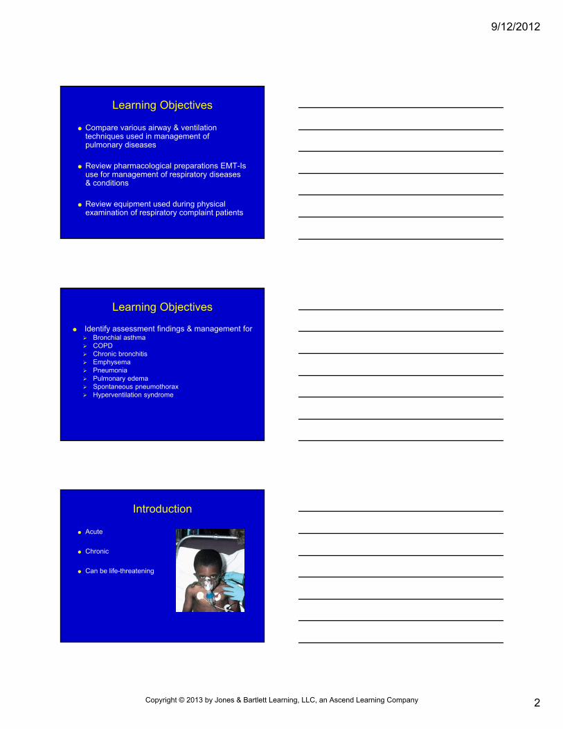
Introduction
Acute
Chronic
Can be life-threatening
Copyright © 2013 by Jones & Bartlett Learning, LLC, an Ascend Learning Company
9/12/2012
3
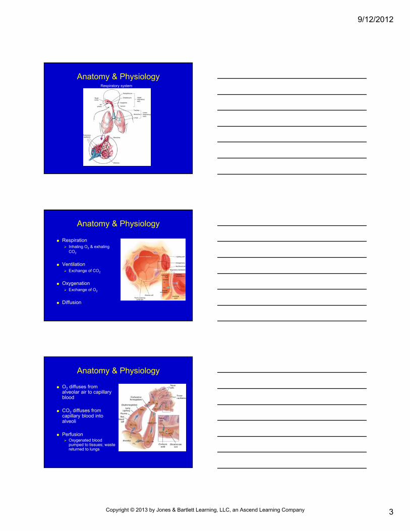
Anatomy & PhysiologyRespiratory system
Anatomy & Physiology
Respiration Inhaling O2 & exhaling
CO2
Ventilation Exchange of CO2
Oxygenation Exchange of O2
Diffusion
Anatomy & Physiology
O2 diffuses from alveolar air to capillary blood
CO2 diffuses from capillary blood into alveoli
Perfusion Oxygenated blood
pumped to tissues; waste returned to lungs
Copyright © 2013 by Jones & Bartlett Learning, LLC, an Ascend Learning Company

9/12/2012
4
General Respiratory System
Pathophysiology Respiratory abnormalities
• Primarily affecting ventilation Upper airway obstruction
Lower airway obstruction
Impaired chest wall movement
Problems with neurological control
General Respiratory System
Pathophysiology Respiratory abnormalities
• Diffusion-related conditions Inadequate O2 in ambient air
Alveolar pathology
Interstitial space pathology
• Perfusion-related factors Inadequate blood volume or hemoglobin levels
Impaired circulatory blood flow
Chest wall pathology
General Respiratory System
Assessment Major focus—signs of life-
threatening distress• Alterations in mental status
• Severe cyanosis
• Absent breath sounds
• Audible stridor
• 1- or 2-word dyspnea
• HR >130 bpm
• Pallor; diaphoresis
• Retractions/accessory muscle use
Copyright © 2013 by Jones & Bartlett Learning, LLC, an Ascend Learning Company
9/12/2012
5
General Respiratory System
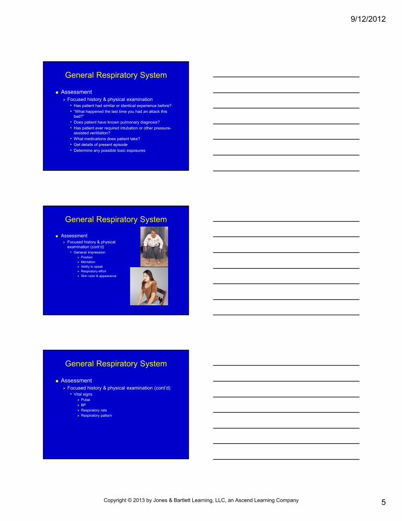
Assessment Focused history & physical examination
• Has patient had similar or identical experience before?
• “What happened the last time you had an attack this bad?”
• Does patient have known pulmonary diagnosis?
• Has patient ever required intubation or other pressure-assisted ventilation?
• What medications does patient take?
• Get details of present episode
• Determine any possible toxic exposures
General Respiratory System
Assessment Focused history & physical
examination (cont’d)• General impression
Position
Mentation
Ability to speak
Respiratory effort
Skin color & appearance
General Respiratory System
Assessment Focused history & physical examination (cont’d)
• Vital signs Pulse
BP
Respiratory rate
Respiratory pattern
Copyright © 2013 by Jones & Bartlett Learning, LLC, an Ascend Learning Company
9/12/2012
6
General Respiratory System
General Respiratory System
Assessment Focused history & physical
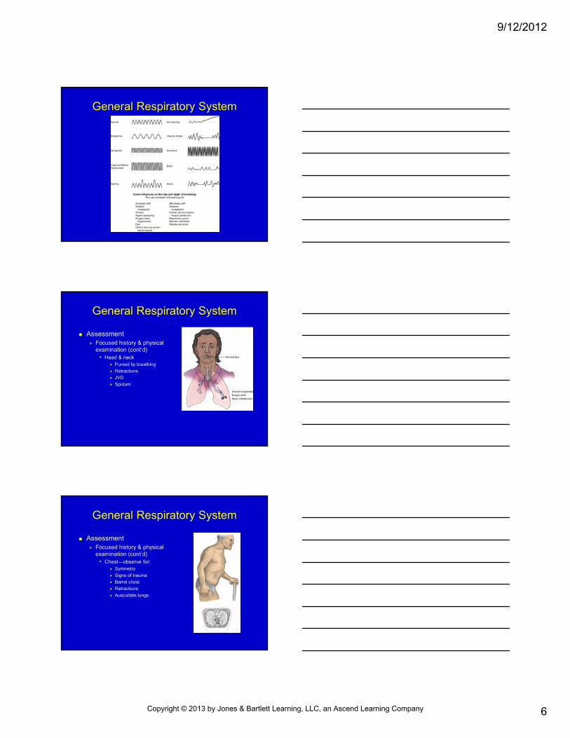
examination (cont’d)• Head & neck
Pursed lip breathing
Retractions
JVD
Sputum
General Respiratory System
Assessment Focused history & physical
examination (cont’d)• Chest—observe for:
Symmetry
Signs of trauma
Barrel chest
Retractions
Auscultate lungs
Copyright © 2013 by Jones & Bartlett Learning, LLC, an Ascend Learning Company
9/12/2012
7
General Respiratory System
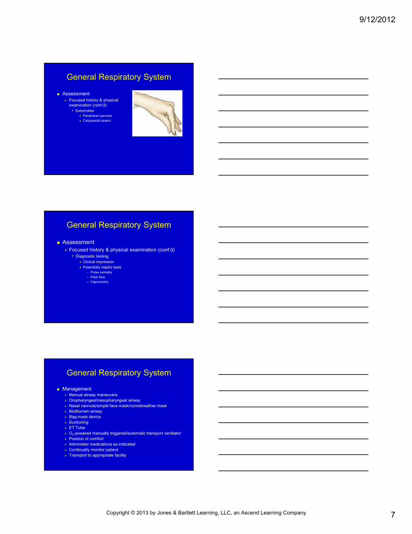
Assessment Focused history & physical
examination (cont’d)• Extremities
Peripheral cyanosis
Carpopedal spasm
General Respiratory System
Assessment Focused history & physical examination (cont’d)
• Diagnostic testing Clinical impression
Potentially helpful tests – Pulse oximetry
– Peak flow
– Capnometry
General Respiratory System
Management Manual airway maneuvers Oropharyngeal/nasopharyngeal airway Nasal cannula/simple face mask/nonrebreather mask Multilumen airway Bag-mask device Suctioning ET Tube O2-powered manually triggered/automatic transport ventilator Position of comfort Administer medications as indicated Continually monitor patient Transport to appropriate facility
Copyright © 2013 by Jones & Bartlett Learning, LLC, an Ascend Learning Company
9/12/2012
8
Obstructive Airway Disease
Epidemiology & causes 4%-5% of U.S. population have asthma Nearly 20% of adult males have chronic bronchitis Most common cause of COPD—cigarette smoking Factors exacerbating underlying conditions
• Stress• Infection• Exercise
External stimuli• Tobacco smoke• Allergens• Drugs• Occupational hazards
Obstructive Airway Disease:Pathophysiology
Obstruction—usually result of: Smooth muscle spasm
Mucus
Inflammation
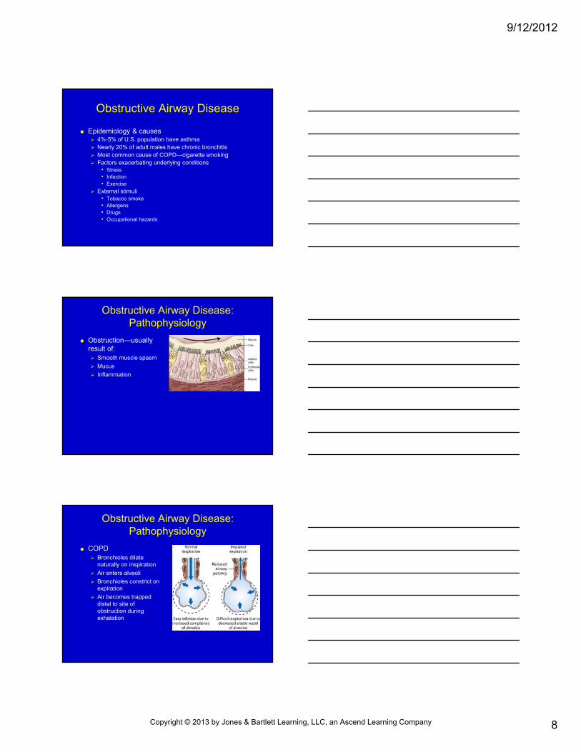
Obstructive Airway Disease:Pathophysiology
COPD Bronchioles dilate
naturally on inspiration
Air enters alveoli
Bronchioles constrict on expiration
Air becomes trapped distal to site of obstruction during exhalation
Copyright © 2013 by Jones & Bartlett Learning, LLC, an Ascend Learning Company
9/12/2012
9
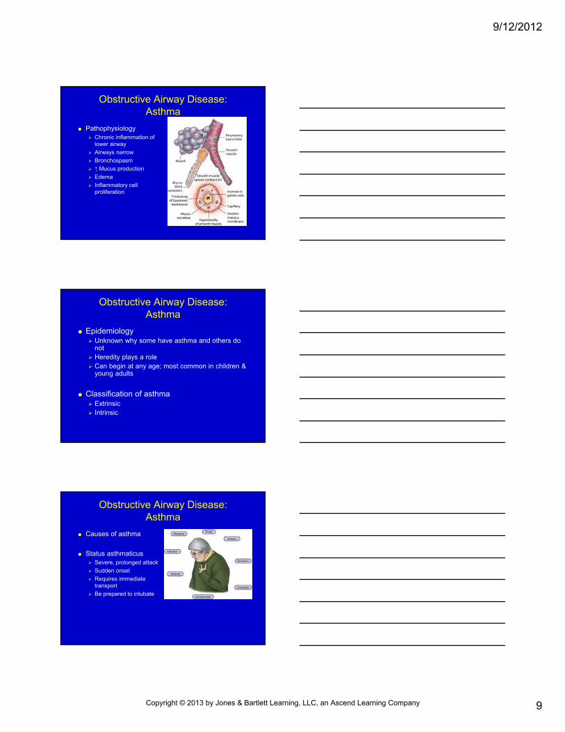
Obstructive Airway Disease:Asthma
Pathophysiology Chronic inflammation of
lower airway
Airways narrow
Bronchospasm
↑ Mucus production
Edema
Inflammatory cell proliferation
Obstructive Airway Disease:Asthma
Epidemiology Unknown why some have asthma and others do
not Heredity plays a role Can begin at any age; most common in children &
young adults
Classification of asthma Extrinsic Intrinsic
Obstructive Airway Disease:Asthma
Causes of asthma
Status asthmaticus Severe, prolonged attack
Sudden onset
Requires immediate transport
Be prepared to intubate
Copyright © 2013 by Jones & Bartlett Learning, LLC, an Ascend Learning Company

9/12/2012
10
Obstructive Airway Disease: COPD
Progressive, irreversible
↓ Inspiratory & expiratory capacity
Chronic bronchitis Excess mucus production
Emphysema Lung tissue damage with
loss of recoil of lung
Obstructive Airway Disease: COPD
Pathophysiology Chronic bronchitis
• Overgrowth of airway mucous glands
• Excess mucus blocks airway
Emphysema • Destruction of walls of
alveoli
• Air is trapped within lungs
Obstructive Airway Disease: COPD
Pathophysiology (cont’d) Cor pulmonale—severe COPD
• Right side of heart may fail because of effort required to move blood though diseased lung
Causes Cigarette smoking Industrial inhalants
• Coal dust• Air pollution• Tuberculosis
Copyright © 2013 by Jones & Bartlett Learning, LLC, an Ascend Learning Company

9/12/2012
11
Obstructive Airway Disease
Assessment findings: Patients may report:
• New cough or change in previous cough pattern & sputum production
• No longer taking medications
• Use of home oxygen or CPAP/BiPAP
• Use of home nebulizer
• ↑ Dyspnea with activity
Patient may be found in tripod position
Breathing loud; wheezing present
No wheezing but short of breath—ominous sign
Obstructive Airway Disease
Other signs & symptoms: Shortness of breath; tachypnea
Coughing
Tightness in chest
Cyanosis
Anxiety, agitation
Diaphoresis; pallor
Cigarette stains on fingertips
Tachycardia
Hypotension
Hyperinflated (barrel) chest
Use of accessory muscles
Audible abnormal breath sounds
↓ O2 saturation
Obstructive Airway Disease
If cor pulmonale present: Neck vein distention
Abdominal bloating
Leg edema
Copyright © 2013 by Jones & Bartlett Learning, LLC, an Ascend Learning Company
9/12/2012
12
Obstructive Airway Disease
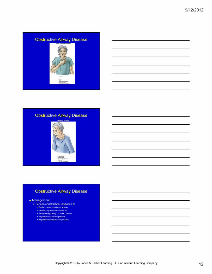
Obstructive Airway DiseasePriority patient
Obstructive Airway Disease
Management Perform endotracheal intubation if:
• Patient cannot maintain airway
• Ventilatory assistance needed
• Severe respiratory distress present
• Significant cyanosis present
• Significant hypotension present
Copyright © 2013 by Jones & Bartlett Learning, LLC, an Ascend Learning Company
9/12/2012
13
Obstructive Airway Disease
Management Respiratory distress but breathing adequately
• Administer high-concentration humidified O2• Place patient in position of comfort• Provide calm & reassurance
Provide timely transport Monitor vital signs, including pulse oximetry, frequently Monitor ECG Treat bronchospasm
• Bronchodilator• Epinephrine
Initiate IV Notify receiving facility early if ventilatory support needed
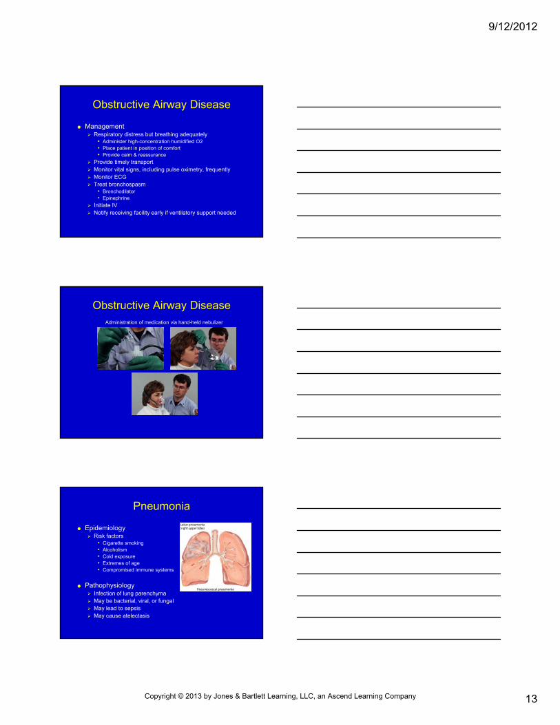
Obstructive Airway DiseaseAdministration of medication via hand-held nebulizer
Pneumonia
Epidemiology Risk factors
• Cigarette smoking• Alcoholism• Cold exposure• Extremes of age• Compromised immune systems
Pathophysiology Infection of lung parenchyma May be bacterial, viral, or fungal May lead to sepsis May cause atelectasis
Copyright © 2013 by Jones & Bartlett Learning, LLC, an Ascend Learning Company
9/12/2012
14
Pneumonia
Assessment findings Typical
• Acute onset of fever, chills• Productive cough• Pleuritic chest pain• Crackles & signs of consolidation may be present
Atypical• Nonproductive cough• Headache• Myalgia• Fatigue• Sore throat• Nausea, vomiting, diarrhea• Fever, chills (continuous)
Pneumonia
Management Consider patient contagious; act accordingly
Airway, ventilatory support
High-concentration O2; may require intubation
IV fluids per local protocol
Reduce body temperature
Consider beta-2 agonists per local protocol
Monitor patient during transport
Provide psychological support
Pulmonary Edema
Epidemiology Lungs fill with fluid in interstitial spaces, alveoli, or
both
Pathophysiological condition
Copyright © 2013 by Jones & Bartlett Learning, LLC, an Ascend Learning Company

9/12/2012
15
Pulmonary Edema
Anatomy & physiology High pressure (cardiogenic)
• Acute myocardial ischemia• Heart valve disease• Chronic hypertension• Myocarditis
High permeability (noncardiogenic)• Acute hypoxemia• Near-drowning• Cardiac arrest• Shock• High altitude exposure• Inhalation of pulmonary irritants• ARDS
Pulmonary Edema
Pathophysiology Changes in high-pressure pulmonary edema
• Ischemia • Ventricle unable to pump blood adequately• ↑ Ventricular pressure • ↑ Pulmonary venous pressure • Vessels leak fluid into interstitial space • Cough & lymphatic drainage fail to drain excess fluid• Fluid accumulation • Significant interstitial fluid cause alveoli to rupture & fill
with fluid
Pulmonary EdemaHigh-pressure (cardiogenic)
Copyright © 2013 by Jones & Bartlett Learning, LLC, an Ascend Learning Company
9/12/2012
16
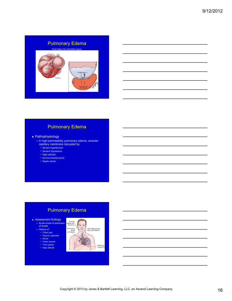
Pulmonary EdemaFluid leaks into interstitial space
Pulmonary Edema
Pathophysiology In high-permeability pulmonary edema, alveolar-
capillary membrane disrupted by:• Severe hypotension
• Severe hypoxemia
• High altitude
• Environmental toxins
• Septic shock
Pulmonary Edema
Assessment findings Acute onset of shortness
of breath
History of:• Chest pain
• Hypoxic episodes
• Shock
• Chest trauma
• Toxic gases
• High altitude
Copyright © 2013 by Jones & Bartlett Learning, LLC, an Ascend Learning Company
9/12/2012
17
Pulmonary Edema
Management Airway, circulatory support
High-concentration O2 preferred if patient will tolerate
Assist ventilation and intubate as required
Initiate IV per local protocol
Position patient upright with legs dangling
Provide calm & reassurance
Pharmacological management• Nitroglycerin
• Furosemide (Lasix)
• Morphine sulfate
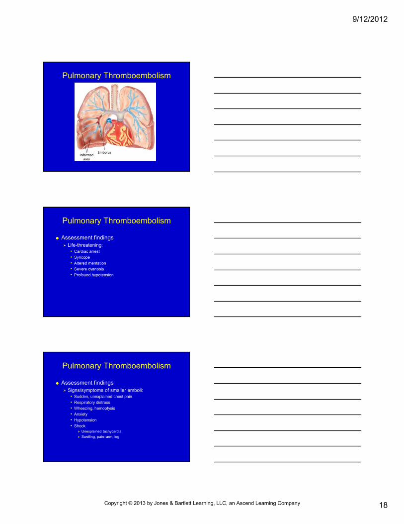
Pulmonary Thromboembolism
Epidemiology Blockage of pulmonary artery
Combination of 2 processes• Formation of venous thrombus
• Fragment breaks off into venous circulation
50,000 deaths annually
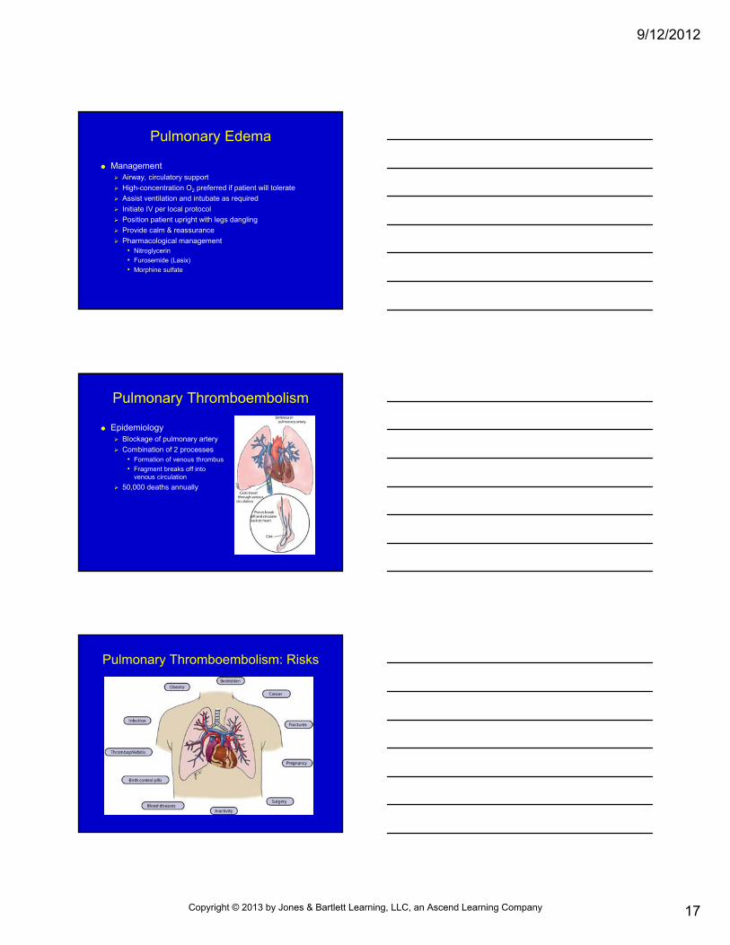
Pulmonary Thromboembolism: Risks
Copyright © 2013 by Jones & Bartlett Learning, LLC, an Ascend Learning Company
9/12/2012
18
Pulmonary Thromboembolism
Pulmonary Thromboembolism
Assessment findings Life-threatening:
• Cardiac arrest
• Syncope
• Altered mentation
• Severe cyanosis
• Profound hypotension
Pulmonary Thromboembolism
Assessment findings Signs/symptoms of smaller emboli:
• Sudden, unexplained chest pain
• Respiratory distress
• Wheezing, hemoptysis
• Anxiety
• Hypotension
• Shock Unexplained tachycardia
Swelling, pain–arm, leg
Copyright © 2013 by Jones & Bartlett Learning, LLC, an Ascend Learning Company
9/12/2012
19
Pulmonary Thromboembolism
Management Airway, ventilatory support
IV fluids
Watch for shock
ECG monitor
Position of comfort
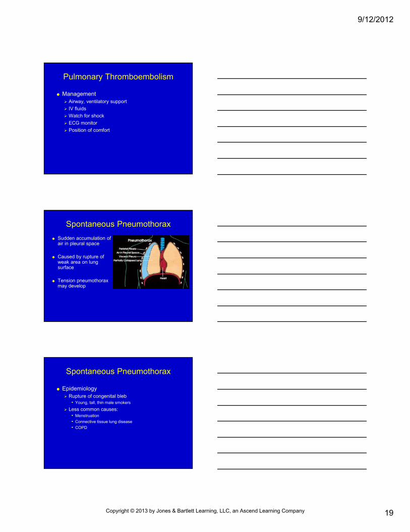
Spontaneous Pneumothorax
Sudden accumulation of air in pleural space
Caused by rupture of weak area on lung surface
Tension pneumothorax may develop
Spontaneous Pneumothorax
Epidemiology Rupture of congenital bleb
• Young, tall, thin male smokers
Less common causes:• Menstruation
• Connective tissue lung disease
• COPD
Copyright © 2013 by Jones & Bartlett Learning, LLC, an Ascend Learning Company
9/12/2012
20
Spontaneous Pneumothorax
Assessment findings Sudden onset of sharp chest pain on affected side Shortness of breath Tachypnea Patient may be coughing, anxious, or agitated Signs/symptoms of tension pneumothorax
• ↑ Respiratory distress• Weak pulse• Cyanosis• Hypotension• ↓ Breath sounds on side of lung involved• Distended neck veins• Tracheal deviation• Subcutaneous emphysema
Spontaneous Pneumothorax
Management Airway & ventilatory support; high-concentration
O2
IV fluids
ECG monitor
Position of comfort; consider lying patient with affected side down
Watch for tension pneumothorax
Hyperventilation Syndrome
Respiratory rate greater than required
Causes: ↑ Frequency of breathing
↑ Volume of air moved
Both
↑ Intake of O2; ↑ elimination of CO2
Copyright © 2013 by Jones & Bartlett Learning, LLC, an Ascend Learning Company
9/12/2012
21
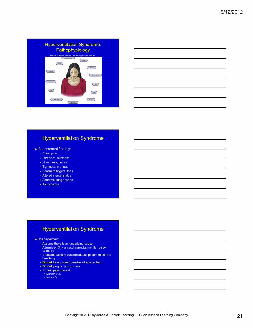
Hyperventilation Syndrome:Pathophysiology
Many disease states cause hyperventilation
Hyperventilation Syndrome
Assessment findings Chest pain
Dizziness, faintness
Numbness, tingling
Tightness in throat
Spasm of fingers, toes
Altered mental status
Abnormal lung sounds
Tachycardia
Hyperventilation Syndrome
Management Assume there is an underlying cause Administer O2 via nasal cannula; monitor pulse
oximetry If isolated anxiety suspected, ask patient to control
breathing Do not have patient breathe into paper bag Do not plug portals of mask If chest pain present:
• Monitor ECG• Initiate IV
Copyright © 2013 by Jones & Bartlett Learning, LLC, an Ascend Learning Company

9/12/2012
22
Summary
Respiratory system responsible for filtering, warming, humidifying, & exchanging more than 10,000 L air/day
Chronic & acute respiratory problems can present as life threatening
Abnormalities of ventilation, diffusion, or perfusion (or combination of these) can lead to respiratory problems
Summary
Ensure safe environment for EMS personnel before initiating patient contact
Major focus of initial assessment: recognition of life-threatening conditions
Focused history & physical examination is aimed at patient’s specific respiratory complaints
Specific conditions may be difficult to diagnose
Summary
Common respiratory problems include: Obstructive airway Pneumonia Pulmonary edema Pulmonary thromboembolism Spontaneous pneumothorax Hyperventilation syndrome
Copyright © 2013 by Jones & Bartlett Learning, LLC, an Ascend Learning Company

9/12/2012
23
Questions?
Copyright © 2013 by Jones & Bartlett Learning, LLC, an Ascend Learning Company
![Chapter 012ems.jbpub.com/shade/intermediate1999/docs/lecture_notes/Chapter... · Title: Microsoft PowerPoint - Chapter_012 [Compatibility Mode] Author: Jennifer.Meltz Created Date:](https://img.dokumen.tips/doc/110x75/5cc041f988c993d1388bf535/chapter-title-microsoft-powerpoint-chapter012-compatibility-mode-author.jpg)