Embed Size (px)
Citation preview

9/10/2012
1
1
Chapter 22
Cardiology
2
Lesson 22.1
Cardiovascular Disease Risk
Factors, Heart Anatomy, and
Physiology
3
Copyright © 2013 by Jones & Bartlett Learning, LLC, an Ascend Learning Company

9/10/2012
2
Learning Objectives
• Identify risk factors and prevention strategies associated with cardiovascular disease.
• Describe the normal anatomy and physiology of the heart.
4
Morbidity Rates
• MI death rates have declined over past several decades due to
– Heightened public awareness
– Increased availability of automated external defibrillators
– Improved cardiovascular diagnosis and therapy
– Use of cardiovascular drugs by persons at high risk
– Improved revascularization techniques
– Improved, more aggressive risk factor modification
5
Risk Factors/Modifications
• Risks for cardiovascular disease– Advanced age
– Male sex
– Diabetes
– Hypertension
– Hypercholesterolemia
– Hyperlipidemia
– Family history of premature cardiovascular disease
– Known coronary artery disease
6
Copyright © 2013 by Jones & Bartlett Learning, LLC, an Ascend Learning Company

9/10/2012
3
Risk Factors/Modifications
• Risks increased with
– Obesity
– Smoking
– Sedentary lifestyle
7
Risk Factors/Modifications
• Modifiable risk factors
– Cessation of smoking
– Medical management and control of blood pressure, diabetes, cholesterol, and lipid disorders
– Exercise
– Weight loss
– Diet
– Stress reduction
8
Risk Factors/Modifications
• Modifying risk factors can slow arterial disease development and reduce rate of
– MI
– Sudden death
– Renal failure
– Stroke
9
Copyright © 2013 by Jones & Bartlett Learning, LLC, an Ascend Learning Company

9/10/2012
4
Prevention Strategies
• Paramedics can support and practice prevention strategies– Educational programs about nutrition in their communities
– Cessation of smoking• Smoking prevention for children
– Early recognition and management of hypertension and cardiac symptoms
– Prompt intervention• CPR
• Early use of automated external defibrillator
10
Heart Anatomy
• Muscular organ with four chambers
• Cone shaped
• Size of man's closed fist
• Lies just to left of midline of thorax
11
12
Copyright © 2013 by Jones & Bartlett Learning, LLC, an Ascend Learning Company

9/10/2012
5
Heart Anatomy
• Enclosed in pericardial sac lined with parietal layers of serous membrane that form wall of heart
– Outer layer (epicardium)
– Middle layer (myocardium)
– Inner layer (endocardium)
13
Heart Anatomy
• Chambers
– Right atrium
• Receives deoxygenated blood from systemic veins
– Right ventricle
– Left atrium
• Receives oxygenated blood from pulmonary veins
– Left ventricle
14
Heart Anatomy
• Valves – Keep blood flowing in right direction
– Atrioventricular (cuspid) valves• Located between atria and ventricles
– Semilunar valves• Located at large vessels leaving ventricles
– Right atrioventricular valve• Tricuspid valve
– Left atrioventricular valve• Bicuspid or mitral valve
15
Copyright © 2013 by Jones & Bartlett Learning, LLC, an Ascend Learning Company

9/10/2012
6
Heart Anatomy
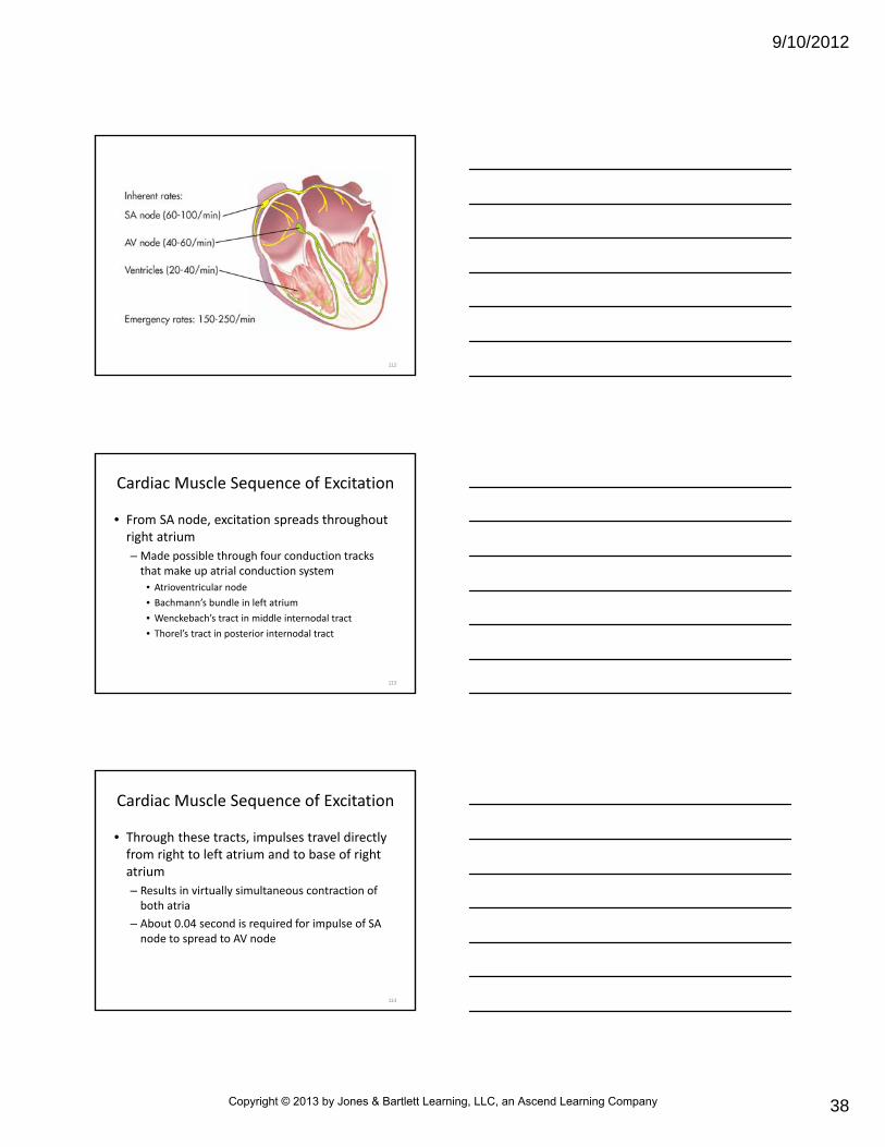
• Valves
– Pulmonary semilunar valve
• Between right ventricle and pulmonary trunk
– Aortic semilunar valve
• Between left ventricle and aorta
16
Heart Anatomy
• When ventricles contract, atrioventricularvalves close to prevent blood from flowing back into atria
• When ventricles relax, semilunar valves close to prevent blood from flowing back into ventricles
17
Blood Supply to Heart
• Coronary arteries
– Sole suppliers of arterial blood to heart
– Deliver 200 to 250 mL of blood to myocardium each minute during rest
– Left coronary artery carries about 85 percent of blood supply to myocardium
– Right coronary artery carries rest
18
Copyright © 2013 by Jones & Bartlett Learning, LLC, an Ascend Learning Company

9/10/2012
7
Blood Supply to Heart
• Coronary arteries
– Begin just above aortic valve where aorta exits heart
– Run along epicardial surface
– Divide into smaller vessels as they penetrate myocardium and endocardial (inner) surface
19
20
Blood Supply to Heart
• Left main coronary artery supplies
– Left ventricle
– Interventricular septum
– Part of right ventricle
– Two main branches
• Left anterior descending
• Circumflex artery
21
Copyright © 2013 by Jones & Bartlett Learning, LLC, an Ascend Learning Company

9/10/2012
8
Blood Supply to Heart
• Right coronary artery supplies
– Right atrium and ventricle
– Part of left ventricle
– Conduction system
– Two major branches
• Right anterior descending
• Marginal branch
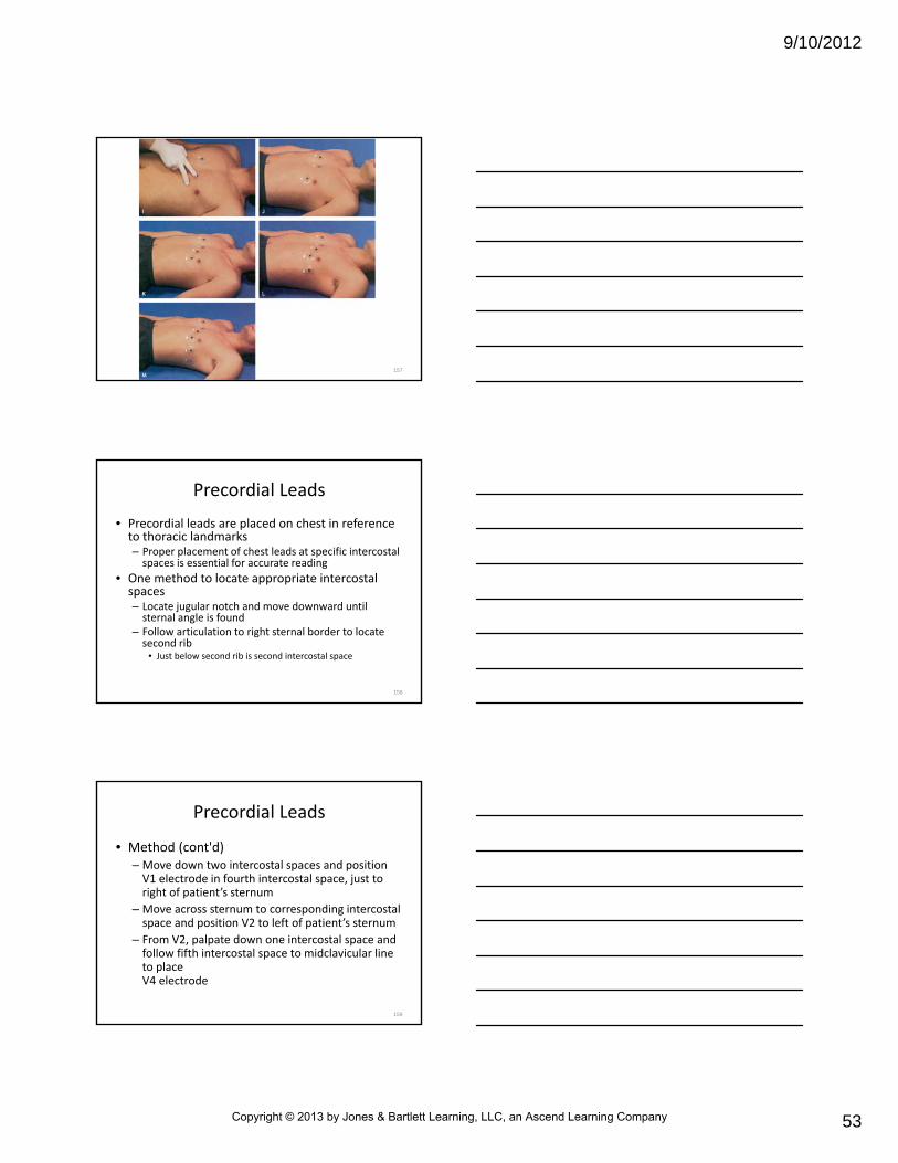
22
Blood Supply to Heart
• Connections (anastomoses) exist between arterioles to provide backup (collateral) circulation
– Play key role in providing alternative routes of blood flow in event of blockage in one or more of coronary vessels
23
24
Copyright © 2013 by Jones & Bartlett Learning, LLC, an Ascend Learning Company

9/10/2012
9
Blood Supply to Heart
• Coronary capillaries
– Allow for exchange of nutrients and metabolic wastes
– Merge to form coronary veins
• Veins deliver most of blood to coronary sinus
• Coronary sinus empties directly into right atrium
• Coronary sinus is major vein draining myocardium
25
Physiology
• Heart is two pumps in one
– Low pressure
• Right ventricle
• Right atrium
• Supplies blood to lungs
– High pressure
• Left ventricle
• Left atrium
• Supplies blood to body
26
Physiology
• Right atrium
– Receives venous blood from systemic circulation and from coronary veins
– Then passes to right ventricle as ventricle relaxes from previous contraction
– Once right ventricle receives about 70 percent of its volume, right atrium contracts
– Blood remaining is pushed into ventricle
27
Copyright © 2013 by Jones & Bartlett Learning, LLC, an Ascend Learning Company

9/10/2012
10
Physiology
• Right ventricle contraction pushes blood against tricuspid valve (forcing it closed) and through pulmonic valve (forcing it open)
– Allows blood to enter lungs via pulmonary arteries
• Blood enters capillaries in the lungs where gas exchange takes place
28
Physiology
• Atrial kick
– From lungs, blood travels through four pulmonary veins back to left atrium
– Mitral valve opens, and blood flows to left ventricle
– Once left ventricle receives about 70 percent of its volume, left atrium contracts
– Remaining blood 20 to 30 percent is pushed into ventricles during atrial contract
29
Physiology
• Blood passing from left atrium to left ventricle opens bicuspid valve when ventricle relaxes to complete left ventricular filling
• As left ventricle contracts, blood is pushed against bicuspid valve (closing it) and against aortic valve (opening it)
– Allows blood to enter the aorta
• From aorta, blood is distributed first to heart itself and then throughout systemic arterial circulation
30
Copyright © 2013 by Jones & Bartlett Learning, LLC, an Ascend Learning Company

9/10/2012
11
Cardiac Cycle
• Heart pumping
– Rhythmic, alternate contraction and relaxation
– Systole
• Contraction
– Diastole
• Relaxation
– Beats about 70 times/min in resting adults
– Responsible for blood movement
31
32
Heart Pumping
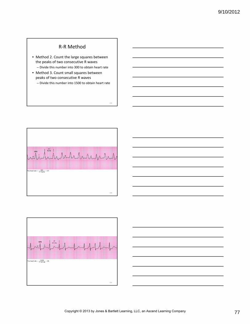
• As ventricles begin to contract, ventricular pressure exceeds atrial pressure– Causes atrioventricular valves to close
– As contraction proceeds, ventricular pressure continues to rise
– Pressure rises until it exceeds that in pulmonary artery on right side of heart and in aorta on left side
• At that time, pulmonary and aortic valves open
• Then blood flows from ventricles into those arteries
33
Copyright © 2013 by Jones & Bartlett Learning, LLC, an Ascend Learning Company

9/10/2012
12
Heart Pumping
• After ventricular contraction, ventricular relaxation begins– Ventricular pressure falls rapidly– When pressure falls below pressure in aorta or pulmonary trunk, blood is forced back toward ventricles
– This closes pulmonic and aortic valves– As ventricular pressure drops below atrial pressure, tricuspid and mitral valves open
– Then blood flows from atria into ventricles– Atrial systole occurs during ventricular diastole
34
Stroke Volume
• Amount of blood ejected from heart with each ventricular contraction
• Depends on
– Preload
• Volume of blood returning to heart
– Afterload
• Resistance against which heart muscles must pump
– Myocardial contractility
• Performance of cardiac muscle
35
Preload
• During diastole, blood flows from atria into ventricles
• End‐diastolic volume
– Volume of blood returning to each ventricle
– Normally reaches 120 to 130 mL
– As ventricles empty during systole, their volume decreases to 50 to 60 mL (end‐systolic volume)
• Amount of blood ejected during each cardiac cycle (stroke volume) in average adult is about 70 mL
36
Copyright © 2013 by Jones & Bartlett Learning, LLC, an Ascend Learning Company

9/10/2012
13
Preload
• Healthy heart capacity to increase stroke volume is great – If large amounts of blood flow into ventricles during diastole, their end‐diastolic volume can be as much as 200 to 250 mL
– In this way, stroke volume can increase to more than double that of normal
– Ability of heart to pump more strongly when it has larger preload is explained by Starling’s law of the heart
37
Preload
• Starling's law
– Myocardial fibers contract more forcefully when they are stretched
– When ventricles are filled with larger‐than‐normal volumes of blood (increased preload), they contract with greater‐than‐normal force to deliver all blood to systemic circulation
38
39
Copyright © 2013 by Jones & Bartlett Learning, LLC, an Ascend Learning Company

9/10/2012
14
How does the behavior of a latex balloon resemble myocardial fibers?
40
Preload
• Most important feature of heart's ability to handle changes in venous blood return
– Changes in arterial pressure have minimal effect on cardiac output
– Heart can pump small or large amount of blood
– Heart adapts as long as total quantity of blood does not exceed limit that heart can pump
41
Preload
• Venous return is most important factor in stroke volume, with arterial pressure causing a lesser effect in form of afterload
– Starling’s law and its effect on stroke volume can be applied only up to certain limit of muscle fiber stretching
• Beyond that limit, muscle fiber stretch actually diminishes strength of contraction
• At that point, heart begins to fail
42
Copyright © 2013 by Jones & Bartlett Learning, LLC, an Ascend Learning Company

9/10/2012
15
Afterload
• Pressure within aorta prior to ventricular contraction
• Result of peripheral vascular resistance
– Total resistance against which blood must be pumped
43
Afterload
• The more afterload, the more difficult it is for left ventricle to pump blood to body
• Amount of blood ejected with ventricular contraction (stroke volume) also is reduced
• As afterload is decreased, stroke volume increases, provided there is enough blood in system
44
Myocardial Contractility
• Unique function of myocardial muscle fibers and influence of autonomic nervous system play major role in function of the heart– Ischemia or various drugs can decrease myocardial contractility
– Ischemia can decrease total number of working myocardial cells
• This occurs in myocardial infarction
– Hypoxia or administration of beta‐blockers can decrease ability of myocardial cells to contract
45
Copyright © 2013 by Jones & Bartlett Learning, LLC, an Ascend Learning Company

9/10/2012
16
Cardiac Output
• Amount of blood pumped by ventricles per minute
– Cardiac output can increase by increasing heart rate, stroke volume, or both
– Cardiac output is calculated as follows
• Cardiac output = stroke volume × heart rate
– Peripheral vascular resistance changes cardiac output by affecting stroke volume
46
Cardiac Output
• Body responds to decreased afterload by constricting venous circulation
– Increases amount of blood returning to heart and causes heart to contract more forcefully (Starling’s law)
• Helps to maintain or increase cardiac output
47
Nervous System Control of Heart
• Autonomic nervous system also controls behavior of heart
– Influences heart rate, conductivity, and contractility
– Innervates atria and ventricles
• Atria are supplied with large numbers of sympathetic and parasympathetic nerve fibers
• Ventricles mainly are supplied by sympathetic nerves
48
Copyright © 2013 by Jones & Bartlett Learning, LLC, an Ascend Learning Company

9/10/2012
17
Nervous System Control of Heart
• Parasympathetic nervous system mainly is concerned with vegetative functions
• Sympathetic nervous system helps prepare body to respond to stress
• These control systems work in check‐and‐balance manner
– Stimulate heart to increase or decrease cardiac output according to metabolic demands of body
49
How is the behavior of the autonomic nervous system similar to how you would
regulate the hot and cold taps in your shower?
50
Parasympathetic Control
• Through vegus nerve
– Control by these nerve fibers has continuous restraining influence on heart
• Decreases heart rate and contractility
– May be stimulated in several ways
• Valsalva maneuver
• Carotid sinus massage
• Pain
• Distention of the urinary bladder
51
Copyright © 2013 by Jones & Bartlett Learning, LLC, an Ascend Learning Company

9/10/2012
18
Parasympathetic Control
• Strong parasympathetic stimulation can decrease heart rate to 20 to 30 beats/minute
– Such stimulation generally has little effect on stroke volume
– Stroke volume may increase with decreased heart rate
• Occurs because longer time interval between heartbeats allows heart to fill with larger amount of blood and thus contract more forcefully
52
Sympathetic Control
• Sympathetic nerve fibers originate in thoracic region of spinal cord
– Form ganglia
• Groups of nerve fibers
– Their postganglionic fibers release chemical norepinephrine
53
Sympathetic Control
• Norepinephrine
– Positive chronotropic effect
• Stimulates an increase in heart rate
– Positive inotropic effect
• Stimulates increase in force of muscle contraction
54
Copyright © 2013 by Jones & Bartlett Learning, LLC, an Ascend Learning Company

9/10/2012
19
Sympathetic Control
• Sympathetic stimulation of heart
– Causes coronary arteries to dilate
– Causes constriction of peripheral vessels
– Effects help to increase blood and O2 supply to heart
– Cardiac effects of norepinephrine result from stimulation of alpha‐ and beta‐adrenergic receptors
55
Sympathetic Control
• Strong sympathetic stimulation of heart may increase heart rate notably
– When rates are significantly high (greater than 150 beats/minute), time available for heart to fill is decreased
• Produces decrease in stroke volume
56
Hormonal Regulation of Heart
• Impulses from sympathetic nerves are sent to adrenal medulla at same time that they are sent to all blood vessels
– Adrenal medulla secretes hormones epinephrine and norepinephrine into circulating blood in response to increased physical activity, emotional excitement, or stress
57
Copyright © 2013 by Jones & Bartlett Learning, LLC, an Ascend Learning Company

9/10/2012
20
Hormonal Regulation of Heart
• Epinephrine
– Has basically same effect on cardiac muscles as norepinephrine
– Increases rate and force of contraction
– Causes blood vessels to constrict in skin, kidneys, gastrointestinal tract, and other organs (viscera)
– Causes dilation of skeletal and coronary blood vessels
– From adrenal glands takes longer to act on heart than direct sympathetic innervation does
• Effect lasts longer
58
Hormonal Regulation of Heart
• Norepinephrine
– Causes constriction of peripheral blood vessels in most areas of body
– Stimulates cardiac muscle
59
Role of Electrolytes
• Myocardial cells are bathed in an electrolyte solution
• Major electrolytes that affect cardiac function
– Calcium
– Potassium
– Sodium
60
Copyright © 2013 by Jones & Bartlett Learning, LLC, an Ascend Learning Company

9/10/2012
21
Role of Electrolytes
• Magnesium is major intracellular cation
• Changes in electrolytes can affect depolarization, repolarization, and myocardial contractility
61
Lesson 22.2
Electrophysiology and the Electrical Conduction
System
62
Learning Objectives
• Discuss electrophysiology as it relates to the normal electrical and mechanical events in the cardiac cycle.
• Outline the activity of each component of the electrical conduction system of the heart.
63
Copyright © 2013 by Jones & Bartlett Learning, LLC, an Ascend Learning Company

9/10/2012
22
Heart Electrophysiology
• Paramedic must understand
– Mechanical and electrical functions of heart
– Why and how electrical conduction system can malfunction
– Effect that lack of O2 to cells (myocardial ischemia) has on cardiac rhythms
64
Heart Electrophysiology
• Two basic groups of cells within myocardium are vital for cardiac function
– One group is specialized cells of electrical conduction system
• Responsible for formation and conduction of electrical current
– Second group is the working myocardial cells
• These cells possess the property of contractility
• They do the actual pumping of the blood
65
Cardiac Cell Electrical Activity
• Ions are charged particles
– Positive or negative
– Charge depends on ability of ion to accept or to donate electrons
• In solutions containing electrolytes, particles with unlike (opposite) charges attract each other, and particles with like charges push away from each other
• Results in tendency to produce ion pairs, which keep solution neutral
66
Copyright © 2013 by Jones & Bartlett Learning, LLC, an Ascend Learning Company
9/10/2012
23
Cardiac Cell Electrical Activity
• Electrically charged particles
– Can be thought of as small magnets
• Require energy to pull them apart if they have opposite charges
• Require energy to push them together if they have like electrical charges
• Separated particles with opposite charges have electrical magnetic‐like force of attraction
• This gives them potential energy
67
68
Cardiac Cell Electrical Activity
• Electrical charge creates membrane potential between inside and outside of cell
– Electrical charge (potential difference) between inside and outside of cells is expressed in millivolts (1 mV = 0.001 volt)
– This potential energy is released when cell membrane separating ions becomes permeable
69
Copyright © 2013 by Jones & Bartlett Learning, LLC, an Ascend Learning Company

9/10/2012
24
Resting Membrane Potential
• When cell is in its resting state, electrical charge difference
– Potential is synonym for voltage
– Inside of cell is negative compared with outside of cell membrane
• Recorded from inside of cell
• Reported as negative number (about –70 to –90 mV)
70
Resting Membrane Potential
• Result of balance between two opposing forces
– Factors
• Concentration gradient of ions (mainly potassium) across a permeable cell membrane
• Electrical forces produced by separation of positively charged ions from their negative ion pair
71
Resting Membrane Potential
• Established by difference between intracellular potassium ion level and extracellular potassium ion level
– Ratio of 148:5 produces large chemical gradient for potassium ions to leave cell
– Negative intracellular charge relative to extracellular charge tends to keep potassium ions in cell
72
Copyright © 2013 by Jones & Bartlett Learning, LLC, an Ascend Learning Company

9/10/2012
25
73
Resting Membrane Potential
• Sodium ions
– Positively charged ions on outside of cell
– Have chemical and electrical gradient
• Tend to cause sodium ions to move intracellularly, making cell more positive on inside compared with outside
74
Diffusion Through Ion Channels
• Cell membrane
– Relatively permeable to potassium
– Somewhat less permeable to calcium chloride
– Minimally permeable to sodium
75
Copyright © 2013 by Jones & Bartlett Learning, LLC, an Ascend Learning Company

9/10/2012
26
Diffusion Through Ion Channels
• Cell membrane
– Appears to have individual protein‐lined channels
• Potassium ion channels
• Sodium ion channels
• These channels allow passage of specific ion or group of ions
76
Diffusion Through Ion Channels
• Permeability is influenced by
– Electrical charge
– Size
– Proteins open and close channels (gating proteins)
77
Diffusion Through Ion Channels
• Potassium ion channels– Smaller than sodium ion channels
– Prevent sodium from passing into cell
– Small enough to pass through sodium ion channels, but cell favors sodium ions entering cell during rapid depolarization
• Creates local area of current known as action potential
• After one patch of membrane is depolarized, electrical charge spreads along cell surface
• Opens more channels
78
Copyright © 2013 by Jones & Bartlett Learning, LLC, an Ascend Learning Company

9/10/2012
27
79
Diffusion Through Ion Channels
• Factors for contribution of unpaired ions to resting membrane potential
– Diffusion of ions through membrane by way of ion channels
• Creates imbalance of charges
– Active transport of ions through membrane by way of sodium‐potassium exchange pump
• Creates imbalance of charges
80
Sodium‐Potassium Exchange Pump • Pumps sodium ions out of cell and potassium ions into cell
– Separates ions across membrane against their concentration gradients
– Potassium ions are transported into cell
• Increases their concentration in cell
– Sodium ions are transported out of cell
• Increases their concentration outside cell
81
Copyright © 2013 by Jones & Bartlett Learning, LLC, an Ascend Learning Company

9/10/2012
28
82
Sodium‐Potassium Exchange Pump • Normally transports three sodium ions out for every two potassium ions taken in
– More positively charged ions are transferred outward than inward
• Repolarizes cell and returns it to its resting state
• Number of negative charges inside cell = number of positive charges outside cell
83
Which of these processes of electrolyte transfer requires
energy to occur?
84
Copyright © 2013 by Jones & Bartlett Learning, LLC, an Ascend Learning Company

9/10/2012
29
How can imbalances in sodium, potassium, or calcium affect the electrical activity of the heart?
85
Pharmacological Actions
• In cardiac muscle, sodium and calcium ions can enter cell through two separate channel systems in cell membrane– Fast channels and slow channels
• Fast channels– Sensitive to small changes in membrane potential
– As cell drifts toward threshold level (point at which cell depolarizes), fast sodium channels open
– Results in rush of sodium ions into cell and in rapid depolarization
86
Pharmacological Actions
• Slow channels
– Has selective permeability to calcium, and to a lesser extent, sodium
• Calcium plays an electrical role by contributing to number of positive charges in cell
• Calcium also plays contractile role
• Calcium is ion required for cardiac muscle contraction to occur
87
Copyright © 2013 by Jones & Bartlett Learning, LLC, an Ascend Learning Company

9/10/2012
30
Cell Excitability
• Excitability
– Nerve and muscle cells are capable of producing action potentials
– When stimulated, series of changes in resting membrane potential normally causes depolarization of small region of cell membrane
– Stimulus may be strong enough to depolarize cell membrane to level called threshold potential
• Explosive series of permeability changes takes place
• Causes action potential to spread over entire cell membrane
88
Propagation of Action Potential
• Action potential at any point on cell membrane acts as stimulus to adjacent regions of cell membrane– Excitation process, once started, is spread along length of cell and onto next cell
– Stimulus that is strong enough to cause cell to reach threshold and depolarize spreads quickly from one cell to another
– Cardiac action potential can be divided into five phases (phases 0 to 4)
89
90
Copyright © 2013 by Jones & Bartlett Learning, LLC, an Ascend Learning Company

9/10/2012
31
Phase 0
• Rapid depolarization phase
– Represents rapid upstroke of action potential
– Occurs when cell membrane reaches threshold potential
– Fast sodium channels open momentarily
• Sodium channels permit rapid entry of sodium into cell
• As positively charged ions flow into cell, inside of cell becomes positively charged compared with outside, leading to muscular contraction
91
Phase 1
• Early rapid repolarization phase
– Fast sodium channels close, flow of sodium into cell stops, and potassium continues to be lost from cell
– Results in decrease in number of positive electrical charges inside cell and drop in membrane potential
• Returns cell membrane to its resting permeability state
92
Phase 2
• Plateau phase or prolonged phase of repolarization of action potential
– Calcium enters myocardial cells
– Triggers large secondary release of calcium from intracellular storage sites and initiates contraction
– Calcium slowly enters cell through slow calcium channels
– At same time, potassium continues to leave cell
93
Copyright © 2013 by Jones & Bartlett Learning, LLC, an Ascend Learning Company

9/10/2012
32
Phase 2
• Plateau phase or prolonged phase of repolarization of action potential
– Inward calcium current maintains cell in prolonged depolarization state
• Allows time for completion of one muscle contraction before another depolarization begins
• Stimulates release of intracellular stores of calcium and aids in contraction process
94
Phase 3
• Terminal phase of rapid repolarization
– Results in inside of cell becoming negative
– Membrane potential also returns to its resting state
– Phase is initiated by closing of slow calcium channels and by increase in permeability with outflow of potassium
– Repolarization is completed by end of this phase
95
Phase 4
• Period between action potentials, when membrane has returned to its resting membrane potential– Inside of cell is negatively charged with respect to outside
– Cell still has excess of sodium inside and of potassium outside
• Activates sodium‐potassium exchange pump• Excess sodium is transported out of cell and potassium is transported back into cell
– Pacemaker cells have slow depolarization from their most negative membrane potential to level at which threshold is reached, and phase 0 begins all over again
96
Copyright © 2013 by Jones & Bartlett Learning, LLC, an Ascend Learning Company

9/10/2012
33
Cardiac Muscle Refractory Period
• Cardiac muscle has refractory period, cells are incapable of repeating a particular action
• Refractory period defined
– Absolute refractory period
• When cardiac muscle cell cannot respond to any stimulation, regardless of how long the stimulus is applied
– Relative refractory period
• When cardiac muscle cell is more difficult than normal to excite
• Cell can still be stimulated
97
Cardiac Muscle Refractory Period
• Ensures that cardiac muscle is fully relaxed before another contraction begins– Refractory period of ventricles is of about same duration as that of action potential
– Refractory period of atrial muscle is much shorter than that of ventricles
• Allows rate of atrial contraction to be much faster than that of ventricles
• If depolarization phase of cardiac muscle is prolonged, refractory period also is prolonged
98
99
Copyright © 2013 by Jones & Bartlett Learning, LLC, an Ascend Learning Company

9/10/2012
34
How are the relative and absolute refractory periods of the heart
similar to the flushing mechanism of your toilet?
100
Heart Electrical Conduction System
• Composed of two nodes and conducting branch
– Contained in walls right atrium
– Named according to their location
– Sinoatrial node (SA node)
• Medial to opening of superior vena cava
– Atrioventricular node (AV node)
• Medial to right atrioventricular valve
101
102
Copyright © 2013 by Jones & Bartlett Learning, LLC, an Ascend Learning Company
9/10/2012
35
Heart Electrical Conduction System
• Atrioventricular junction formed by
– AV node
– Bundle of His
– Serves as only electrical link between atria and ventricles in normal heart
– Bundle of His passes through small opening in heart and reaches interventricular septum
• There it divides into right bundle branch and left bundle branch
103
Heart Electrical Conduction System
• Left bundle branch subdivides into anterior‐superior and posterior‐inferior fascicles
– Provide pathways for impulse conduction
– Third fascicle of left bundle branch also innervates interventricular septum and base of heart
104
Heart Electrical Conduction System
• Right and left bundle branches extend beneath endocardium on either side of septum to apical portions of right and left ventricles
– Bundle branches subdivide into smaller branches
– Smallest branches are called Purkinje fibers
105
Copyright © 2013 by Jones & Bartlett Learning, LLC, an Ascend Learning Company

9/10/2012
36
Heart Electrical Conduction System
• Terminal Purkinje fibers spread electrical impulses from cell to cell through myocardial fibers
– Results in contraction of heart muscle
– Rapid conduction along these fibers causes depolarization of all right and left ventricular cells
• Cells contract at more or less same time, ensuring single coordinated contraction
106
Pacemaker Activity
• In skeletal and most smooth muscle, individual cells contract only in response to hormones or nerve impulses from CNS
– Cardiac fibers have pacemaker cells
• Can generate electrical impulses spontaneously (known as automaticity)
107
Pacemaker Activity
• Pacemaker cells can depolarize in repetitive manner
– Rhythmic activity occurs because these tissues do not have stable resting membrane potential (RMP)
• Gradually decreases from its maximum repolarizationpotential
• Continues until RMP reaches critical threshold, leading to depolarization
108
Copyright © 2013 by Jones & Bartlett Learning, LLC, an Ascend Learning Company
9/10/2012
37
Pacemaker Activity
• Sometimes sinoatrial node may fail to generate electrical impulse
– Other pacemaker cells take over
• Capable of spontaneous depolarization and subsequent spread of action potential
• Their rate is usually slower
109
Cardiac Muscle Sequence of Excitation
• Under normal conditions, chief pacemaker of heart is SA node
– SA node reaches its threshold for depolarization at faster rate than other pacemaker cells
– Rapid rate of SA node normally prevents discharge of slower pacemakers from becoming dominant
– If impulses from SA node do not develop normally, next pacemaker to reach its threshold level would take over pacemaker duties
110
Cardiac Muscle Sequence of Excitation
• Because of automaticity, cardiac cells can act as “fail‐safe” means for initiating electrical impulses– Backup cells (intrinsic pacemakers) are arranged in cascade fashion: farther from the SA node, slower the intrinsic firing rate
– In order, location of cells with pacemaker capabilities and rates of spontaneous discharge are:
• SA node (60 to 100 discharges/minute)
• AV junctional tissue (40 to 60 discharges/minute)
• Ventricles, including bundle branches and Purkinje fibers (20 to 40 discharges/minute)
111
Copyright © 2013 by Jones & Bartlett Learning, LLC, an Ascend Learning Company
9/10/2012
38
112
Cardiac Muscle Sequence of Excitation
• From SA node, excitation spreads throughout right atrium
– Made possible through four conduction tracks that make up atrial conduction system
• Atrioventricular node
• Bachmann’s bundle in left atrium
• Wenckebach’s tract in middle internodal tract
• Thorel’s tract in posterior internodal tract
113
Cardiac Muscle Sequence of Excitation
• Through these tracts, impulses travel directly from right to left atrium and to base of right atrium
– Results in virtually simultaneous contraction of both atria
– About 0.04 second is required for impulse of SA node to spread to AV node
114
Copyright © 2013 by Jones & Bartlett Learning, LLC, an Ascend Learning Company
9/10/2012
39
Cardiac Muscle Sequence of Excitation
• From there, propagation of action potentials within AV node is slow compared with rate in rest of conducting system
– As a result, delay of 0.11 second occurs from time action potentials reach AV node until they pass to atrioventricular bundle
• Total delay of 0.15 second allows atrial contraction to be completed before ventricular contraction begins
115
Cardiac Muscle Sequence of Excitation
• After leaving AV node, impulse picks up speed
– Travels rapidly through bundle of His and left and right bundle branches
– Action potential passes quickly through individual Purkinje fibers
116
Cardiac Muscle Sequence of Excitation
• Impulse ends in near simultaneous stimulation and contraction of left and right ventricles
– Ventricular contraction begins at apex
– Once stimulated, special arrangement of muscle layers the wall of heart produces a wringing action that proceeds toward base of heart
117
Copyright © 2013 by Jones & Bartlett Learning, LLC, an Ascend Learning Company

9/10/2012
40
ANS Effects on Pacemaker Cells
• Effects of autonomic nervous system stimulation on heart rate are mediated by acetylcholine and norepinephrine
– Acetylcholine causes cell membrane of the SA node to become more permeable to potassium ions
• Delays pacemaker reaching threshold, decreases heart rate
118
ANS Effects on Pacemaker Cells
• Parasympathetic effects also may result from stimulation of cardiac branch of vagus nerve
– Causes heart rate to slow
– Example of vagal stimulation is carotid sinus massage
• Excessive vagal stimulation may result in asystole(absence of electrical and mechanical activity in heart)
• Asystole at times referred to as “ultimate bradycardia”
119
What else can cause unintentional vagal stimulation?
120
Copyright © 2013 by Jones & Bartlett Learning, LLC, an Ascend Learning Company

9/10/2012
41
ANS Effects on Pacemaker Cells
• Norepinephrine– Increases heart rate by increasing rate of depolarization
– Result is increase in pacemaker discharge rate in SA node
– Increases flow of potassium and calcium ions into cell during depolarization of action potential
• As a result, sympathetic stimulation leads to increase in heart rate
• Force of cardiac contractions also increases
121
Ectopic Electrical Impulse Formation
• Ectopic beat
– When heart contracts from cells other than those in SA node
– Sometimes called premature beats because they occur early in cycle before SA node normally would discharge
– New pacemaker is called ectopic focus
122
Ectopic Electrical Impulse Formation
• Depending on location of ectopic focus, origins of these premature complexes or contractions may be
– Atrial
• Premature atrial contractions (PACs)
– Junctional
• Premature junctional contractions (PJCs)
– Ventricular
• Premature ventricular contractions (PVCs)
123
Copyright © 2013 by Jones & Bartlett Learning, LLC, an Ascend Learning Company

9/10/2012
42
Ectopic Electrical Impulse Formation
• Ectopic focus may be intermittent or may be sustained and assume pacemaker duties of heart (i.e., pacemaker site that fires fastest controls heart)
• Two basic ways ectopic impulses are generated are by enhanced automaticity and reentry
124
Enhanced Automaticity
• Caused by acceleration in depolarization
– Commonly results from abnormally high leakage of sodium ions into cells, causing cells to reach threshold prematurely
– As result, rate of electrical impulse formation in potential pacemakers increases beyond their inherent rate
125
Enhanced Automaticity
• Responsible for dysrhythmias (abnormal rhythms) in Purkinje fibers and other myocardial cells– May occur following release of:
• Excess catecholamines (i.e., norepinephrine and epinephrine)
• Digitalis toxicity
• Hypoxia
• Hypercapnia
• Myocardial ischemia or infarction
• Increased venous return (preload)
• Hypokalemia or other electrolyte abnormalities
• Atropine administration
126
Copyright © 2013 by Jones & Bartlett Learning, LLC, an Ascend Learning Company

9/10/2012
43
Reentry
• Reactivation of myocardial tissue for second or subsequent time by same
– Occurs when progression of electrical impulse is delayed, blocked, or both in one or more segments of electrical conduction system of heart
• Can enter cardiac cells that have just become repolarized
• This reentry may produce single or repetitive ectopic beats
127
128
Reentry
• Reentry dysrhythmias can occur in SA node, atria, AV junction, bundle branches, or Purkinje fibers
• Most common mechanism in producing ectopic beats, including cases of– PVCs
– Ventricular tachycardia (VT)
– Ventricular fibrillation (VF)
– Atrial fibrillation
– Atrial flutter
– Paroxysmal supraventricular tachycardia (PSVT)
129
Copyright © 2013 by Jones & Bartlett Learning, LLC, an Ascend Learning Company

9/10/2012
44
Reentry
• Reentry mechanism requires that at some point, conduction through heart takes parallel pathways– Each pathway has different conduction speeds and refractory characteristics
– Example: premature impulse may find one branch of conducting pathway still refractory from passage of last normal impulse
• If this occurs, impulse may pass (somewhat slowly) along parallel conducting pathway
130
Reentry
• By time impulse reaches previously blocked pathway, blocked pathway may have had time to recover its ability to conduct
– If the two parallel paths connect at an area of excitable myocardial tissue, depolarization process from slower path may enter now repolarized tissue
• Can give rise to new impulse spawned from original impulse
• Common causes of delayed or blocked electrical impulses include myocardial ischemia, certain drugs, hyperkalemia
131
Lesson 22.3
ECG Interpretation
132
Copyright © 2013 by Jones & Bartlett Learning, LLC, an Ascend Learning Company

9/10/2012
45
Learning Objectives
• Describe basic monitoring techniques that permit electrocardiogram (ECG) interpretation.
• Explain the relationship of the electrocardiogram tracing to the electrical activity of the heart.
• Describe in sequence the steps in electrocardiogram interpretation.
• Identify the characteristics of normal sinus rhythm.
133
ECG Monitoring
• Graphic representation of electrical activity of heart
– Produced by electrical events in atria and ventricles
– Important diagnostic tool
– Helps to identify cardiac abnormalities
• Abnormal heart rates and rhythms
• Abnormal conduction pathways
• Hypertrophy or atrophy of portions of the heart
• Approximate location of ischemic or infarcted cardiac muscle
134
ECG Monitoring
• Evaluation of ECG requires systematic approach
– Paramedic analyzes ECG, then relates it to clinical assessment of patient
– ECG tracing is only reflection of electrical activity of heart
– Does not provide information on mechanical events such as force of contraction or BP
135
Copyright © 2013 by Jones & Bartlett Learning, LLC, an Ascend Learning Company

9/10/2012
46
ECG Monitoring Basic Concepts
• Summation of all action potentials transmitted through heart during cardiac cycle can be measured on body surface
– Measurement is obtained by applying electrodes to patient’s skin that are connected to ECG machine
– Voltage changes are fed to machine, amplified, and displayed visually on oscilloscope screen, graphically on ECG paper, or both
136
ECG Monitoring Basic Concepts
• Voltage may be
– Positive
• Seen as upward deflection on ECG tracing
– Negative
• Seen as downward deflection on ECG tracing
– Isoelectric
• When no electrical current is detected (seen as a straight baseline on ECG tracing)
137
138
Copyright © 2013 by Jones & Bartlett Learning, LLC, an Ascend Learning Company

9/10/2012
47
ECG Leads
• ECG machines offer many views of electrical activity of heart
– Monitor voltage changes between electrodes (leads) applied to body
– Modern ECG views electrical activity of heart from 12 leads
• 3 standard limb leads
• 3 augmented limb leads
• 6 precordial (chest) leads
139
ECG Leads
• Standard limb leads: I, II, III
• Augmented limb leads: aVR, aVL, and aVF
• Precordial leads: V1 through V6
• Each lead assesses electrical activity from slightly different view and produces different ECG tracings
140
Standard Limb Leads
• Bipolar leads
– Use two electrodes of opposite polarity (one pole being positive and one pole being negative) to form lead
– Standard limb leads record difference in electrical potential between left arm (+), right arm (–), and left leg (–) electrodes
– Lead I records difference in electrical potential between left arm (+) and right arm (–) electrodes
141
Copyright © 2013 by Jones & Bartlett Learning, LLC, an Ascend Learning Company

9/10/2012
48
Standard Limb Leads
• Lead II
– Records difference in electrical potential between left leg (+) and right arm (–) electrodes
• Lead III
– Records difference in electrical potential between left leg (+) and left arm (–) electrodes
142
Standard Limb Leads
• Imaginary lines (axes) join positive and negative electrodes of each lead
– Form straight line between positive and negative poles
– These lines form equilateral triangle with heart at center (Einthoven’s triangle)
143
144
Copyright © 2013 by Jones & Bartlett Learning, LLC, an Ascend Learning Company

9/10/2012
49
Standard Limb Leads
• Placement of electrodes of bipolar leads– Lead I
• Positive electrode: left arm
• Negative electrode: right arm
– Lead II• Positive electrode: left leg
• Negative electrode: right arm
– Lead III• Positive electrode: left leg
• Negative electrode: left arm
145
Augmented Limb Leads
• Record difference in electrical potential
• Are unipolar leads
– Have one electrode for positive pole
– Have no distinct negative pole
• Made by combining two negative electrodes
• Use three electrodes to provide their view of heart
146
Augmented Limb Leads
• Magnify voltage of positive lead (which is usually small)
– Increases size of complexes seen on ECG
• Use same set of electrodes as standard limb leads
147
Copyright © 2013 by Jones & Bartlett Learning, LLC, an Ascend Learning Company

9/10/2012
50
Augmented Limb Leads
• Placement of electrodes– aVL
• Positive electrode: left arm
• Negative electrode: left leg and right arm
– aVR• Positive electrode: right arm
• Negative electrode: left leg and left arm
– aVF• Positive electrode: left leg
• Negative electrode: left arm and right arm
148
Augmented Limb Leads
• Intersect at different angles than standard limb leads
• Produce three other intersecting lines of reference
• When combined with lines of reference of standard limb leads, form six lines of reference known as hexaxial reference system
– Important for advanced ECG interpretation
149
150
Copyright © 2013 by Jones & Bartlett Learning, LLC, an Ascend Learning Company

9/10/2012
51
Precordial Leads
• 6 precordial leads or chest leads are unipolar leads that record electrical activity of heart in horizontal plane
• These leads are used in 12‐lead ECG monitoring and measure amplitude of heart’s electrical current
• Precordial leads are projected through anterior chest wall (through AV node) toward patient’s back
151
Precordial Leads
• Projection of leads separates body into upper and lower halves, providing transverse or horizontal plane
• Electrodes on patient’s chest are considered positive, but they are considered negative posteriorly
• Chest leads are numbered from V1 to V6
152
153
Copyright © 2013 by Jones & Bartlett Learning, LLC, an Ascend Learning Company

9/10/2012
52
Precordial Leads
• When properly positioned on chest, chest leads surround heart from right to left side
• Leads V1 and V2 are positioned over right side of heart
– V5 and V6 over left side of heart
– V3 and V4 over interventricular septum
• Right and left ventricle
• AV bundle
• Right and left bundle branches
154
155
156
Copyright © 2013 by Jones & Bartlett Learning, LLC, an Ascend Learning Company
9/10/2012
53
157
Precordial Leads
• Precordial leads are placed on chest in reference to thoracic landmarks– Proper placement of chest leads at specific intercostalspaces is essential for accurate reading
• One method to locate appropriate intercostal spaces– Locate jugular notch and move downward until sternal angle is found
– Follow articulation to right sternal border to locate second rib
• Just below second rib is second intercostal space
158
Precordial Leads
• Method (cont'd)– Move down two intercostal spaces and position V1 electrode in fourth intercostal space, just to right of patient’s sternum
– Move across sternum to corresponding intercostalspace and position V2 to left of patient’s sternum
– From V2, palpate down one intercostal space and follow fifth intercostal space to midclavicular line to place V4 electrode
159
Copyright © 2013 by Jones & Bartlett Learning, LLC, an Ascend Learning Company

9/10/2012
54
Precordial Leads
• Method (cont'd)– Place lead V3 midway between V2 and V4
– Place V5 in anterior axillary line in straight line with V4 (where arm joins chest)
– Place V6 in midaxillary line, level with V4 and V5• May be more convenient to place V6 first, and then V5
• In women, place V4 to V6 electrodes under left breast to avoid any errors in ECG tracing that may occur from breast tissue
• Lift breast away using back of hand
160
161
Routine ECG Monitoring
• Routine monitoring of cardiac rhythm in prehospital setting, emergency department, or coronary care unit usually is obtained in lead II or MCL1
– Best leads to monitor for dysrhythmias because of their ability to display P waves (atrialdepolarization) on ECG tracing
162
Copyright © 2013 by Jones & Bartlett Learning, LLC, an Ascend Learning Company

9/10/2012
55
Routine ECG Monitoring
• Much information can be gathered from single monitoring lead, and in many cases, cardiac monitoring by a single lead is sufficient– Paramedic also can determine how long conduction lasts in different parts of heart
– Single‐lead monitoring does have limitations and may fail to reveal various cardiac abnormalities
– In most EMS systems that provide advanced life support, 12‐lead ECG is standard in monitoring patients with chest pain of cardiac origin
163
Monitoring Electrodes Application• Most commonly used electrodes for continuous ECG monitoring are pre‐gelled stick‐on disks
– Can be applied easily to chest wall
164
Monitoring Electrodes Application
• Observe guidelines to minimize artifacts in signal and to make effective contact between electrode and skin– Choose appropriate area of skin, avoiding large muscle masses and large quantities of hair, which may prevent electrode from lying flat against skin
– Cleanse area with alcohol to remove dirt and body oil• When attaching electrodes to extremities, use inner surfaces of arms and legs
• If necessary, trim excess body hair before placing electrodes• If patient is extremely diaphoretic, use tincture of benzoin to aid in securing application or use diaphoretic electrodes
165
Copyright © 2013 by Jones & Bartlett Learning, LLC, an Ascend Learning Company

9/10/2012
56
Why should alcohol or benzoin not be used under
defibrillator pads?
166
Monitoring Electrodes Application
• Guidelines (cont'd)
– Attach electrodes to prepared site
– Attach ECG cables to electrodes
• Most cables are marked for right arm, left arm, and left leg application
– Turn on ECG monitor and obtain baseline tracing
• If signal is poor, recheck cable connections and effectiveness of patient’s skin contact with electrodes
• Other common causes of poor signal include body hair, dried conductive gel, poor electrode placement, and diaphoresis
167
What measures can you take to decrease potential discomfort or
embarrassment of a female patient while performing a 12‐lead ECG tracing?
168
Copyright © 2013 by Jones & Bartlett Learning, LLC, an Ascend Learning Company

9/10/2012
57
What effect will improper lead placement have on the view of the heart and subsequent analysis of the
ECG tracing?
169
ECG Graph Paper
• Paper used in recording ECGs is standardized to allow comparative analysis of an ECG wave
– Divided into squares 1 mm in height and width
– Paper is divided further by darker lines every fifth square vertically and horizontally
– Each large square is 5 mm high and 5 mm wide
170
171
Copyright © 2013 by Jones & Bartlett Learning, LLC, an Ascend Learning Company

9/10/2012
58
ECG Graph Paper
• As graph paper moves past needle or pen of ECG machine, it measures time and amplitude
– Time is measured on horizontal plane (side to side)
– When ECG is recorded at standard paper speed of 25 mm per second
• Each small square = 1 mm (0.04 second)
• Each large square (the dark vertical lines) = 5 mm (0.20 second)
• Squares measure length of time it takes electrical impulse to pass through specific part of heart
172
ECG Graph Paper
• Amplitude is measured on vertical axis (top to bottom) of graph paper
– Each small square = 0.1 mV
– Each large square (five small squares) = 0.5 mV
173
ECG Graph Paper
• Sensitivity of 12‐lead ECG machine is standardized
– When properly calibrated, a 1‐mV electrical signal produces 10‐mm deflection (two large squares) on ECG tracing
– ECG machines equipped with calibration buttons should have calibration curve placed at beginning of first tracing (generally 1‐mV burst, represented by 10‐mm “block” wave)
174
Copyright © 2013 by Jones & Bartlett Learning, LLC, an Ascend Learning Company

9/10/2012
59
ECG Graph Paper
• Time‐interval markings denoted by short vertical lines and usually located on top of ECG graph paper
– When ECG is recorded at standard paper speed of 25 mm/second, distance between each short vertical line = 75 mm (3 seconds)
• Each 3‐second interval contains 15 large squares (0.2 second x 15 squares = 3 seconds)
• Used as method of heart rate calculation
175
ECG to Electrical Activity Relationship
• Each waveform seen on oscilloscope or recorded on ECG graph paper represents conduction of electrical impulse through certain part of heart
– All waveforms begin and end at isoelectric line
• Represents absence of electrical activity in cardiac tissue
• Deflection above baseline is positive
• Indicates electrical flow toward positive electrode
• Deflection below baseline is negative
• Indicates electrical flow away from positive electrode
176
ECG to Electrical Activity Relationship
• Normal ECK consists of a P wave, QRS complex, and T wave
• U wave
– May sometimes be seen after T wave
– Represents repolarization of Purkinje fibers
– May be associated with electrolyte abnormalities
– If present, usually is positive deflection
177
Copyright © 2013 by Jones & Bartlett Learning, LLC, an Ascend Learning Company

9/10/2012
60
ECG to Electrical Activity Relationship
• Other key parts of ECG that should be evaluated include P‐R interval, ST segment, Q‐T interval
– Combination of these waves represents single heartbeat, or one complete cardiac cycle
– Electrical events of cardiac cycle are followed by their mechanical counterparts
– Descriptions of ECG waveform components refer to those that would be seen in lead II monitoring
178
179
P Wave
• First positive (upward) deflection on ECG
– Represents atrial depolarization
– Usually is rounded
– Precedes QRS complex
– Begins with first positive deflection from baseline
– Ends at point where wave returns to baseline
180
Copyright © 2013 by Jones & Bartlett Learning, LLC, an Ascend Learning Company

9/10/2012
61
P Wave
• Duration normally is 0.10 second or less
• Amplitude is 0.5 to 2.5 mm
• Usually followed by QRS complex
• If conduction disturbances are present, QRS complex does not always follow each P wave
181
P‐R Interval
• Time it takes for electrical impulse to be conducted through atria and AV node up to instant of ventricular depolarization
• Measured from beginning of P wave to beginning of next deflection on baseline (onset of QRS complex)
• Normal = 0.12 to 0.20 second
– Three to five small squares on graph paper
182
P‐R Interval
• P‐R interval depends on heart rate and conduction characteristics of AV node
– When heart rate is fast, P‐R interval normally is of shorter duration than when heart rate is slow
– Normal P‐R interval indicates that electrical impulse has been conducted through atria, AV node, and bundle of His normally and without delay
183
Copyright © 2013 by Jones & Bartlett Learning, LLC, an Ascend Learning Company

9/10/2012
62
184
185
QRS Complex
• Generally is composed of three individual waves: Q, R, and S waves– Begins at point where first wave of complex deviates from baseline
– Ends where last wave of complex begins to flatten at, above, or below baseline
• Direction of Q wave may be predominantly– Positive (upright)
– Negative (inverted)
– Biphasic (partly positive, partly negative)
186
Copyright © 2013 by Jones & Bartlett Learning, LLC, an Ascend Learning Company

9/10/2012
63
QRS Complex
• Shape is narrow and sharply pointed (when conduction is normal)
• Duration generally is 0.08‐0.10 second (2 to 2.5 small squares on graph paper) or less
• Amplitude normally varies from less than 5 mm to greater than 15 mm
187
QRS Complex
• Q wave
– First negative (downward) deflection of QRS complex on ECG
– May not be present in all leads
– Represents depolarization of interventricularseptum or a pathological change
188
QRS Complex
• R wave
– First positive deflection after P wave
– Subsequent positive deflections in QRS complex that extend above baseline and that are taller than first R wave are called R prime (R'), R double prime (R"), and so on
189
Copyright © 2013 by Jones & Bartlett Learning, LLC, an Ascend Learning Company

9/10/2012
64
QRS Complex
• S wave– Negative deflection that follows R wave
– Subsequent negative deflections are called S prime (S’), S double prime (S”), and so on
– May be only one Q wave
– Can be more than one R wave and one S wave in QRS complex
– R and S waves represent sum of electrical forces resulting from depolarization of right and left ventricles
190
191
What is the significance of a QRS duration greater than
0.10 second?
192
Copyright © 2013 by Jones & Bartlett Learning, LLC, an Ascend Learning Company

9/10/2012
65
QRS Complex
• Follows P wave
• Marks approximate beginning of mechanical contraction of ventricles, which continues through onset of T wave
• Represents ventricular depolarization– Includes conduction of electrical impulse from AV node through bundle of His, Purkinje fibers, and the right and left bundle branches
• Impulse results in ventricular depolarization
193
Will the P wave be visible if it occurs during the QRS wave?
Why?
194
ST Segment
• Represents early phase of repolarization of right and left ventricles
– Immediately follows QRS complex
– Ends with onset of T wave
– J point
• Point at which it takes off from QRS complex is called J point
– In normal ECG, ST segment begins at baseline and has slight upward slope
195
Copyright © 2013 by Jones & Bartlett Learning, LLC, an Ascend Learning Company

9/10/2012
66
ST Segment
• Position commonly is judged as normal or abnormal using baseline of P‐R or T‐P interval as reference
– ST segment elevation
• Deviations above this baseline
– ST segment depression
• Deviations below baseline
196
197
ST Segment
• Certain conditions can cause depression or elevation of P‐R interval
– Affects reference for ST segment abnormalities
• Usually baseline from end of T wave to beginning of P wave maintains its isoelectric position and can be used as reference
– Abnormal ST segments may be seen in infarction, ischemia, and pericarditis; after digitalis administration; and in other disease states
198
Copyright © 2013 by Jones & Bartlett Learning, LLC, an Ascend Learning Company

9/10/2012
67
T Wave
• Represents repolarization of ventricular myocardial cells
– Occurs during last part of ventricular contraction
– Identified as first deviation from ST segment and ends where T wave returns to baseline (Figure 22‐28)
• May be above or below isoelectric line
199
200
T Wave
• Slightly rounded and slightly asymmetrical
• Deep and symmetrically inverted T waves may indicate cardiac ischemia
– Elevated more than half the height of QRS complex (peaked T wave) may indicate new onset of ischemia of myocardium or hyperkalemia
201
Copyright © 2013 by Jones & Bartlett Learning, LLC, an Ascend Learning Company

9/10/2012
68
Q‐T Interval
• Measured from beginning of QRS complex to end of T wave
– Represents time from beginning of ventricular depolarization until end of ventricular repolarization
• During initial phase, heart is completely unable to respond to electrical stimuli
– Absolute refractory period
202
Q‐T Interval
• During latter portion (from peak of T wave onward), heart may be able to respond to premature stimuli
– Relative refractory period
– During this period, premature impulses may depolarize heart
203
204
Copyright © 2013 by Jones & Bartlett Learning, LLC, an Ascend Learning Company

9/10/2012
69
Q‐T Interval
• Commonly prescribed medications that may prolong Q‐T interval
– Quinidine
– Procainamide
– Amiodarone
– Disopyramide
205
Q‐T Interval
• Antidysrhythmics, by virtue of their effect on Q‐T interval, may lead to potentially lethal dysrhythmias
– Ventricular tachycardia
– Ventricular fibrillation
– Torsades de pointes
• Unusual bidirectional ventricular dysrhythmia
206
Artifacts
• Marks on ECG display or tracing caused by activities other than electrical activity of heart
– Common causes
• Improper grounding of ECG machine
• Patient movement
• Loss of electrode contact with patient’s skin
• Patient shivering or tremors
• External chest compression
207
Copyright © 2013 by Jones & Bartlett Learning, LLC, an Ascend Learning Company

9/10/2012
70
Artifacts
• Two types of artifacts deserve special mention
– Alternating current interference (60‐cycle interference)
– Biotelemetry‐related interference
208
209
Artifacts
• Alternating current interference– May occur in poorly grounded ECG machine
– May occur when ECG is obtained near high‐tension wires, transformers, and some household appliances
• Results in thick baseline made up of 60‐cycle waves
• P waves may not be discernible because of interference
• QRS complex usually is visible
– May be caused by patient or lead cable touching metal object such as bed rail
• Placing blanket between metal object and patient may correct interference
210
Copyright © 2013 by Jones & Bartlett Learning, LLC, an Ascend Learning Company

9/10/2012
71
Artifacts
• Biotelemetry‐related interference
– May occur when biotelemetry ECG signals are poorly received
• May result from weak batteries or from ECG transmission in areas with poor signaling conditions
• Interference also may result if transmitter is located distance away from base station receiver
• Biotelemetry‐related interference may produce sharp spikes and waves that have jagged appearance
211
Steps in Rhythm Analysis
• Evaluation of ECG requires systematic approach to analyzing given rhythm– Numerous methods can be used for rhythm interpretation
– Text uses method that first looks at QRS complex• Most important observation in life‐threatening dysrhythmias
• Followed by P waves and relationship between P waves and QRS
• Rate
• Rhythm
• P‐R interval
212
Steps in Rhythm Analysis
• Questions paramedic must ask in any rhythm analysis to determine presence or potential for life‐threatening rhythm disturbances – Is the patient sick?
– What is the heart rate?
– Are there normal looking QRS complexes?
– Are there normal looking P waves?
– What is the relationship between the P waves and QRS complexes?
213
Copyright © 2013 by Jones & Bartlett Learning, LLC, an Ascend Learning Company

9/10/2012
72
Analyze the QRS Complex
• Analyze QRS complex for regularity and width
– QRS complexes ≤ 0.10 second wide (less than three small squares) are supraventricular in origin
• These complexes are normal
– Complexes ≥ 0.12 second wide may indicate conduction abnormality in ventricles
• May indicate that focus originates in ventricles and is abnormal
214
215
Analyze the P Waves
• Normal P wave in lead II is positive and smoothly rounded and usually precedes each QRS complex, indicating that pacemaker originates in SA node– Paramedic should observe the following five components when evaluating P waves
• Are P waves present?• Are P waves occurring at regular intervals? • Is there one P wave for each QRS complex, and is there a QRS complex following each P wave?
• Are P waves upright or inverted?• Do they all look alike? (P waves that look alike and are regular are likely from same pacemaker.)
216
Copyright © 2013 by Jones & Bartlett Learning, LLC, an Ascend Learning Company

9/10/2012
73
217
Analyze the Rate
• Analysis of heart rate may be done in a number of ways
– Methods for calculating heart rate
• Heart rate calculator rulers
• Triplicate method
• R‐R method
• 6‐second count method
218
Analyze the Rate
• Determined by analyzing ventricular rate (QRS complex)
– Normal adult heart rate is between 60 and 99 beats/minute
• If ventricular rate is less than 60 beats/minute, considered bradycardia
• If rate is greater than or equal to 100 beats/minute, considered tachycardia
219
Copyright © 2013 by Jones & Bartlett Learning, LLC, an Ascend Learning Company

9/10/2012
74
Take a poll of your classmates. How many have a resting
heart rate less than 60 beats/minute?
220
Heart Rate Calculator Rulers
• Available from number of manufacturers
– Follow directions that come with rulers
– Are reasonably accurate if rhythm is regular
– Mechanical device or tool should not be relied on solely to determine heart rate
• There will be occasions when device or tool is not readily available
221
222
Copyright © 2013 by Jones & Bartlett Learning, LLC, an Ascend Learning Company

9/10/2012
75
Triplicate Method
• Accurate only under two circumstances
– Rhythm is regular
– Heart rate greater than 50 beats/minute
223
Triplicate Method
• Requires memorizing two sets of numbers
– 300‐150‐100
– 75‐60‐50
• Numbers are derived from distance between heavy black lines (each representing 1/300 minute)
• Two 1/300‐minute units = 2/300 minute = 1/150 minute, or heart rate of 150 beats/minute
• Three 1/300‐minute units = 3/300 minute = 1/100 minute, or heart rate of 100 beats/minute
224
225
Copyright © 2013 by Jones & Bartlett Learning, LLC, an Ascend Learning Company

9/10/2012
76
Triplicate Method
• Using triplicates, the paramedic can calculate heart rate as follows
– Select an R wave that lines up with dark vertical line
– Number next six dark vertical lines consecutively from left to right as 300‐150‐100 and 75‐60‐50
– Identify where next R wave falls with reference to six dark vertical lines
• If R wave falls on 75, heart rate = 75 beats/minute
• If R wave falls halfway between 100 and 150, heart rate is about 125 beats/minute
226
R‐R Method
• May be used several different ways to calculate heart rate– Rhythm must be regular to obtain accurate reading
– Method works equally well for slow rates
• Method 1. Measure distance in seconds between peaks of two consecutive R waves– Divide this number into 60 to obtain heart rate
227
228
Copyright © 2013 by Jones & Bartlett Learning, LLC, an Ascend Learning Company
9/10/2012
77
R‐R Method
• Method 2. Count the large squares between the peaks of two consecutive R waves
– Divide this number into 300 to obtain heart rate
• Method 3. Count small squares between peaks of two consecutive R waves
– Divide this number into 1500 to obtain heart rate
229
230
231
Copyright © 2013 by Jones & Bartlett Learning, LLC, an Ascend Learning Company

9/10/2012
78
6‐Second Count Method
• Least accurate method of determining heart rate– Useful for quickly obtaining an approximate rate in regular and irregular rhythms
• Short vertical lines at top of most ECG graph papers are divided into 3‐second intervals when run at standard speed of 25 mm/second– Two of these intervals = 6 seconds
– Heart rate is calculated by counting number of QRS complexes in 6‐second interval
• This number is multiplied by 10
232
Which of these rate calculation methods is fastest?
Which is most accurate?
233
234
Copyright © 2013 by Jones & Bartlett Learning, LLC, an Ascend Learning Company

9/10/2012
79
Step 4: Analyze the Rhythm
• To analyze ventricular rhythm, compare R‐R intervals on ECG tracing in systematic way from left to right– Measurement may be taken using ECG calipers or pen and paper
– Using calipers, place one tip of caliper on peak of one R wave and adjust other tip so that it rests on peak of adjacent R wave
– Use caliper to map distance of R‐R interval to evaluate evenness and regularity
• P waves may be mapped for regularity in this same way
235
Step 4: Analyze the Rhythm
• In absence of calipers, use similar method of evaluating R‐R interval using pen and paper
– Place straight edge of paper near peaks of R waves and mark off distance between two other consecutive R waves
– Compare this R‐R interval with other R‐R intervals in ECG tracing
236
237
Copyright © 2013 by Jones & Bartlett Learning, LLC, an Ascend Learning Company

9/10/2012
80
Step 4: Analyze the Rhythm
• If distances between R waves are equal or vary by less than 0.16 second (four small squares), rhythm is regular
– If shortest and longest R‐R intervals vary by more than 0.16 second, rhythm is irregular
– Irregular rhythms may be classified further
– May be classified as regularly irregular
238
Step 4: Analyze the Rhythm
• In this case, irregularity has pattern, also called “group beating”
• Irregular rhythms also may be occasionally irregular– In this case, only one or two R‐R intervals are unequal
• Irregular rhythms may be irregularly irregular– In this case, rhythm is totally irregular
– No relationship is seen between R‐R intervals
239
240
Copyright © 2013 by Jones & Bartlett Learning, LLC, an Ascend Learning Company

9/10/2012
81
Step 5: Analyze the P‐R Interval
• P‐R interval indicates time it takes for electrical impulse to be conducted through atria and AV node
– Interval should be constant across ECG tracing
– Prolonged P‐R interval (greater than 0.20 second) indicates delay in conduction of impulse through AV node or bundle of His
– Delay is called atrioventricular block
241
Step 5: Analyze the P‐R Interval
• Short P‐R interval (less than 0.12 second) indicates impulse progressed from atria to ventricles through pathways other than AV node
– Known as accessory pathway syndrome, most common of which is Wolff‐Parkinson‐White syndrome
242
243
Copyright © 2013 by Jones & Bartlett Learning, LLC, an Ascend Learning Company

9/10/2012
82
Using Five Steps to Analyze Rhythm
• Normal sequence of atrial and ventricular activation as it relates to ECG tracing is as follows
– Each P wave (atrial depolarization) is followed by normal QRS complex (ventricular depolarization) and T wave (ventricular repolarization)
– All QRS complexes are preceded by P waves
– P‐R interval is within normal limits, and R‐R interval is regular
– Five steps in ECG rhythm interpretation can be applied to rhythm
244
245
Lesson 22.4
Rhythm, Site of Origin, Causes, Clinical Significance,and Prehospital Management
246
Copyright © 2013 by Jones & Bartlett Learning, LLC, an Ascend Learning Company

9/10/2012
83
Learning Objective
• When shown an electrocardiogram tracing, identify the rhythm, site of origin, possible causes, clinical significance, and prehospital management that is indicated.
247
Dysrhythmias
• Causes
– Myocardial ischemia or necrosis
– Autonomic nervous system imbalance
– Distention of heart chambers
– Acid‐base abnormalities
– Hypoxemia
– Electrolyte imbalance
248
Dysrhythmias
• Causes
– Drug effects or toxicity
– Electrical injury
– Hypothermia
– CNS injury
249
Copyright © 2013 by Jones & Bartlett Learning, LLC, an Ascend Learning Company

9/10/2012
84
Dysrhythmias
• In addition to these potential causes of dysrhythmias, some cardiac rhythm disturbances are normal, even in patients who have healthy hearts
– Regardless of cause or type of dysrhythmia, management should focus on patient and underlying cause
– Management should not focus merely on dysrhythmia
250
Dysrhythmia Classifications
• Factors
– Changes in automaticity versus disturbances in conduction
– Cardiac arrest (lethal) rhythms
– Noncardiac arrest (nonlethal) rhythms
– Site of origin
251
Dysrhythmia Classifications
• Dysrhythmias originating in sinoatrial node
– Sinus bradycardia
– Sinus tachycardia
– Sinus dysrhythmia
– Sinus arrest
252
Copyright © 2013 by Jones & Bartlett Learning, LLC, an Ascend Learning Company

9/10/2012
85
Dysrhythmia Rhythm Groups
• Dysrhythmias originating in the atria
– Wandering pacemaker
– Multifocal atrial tachycardia
– Premature atrial complex
– Paroxysmal supraventricular tachycardia
– Atrial flutter
253
Dysrhythmia Rhythm Groups
• Dysrhythmias originating in the atrioventricular node and surrounding tissues
– Premature junctional contraction
– Junctional escape complexes or rhythms
– Accelerated junctional rhythm
254
Dysrhythmia Rhythm Groups
• Dysrhythmias originating in the ventricles
– Ventricular escape complexes or rhythms
– Premature ventricular complex
– Ventricular tachycardia
– Ventricular fibrillation
– Asystole
– Artificial pacemaker rhythms
255
Copyright © 2013 by Jones & Bartlett Learning, LLC, an Ascend Learning Company

9/10/2012
86
Dysrhythmia Rhythm Groups
• Dysrhythmias that are disorders of conduction
– Atrioventricular blocks
• First‐degree atrioventricular block
• Second‐degree atrioventricular block type I (or Wenckebach)
• Second‐degree atrioventricular block type II
• Third‐degree atrioventricular block
– Disturbances of ventricular conduction
– Pulseless electrical activity
– Preexcitation syndrome: Wolff‐Parkinson‐White syndrome and Lown‐Ganong‐Levine syndrome
256
Use of Algorithms for Classification
• Algorithms are lists used to summarize information– Some contain prehospital and in‐hospital management recommendations
• Algorithms guidelines:– First, manage patient, not monitor– Algorithms for cardiac arrest presume that condition under discussion continually persists
• Patient remains in cardiac arrest and that CPR is always performed
– Apply different interventions when appropriate indications exist
257
Use of Algorithms for Classification
• Algorithm guidelines
– Designed to outline most common assessments and actions performed for majority of patients, but are not designed to be all‐inclusive or restrictive
• Flow diagrams present treatments mostly in sequential order of priority
• Next to treatment or pharmacological agent may be class recommendation
• Footnotes to algorithm contain additional important information related to assessment, treatment, and evaluation
258
Copyright © 2013 by Jones & Bartlett Learning, LLC, an Ascend Learning Company

9/10/2012
87
Use of Algorithms for Classification
• Algorithm guidelines
– Adequate airway, ventilation, oxygenation, chest compression, and defibrillation are more important than administration of medications
• Measures take precedence over initiating IV line or injecting pharmacological agents
259
Use of Algorithms for Classification
• Algorithm guidelines– In unlikely event that IV or IO access is not available, some medications (naloxone, atropine, vasopressin epinephrine, and lidocaine [N‐A‐V‐E‐L]), can be administered via an endotracheal tube
• Endotracheal dose is 2 to 2½ times IV dose for adults
• ET route is least preferred method of drug administration
– With few exceptions, IV medications should always be administered rapidly in bolus method
260
Use of Algorithms for Classification
• Algorithm guidelines
– After each IV medication, give a 20‐ to 30‐mL bolus of IV fluid
• Immediately elevate extremity
• Enhances delivery of drugs to central circulation
• This delivery may take 1 to 2 minutes
– Last, manage patient, not monitor
261
Copyright © 2013 by Jones & Bartlett Learning, LLC, an Ascend Learning Company

9/10/2012
88
Dysrhythmias Originating in SA Node
• Most sinus dysrhythmias result from increases or decreases in vagal tone (parasympathetic nervous system)– SA node generally receives sufficient inhibitory parasympathetic impulses from vagus nerve to keep SA node within normal rate of 60 to 100
• If vagal nerve activity increases, heart rate slows and results in sinus bradycardia
• If vagus nerve is slowed or blocked, heart rate increases and results in sinus tachycardia
262
Dysrhythmias Originating in SA Node
• Dysrhythmias that originate in SA node include
– Sinus bradycardia
– Sinus tachycardia
– Sinus dysrhythmia
– Sinus arrest
263
Dysrhythmias Originating in SA Node
• ECG features common to all SA node dysrhythmias include
– Normal duration of QRS complex (in absence of bundle branch block)
– Upright P waves in lead II
– Similar appearance of all P waves
– Normal duration of P‐R interval (in absence of atrioventricular block)
264
Copyright © 2013 by Jones & Bartlett Learning, LLC, an Ascend Learning Company

9/10/2012
89
Sinus Bradycardia
• Possible causes
– Intrinsic sinus node disease
– Increased parasympathetic vagal tone
– Hypothermia
– Hypoxia
– Drug effects (e.g., digitalis, beta‐blockers, and calcium channel blockers)
– Myocardial infarction
265
Sinus Bradycardia
• ECG characteristics:– QRS complex: less than 0.12 second, provided there is no ventricular conduction disturbance
– P waves: normal and upright; one P wave before each QRS complex
– Rate: less than 60 beats/minute
– Rhythm: regular
– P‐R interval: 0.12 to 0.20 second and constant (normal), provided no atrioventricular block is present
266
Sinus Bradycardia
• Clinical significance– Decreased rate may compromise cardiac output
– May result in hypotension or other signs of shock, angina pectoris, or central nervous system symptoms (e.g., light‐headedness, vertigo, and syncope)
– Can result from nausea and vomiting
– Dysrhythmia is associated with overstimulation of vagus nerve that can result in fainting (vasovagal syncope)
267
Copyright © 2013 by Jones & Bartlett Learning, LLC, an Ascend Learning Company

9/10/2012
90
Sinus Bradycardia
• Clinical significance:
– May be beneficial
– May reduce myocardial O2 consumption during myocardial infarction, provided patient is well perfused
– May follow application of carotid sinus pressure (carotid sinus massage)
– Dysrhythmia is common during sleep and in well‐conditioned athletes
268
Sinus Bradycardia
• Management:
– Prehospital intervention usually unnecessary unless
• Hypotension
• Altered mental status caused by inadequate perfusion
• Ventricular irritability
– Aimed at increasing heart rate to improve cardiac output
– Inotropic support also may be required
269
270
Copyright © 2013 by Jones & Bartlett Learning, LLC, an Ascend Learning Company

9/10/2012
91
Sinus Bradycardia
• Management
– O2
– Transcutaneous pacing (use of an artificial pacemaker)
– Atropine
– Dopamine infusion
– Epinephrine infusion
271
272
Sinus Bradycardia
• Transcutaneous pacing
– Class I intervention for all symptomatic bradycardias
• If patient fails to respond to atropine or is critically unstable, begin pacing immediately
• Indicated for symptomatic bradycardias related to conduction delay or block at or below His‐Purkinje level (infranodal)
273
Copyright © 2013 by Jones & Bartlett Learning, LLC, an Ascend Learning Company

9/10/2012
92
Sinus Bradycardia
• Management
– For mild symptoms, atropine may be administered intravenously
• Administration may be repeated every 3 to 5 minutes as needed
• Frequency is based on patient’s condition
• Should be administered at shorter intervals, every 3 minutes, for severely unstable patients
274
Sinus Bradycardia
• Management:– If hypotension persists after atropine administration and transcutaneous pacing, dopamine infusion may be needed
• Can be used for symptomatic bradycardia
• Occurs after atropine administration and transcutaneous pacing fail to improve patient’s condition
• May be administered earlier if patient displays severe symptoms and is deteriorating quickly
275
Sinus Tachycardia
• Results from increase in rate of sinus node discharge
– Sinus tachycardia is common, may result from multiple factors, including
• Exercise
• Fever
• Anxiety
• Ingestion of caffeine or alcohol
• Smoking
• Hypovolemia
• Hyperthyroidism
• Anemia
• Congestive heart failure
• Administration of atropine or any vagolyticor sympathomimetic drug (e.g., cocaine, phencyclidine, and epinephrine)
276
Copyright © 2013 by Jones & Bartlett Learning, LLC, an Ascend Learning Company

9/10/2012
93
277
Sinus Tachycardia
• ECG characteristics– QRS complex: less than 0.12 second, provided there is no ventricular conduction disturbance
– P waves: normal and upright; one before each QRS complex
– Rate: greater than or equal to 100 beats/minute
– Rhythm: regular
– P‐R interval: 0.12 to 0.20 second (normal), provided no atrioventricular conduction block is present
278
Sinus Tachycardia
• Clinical significance
– In healthy individuals, generally benign rhythm disturbance
– If tachycardia is associated with MI, may increase O2 requirements of heart, increase MI, and predispose patient to more serious rhythm disturbances
279
Copyright © 2013 by Jones & Bartlett Learning, LLC, an Ascend Learning Company

9/10/2012
94
Sinus Tachycardia
• Management
– Sinus tachycardia usually does not require treatment
– When underlying cause is removed, tachycardia usually resolves gradually and spontaneously
280
Sinus Dysrhythmia
• Present when difference between longest and shortest R‐R intervals is greater than 0.16 second
• Usually is normal– Related to respiratory cycle and to changes in intrathoracic pressure
• Cause heart rate to increase during inspiration and to decrease during expiration
• Although sometimes occurs normally in healthy persons, more common in patients with heart disease or MI
• More common in patients receiving certain drugs such as digoxin and morphine
281
282
Copyright © 2013 by Jones & Bartlett Learning, LLC, an Ascend Learning Company

9/10/2012
95
Sinus Dysrhythmia
• ECG characteristics– QRS complex: less than 0.12 second, provided no ventricular conduction disturbance is present
– P waves: normal and upright; one P wave before each QRS complex
– Rate: usually 60 to 99 beats/minute (varies with respiration)
– Rhythm: irregular (changes occur in cycles and usually follow patient’s respiratory pattern)
– P‐R interval: 0.12 to 0.20 second and constant (normal)
283
Sinus Dysrhythmia
• Clinical significance
– Common in people of all ages
– May be associated with palpitations, dizziness, syncope (rare)
• Management
– Usually is not serious dysrhythmia
– Seldom requires treatment
284
Sinus Arrest
• Results from depression in automaticity of SA node
– Failure of sinus node causes short periods of cardiac standstill
– Occurs until lower‐level pacemakers discharge (escape beats) or sinus node resumes its normal function
285
Copyright © 2013 by Jones & Bartlett Learning, LLC, an Ascend Learning Company

9/10/2012
96
286
Sinus Arrest
• Sinus arrest may be precipitated by:
– Increase in parasympathetic tone on SA node
– Hypoxia or ischemia
– Excessive administration of digitalis or propranolol
– Hyperkalemia
– Damage to SA node (acute MI, degenerative fibrotic disease that affects heart)
287
Sinus Arrest
• ECG characteristics
– QRS complex
• Less than 0.12 second, provided there is no bundle branch conduction disturbance
– P waves
• Normal and upright
• If electrical impulse is not generated by SA node or blocked from entering atria, atrial depolarization does not occur and P wave is dropped
288
Copyright © 2013 by Jones & Bartlett Learning, LLC, an Ascend Learning Company

9/10/2012
97
Sinus Arrest
• ECG characteristics– Rate
• Normal to slow, depending on the frequency and duration of sinus arrest
– Rhythm• Irregular when sinus arrest is present
– P‐R interval• P‐R intervals (when the P wave is present) of the underlying rhythm are normal (0.12 to 0.20 second) in the absence of AV block
• Junctional escape beats may occur with no P waves
289
Sinus Arrest
• Clinical significance– Frequent or prolonged episodes of sinus arrest may decrease cardiac output
• Overall heart rate slows, atria do not contract, ventricular filling is reduced
• If escape pacemaker does not take over, ventricular asystole may result
• Causes light‐headedness followed by syncope
• Danger that sinus node activity will cease completely
• Danger that escape pacemaker may not take over pacing, result in asystole
290
Sinus Arrest
• Management
– If patient is asymptomatic, need close observation
– In patients with bradycardia that produces symptoms, management may include administration of atropine or transcutaneouscardiac pacing
291
Copyright © 2013 by Jones & Bartlett Learning, LLC, an Ascend Learning Company

9/10/2012
98
Dysrhythmias Originating in Atria
• Atrial dysrhythmias may begin in tissues of atria or in AV junction
– Common causes
• Ischemia
• Hypoxia
• Atrial dilation caused by congestive heart failure
• Mitral valve abnormalities
• Increased pulmonary artery pressures
292
Dysrhythmias Originating in Atria
• Atrial dysrhythmias include
– Wandering pacemaker
– Premature atrial complexes
– Paroxysmal supraventricular tachycardia
– Atrial flutter
– Atrial fibrillation
293
Dysrhythmias Originating in Atria
• ECG features common to all atrial dysrhythmias (provided there is no ventricular conduction disturbance) include
– Normal QRS complexes
– P waves (if present) that differ in appearance from sinus P waves
– Abnormal, shortened, or prolonged P‐R intervals
294
Copyright © 2013 by Jones & Bartlett Learning, LLC, an Ascend Learning Company

9/10/2012
99
Wandering Pacemaker
• Occurs when pacemaker shifts from sinus node to another pacemaker site in atria or AV junction
– Shift in site usually is transient, back and forth along SA node, atria, AV junction
295
296
Wandering Pacemaker
• Type of sinus dysrhythmia
– May be normal in very young, older adults, and well‐conditioned athletes
– Dysrhythmia generally is caused by inhibitory vagal effect on SA node and AV junction (often related to respiration)
– Vagal simulation can cause pacemaker rates to slow
– Other causes include associated underlying heart disease and administration of digitalis
297
Copyright © 2013 by Jones & Bartlett Learning, LLC, an Ascend Learning Company

9/10/2012
100
Wandering Pacemaker
• ECG characteristics
– QRS complex
• Usually less than 0.12 second, provided no conduction block occurs in bundle branches
– P waves
• Change in P wave morphology from beat to beat
• In lead II, P waves may be upright, rounded, notched, inverted, biphasic, or buried in QRS complex
298
Wandering Pacemaker
• ECG characteristics
– Rate
• Usually 60 to 99 beats/minute
• May slow gradually when pacemaker site shifts from SA node to atria or AV junction
• May increase when pacemaker site shifts back to SA node
– Rhythm
• Irregular P‐R
– P‐R interval
• Varies
299
Wandering Pacemaker
• Clinical significance
– Usually does not produce serious signs and symptoms
– Other atrial dysrhythmias (such as atrialfibrillation) occasionally are associated with this dysrhythmia
300
Copyright © 2013 by Jones & Bartlett Learning, LLC, an Ascend Learning Company

9/10/2012
101
Wandering Pacemaker
• Management
– Sometimes is benign rhythm
• No management is required
301
Wandering Pacemaker
• Management
– Multifocal atrial tachycardia may be precipitated by
• Acute exacerbation of emphysema
• Congestive heart failure
• Acute mitral valve regurgitation
• Management aimed at underlying cause
– O2 administration is usually initial treatment of choice
302
303
Copyright © 2013 by Jones & Bartlett Learning, LLC, an Ascend Learning Company
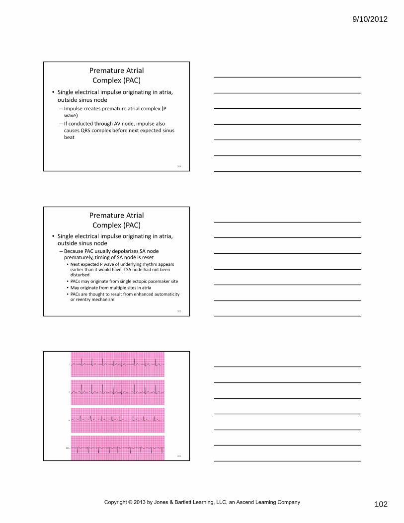
9/10/2012
102
Premature Atrial Complex (PAC)
• Single electrical impulse originating in atria, outside sinus node
– Impulse creates premature atrial complex (P wave)
– If conducted through AV node, impulse also causes QRS complex before next expected sinus beat
304
Premature Atrial Complex (PAC)
• Single electrical impulse originating in atria, outside sinus node– Because PAC usually depolarizes SA node prematurely, timing of SA node is reset
• Next expected P wave of underlying rhythm appears earlier than it would have if SA node had not been disturbed
• PACs may originate from single ectopic pacemaker site
• May originate from multiple sites in atria
• PACs are thought to result from enhanced automaticity or reentry mechanism
305
306
Copyright © 2013 by Jones & Bartlett Learning, LLC, an Ascend Learning Company

9/10/2012
103
Premature Atrial Complex
• Causes– Increase in catecholamines and sympathetic tone
– Use of caffeine, tobacco, or alcohol
– Use of sympathomimetic drugs (epinephrine, albuterol, norepinephrine)
– Electrolyte imbalance
– Hypoxia
– Digitalis toxicity
– Cardiovascular disease
– In some cases, no apparent cause
307
Premature Atrial Complex
• ECG characteristics
– QRS complex
• Usually less than 0.12 second
• QRS complex may be greater than 0.12 second and appear bizarre if PAC is conducted abnormally
• QRS complex may be absent as a result of temporary complete AV block (nonconducted PAC) that occurs during refractory period of AV node or ventricles
308
Premature Atrial Complex
• ECG characteristics
– P waves
• P wave of PAC differs in shape from sinus P wave
• Occurs earlier than next expected sinus P wave and may be so early that it is superimposed or hidden in preceding T wave
• Evaluate preceding T wave to see if shape is altered by presence of P wave
– Rate
• Depends on underlying rhythm
309
Copyright © 2013 by Jones & Bartlett Learning, LLC, an Ascend Learning Company

9/10/2012
104
Premature Atrial Complex
• ECG characteristics
– Rhythm
• Usually underlying rhythm is sinus and regular with irregular premature beats when PACs occur
– P‐R interval
• Usually in normal range but differs from those of underlying rhythm
• P‐R interval of PAC varies from 0.20 second when pacemaker site is near SA node to 0.12 second when pacemaker site is near AV junction
310
What will you feel when you palpate the pulse of a patient
with PACs?
311
Premature Atrial Complex
• Clinical significance– Isolated PACs in healthy patients are not significant
– Frequent PACs that occur in patients with heart disease may lead to serious supraventricular dysrhythmias such as
• Multifocal atrial tachycardia
• Atrial tachycardia
• Atrial flutter
• Atrial fibrillation
• Paroxysmal supraventricular tachycardia
312
Copyright © 2013 by Jones & Bartlett Learning, LLC, an Ascend Learning Company

9/10/2012
105
Premature Atrial Complex
• Management
– Prehospital care usually only requires observation
– Frequent or nonconducted PACs may cause symptomatic bradycardia
• Transcutaneous cardiac pacing or atropine may be indicated
313
Supraventricular Tachycardia
• Complex group of dysrhythmias
• Can be broadly defined as any tachycardia that directly or indirectly involves atria or AV node (above bundle of His)
• Result from rapid atrial or junctionaldepolarization that overrides rate of SA node
314
Supraventricular Tachycardia
• AV nodal reentry tachycardia (AVNRT)
– Most common type of reentry supraventricular tachycardia (SVT)
– Usually caused by PAC
– Paroxysmal supraventricular tachycardia (PSVT)
– When dysrhythmia begins and ends abruptly
315
Copyright © 2013 by Jones & Bartlett Learning, LLC, an Ascend Learning Company

9/10/2012
106
Supraventricular Tachycardia
• Most are thought to result from reentry mechanism that involves abnormal pathways in AV node
– In patients prone to reentry SVTs, AV node is functionally divided into two pathways
• Slow (alpha) pathway with longer refractory period, and fast (beta) pathway with a shorter refractory period
• These pathways permit impulses to be conducted from atrium to ventricle (antegrade conduction), or from ventricle to atrium (retrograde conduction)
316
317
Supraventricular Tachycardia
• Reentry SVTs occur when premature impulse becomes blocked in fast pathway and then travels slow pathway
– During this process, fast pathway recovers while slow pathway is firing
– Produces reentry tachycardia in which electrical impulses are caught in cycle that continuously circulates around AV node
318
Copyright © 2013 by Jones & Bartlett Learning, LLC, an Ascend Learning Company

9/10/2012
107
Supraventricular Tachycardia
• Reentry SVTs occur when premature impulse becomes blocked in fast pathway and then travels slow pathway– Cycle and tachycardia continue until reentry pathway is interrupted
– Most characterized by repeated episodes (paroxysms) of atrial tachycardia
• Often have sudden onset (lasting minutes to hours) and abrupt termination
319
320
Supraventricular Tachycardia
• AV reentry tachycardia (AVRT)
– Second most common type of reentry SVT
– Reentry circuit is involved in AV node
– Patients with AVRT are born with conducting tissue (accessory pathway) in heart muscle
– Accessory pathway bridges atrium and ventricles outside of AV node
321
Copyright © 2013 by Jones & Bartlett Learning, LLC, an Ascend Learning Company

9/10/2012
108
322
323
Supraventricular Tachycardia
• Two pathways of reentry circuit can be composed of
– One accessory pathway and AV node
– Two accessory pathways without participation of AV node
• Pathways can conduct impulses either antegrade, retrograde, or in both directions
• Abnormal conduction results in preexcitation of ventricles
324
Copyright © 2013 by Jones & Bartlett Learning, LLC, an Ascend Learning Company

9/10/2012
109
Supraventricular Tachycardia
• Atrial tachycardia (AT)
– Rhythm disturbance that arises from irritable site in atria
– Ectopic focus overrides SA node, producing tachycardia
– Does not require AV junction, accessory pathways, or ventricular tissue to sustain fast rate
325
Supraventricular Tachycardia
• Atrial tachycardia (AT)
– Dysrhythmia presents very similar to sinus tachycardia
• P waves differ some in shape
• Morphology of P wave in AT depends on location in atrium responsible for fast rate
– Paroxysmal atrial tachycardia (PAT)
• AT that begins and ends abruptly
326
Supraventricular Tachycardia
• May occur at any age
– Dysrhythmias are common in young adults and are more common in women than in men
• SVTs are not commonly associated with underlying heart disease, are rare in patients with MI
– Can precipitate angina pectoris or MI in patients with heart disease
327
Copyright © 2013 by Jones & Bartlett Learning, LLC, an Ascend Learning Company

9/10/2012
110
Supraventricular Tachycardia
• Precipitating factors
– Stress
– Overexertion
– Tobacco use
– Caffeine consumption
– Illicit drug use (e.g., cocaine)
• Common in patients with Wolff‐Parkinson‐White syndrome
328
Supraventricular Tachycardia
• ECG characteristics
– QRS complex
• Less than 0.12 second, provided no ventricular conduction disturbance is present
– P waves
• Ectopic P waves differ from normal sinus P waves
• In lead II, P waves may be normal and upright if pacemaker site is near SA node but inverted if they originate near AV junction
• P waves frequently are buried in preceding T or U waves or QRS complexes and cannot be identified
329
Supraventricular Tachycardia
• ECG characteristics
– Rate
• 150 to 250 beats/minute
– Rhythm
• Regular except at onset and termination
– P‐R interval
• If P waves are discernible, P‐R interval often is shortened but may be normal or, rarely, prolonged
330
Copyright © 2013 by Jones & Bartlett Learning, LLC, an Ascend Learning Company

9/10/2012
111
Supraventricular Tachycardia
• Clinical significance– May occur in patients who have healthy hearts
– Patients may tolerate well for short periods
– Often accompanied by palpitations, nervousness, and anxiety
• Patient often complains of “racing heart”
– Rapid ventricular rate may prevent ventricles from filling fully
• Can compromise cardiac output in patients with existing heart disease
331
Supraventricular Tachycardia
• Clinical significance
– Decreased perfusion may cause
• Confusion
• Vertigo
• Lightheadedness
• Syncope
332
Supraventricular Tachycardia
• May precipitate
– Angina pectoris
– Hypotension
– Congestive heart failure
• Increases O2 requirement of heart
– May increase MI and may increase frequency and severity of patient’s chest pain
333
Copyright © 2013 by Jones & Bartlett Learning, LLC, an Ascend Learning Company

9/10/2012
112
Supraventricular Tachycardia
• Management
– Manage promptly
• Helps reverse consequences of reduced cardiac output and increased workload on heart
– If patient is stable (conscious with normal BP, without chest pain, congestive heart failure, or pulmonary edema), attempt techniques to terminate SVT
334
335
What rhythms are produced by supraventricular activity?
336
Copyright © 2013 by Jones & Bartlett Learning, LLC, an Ascend Learning Company

9/10/2012
113
SVT Management
• Vagal maneuvers
– Can slow heart and decrease force of atrialcontraction
– Stimulate parasympathetic nerve fibers in wall of atria and in specialized tissues of SA and AV nodes
– Can interrupt and terminate some SVTs
– Should be attempted only under medical direction
337
SVT Management
• Vagal maneuvers– Patient must be stable
– Continuous ECG monitoring and an IV line must be in place before beginning
– Atropine and airway equipment should be readily available
– Include• Valsalva maneuver
• Ice pack maneuver
• Unilateral carotid sinus pressure
338
SVT Management
• Valsalva maneuver
– Place patient in sitting or semi‐sitting position with head tilted down
– Instruct patient to take in deep breath and to bear down as if to have bowel movement
• Forced expiration against closed glottis stimulates vagusnerve and may terminate tachycardia
– Procedure may be repeated if unsuccessful
339
Copyright © 2013 by Jones & Bartlett Learning, LLC, an Ascend Learning Company

9/10/2012
114
SVT Management
• Ice pack maneuver
– Placing ice pack on patient’s anterior neck may stimulate vagus nerve because of mammalian diving reflex
• Do not attempt if ischemic heart disease is present or suspected
• Procedure may be repeated (per medical direction) if unsuccessful
340
SVT Management
• Unilateral carotid sinus pressure
– Stimulates carotid bodies located in carotid arteries
– Body interprets this localized pressure as increase in BP
• Activates autonomic nervous system and stimulates vagus nerve
• Heart rate slows in attempt to lower BP
341
SVT Management
• Unilateral carotid sinus pressure
– Auscultate carotid arteries for presence of bruit before applying carotid sinus pressure
– Should not be used if
• Bruits are present
• Patient is an older adult
• Patient is known to have carotid artery disease or cerebral vascular disease
342
Copyright © 2013 by Jones & Bartlett Learning, LLC, an Ascend Learning Company

9/10/2012
115
SVT Management
• Unilateral carotid sinus pressure
– Possible complications
• Cerebral emboli
• Stroke
• Syncope
• Sinus arrest
• Asystole
• Increased degree of AV block
343
SVT Management
• Unilateral carotid sinus pressure• Procedure
– Position yourself behind patient, who is lying supine with neck extended and head turned away from side of applied pressure
– Gently palpate each carotid artery to confirm presence of equal pulses
– If pulses are unequal or if one is absent, do not apply carotid sinus pressure
– Auscultate (while patient holds his or her breath for 4 to 5 seconds) for presence of bruits
344
SVT Management
• Unilateral carotid sinus pressure
• Procedure (cont'd)
– Place index and middle fingers over artery on neck just below angle of jaw
• Compress artery firmly against vertebral column while massaging area
• Inform patient that he or she may experience some pain or discomfort
• Maintain pressure no longer than 5 to 10 seconds
345
Copyright © 2013 by Jones & Bartlett Learning, LLC, an Ascend Learning Company
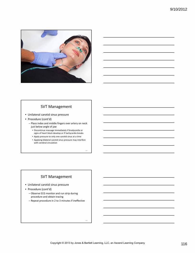
9/10/2012
116
346
SVT Management
• Unilateral carotid sinus pressure
• Procedure (cont'd)
– Place index and middle fingers over artery on neck just below angle of jaw
• Discontinue massage immediately if bradycardia or signs of heart block develop or if tachycardia breaks
• Apply pressure to only one carotid sinus at a time
• Applying bilateral carotid sinus pressure may interfere with cerebral circulation
347
SVT Management
• Unilateral carotid sinus pressure
• Procedure (cont'd)
– Observe ECG monitor and run strip during procedure and obtain tracing
– Repeat procedure in 2 to 3 minutes if ineffective
348
Copyright © 2013 by Jones & Bartlett Learning, LLC, an Ascend Learning Company

9/10/2012
117
SVT Management
• Pharmacological therapy
– If vagal maneuvers fail or are contraindicated and patient remains stable, antidysrhythmics may end symptomatic SVT
• Administration of adenosine (initial drug of choice)
• Verapamil
• Diltiazem
• Beta blockers (metoprolol)
349
SVT Management
• Pharmacological therapy
– Drug treatment recommended by AHA for stable narrow‐complex SVTs is based on rhythm interpretation and stability of patient
– Persons with heart disease may experience rapid heart rate and yet remain clinically stable
• Often are chronically ill with heart disease and have adjusted to reduced level of cardiac function
350
SVT Management
• Pharmacological therapy
– Based on patient history and physical examination, paramedic should be able to distinguish between acute and chronic or stable congestive heart failure
– Sudden onset of signs and symptoms of congestive heart failure with impaired cardiac function indicates that patient is unstable
351
Copyright © 2013 by Jones & Bartlett Learning, LLC, an Ascend Learning Company

9/10/2012
118
SVT Management
• Pharmacological therapy– Junctional tachycardia in adults is rare, not paroxysmal
– Ectopic atrial tachycardia is not paroxysmal• Will often continue after drug treatment to block conduction through AV node
– Not responsive to electrical cardioversion• Ectopic atrial tachycardia
• Multifocal atrial tachycardia
• Sinus tachycardia
352
SVT Management
• Pharmacological therapy
– In symptomatic SVT, vagal maneuvers and adenosine should be used first to end tachydysrhythmia
– If adenosine is not effective and if patient is hemodynamically stable without evidence of congestive heart failure, secondary drug treatment options include
• Calcium channel blockers
• Beta‐blockers
• Amiodarone
353
What signs or symptoms would indicate hemodynamic instability in
these patients (atrial fibrillation/flutter)?
354
Copyright © 2013 by Jones & Bartlett Learning, LLC, an Ascend Learning Company

9/10/2012
119
SVT Management
• Pharmacological therapy– Consecutive use of calcium channel blockers, beta‐blockers, and primary antidysrhythmics is discouraged
– Use only one antidysrhythmic agent• Using several can result in more dysrhythmias and a drop in BP
– Avoid negative inotropic drugs (verapamil, beta‐blockers, flecainide, procainamide, propafenone, and sotalol) in hemodynamically unstable patients
355
SVT Management
• Pharmacological therapy
– Several drug treatments are available for narrow complex tachycardias (ventricular rate greater than or equal to 150)
– When serious signs and symptoms point to poor perfusion and clinical instability, synchronized electrical cardioversion is treatment of choice to terminate rhythm
356
SVT Management
• Pharmacological therapy
– Cardioversion should begin with synchronized shock of 50 J or equivalent biphasic energy
• Atrial fibrillation should first be managed with a shock of 100 J
• If this fails, energy may be increased to 100, 200, 300, and then 360 J monophasic (or equivalent biphasic)
– Sedation should be considered before cardioversion, if time permits
357
Copyright © 2013 by Jones & Bartlett Learning, LLC, an Ascend Learning Company

9/10/2012
120
Lesson 22.5
Rhythm, Site of Origin, Causes,
Clinical Significance, and Prehospital Management
358
Learning Objectives
• When shown an electrocardiogram tracing, identify the rhythm, site of origin, possible causes, clinical significance, and prehospital management that is indicated.
359
Atrial Flutter
• Almost always result of rapid atrial reentry focus• Atrial flutter not slowed by preexisting atrioventricular block usually manifests a 2:1 atrioventricular conduction ratio and may look like SVT– 2:1 AV conduction ratio means that 50 percent of atrial impulses are conducted through ventricles
– 3:1, 4:1, and greater conduction ratios are not uncommon
• Ratios produce discrepancy between atrial and ventricular rates
• Conduction ratios may be constant or variable
360
Copyright © 2013 by Jones & Bartlett Learning, LLC, an Ascend Learning Company

9/10/2012
121
Atrial Flutter
• May be seen with atrial fibrillation (atrial fib‐flutter)
– Rarely, atrial flutter may conduct 1:1
• Results in extremely fast ventricular rates with rapid hemodynamic deterioration
361
362
Atrial Flutter
• Seen in middle‐aged and older patients who have heart disease
• Can occur in patients with healthy hearts– Commonly associated with
• Cardiomyopathy
• Cardiac hypertrophy
• Digitalis toxicity (rare)
• Hypoxia
• Congestive heart failure
• Pericarditis
• Myocarditis
363
Copyright © 2013 by Jones & Bartlett Learning, LLC, an Ascend Learning Company

9/10/2012
122
Atrial Flutter
• ECG characteristics
– QRS complex
• Less than 0.12 second, unless ventricular conduction disturbance (aberrancy) present
– P waves
• Normal P waves are absent
• Flutter waves (f waves) usually resemble sawtooth or picket fence pattern
• Represent atrial depolarization in abnormal direction followed by atrial repolarization
364
Atrial Flutter
• ECG characteristics– Rate
• Atrial rate is 250 to 300 beats/minute
• Ventricular rate is regular but often is < atrial rate
– Rhythm• Atrial rhythm is regular
• Ventricular rate is usually regular
• Ventricular rate may be irregular if AV conduction ratio varies
– P‐R interval• Usually constant but may vary
365
Atrial Flutter
• Clinical significance
– With normal ventricular rate, atrial flutter usually well tolerated by patient
– Rapid ventricular rate produces same signs and symptoms of decreased cardiac output as seen in patients with SVT
366
Copyright © 2013 by Jones & Bartlett Learning, LLC, an Ascend Learning Company
9/10/2012
123
Atrial Flutter
• Clinical significance
– In some flutter rhythms (particularly a 2:1 atrialflutter), atria do not contract regularly and empty before each ventricular contraction
– Loss of “atrial kick” results in incomplete filling of ventricles
• May further decrease cardiac output
367
Atrial Fibrillation
• Results from multiple areas of reentry within atria
• Can result from ectopic atrial pacemakers
– Activity of SA node is suppressed completely by atrial fibrillation
368
Atrial Fibrillation
• Produces chaotic impulses too numerous for all to be conducted by AV node through ventricles
– AV conduction is random
• Results in irregular but usually rapid ventricular response
• Medications such as digoxin, beta blockers, or calcium channel blockers often are prescribed to slow ventricular rate
369
Copyright © 2013 by Jones & Bartlett Learning, LLC, an Ascend Learning Company

9/10/2012
124
370
Atrial Fibrillation
• Sudden onset (paroxysmal) atrial fibrillation may occur in young adults after heavy alcohol ingestion (“holiday heart” syndrome) or acute stress– In these cases, fibrillation usually is self‐limited and resolves without treatment
– Chronic atrial fibrillation may be intermittent
– Often associated with rheumatic heart disease, congestive heart failure, and coronary heart disease
371
Atrial Fibrillation
• Chronic atrial fibrillation usually requires drug therapy with digitalis (or calcium channel–blocker or beta‐blocker) therapy
– Slows ventricular rate to 80 to 100 beats/minute
• May be stable rhythm that does not require management
372
Copyright © 2013 by Jones & Bartlett Learning, LLC, an Ascend Learning Company

9/10/2012
125
Atrial Fibrillation
• May occur in cardiomyopathy, acute myocarditis and pericarditis, and chest trauma
• Rarely caused by digitalis toxicity
– Slow, regular ventricular response with atrialfibrillation could be result of digitalis toxicity
373
Atrial Fibrillation
• ECG characteristics– QRS complex
• Less than 0.12 second, provided there is no ventricular conduction disturbance
– P waves• Absent P waves and organized atrial contractions
• Fibrillation waves (f waves) may be fine less than 1 mm) or coarse (more than 1 mm)
• Fine f waves may be so small that they appear as wavy or flat (isoelectric) line or absent
• F waves are irregularly shaped, rounded (or pointed), and dissimilar
374
Atrial Fibrillation
• ECG characteristics
– Rate
• Atrial rate = 350 to 700 beats/minute (cannot be counted)
• Ventricular rate varies greatly, depending on conduction through AV node (average 150 to 180 beats/minute, if uncontrolled)
– Rhythm
• Irregularly irregular
– P‐R interval
• None
375
Copyright © 2013 by Jones & Bartlett Learning, LLC, an Ascend Learning Company

9/10/2012
126
Atrial Fibrillation
• Clinical significance
– Atrial kick is lost in atrial fibrillation
• Can reduce cardiac output by as much as 15 percent
• Coupled with rapid ventricular response, may cause cardiovascular decompensation (angina pectoris, myocardial infarction, congestive heart failure, or cardiogenic shock)
376
Atrial Fibrillation
• Management of atrial fibrillation/atrial flutter
– Risk of emboli formation exists when atrial fibrillation or atrial flutter has been present for 48 hours or more
– Formation of emboli in heart increases risk of “throwing a clot” or systemic embolization
– Most often occurs when atrial fibrillation is converted suddenly to sinus rhythm
377
Why does atrial fibrillation put the patient at increased risk for
emboli?
378
Copyright © 2013 by Jones & Bartlett Learning, LLC, an Ascend Learning Company

9/10/2012
127
Atrial Fibrillation
• Management of atrial fibrillation/atrial flutter
– Algorithm cautions against converting atrialfibrillation or atrial flutter without first giving patient drugs that prevent blood from clotting
– Electrical cardioversion and use of antidysrhythmic agents that may convert rhythm should be avoided unless patient is unstable or hemodynamically compromised
379
Atrial Fibrillation
• Management of atrial fibrillation/atrial flutter
– Using drugs to control heart rate is recommended initial treatment for stable, rapid atrial fibrillation or atrial flutter regardless of how long patient has had it
– Specific drug treatment depends on patient’s condition and how stable patient is
– Using several different drugs can cause dysrhythmia to develop (proarrhythmia)
– Paramedic should use only one drug from list of suggested drug treatments
380
Atrial Fibrillation
• Management of atrial fibrillation/atrial flutter– In patients with rapid atrial fibrillation or flutter with rapid ventricular response, rate may be controlled with diltiazem or beta blockers
– Amiodarone has potential for rhythm conversion• Should be reserved for use within first 48 hours of dysrhythmia onset when other medications for rate control have failed
– Use of calcium channel–blocking agents and beta‐blocking agents warrants caution in presence of congestive heart failure because of their negative inotropic properties
381
Copyright © 2013 by Jones & Bartlett Learning, LLC, an Ascend Learning Company

9/10/2012
128
Atrial Fibrillation
• Management of atrial fibrillation/atrial flutter
– Beta‐blocking agents also should be used with caution in patients with asthma and COPD
– Another drug that may be effective to convert rhythm is digoxin
• Digoxin should only be used if patient developed atrialfibrillation within 48 hours or less
382
Atrial Fibrillation
• Management of atrial fibrillation/atrial flutter– If patient reports history of Wolff‐Parkinson‐White (WPW) syndrome or if paramedic made that field impression before he or she developed atrial fibrillation, treatment will change
– If patient has WPW, do not give adenosine, diltiazem, verapamil, digoxin, or, in most cases, beta blockers
• May cause dangerous increase in heart rate
• If patient has had dysrhythmia for longer than 48 hours, paramedic should avoid elective cardioversion unless anticoagulation drugs have been given
383
Atrial Fibrillation
• Management of atrial fibrillation/atrial flutter– Cardiovert patient immediately when serious signs/symptoms occur
• Chest pain
• Shortness of breath
• Pulmonary congestion
• Decreased level of consciousness
• Hypotension
– Initial attempt at cardioversion for atrial flutter should consist of synchronized shock of 50 J or equivalent biphasic energy
384
Copyright © 2013 by Jones & Bartlett Learning, LLC, an Ascend Learning Company

9/10/2012
129
Atrial Fibrillation
• Management of atrial fibrillation/atrial flutter
– If needed, energy may be increased to 100, 200, 300, and 360 J monophasic (or equivalent biphasic)
• Because atrial fibrillation is more difficult rhythm to convert and lower joule settings have been known to cause asystole, recommendations are initially to use synchronized shock of 100 J monophasic (or equivalent biphasic), followed by 200, 300, and 360 J if necessary
385
Dysrythmias Sustained/Originating in AV Junction
• AV node or area surrounding AV node may assume role of secondary pacemaker
– When SA node and atria cannot generate electrical impulses needed to begin depolarization because of factors such as
• Hypoxia
• Ischemia
• Myocardial infarction
• Drug toxicity
386
Dysrythmias Sustained/Originating in AV Junction
• Rhythms that start in AV node or AV junctionalarea are junctional rhythms– Usually is benign dysrhythmia
– Paramedic must assess rhythm to determine patient’s tolerance of rhythm disturbance
– Dysrhythmias that originate in AV junction include• Premature junctional contractions
• Junctional escape complexes or junctional escape rhythms
• Accelerated junctional rhythm
387
Copyright © 2013 by Jones & Bartlett Learning, LLC, an Ascend Learning Company
9/10/2012
130
Dysrythmias Sustained/Originating in AV Junction
• In junctional rhythms, electrical impulses travel in normal pathway from AV junction through bundle of His and bundle branches to Purkinje fibers
– Pathway ends in ventricular muscle
– Conduction through ventricles proceeds normally
– QRS complex usually is within normal limits of 0.04 to 0.10 second
388
Dysrythmias Sustained/Originating in AV Junction
• Impulse that depolarizes atria travels in backward or retrograde motion
– Retrograde depolarization of atria results in one of following three P wave characteristics
• Inverted P waves in lead II with short P‐R interval
• Absent P waves
• P waves following QRS complex
389
Premature Junctional Contraction
• Results from single electrical impulse from AV junction
– Impulse occurs before next expected sinus impulse
• Isolated premature junctional contractions (PJCs) may occur in healthy person without apparent cause
– More often result of heart disease or drug toxicity
– Usually PJCs result from enhanced automaticity or reentry mechanism
390
Copyright © 2013 by Jones & Bartlett Learning, LLC, an Ascend Learning Company

9/10/2012
131
391
Premature Junctional Contraction
• Causes:
– Digitalis toxicity
– Other cardiac medications (quinidine, procainamide)
– Increased vagal tone on SA node
– Sympathomimetic drugs (e.g., cocaine and methamphetamines)
– Hypoxia
– Congestive heart failure
– Damage to the AV junction
392
Premature Junctional Contraction
• ECG characteristics
– QRS complex
• Usually less than 0.12 second, provided there is no ventricular conduction disturbance
– P waves
• May be associated with PJCs
• P waves can occur before, during, or after QRS complex or can be absent
• If present, P waves are abnormal, differing in size, shape, and direction from normal P waves
393
Copyright © 2013 by Jones & Bartlett Learning, LLC, an Ascend Learning Company

9/10/2012
132
Premature Junctional Contraction
• ECG characteristics
– Rate
• Heart rate is that of underlying rhythm
– Rhythm
• Usually regular, except when premature junctionalcontractions are present
– P‐R interval
• Usually less than 0.12 second if P wave precedes QRS complex
394
Premature Junctional Contraction
• Clinical significance
– Occasional PJCs usually are not significant
• Management
– No management required
395
Premature Junctional Contraction
• Junctional escape beat or rhythm (series of beats) occurs when rate of SA node falls below that of AV junction– Dysrhythmia also may occur when electrical impulses from SA node or atria fail to reach AV junction because of sinoatrial or atrioventricular block
– Escape complex or rhythm provided by AV junction serves as safety mechanism
• Prevents cardiac standstill• AV junction begins firing at inherent rate of 40 to 60 beats/minute within about 1.0 to 1.5 seconds of not receiving impulse from SA node
396
Copyright © 2013 by Jones & Bartlett Learning, LLC, an Ascend Learning Company

9/10/2012
133
397
Premature Junctional Contraction
• Junctional escape complex (isolated impulse) or junctional escape rhythm (series of impulses) is normal response
– May result from
• Increased vagal tone on SA node
• Pathological slowing of sinoatrial discharge
• Complete atrioventricular block
398
Premature Junctional Contraction
• ECG characteristics– QRS complex
• Usually less than 0.12 second, provided no preexisting bundle branch block is present
– P waves• May be present (with or without relationship to QRS complex) or absent
• If P waves are present, may occur before, after, or during QRS complex
• Depending on pacemaker site, P waves may differ from normal P waves in size, shape, direction and may be upright or inverted
399
Copyright © 2013 by Jones & Bartlett Learning, LLC, an Ascend Learning Company

9/10/2012
134
Premature Junctional Contraction
• ECG characteristics
– Rate
• Usually 40 to 60 beats/minute but may be less
– Rhythm
• Ventricular rhythm usually is regular in junctional rhythm
• May be irregular if isolated junctional escape complex is present
– P‐R interval
• If P waves precede QRS complex, P‐R interval commonly is shortened (less than 0.12 second) and constant
400
Premature Junctional Contraction
• Clinical significance
– Junctional bradycardias can cause decreased cardiac output
• Patients can show signs and symptoms that are similar to those of other bradycardias
• Patients tolerate junctional rhythms of 50 beats/minute or greater
401
Premature Junctional Contraction
• Management
– Stable patients do not need to be treated
– If patient is symptomatic or if ventricular irritability is present, drug therapy (beginning with atropine) may be indicated
– In severe cases and in patients unresponsive to atropine, external pacing may be necessary
– If SA node is diseased or damaged, patient may need permanent pacemaker
402
Copyright © 2013 by Jones & Bartlett Learning, LLC, an Ascend Learning Company

9/10/2012
135
Accelerated Junctional Rhythm
• Results from increased automaticity of AV junction– Increase causes it to discharge faster than its intrinsic rate
• Intrinsic rate 40 to 60 beats/minute
– Rate in turn overrides main (SA node) pacemaker
– Rate of this dysrhythmia (usually 60 to 99 beats/minute) does not truly constitute tachycardia
• Dysrhythmia is termed accelerated junctional rhythm
403
404
Accelerated Junctional Rhythm
• Result of digitalis toxicity
• Other causes
– Excessive catecholamine administration
– Damage to AV junction
– Inferior wall MI
– Rheumatic fever
405
Copyright © 2013 by Jones & Bartlett Learning, LLC, an Ascend Learning Company

9/10/2012
136
Accelerated Junctional Rhythm
• ECG characteristics
– QRS complex
• Usually less than 0.12 second, provided no preexisting bundle branch block
– P waves
• May be present (with/without relationship to QRS complex), absent (retrograde atrioventricular block), or buried in QRS complex
• If present, P waves usually are inverted and appear before/after QRS complex
406
Accelerated Junctional Rhythm
• ECG characteristics– Rate
• Usually 60 to 99 beats/minute
– Rhythm• Regular
– P‐R interval• If P wave occurs before QRS complex, P‐R interval will be less than 0.12 second
• If P wave follows QRS complex, technically is R‐P interval and usually less than 0.20 second
407
Accelerated Junctional Rhythm
• Clinical significance
– Well tolerated by patient
– Presence of heart disease and lack of O2 to heart muscle may cause more serious dysrhythmias
• Management
– Generally requires no immediate treatment
408
Copyright © 2013 by Jones & Bartlett Learning, LLC, an Ascend Learning Company
9/10/2012
137
Do you need to start an IV line on these patients, since no drug
therapy is indicated?
409
Dysrhythmias Originating in Ventricles
• Ventricular dysrhythmias
– Usually considered life‐threatening
– Result from failure of atria, AV junction, or both to initiate electrical impulse
• Can result from enhanced automaticity or reentry pathways in ventricles
410
Dysrhythmias Originating in Ventricles
• Ventricular dysrhythmias
– Enhanced automaticity and reentry can lead to:
• Premature ventricular complexes
• Ventricular tachycardia
• Ventricular fibrillation
411
Copyright © 2013 by Jones & Bartlett Learning, LLC, an Ascend Learning Company

9/10/2012
138
Dysrhythmias Originating in Ventricles
• Associated with MI or infarction
– Ventricle is least efficient pacemaker of heart
– Usually generates only 20 to 40 impulses per minute
– Ventricle may discharge at rates up to 99 impulses per minute (accelerated idioventricular rhythm) or even faster (ventricular tachycardia) because of increased automaticity
412
Dysrhythmias Originating in Ventricles
• Associated with MI or infarction
– Dysrhythmias originating in ventricles include
• Ventricular escape complexes or rhythms
• Premature ventricular complexes
• Ventricular tachycardia
• Ventricular fibrillation
• Asystole
• Artificial pacemaker rhythm
413
Dysrhythmias Originating in Ventricles
• Electrical impulses of ventricular origin start in lower portion of heart (ventricular muscle, bundle branches, or Purkinje fibers)
– Electrical impulse must travel in a retrograde conduction pathway to depolarize atria
– Impulse may travel in antegrade direction to depolarize ventricles, depending on site of initiation of impulse
414
Copyright © 2013 by Jones & Bartlett Learning, LLC, an Ascend Learning Company
9/10/2012
139
Dysrhythmias Originating in Ventricles
• Regardless of direction of depolarization, normal, rapid conducting pathways are bypassed, producing three electrocardiogram features– QRS complexes are wide and bizarre in appearance
• They are 0.12 second or greater in duration
– P waves may be hidden in QRS complex• Because atria are depolarized at about same time as ventricles
• May be superimposed on every second or third QRS complex when ventricular tachycardia with AV dissociation (P waves that have no set relation to QRS complexes) is present
415
Dysrhythmias Originating in Ventricles
• Regardless of direction of depolarization, normal, rapid conducting pathways are bypassed, producing three electrocardiogram features
– ST segments usually deviate from baseline
• T waves frequently are sloped off in opposite direction of QRS complex
416
Ventricular Escape Complexes/Rhythms
• Also known as idioventricular rhythm
• Dysrhythmia results when impulses from higher pacemakers fail to fire or to reach ventricles
– Results when rate of discharge of higher pacemaker sites falls to less than that of ventricles
– Like junctional escape complex or rhythm, this dysrhythmia serves as compensatory mechanism to prevent cardiac standstill
417
Copyright © 2013 by Jones & Bartlett Learning, LLC, an Ascend Learning Company

9/10/2012
140
418
419
Ventricular Escape Complexes/Rhythms
• Occur in two ways
– Rate of impulse formation of dominant pacemaker (usually SA node) can fall below that of ventricles
– Escape pacemaker in AV junction can fail or fall below that of pacemaker in ventricles
• Often seen as first rhythm after defibrillation
420
Copyright © 2013 by Jones & Bartlett Learning, LLC, an Ascend Learning Company

9/10/2012
141
Ventricular Escape Complexes/Rhythms
• ECG characteristics
– QRS complex
• Generally exceed 0.12 second and are bizarre in appearance
• Shape of QRS complex may vary in any given lead
– P waves
• May be absent
• If they are present and have no set relationship to QRS complex, then third‐degree AV block should be suspected
421
Ventricular Escape Complexes/Rhythms
• ECG characteristics
– Rate
• Usually 20 to 40 beats/minute; may be lower
– Rhythm
• Ventricular rhythm usually regular but may be irregular
– P‐R interval
• If P waves are present, P‐R interval is variable and irregular
422
Ventricular Escape Complexes/Rhythms
• Clinical significance– Ventricular escape rhythm generally produces symptoms
– Manifested by• Hypotension
• Decreased cardiac output
• Decreased perfusion of brain and other vital organs, often resulting in syncope and shock
– Patient assessment is essential because escape rhythm may be perfusing or nonperfusing (pulseless electrical activity)
423
Copyright © 2013 by Jones & Bartlett Learning, LLC, an Ascend Learning Company

9/10/2012
142
Ventricular Escape Complexes/Rhythms
• Management
– Perfused rhythm: direct at increasing heart rate by administering O2, transcutaneous cardiac pacing, and/or dopamine
– Escape rhythm: lidocaine is lethal and contraindicated
– Nonperfused rhythm: initiate basic life support measures and follow treatment guidelines for pulseless arrest
424
425
Why might lidocaine be harmful in this situation
(ventricular escape rhythm)?
426
Copyright © 2013 by Jones & Bartlett Learning, LLC, an Ascend Learning Company
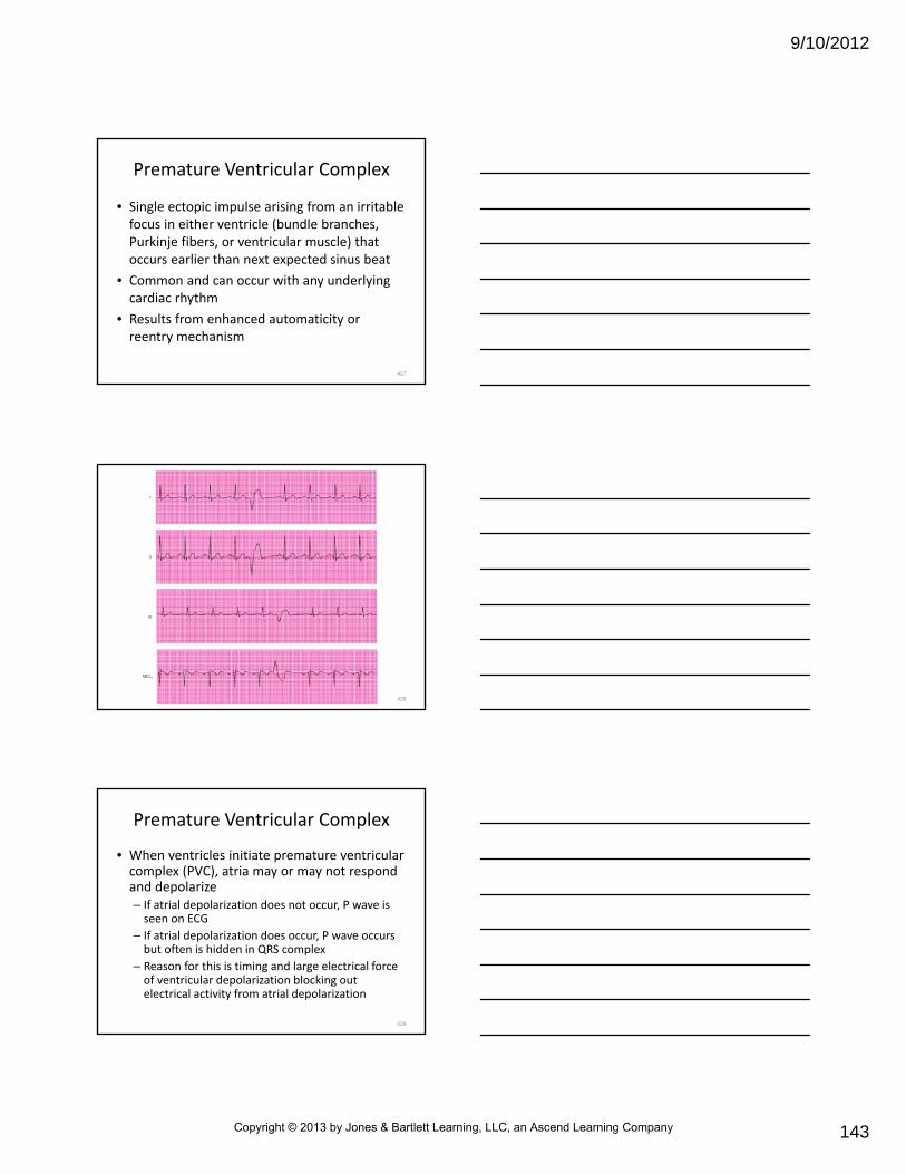
9/10/2012
143
Premature Ventricular Complex
• Single ectopic impulse arising from an irritable focus in either ventricle (bundle branches, Purkinje fibers, or ventricular muscle) that occurs earlier than next expected sinus beat
• Common and can occur with any underlying cardiac rhythm
• Results from enhanced automaticity or reentry mechanism
427
428
Premature Ventricular Complex
• When ventricles initiate premature ventricular complex (PVC), atria may or may not respond and depolarize– If atrial depolarization does not occur, P wave is seen on ECG
– If atrial depolarization does occur, P wave occurs but often is hidden in QRS complex
– Reason for this is timing and large electrical force of ventricular depolarization blocking out electrical activity from atrial depolarization
429
Copyright © 2013 by Jones & Bartlett Learning, LLC, an Ascend Learning Company

9/10/2012
144
Premature Ventricular Complex
• Altered sequence of ventricular depolarization results in wide, bizarre QRS complex– Depolarization may be deflected in opposite direction from QRS complex in underlying rhythm
– May be deflected in same direction• Depends on location of focus and lead selected
– T wave that immediately follows PVC usually is deflected in opposite direction from QRS complex of PVC because of altered sequence of repolarization
430
Premature Ventricular Complex
• Usually does not depolarize SA node or interrupt its rhythm
• Ectopic impulse usually is followed by full compensatory pause
431
Premature Ventricular Complex
• Compensatory pauses are confirmed by measuring interval between R wave before PVC and R wave after it
– If pause is compensatory, distance is at least two times R‐R interval of underlying rhythm
– At times, PVC falls between two sinus beats without interrupting rhythm
• Called interpolated PVC
432
Copyright © 2013 by Jones & Bartlett Learning, LLC, an Ascend Learning Company

9/10/2012
145
433
Premature Ventricular Complex
• May originate from single ectopic pacemaker site (unifocal PVC) or from multiple sites in ventricles (multifocal PVC)
• Unifocal PVCs look alike
• Multifocal PVCs have varying shapes and sizes
434
435
Copyright © 2013 by Jones & Bartlett Learning, LLC, an Ascend Learning Company
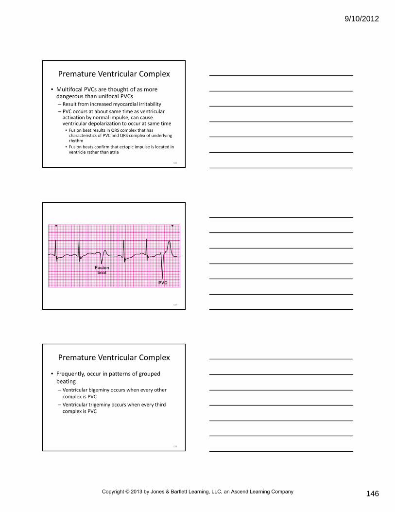
9/10/2012
146
Premature Ventricular Complex
• Multifocal PVCs are thought of as more dangerous than unifocal PVCs– Result from increased myocardial irritability
– PVC occurs at about same time as ventricular activation by normal impulse, can cause ventricular depolarization to occur at same time
• Fusion beat results in QRS complex that has characteristics of PVC and QRS complex of underlying rhythm
• Fusion beats confirm that ectopic impulse is located in ventricle rather than atria
436
437
Premature Ventricular Complex
• Frequently, occur in patterns of grouped beating
– Ventricular bigeminy occurs when every other complex is PVC
– Ventricular trigeminy occurs when every third complex is PVC
438
Copyright © 2013 by Jones & Bartlett Learning, LLC, an Ascend Learning Company
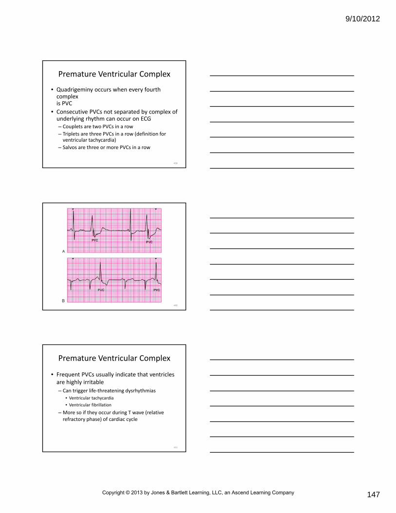
9/10/2012
147
Premature Ventricular Complex
• Quadrigeminy occurs when every fourth complex is PVC
• Consecutive PVCs not separated by complex of underlying rhythm can occur on ECG– Couplets are two PVCs in a row
– Triplets are three PVCs in a row (definition for ventricular tachycardia)
– Salvos are three or more PVCs in a row
439
440
Premature Ventricular Complex
• Frequent PVCs usually indicate that ventricles are highly irritable
– Can trigger life‐threatening dysrhythmias
• Ventricular tachycardia
• Ventricular fibrillation
– More so if they occur during T wave (relative refractory phase) of cardiac cycle
441
Copyright © 2013 by Jones & Bartlett Learning, LLC, an Ascend Learning Company

9/10/2012
148
Premature Ventricular Complex
• During this period, heart muscle is at its greatest electrical instability
– Because in relative refractory period, some of ventricular muscle fibers may be partially repolarized, completely repolarized, or completely refractory
– Stimulation of ventricles in vulnerable period by electrical impulse may cause ventricular fibrillation or ventricular tachycardia
• Occurrence of ventricular depolarization during relative refractory period is known as R‐on‐T phenomenon
442
443
Premature Ventricular Complex
• Isolated PVCs do occur in healthy persons without apparent cause
– Usually are of no significance
– Usually are result of
• Myocardial ischemia
• Hypoxia
• Acid‐base and electrolyte imbalance
• Hypokalemia
• Congestive heart failu
444
Copyright © 2013 by Jones & Bartlett Learning, LLC, an Ascend Learning Company

9/10/2012
149
Premature Ventricular Complex
• Isolated PVCs do occur in healthy persons without apparent cause
– Usually are result of
• Increased catecholamine and sympathetic tone (as in emotional stress)
• Ingestion of stimulants (alcohol, caffeine, tobacco)
• Drug toxicity
• Sympathomimetic drugs (cocaine; stimulants such as phencyclidine, epinephrine, and methamphetamine)
445
Premature Ventricular Complex
• ECG characteristics– QRS complex
• 0.12 second or more• Frequently distorted and bizarre P waves may be present or absent
• If present, usually are of underlying rhythm and have no relationship to PVC
– Rate: depends on underlying rhythm and number of PVCs
– Rhythm: PVCs interrupt regularity of underlying rhythm
– P‐R interval: none
446
Premature Ventricular Complex
• Clinical significance
– PVCs that occur in patients without heart disease usually do not produce serious signs and symptoms
– May complain of “skipped beats”
– PVCs that occur with heart disease (myocardial ischemia) may result from enhanced automaticity, reentry mechanism, or both
– May trigger lethal ventricular dysrhythmias
447
Copyright © 2013 by Jones & Bartlett Learning, LLC, an Ascend Learning Company

9/10/2012
150
Premature Ventricular Complex
• Clinical significance
– Do not permit complete ventricular filling
– May produce diminished or nonpalpable pulse (nonperfusing PVC)
– If occurs often enough and occur early enough in cardiac cycle, cardiac output drops
448
Premature Ventricular Complex
• Clinical significance
– Warning signs of serious ventricular dysrhythmias in patients with myocardial ischemia include
• Frequent PVCs
• Presence of multifocal PVCs
• Early PVCs (R‐on‐T phenomenon)
• Patterns of grouped beating
449
Premature Ventricular Complex
• Management– PVCs that occur in patients without symptoms and without known heart disease seldom require treatment
– In patients with myocardial ischemia, frequent PVCs must be treated promptly with O2 and antidysrhythmic drugs (e.g., beta blockers)
– At hospital, serum potassium should be checked immediately
– Hypokalemia should be treated promptly
450
Copyright © 2013 by Jones & Bartlett Learning, LLC, an Ascend Learning Company

9/10/2012
151
Ventricular Tachycardia (VT)
• Dysrhythmia defined by three or more consecutive ventricular complexes occurring at rate of 100+ beats/minute – Overrides primary pacemaker
– Starts suddenly and triggered by PVC
– During ventricular tachycardia, atria and ventricles are not beating in step with each other
– If ventricular tachycardia continues, patient’s condition may become unstable
– Ventricular tachycardia can produce unconsciousness
451
452
Ventricular Tachycardia• Can lead to loss of perfusing pulse
• Some patients in ventricular tachycardia may be able to walk and talk
• Misconception that ventricular tachycardia cannot be associated with reasonable blood pressure may result in patient being inappropriately managed
• Origin of ventricular tachycardia is enhanced automaticity or reentry
453
Copyright © 2013 by Jones & Bartlett Learning, LLC, an Ascend Learning Company

9/10/2012
152
Ventricular Tachycardia
• Usually occurs in presence of myocardial ischemia or significant cardiac disease
• Other causes
– Acid‐base and electrolyte imbalance
– Hypokalemia
– Congestive heart failure
– Increased catecholamine and sympathetic tone (as in emotional stress)
454
Ventricular Tachycardia
• Other causes
– Ingestion of stimulants (alcohol, caffeine, tobacco)
– Drug toxicity (digitalis, tricyclic antidepressants)
– Sympathomimetic drugs (cocaine, methamphetamines)
– Prolonged Q‐T interval (may be caused by drugs, metabolic problems, or be congenital)
455
Ventricular Tachycardia
• ECG characteristics– QRS complex
• 0.12 second or more and usually distorted and bizarre
• QRS complexes generally are identical, but if fusion beats are present, one or more QRS complexes may differ in size, shape, direction
– P waves• May be absent
• If present, P waves have no set relation to QRS complex (AV dissociation)
• P waves occur at slower rate than ventricular focus and superimposed on QRS complexes
456
Copyright © 2013 by Jones & Bartlett Learning, LLC, an Ascend Learning Company

9/10/2012
153
Ventricular Tachycardia
• ECG characteristics
– Rate
• Usually between 100 and 250 beats/minute
– Rhythm
• Usually regular (unless drug induced) but may be slightly irregular
– P‐R interval
• If P waves are present, P‐R interval varies widely
457
Ventricular Tachycardia
• Clinical significance– Usually indicates significant heart disease
– Rapid rate and loss of atrial kick cause drop in cardiac output and decreased coronary artery and cerebral perfusion
– Severity of symptoms varies with rate of ventricular tachycardia and how much heart disease is present
– May be perfusing or nonperfusing• May produce pulse or it may not
• Ventricular tachycardia also may lead to ventricular fibrillation
458
VT Management
• Treatment is based on signs and symptoms and presence/absence of torsades de pointes
• As with other SVTs, obtain history and identify rhythm
• If patient is stable, 12 lead ECG should be obtained
459
Copyright © 2013 by Jones & Bartlett Learning, LLC, an Ascend Learning Company

9/10/2012
154
460
VT Management
• Depends on whether QRS complex is monomorphic(having same morphology or fixed shape) or polymorphic (having varying morphology)– Any wide‐complex tachycardia that occurs with serious signs and symptoms requires immediate cardioversion
• Chest pain
• Dyspnea
• Decreased level of consciousness
• Hypotension
• Other signs of shock
• Patients with VT without pulse should be treated as if rhythm were ventricular fibrillation
461
VT Management
• Monomorphic VT treatment guidelines
– Based on heart function (cardiac ejection fraction)
– Signs/symptoms of failing heart function
• Pulmonary congestion
• Decreased level of consciousness
– Managed with amiodarone
• Alternative drugs are procainamide and sotalol
462
Copyright © 2013 by Jones & Bartlett Learning, LLC, an Ascend Learning Company

9/10/2012
155
VT Management
• Unstable patients with monomorphic VT should receive immediate synchronized cardioversion
– Begin with initial shock of 100 J
– If no response to first shock, dose should be increased (in stepwise fashion) at 200 J, 300 J, and 360 J monophasic or equivalent biphasic energy levels
• When unstable VT, precordial thump may be performed if cardioversion is not immediately available
463
VT Management
• Precordial thump
– For monitored adult patients whose rhythm is unstable VT
• Must have defibrillator readily available
• May cause VT to deteriorate to asystole, VF, or pulseless electrical activity
– May terminate a dysrhythmia by causing ventricular depolarization and resumption of organized rhythm
464
VT Management
• Precordial thump
– To deliver, paramedic's arm and wrist should be parallel to long axis of sternum to avoid rib fractures
– Thump delivered to midsternum with heel of fist from 10 to 12 inches
– Conscious patient should be advised of procedure
465
Copyright © 2013 by Jones & Bartlett Learning, LLC, an Ascend Learning Company

9/10/2012
156
How will you manage a patient with VT with chest pain or difficulty breathing if you can’t establish an
IV?
466
VT Management• Polymorphic VT can degenerate into ventricular fibrillation quickly, requires immediate intervention– If patient has polymorphic VT and is unstable (as is often the case), rhythm should be treated as ventricular fibrillation with high‐energy unsynchronized shocks
• Synchronization is not usually possible with irregular wave forms, such as polymorphic VT
• If patient’s rhythm is torsades, rhythm may be result of prolonged Q‐T interval
• Medications that prolong Q‐T interval should be discontinued
• Electrolyte imbalances should be corrected, and IV magnesium sulfate should be given
467
Lesson 22.6
Rhythm, Site of Origin, Causes,
Clinical Significance, and Prehospital Management
468
Copyright © 2013 by Jones & Bartlett Learning, LLC, an Ascend Learning Company
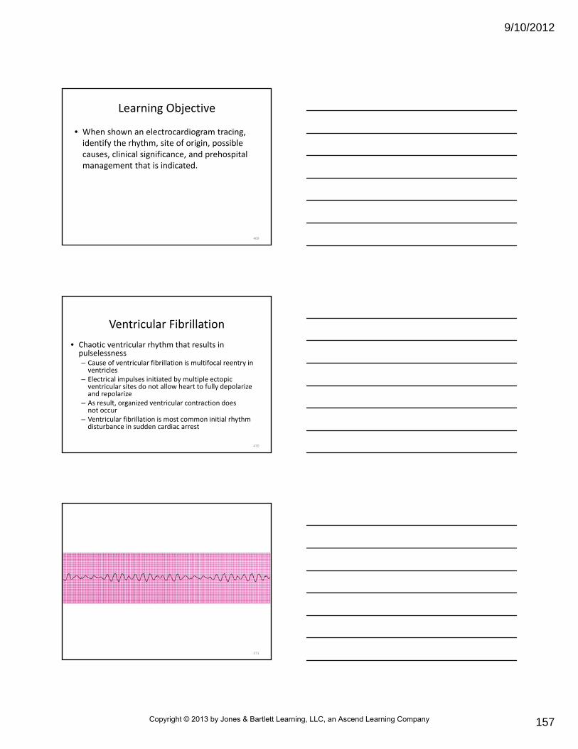
9/10/2012
157
Learning Objective
• When shown an electrocardiogram tracing, identify the rhythm, site of origin, possible causes, clinical significance, and prehospital management that is indicated.
469
Ventricular Fibrillation
• Chaotic ventricular rhythm that results in pulselessness– Cause of ventricular fibrillation is multifocal reentry in ventricles
– Electrical impulses initiated by multiple ectopic ventricular sites do not allow heart to fully depolarize and repolarize
– As result, organized ventricular contraction does not occur
– Ventricular fibrillation is most common initial rhythm disturbance in sudden cardiac arrest
470
471
Copyright © 2013 by Jones & Bartlett Learning, LLC, an Ascend Learning Company

9/10/2012
158
Ventricular Fibrillation
• Most commonly associated with significant heart disease
– May be precipitated by
• PVCs
• R‐on‐T phenomenon (rarely)
• Sustained VT
472
Ventricular Fibrillation
• Other causes
– Myocardial ischemia
– Acute MI
– Third‐degree atrioventricular block with a slow ventricular escape rhythm
– Cardiomyopathy
– Digitalis toxicity
– Hypoxia
– Acidosis
– Electrolyte imbalance (hypokalemia, hyperkalemia, submersion)
– Electrical injury
– Drug overdose or toxicity (cocaine, tricyclicantidepressants)
473
Ventricular Fibrillation
• ECG characteristics– QRS complex: absent
– P waves: absent
– Rate• No coordinated ventricular contractions are present
• Unsynchronized ventricular impulses occur at rates from 300 to 500 beats/minute
– Rhythm: irregularly irregular
– P‐R interval: absent
474
Copyright © 2013 by Jones & Bartlett Learning, LLC, an Ascend Learning Company

9/10/2012
159
Ventricular Fibrillation
• Because organized depolarizations of atria and ventricles are absent, P waves, QRS complexes, ST segments, and T waves are absent– Ventricular fibrillatory waves are seen on oscilloscope as bizarre, rounded or pointed
• Appear considerably different in shape
• Vary at random from positive to negative
• Represent twitching of small individual groups of muscle fibers
475
Ventricular Fibrillation
• Fibrillatory waves
– Less than 3 mm in amplitude are called fine ventricular fibrillation
– Greater than 3 mm are called coarse ventricular fibrillation
– May be so fine they appear as flat line, resembling ventricular asystole
476
477
Copyright © 2013 by Jones & Bartlett Learning, LLC, an Ascend Learning Company

9/10/2012
160
Ventricular Fibrillation
• Clinical significance
– Causes all life functions to cease because of lack of circulating blood flow
– May result in light‐headedness
– Usually followed within seconds by loss of consciousness, apnea, and, if untreated, death
478
Ventricular Fibrillation
• Management
– For adult resuscitation, management is most important sequence because most adult cardiac arrests result from
• Ventricular fibrillation
• Pulseless ventricular tachycardia
– Vast majority of successful resuscitations result from appropriate management of both
479
Ventricular Fibrillation
• Management
– Ventricular fibrillation and nonperfusingventricular tachycardia are managed alike:
• Basic life support (if defibrillator is not immediately available)
• Defibrillation
• Airway management to secure trachea
• IV/IO access
• Pharmacological therapy (epinephrine, amiodarone, lidocaine, and in some cases magnesium)
480
Copyright © 2013 by Jones & Bartlett Learning, LLC, an Ascend Learning Company

9/10/2012
161
Ventricular Asystole
• Ventricular asystole (cardiac standstill) refers to absence of all ventricular activity
– May be cause of cardiac arrest
– May occur in complete heart block with no escape pacemaker
– Usually associated with extensive heart disease
– Often follows ventricular tachycardia, ventricular fibrillation, pulseless electrical activity, or agonalescape rhythm in dying heart
481
482
Ventricular Asystole
• ECG characteristics
– QRS complexes: absent
– P waves: absent or present
– Rate: absent
– Rhythm: absent
– P‐R interval: absent
483
Copyright © 2013 by Jones & Bartlett Learning, LLC, an Ascend Learning Company

9/10/2012
162
Ventricular Asystole
• Clinical significance
– Ventricular asystole produces no cardiac output and is ominous dysrhythmia
– Asystole often confirms death
– Chance for resuscitation is small
484
Ventricular Asystole
• Management– Basic life support with effective CPR, advanced airway, and pharmacological therapy (epinephrine, atropine, and possibly sodium bicarbonate)
– If fine ventricular fibrillation is suspected, defibrillation indicated
• Defibrillating asystole “just in case” not recommended
• Termination of resuscitation efforts in prehospital setting after meeting medical protocol indicated for this patient situation
485
Ventricular Asystole
• Management
– Potential reversible causes of asystole should be considered before cessation of resuscitative efforts
• Include hypoxia, hyperkalemia, hypothermia, drug overdose, acidosis
486
Copyright © 2013 by Jones & Bartlett Learning, LLC, an Ascend Learning Company

9/10/2012
163
Artificial Pacemaker Rhythms
• Generate rhythm by regular electrical stimulation of heart through electrode implanted in heart– Electrode is connected to power source (battery cell implanted
subcutaneously, typically right or left side of chest)
– Tip of pacemaker wire is at apex of right ventricle (ventricular pacemaker), in right atrium (atrial pacemaker), or in both locations (dual‐chamber pacemaker)
– Devices are placed in patients with complete heart block
– Used by patients who have episodes of severe symptomatic bradycardia
487
488
Artificial Pacemaker Rhythms
• Fixed‐rate or asynchronous pacemakers
– Fire continuously at preset rate regardless of patient’s own electrical activity
– Rarely are used today
• Demand pacemakers
– Fire only if patient’s own rate drops below preset rate of pacemaker
– Act as escape rhythm
489
Copyright © 2013 by Jones & Bartlett Learning, LLC, an Ascend Learning Company

9/10/2012
164
Artificial Pacemaker Rhythms
• Atrial synchronous ventricular pacemakers
– Synchronized with patient’s atrial rhythm
– Paces ventricle after patient’s atria contract
– Useful in patients with normal sinus node activity but various degrees of AV block
• Atrioventricular sequential pacemakers
– Pace atria first and then ventricles when normal impulses are absent or slowed in either or both chambers
490
Artificial Pacemaker Rhythms
• If regular atrial activity is too slow, both chambers are paced sequentially to maintain atrial kick
– If atrial rate is adequate, atrial pacer does not fire
– Ventricular pacemaker still fires if ventricular rate is below preset rate
– Pacemaker is ideal for sick sinus syndrome and sinus arrest
491
Artificial Pacemaker Rhythms
• Rate‐responsive pacemakers
– Can adjust their pacing rates to patient’s needs
– Sense when cardiac output should be increased
– Several methods of sensing metabolic activity are used
– Most popular detect patient movement to determine best firing rate
492
Copyright © 2013 by Jones & Bartlett Learning, LLC, an Ascend Learning Company

9/10/2012
165
Artificial Pacemaker Rhythms
• Can increase cardiac output and increase tolerance of physical activity
– May increase patient’s pacing rate inappropriately
• Sense muscle movement not caused by increased patient activity
493
Artificial Pacemaker Rhythms
• ECG characteristics
– QRS complex• If pacemaker induced, QRS complexes are 0.12 second or greater
• Their appearance usually is bizarre, resembling PVC
• Pacemaker has electrical capture if each pacemaker spike elicits QRS complex
• If only atria are being paced, QRS complexes usually are normal, provided no bundle branch block is present
• With demand pacemakers, some of patient’s own QRS complexes may be present
• These normal QRS complexes occur without pacemaker spikes
494
Artificial Pacemaker Rhythms
• ECG characteristics
– P waves
• May be present or absent, normal or abnormal
• Relationship of P waves to pacemaker (QRS) complex varies by type of artificial pacemaker
• Pacemaker spikes precede QRS complexes induced by ventricular pacemakers
• Dual‐chambered pacemakers also produce atrial spike followed by P wave
495
Copyright © 2013 by Jones & Bartlett Learning, LLC, an Ascend Learning Company

9/10/2012
166
Artificial Pacemaker Rhythms
• ECG characteristics
– P waves
• Pacemaker spike is narrow deflection on oscilloscope and represents electrical discharge of pacemaker
• Pacemaker spikes indicate only that pacemaker is discharging
• Provide no information about ventricular contraction or perfusion
496
Artificial Pacemaker Rhythms
• ECG characteristics
– Rate
• Varies according to preset rate of pacemaker
• Typically rate is 60 to 80 beats/minute
– Rhythm
• Regular if pacing is constant; irregular if pacing occurs only on demand
– P‐R interval
• Presence and duration of P‐R intervals depend on underlying rhythm and vary by type of artificial pacemaker
497
Artificial Pacemaker Rhythms
• Clinical significance– Pacemaker spikes
• Indicate that patient’s heart rate is being regulated by artificial pacemaker
• Spikes followed by QRS complexes indicate electrical capture
• If spikes do not elicit QRS complex, pacemaker is not capturing ventricle electrically
• Will be no ventricular contraction
• Large percentage of pacemaker failures occur within first month after implantation
498
Copyright © 2013 by Jones & Bartlett Learning, LLC, an Ascend Learning Company
9/10/2012
167
Artificial Pacemaker Rhythms
• Management
– Pacemaker failure is true emergency
• Requires immediate recognition and rapid transport for definitive care
• May include battery replacement or temporary pacemaker insertion
• Do not delay transport while attempting to stabilize patient
499
If the pacemaker fails, what rhythms might you see on the
monitor?
500
Artificial Pacemaker Rhythms
• Management– When examining an unconscious patient, be alert for battery packs implanted under skin, and any medical alert information
– Manage all dysrhythmias per appropriate algorithm
– Manage ventricular irritability with appropriate drug therapy without fear of suppressing ventricular response to pacemaker rhythm, as long as there is no pacemaker failure
501
Copyright © 2013 by Jones & Bartlett Learning, LLC, an Ascend Learning Company

9/10/2012
168
Artificial Pacemaker Rhythms
• Management
– Defibrillate patients with artificial pacemakers in usual manner
• Do not deliver charge directly over implanted battery pack
– Transcutaneous cardiac pacing may be used in usual manner
502
Artificial Pacemaker Rhythms
• Management
– Battery packs are located in subcutaneous tissues of abdominal wall
– Emergency cardiac care can be given as usual
– Devices present no danger to rescuers
• Paramedics should wear gloves to help avoid unpleasant sensations when device discharges
503
Conduction Dysrhythmia Disorders• Heart block
– Delay or blockage of electrical impulse conduction in heart
– Can occur anywhere in atria between the SA node and AV node or in ventricles between the AV node and the Purkinje fibers
– Conduction defects can be caused by diseased tissue in conduction system
– May be caused by physiological block, as occurs in atrial fibrillation or atrial flutter
504
Copyright © 2013 by Jones & Bartlett Learning, LLC, an Ascend Learning Company

9/10/2012
169
Conduction Dysrhythmia Disorders• Heart block causes
– Atrioventricular junctional ischemia
– Atrioventricular junctional necrosis
– Degenerative disease of conduction system
– Electrolyte imbalances (e.g., hyperkalemia), and drug toxicity, especially with digitalis
505
Conduction Dysrhythmia Disorders• Classifications
– Conduction blocks may be classified based on several characteristics
• Site of block (e.g., left bundle branch block)
• Degree of block (e.g., second‐degree atrioventricularblock)
• Category of atrioventricular conduction disturbances (e.g., type I)
506
Conduction Dysrhythmia Disorders• Classifications
– Evaluation of heart block must consider
• Specific rates of atria and ventricles
• Patient’s clinical presentation
• Findings of complete history and physical examination before one determines clinical severity of atrioventricular conduction disturbances
507
Copyright © 2013 by Jones & Bartlett Learning, LLC, an Ascend Learning Company

9/10/2012
170
First‐Degree AV Block
• First‐degree AV block
– Not true block
• Delay in conduction, usually at level of AV node
– Not considered rhythm in itself
• Usually superimposed on another rhythm
• Must identify underlying rhythm (e.g., sinus bradycardia with first‐degree atrioventricular block)
508
509
First‐Degree AV Block
• May occur for no apparent reason
• Sometimes associated with
– Myocardial ischemia
– Acute MI
– Increased vagal (parasympathetic) tone
– Digitalis toxicity
510
Copyright © 2013 by Jones & Bartlett Learning, LLC, an Ascend Learning Company

9/10/2012
171
First‐Degree AV Block
• ECG characteristics:– QRS complex: typically normal (less than 0.12 second), with atrioventricular conduction ratio of 1:1 (QRS complex follows each P wave)
– P waves: present, identical waves that precede each QRS complex
– Rate: that of underlying sinus or atrial rhythm
– Rhythm: that of underlying rhythm
– P‐R interval: prolonged (more than 0.20 second), constant P‐R interval is hallmark of first‐degree AV block and often is only alteration in ECG
511
First‐Degree AV Block
• Clinical significance– Has little or no clinical significance because all impulses are conducted to ventricles
– Rarely, newly developed first‐degree AV block progresses to more serious AV block
– When first‐degree AV block is present with bundle branch block, can signal risk of complete heart block
• Management– Usually does not require treatment
512
Second‐Degree AV Block, Type I
• Intermittent block
– Usually occurs at level of AV node
– Conduction delay progressively increases from beat to beat until conduction to ventricle is blocked
– Produces characteristic cyclical pattern in which P‐R intervals get progressively longer until P wave occurs that is not followed by a QRS complex
– By time SA node fires again, atrioventricularconduction has had time to recover
– Then sequence starts over
513
Copyright © 2013 by Jones & Bartlett Learning, LLC, an Ascend Learning Company

9/10/2012
172
514
Second‐Degree AV Block, Type I
• Often occurs in acute MI or acute myocarditis
• Other causes
– Increased vagal tone
– Ischemia
– Drug toxicity (digitalis, propranolol, verapamil)
– Head injury
– Electrolyte imbalance
515
Second‐Degree AV Block, Type I
• ECG characteristics
– QRS complex
• Usually less than 0.12 second
• Commonly, atrioventricular conduction ratio (P waves to QRS complexes) is 5:4, 4:3, 3:2, or 2:1; pattern may be constant or variable
• Constant 2:1 block makes it difficult to distinguish between type I and type II blocks
• If QRS complex is narrow‐it is likely type I, 2:1 block
• If QRS complex is wide‐ is likely a type II, 2:1 block
516
Copyright © 2013 by Jones & Bartlett Learning, LLC, an Ascend Learning Company

9/10/2012
173
Second‐Degree AV Block, Type I
• ECG characteristics
– P waves
• Upright and uniform and preceding QRS complex when QRS complex occurs
– Rate
• Atrial rate is that of underlying sinus or atrial rhythm
• Ventricular rate may be normal or slow but always slightly less than atrial rate
517
Second‐Degree AV Block, Type I
• ECG characteristics
– Rhythm
• Atrial rhythm is regular; ventricular rhythm is irregular (characteristic group beating)
– P‐R interval
• Progressively lengthens before nonconducted P wave
• P‐P interval is constant, but R‐R interval decreases until dropped beat (producing grouping of QRS complexes)
518
Second‐Degree AV Block, Type I
• Clinical significance
– Usually is transient and reversible phenomenon
– Can progress to more serious AV block
– If dropped beats occur often, patient may show signs and symptoms of decreased cardiac output
519
Copyright © 2013 by Jones & Bartlett Learning, LLC, an Ascend Learning Company

9/10/2012
174
Second‐Degree AV Block, Type I
• Management
– No management required if patient is asymptomatic
– If dropped beats compromise heart rate and cardiac output, administration of atropine, transcutaneous cardiac pacing, or both may be indicated
520
Second‐Degree AV Block, Type II
• Intermittent block– Occurs when atrial impulses are not conducted to ventricles
– Characterized by consecutive P waves being conducted with constant P‐R interval before dropped beat
– Variation of AV block usually occurs in regular sequence with conduction ratios (P waves to QRS complexes), such as 2:1, 3:2, and 4:3
– Usually occurs below bundle of His
521
522
Copyright © 2013 by Jones & Bartlett Learning, LLC, an Ascend Learning Company

9/10/2012
175
523
Second‐Degree AV Block, Type II
• When at least two consecutive P waves fail to be conducted to ventricles, AV block is referred to as high‐grade AV block
• Clinically, serious high‐grade AV blocks and those less serious are distinguished by atrial and ventricular rates– 2:1 block might be considered high grade (clinically significant) when patient’s underlying atrial rate is 60 beats/minute
– Much less concern if patient’s atrial rate is 120 beats/minute
524
525
Copyright © 2013 by Jones & Bartlett Learning, LLC, an Ascend Learning Company

9/10/2012
176
Second‐Degree AV Block, Type II
• Type II 2:1 AV block sometimes may be difficult to distinguish from type I 2:1 AV block– When assessing patient who has two atrial complexes for each QRS complex, evaluate normal cycle
• If normally conducted cycle has prolonged P‐R interval (> 0.20 second), narrow QRS complex (< 0.12 second, indicating absence of bundle branch block), and adequate escape rate, patient probably has a type I 2:1 atrioventricular block
• If conducted QRS complex has normal P‐R interval, wide QRS complex (> 0.12 second, which indicates presence of bundle branch block), and adequate escape rate, type II 2:1 AV block is most likely
526
527
Second‐Degree AV Block, Type II• Usually associated with acute MI that occurs in septum
• Normally does not result solely from increased parasympathetic tone or drug toxicity
528
Copyright © 2013 by Jones & Bartlett Learning, LLC, an Ascend Learning Company

9/10/2012
177
Second‐Degree AV Block, Type II
• ECG characteristics– QRS complex
• May be abnormal (equal to or greater than 0.12 second) because of bundle branch block
– P waves• Upright and uniform• Some P waves will not be followed by QRS complexes
– Rate• Atrial rate unaffected and is that of underlying sinus, atrial, or junctional rhythm
• Ventricular rate less than that of atrial rate and is often bradycardic
529
Second‐Degree AV Block, Type II
• ECG characteristics
– Rhythm
• Regular or irregular, depending on whether the conduction ratio is constant or variable
– P‐R interval
• Usually is constant for conducted beats and may be greater than 0.20 second
530
Second‐Degree AV Block, Type II
• Clinical significance
– Serious dysrhythmia
– Usually thought of malignant in emergency setting
– Slow ventricular rates may result in signs and symptoms of hypoperfusion
– May progress to more severe heart block
• Ventricular asystole
531
Copyright © 2013 by Jones & Bartlett Learning, LLC, an Ascend Learning Company

9/10/2012
178
Second‐Degree AV Block, Type II
• Regardless of patient’s initial condition, pacemaker insertion is needed
– Prehospital care for symptomatic patients may consist of transcutaneous cardiac pacing and possibly administration of atropine
532
Third‐Degree Heart Block
• Also known as complete heart block
– Results from complete electrical block at or below AV node (infranodal)
– Is present when opportunity for conduction between atria and ventricles is present but conduction does not occur
– SA node serves as pacemaker for atria
533
534
Copyright © 2013 by Jones & Bartlett Learning, LLC, an Ascend Learning Company

9/10/2012
179
Third‐Degree Heart Block
• Ectopic focus serves as pacemaker in ventricles
– Result is that P waves and QRS complexes occur rhythmically
– Rhythms are unrelated to each other (AV dissociation)
– Only electrical link between atria and ventricles is AV node and bundle of His
535
Third‐Degree Heart Block
• Common causes– Vagal tone (which may produce transient atrioventricular dissociation)
– Septal necrosis
– Acute myocarditis
– Digitalis
– Beta‐blocker
– Calcium channel blocker toxicity
– Electrolyte imbalance
536
Third‐Degree Heart Block
• ECG characteristics
– QRS complex
• May be less than 0.12 second if escape focus is below AV node and above bifurcation of bundle branches or 0.12 second or greater if escape focus is ventricular
• Narrow QRS complex is less common than wide QRS complex
– P waves:
• Present but with no relationship to QRS complexes
• In cases of atrial flutter or fibrillation, complete heart block is manifested by slow, regular ventricular response
537
Copyright © 2013 by Jones & Bartlett Learning, LLC, an Ascend Learning Company

9/10/2012
180
Third‐Degree Heart Block
• ECG characteristics– Rate
• Atrial rate is that of underlying sinus or atrial rhythm
• Ventricular rate typically is 40 to 60 beats/minute if escape focus is junctional and less than 40 beats/minute if escape focus is in ventricles
– Rhythm• Atrial and ventricular rhythms usually are regular
• Rhythms are independent of each other
– P‐R interval: • No relation exists between atrial and ventricular activity
538
539
Third‐Degree Heart Block
• Clinical significance
– Patient may have signs and symptoms of severe bradycardia and decreased cardiac output
• Result of slow ventricular rate and asynchronous action of atria and ventricles
• Third‐degree AV block associated with wide QRS complexes is ominous sign
• Potentially lethal
• Patients often present as unstable
540
Copyright © 2013 by Jones & Bartlett Learning, LLC, an Ascend Learning Company

9/10/2012
181
Third‐Degree Heart Block
• Management
– Pacemaker insertion is definitive treatment
– Initial prehospital care includes transcutaneouscardiac pacing or administration of dopamine infusion to increase ventricular rate if needed and administration of epinephrine infusion
541
Third‐Degree Heart Block
• Management
– Transcutaneous cardiac pacing is class I intervention for all symptomatic bradycardias
– Pacing should be applied as soon as possible if patient’s condition is unstable
– Atropine is unlikely to help patients with complete heart block
– Vagus nerve innervates atria, and focus controlling heart is most often in ventricles
– Atropine will likely have no effect on ventricular rate
542
Ventricular Conduction Disturbances• Bundle branch blocks and hemiblocks
– Delays or interruptions in transmission of electrical impulses
– Occur below level of bifurcation of bundle of His
– Identifying blocks is important
– Helps to identify patient who is at increased risk of severe bradycardia and third‐degree heart block
– Especially true when patient has other forms of AV block
543
Copyright © 2013 by Jones & Bartlett Learning, LLC, an Ascend Learning Company

9/10/2012
182
Ventricular Conduction Disturbances
• Common causes– Acute heart failure
– Acute myocardial infarction
– Aortic stenosis
– Cardiomyopathy
– Hyperkalemia
– Infection (e.g., carditis)
– Ischemic heart disease
– Trauma
544
Bundle Branch Anatomy
• Bundle of His
– Begins at AV node and divides to form left and right bundle branches
• Right bundle branch continues toward apex and spreads throughout right ventricle
• Left bundle branch subdivides into anterior and posterior fascicles and spreads throughout left ventricle
• Conduction of electrical impulses through Purkinje fibers stimulates ventricles to contract
545
546
Copyright © 2013 by Jones & Bartlett Learning, LLC, an Ascend Learning Company

9/10/2012
183
Bundle Branch Anatomy
• With normal conduction, first part of ventricle to be stimulated is left side of septum
– Electrical impulse then traverses septum to stimulate other side
– Shortly thereafter, left and right ventricles are stimulated at same time
– Left ventricle is normally much larger and thicker than right ventricle
• Its electrical activity predominates over right ventricle
547
Common ECG Findings
• When an electrical impulse is blocked from passing through right or left bundle branch, aberration (abnormal conduction) occurs, and one ventricle depolarizes and contracts before other– Ventricular activation no longer occurs at same time
– QRS complex widens (often with slurred or notched appearance known as rabbit ears)
– Hallmark of bundle branch block is QRS complex that is 0.12 second or more
548
Common ECG Findings
• Criteria for bundle branch block recognition
– QRS complex 0.12 second or more
– QRS complexes produced by supraventricularactivity
549
Copyright © 2013 by Jones & Bartlett Learning, LLC, an Ascend Learning Company

9/10/2012
184
Common ECG Findings
• Ventricular conduction disturbances are identified best by monitoring leads V1 and V6 with 12‐lead machine
– Leads permit easiest differentiation of right and left bundle branch blocks
– Lead V1 looks at right and left bundle branches and should be monitored during transport of these patients
550
Normal Conduction
• In normal ventricular stimulation, electrical impulse reaches septum first– Then travels from left endocardium to right endocardium of septum
• Generates small R wave in V1
• Rest of impulses mainly are conducted away from V1 electrode
• Yields negative deflection
• During normal conduction, V1 mainly is negative
• QRS complex also usually is 0.08 to 0.10 second wide (same as any other narrow QRS complex)
551
552
Copyright © 2013 by Jones & Bartlett Learning, LLC, an Ascend Learning Company

9/10/2012
185
Right Bundle Branch Block
• In right bundle branch block (RBBB), left bundle branch performs normally
– Left branch activates left side of heart before right
– When left ventricle is activated initially, impulse travels away from V1 electrode
– Yields negative deflection (S wave)
553
554
Right Bundle Branch Block
• Electrical impulse then travels across interventricularseptum and activates right ventricle
• Because impulse is coming back toward V1 electrode, large positive deflection (R wave) occurs– Results in RSR' pattern seen in V1 in patients with right bundle branch block
– QRS (or in this case, RSR) complex is at least 0.12 second
– Whenever two criteria for bundle branch block are met and V1 displays an RSR’ pattern, RBBB should be suspected
555
Copyright © 2013 by Jones & Bartlett Learning, LLC, an Ascend Learning Company

9/10/2012
186
Left Bundle Branch Block
• Left bundle branch block (LBBB) more serious
– Fibers that usually stimulate interventricularseptum are blocked
– This blockage alters normal septal activation and sends it in opposite direction
– Septum is depolarized by right bundle branch and right ventricle is then activated
– Because impulse is leading away from V1, lead shows deep, wide S wave (QS pattern)
556
557
558
Copyright © 2013 by Jones & Bartlett Learning, LLC, an Ascend Learning Company

9/10/2012
187
Left Bundle Branch Block
• Activation takes at least 0.12 second
• Whenever two criteria for bundle branch block are met and QS pattern is seen in V1, LBBB should be suspected
• Patients with new or presumed new LBBB have lost a lot of myocardium
– Left ventricular failure may develop and may lead to death
559
BBB and Hemiblock Management
• No specific treatment necessary for persistent bundle branch blocks or hemiblocks– New onset LBBB with chest pain may be acute coronary syndrome
– LBBB should be treated as STEMI
– If other conditions are causing block, these conditions should be treated
• Hypoxia
• Ischemia
• Electrolyte imbalance
• Drug toxicity
560
BBB and Hemiblock Management
• Some medications administered to patients with cardiac disease can slow electrical impulse conduction through AV node– Procainamide
– Digoxin
– Verapamil
– Diltiazem
– To administer these medicines safely, ensure the patient is not at high risk of developing complete heart block
561
Copyright © 2013 by Jones & Bartlett Learning, LLC, an Ascend Learning Company

9/10/2012
188
BBB and Hemiblock Management
• High risk
– Any patient with type II AV block
– Any patient with evidence of disease in both bundle branches
– Any patient with two or more blocks of any kind
• Prolonged P‐R interval and anterior hemiblock
• RBBB and anterior hemiblock
• Type I AV block
• LBBB
562
BBB and Hemiblock Management
• Prehospital care
– Management of any accompanying signs and symptoms
– Transport
– Constant ECG monitoring
– Anticipation of need for external pacing
563
BBB and Hemiblock Management
• Emergency pacing recommended for
– Hemodynamically compromising bradycardias
– Bradycardias with malignant escape rhythms unresponsive to pharmacological therapy
– Overdrive pacing of refractory SVT or VT unresponsive to pharmacological therapy or cardioversion
– Bradyasystolic cardiac arrest (rare)
564
Copyright © 2013 by Jones & Bartlett Learning, LLC, an Ascend Learning Company

9/10/2012
189
BBB and Hemiblock Management
• AHA recommends pacing readiness of acute MI for patients with
– Symptomatic sinus node dysfunction
– Type II second‐degree AV block
– Third‐degree heart block
– Newly acquired left, right, or alternating bundle branch block or bifascicular block
565
Lesson 22.7
Rhythm, Site of Origin, Causes,
Clinical Significance,and Prehospital Management
566
Learning Objective
• When shown an electrocardiogram tracing, identify the rhythm, site of origin, possible causes, clinical significance, and prehospital management that is indicated.
567
Copyright © 2013 by Jones & Bartlett Learning, LLC, an Ascend Learning Company

9/10/2012
190
Pulseless Electrical Activity
• Also known as electromechanical dissociation
– Absence of detectable pulse and presence of some type of electrical activity other than VT or ventricular fibrillation
– Outcome of pulseless electrical activity almost always is poor unless underlying cause is identified and corrected
• Maintain circulation with basic and advanced life support techniques while searching for correctable cause
568
569
Pulseless Electrical Activity
• Correctable causes of pulseless electrical activity
– Cardiac tamponade
– Tension pneumothorax
– Hypoxemia
– Acidosis
– Hyperkalemia
– Hypothermia
– Overdoses of tricyclic antidepressants, beta‐blockers, and digitalis
570
Copyright © 2013 by Jones & Bartlett Learning, LLC, an Ascend Learning Company

9/10/2012
191
Pulseless Electrical Activity
• Less correctable causes include
– Massive myocardial damage from infarction
– Prolonged ischemia during resuscitation
– Profound hypovolemia
– Massive pulmonary embolism
571
Pulseless Electrical Activity
• Patients in profound shock may have pulselesselectrical activity
– Manage tension pneumothorax with needle decompression
– If hypoxic, manage by improving oxygenation and ventilation
– If acute hypovolemia (because of hemorrhage), begin fluid resuscitation with volume expanders
572
Pulseless Electrical Activity
• Manage acidosis by ensuring adequate CPR and hyperventilation
• If preexisting acidosis or hyperkalemia is suspected, use of sodium bicarbonate may be indicated
• Calcium is specific therapy for hyperkalemiaand calcium channel blocker toxicity
573
Copyright © 2013 by Jones & Bartlett Learning, LLC, an Ascend Learning Company

9/10/2012
192
Pulseless Electrical Activity
• Both conditions can produce pulselesselectrical activity
– Besides calcium channel blockers, other drugs when taken in toxic amounts can produce wide‐complex pulseless electrical activity
– Overdoses can be managed with specific therapy
• May be effective in reestablishing perfusing rhythm
574
Preexcitation Syndromes
• Associated with abnormal conduction pathway between atria and ventricles
– Pathway bypasses AV node, bundle of His, or both
• Allows electrical impulses to initiate depolarization of ventricles earlier than usual
• Most common preexcitation syndrome is Wolff‐Parkinson‐White syndrome
575
Wolff‐Parkinson‐White Syndrome• In some hearts, accessory muscle bundle (known as bundle of Kent or Kent fibers) connects lateral wall of atrium and ventricle, bypassing AV node– Produces early activation of ventricle (Wolff‐Parkinson‐White syndrome)
• Minor clinical significance unless tachycardia is present– Can become life threatening
576
Copyright © 2013 by Jones & Bartlett Learning, LLC, an Ascend Learning Company

9/10/2012
193
Wolff‐Parkinson‐White Syndrome
• May occur in young, healthy persons (mainly men) without apparent cause
• May occur in multiple members of family
• May be present in successive generations
577
Wolff‐Parkinson‐White Syndrome• ECG characteristics
– QRS complex• May be normal or wide (depending on whether conduction is retrograde or anterograde along bundle of Kent)
• Conduction that occurs normally down AV node and simultaneously in anterograde fashion along accessory pathway results in meeting of two waves of depolarization that forms fusion (delta wave)
• Delta wave is evidenced by slurring or notching of onset of QRS complex and is diagnostic finding in Wolff‐Parkinson‐White syndrome
• Not all leads show delta wave• QRS widening may simulate right or left bundle branch block
578
Wolff‐Parkinson‐White Syndrome
• ECG characteristics– QRS P waves
• Normal
– Rate• Normal unless associated with rapid SVT
– Rhythm• Regular
– P‐R interval• Usually less than 0.12 second because normal delay at AV node does not occur
579
Copyright © 2013 by Jones & Bartlett Learning, LLC, an Ascend Learning Company

9/10/2012
194
Wolff‐Parkinson‐White Syndrome• Characteristic ECG findings
– Short P‐R interval
– Delta wave
– QRS widening
580
581
Wolff‐Parkinson‐White Syndrome
• Clinical significance
– Patients are highly susceptible to bouts of paroxysmal SVT
• Reason: accessory pathway provides ready‐made reentry circuit
• Allows continued transmission of impulse from atria to ventricles
• Majority of tachydysrhythmias seen occur with wave of depolarization progressing from AV node to bundle of His to accessory pathway
582
Copyright © 2013 by Jones & Bartlett Learning, LLC, an Ascend Learning Company

9/10/2012
195
Wolff‐Parkinson‐White Syndrome
• Clinical significance
– In accessory pathway, impulse is conducted in retrograde direction to atria
– Majority of tachydysrhythmias seen in Wolff‐Parkinson‐White syndrome are narrow complexes
– Patients may have attacks of paroxysmal tachydysrhythmias for many years
• Attacks are not always benign
• AV node may be bypassed
583
Wolff‐Parkinson‐White Syndrome
• Clinical significance
– Conduction rates also can greatly exceed those in patients whose AV node is part of reentry circuit
• Leads to rapid tachycardias
• Can precipitate congestive heart failure and even death from ventricular fibrillation
584
Wolff‐Parkinson‐White Syndrome
• Management
– Recognition is crucial
– Differentiating syndrome from VT and uncomplicated SVT also is key
– Many emergency drugs used to manage other reentry tachycardias are contraindicated
585
Copyright © 2013 by Jones & Bartlett Learning, LLC, an Ascend Learning Company

9/10/2012
196
Wolff‐Parkinson‐White Syndrome
• Management
– Administer AV nodal blocking agents:
• Adenosine
• Calcium channel blockers
• Digoxin
• Beta blockers
• Can cause paradoxical increase in ventricular response to rapid atrial impulses of atrial fibrillation
586
Wolff‐Parkinson‐White Syndrome
• Management
– Must be based on patient’s signs and symptoms
– If patient’s heart rate is normal, no emergency care required
– If patient has rapid tachycardia, emergency treatment aimed at blocking conduction through accessory pathways from atria to ventricle
587
Wolff‐Parkinson‐White Syndrome
• Management– Pharmacological therapy for specific dysrhythmias, vagal maneuvers, and cardioversion for severe clinical deterioration
• Verapamil and diltiazem are contraindicated in wide‐QRS complex tachycardia because these drugs can speed conduction down accessory pathway (more than 280 beats/minute)
• This increase in conduction may lead to ventricular fibrillation and sudden death
• Patients with wide‐QRS complex tachycardia, drug treatment depends on signs and symptoms and their severity
588
Copyright © 2013 by Jones & Bartlett Learning, LLC, an Ascend Learning Company

9/10/2012
197
Wolff‐Parkinson‐White Syndrome
• Management
– Amiodarone is first drug of choice for presumed SVT and SVT with aberrant conduction
• Procainamide is alternative drug that may be used
– Perform cardioversion without delay in patients with rapid ventricular rates (more than 150beats/minute) who are unstable
589
12‐Lead Monitoring
• Has become standard in many EMS systems that provide advanced life support
– 12‐lead acquisition and interpretation now recognized as key skill for paramedics
– Knowing how to interpret 12‐lead ECG can affect transport decisions, and medication and treatment selections
– Will help paramedics predict how likely patient may become unstable
590
12‐Lead Monitoring
• 12‐lead ECG monitoring used to:
– Determine presence and location of bundle branch blocks
– Determine electrical axis and presence of fascicular blocks
– Identify ST segment and T wave changes relative to myocardial ischemia, injury, infarction
– Identify ventricular tachycardia in wide‐complex tachycardia
591
Copyright © 2013 by Jones & Bartlett Learning, LLC, an Ascend Learning Company

9/10/2012
198
Lead Review
• Imaginary lines join positive and negative electrodes of each lead, forming straight line between positive and negative poles– Straight line is known as axis of ECG lead
– Axes of standard limb leads represent average direction of electrical activity of heart
– If these axes are moved so they cross common midpoint without changing their orientation, they form triaxial reference system (three intersecting lines of reference)
592
Lead Review
• Lead I is lateral (leftward) lead
– Assesses electrical activity of heart from vantage point that is defined as 0 degrees on a circle
• Circle is divided into an upper negative 180 degrees and lower positive 180 degrees
• Leads II and III are inferior leads
– Assess electrical activity of heart from vantage points of +60 degrees and +120 degrees, respectively
593
Lead Review
• Augmented limb leads
– Record difference in electrical potential between positive electrode of extremity lead and vantage point
• Vantage point of zero electrical potential is at center of electrical field of heart
• As result, axis of each lead is formed by line from electrode site (on right arm, left arm, or left leg) to center of heart
• AVR, AVL, and AVF leads intersect at different angles than standard limb leads and produce three other intersecting lines of reference
594
Copyright © 2013 by Jones & Bartlett Learning, LLC, an Ascend Learning Company

9/10/2012
199
595
Lead Review
• When six lines of reference are combined (one every 30 degrees), they form hexaxial reference system– Lead aVL
• Acts as lateral (leftward) lead
• Records electrical activity of heart from vantage point that looks down from left arm
– Lead aVF• Acts as an inferior lead
• Records electrical activity of heart from vantage point that looks up from left lower extremity
596
Lead Review
• When six lines of reference are combined (one every 30 degrees), they form hexaxialreference system
– Lead aVR
• Distant recording electrode
• Looks down at heart from right arm
– Lateral, or left‐sided limb leads, are I and aVL
– Inferior leads are II, III, aVF
597
Copyright © 2013 by Jones & Bartlett Learning, LLC, an Ascend Learning Company
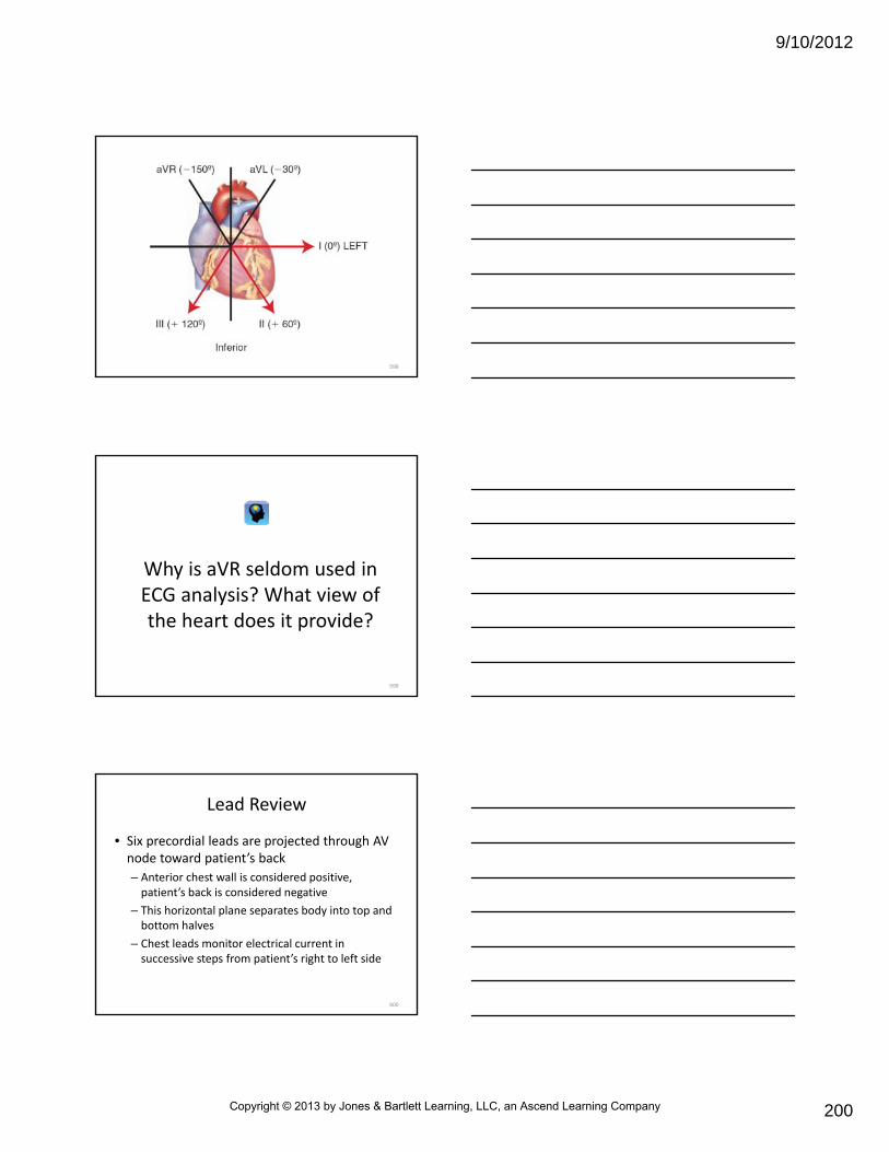
9/10/2012
200
598
Why is aVR seldom used in ECG analysis? What view of the heart does it provide?
599
Lead Review
• Six precordial leads are projected through AV node toward patient’s back
– Anterior chest wall is considered positive, patient’s back is considered negative
– This horizontal plane separates body into top and bottom halves
– Chest leads monitor electrical current in successive steps from patient’s right to left side
600
Copyright © 2013 by Jones & Bartlett Learning, LLC, an Ascend Learning Company

9/10/2012
201
601
Lead Review
• Leads V1 and V2
– Right chest leads that view septum of the heart (septal leads)
• Leads V3 to V4
– View anterior wall of left ventricle (anterior leads)
• Leads V5 to V6
– View lateral wall of left ventricle (lateral leads)
602
603
Copyright © 2013 by Jones & Bartlett Learning, LLC, an Ascend Learning Company

9/10/2012
202
Normal 12‐Lead ECG
• Recorded electronically on ECG paper in four separate columns
– Column 1 records leads I, II, and III
– Column 2 records aVR, aVL, and aVF
– Column 3 records V1, V2, and V3
– Column 4 records V4, V5, and V6
604
605
Normal 12‐Lead ECG
• If electrical current moves toward positive electrode, ECG complex will have positive or upward deflection from baseline
• If electrical current moves away from positive electrode, ECG complex will have negative or downward deflection from baseline
606
Copyright © 2013 by Jones & Bartlett Learning, LLC, an Ascend Learning Company

9/10/2012
203
Normal 12‐Lead ECG
• In normal 12‐lead ECG, limb leads I, II, III, aVF, and AVL will have positive R wave
– Because positive wave of depolarization within heart cells moves toward positive electrodes
– aVR is only limb lead that will have negative complex
– Chest leads are always positive
607
Normal 12‐Lead ECG
• Normal R wave progression
– Leads V1 through V6 display progressive nature of ventricular depolarization
• Chest lead V1 will normally be negative (at or below baseline)
• V2 will be more positive
• V3 will be progressively upright
• V4 through V6 will be directly upright
608
12‐Lead ECG and BBBs
• Heart blocks can occur in any one of three areas: SA node, AV node, or bundle branches
– Blocks interrupt normal passage of electrical stimulation in heart
– Sudden appearance of AV block or bundle branch block may indicate impending MI
609
Copyright © 2013 by Jones & Bartlett Learning, LLC, an Ascend Learning Company

9/10/2012
204
12‐Lead ECG and BBBs
• In LBBB, left ventricle depolarizes late
• In RBBB, right ventricle depolarizes late
– Results in wide QRS complex more than 0.12 second
– With LBBB, block often will yield QS pattern in V1
– With RBBB, delay yields negative S wave and large positive R wave
• Results in RSR' pattern
610
12‐Lead ECG & BBBs
• When bundle branch block suspected, look at chest leads V1 and V2 (right chest) and leads V5 and V6 (left chest)
– If QRS is wide with RSR' pattern in V1 or V2, RBBB may be present
– If wide QRS complex has QS pattern visible in V5 or V6, LBBB may be present
611
612
Copyright © 2013 by Jones & Bartlett Learning, LLC, an Ascend Learning Company

9/10/2012
205
Determination of Axis
• Electrical axis is direction of electrical impulse flow in heart that stimulates contraction– Direction of ventricular depolarization is known as mean QRS vector
– Current travels down from AV node and to left side of heart
• Mean QRS vector points downward and toward patient’s left side
• Position of QRS vector can be visualized in circle that lies on top of patient’s chest
• Circle is divided into degrees with AV node at center• Normal QRS vector is between 0 and +90 degrees
613
614
Determination of Axis
• QRS vector flows slightly to left of ventricular septum
– Because left ventricle has more and larger cardiac cells
– Each person has unique QRS vector that remains constant throughout life
– If cardiac status changes, electrical axis and position of heart can deviate from normal to right to left of its normal position
615
Copyright © 2013 by Jones & Bartlett Learning, LLC, an Ascend Learning Company

9/10/2012
206
Determination of Axis
• Axis is calculated automatically by modern 12‐lead ECG machines
– Paramedic can then interpret degree of axis by memorization or by referring to axis chart
– If ECG machine cannot calculate axis, axis can be determined by quadrant or by assessing leads I, II, and III
616
617
Determining Axis by Quadrant
• Axis can be approximated quickly by quadrant
– Two key leads that can be used for approximating axis are leads I and aVF
– Normal axis lies with quadrant of 0 degrees and +90 degrees
– Deviation of axis to left (left axis deviation) lies within quadrant of 0 degrees and –90 degrees
618
Copyright © 2013 by Jones & Bartlett Learning, LLC, an Ascend Learning Company

9/10/2012
207
619
Determining Axis by Quadrant
• Deviation of axis to right (right axis deviation) lies within quadrant of +90 degrees and ±180 degrees
– Can also be indeterminate axis when axis lies within quadrant of –90 degrees and ±180 degrees
– By looking at net deflection of mean QRS vector in leads I and aVF, approximation of axis can be determined
620
Determining Axis by Leads I, II, III • Axis can be evaluated by looking at QRS complexes in leads I, II, and III, considered– Normal when QRS deflection is positive (upright) in all bipolar leads
– Physiological left (which may be normal in some patients) when QRS deflection is positive in leads I and II but negative (inverted) in lead III
– Pathological left when QRS deflection is positive in lead I and negative in leads II and III (indicating an anterior hemiblock)
621
Copyright © 2013 by Jones & Bartlett Learning, LLC, an Ascend Learning Company

9/10/2012
208
Determining Axis by Leads I, II, III
• Axis can be evaluated by looking at QRS complexes in leads I, II, and III, considered
– Right axis when QRS deflection is negative in lead I, negative or positive in lead II, and positive in lead III (pathological in any adult and may be indicative of posterior hemiblock)
– Extreme right (“no man’s land”) when QRS deflection is negative in all three leads (indicating rhythm is ventricular in origin)
622
Why is the QRS deflection opposite the underlying rhythm?
623
Axis and Hemiblocks
• Hemiblock
– Failure in conduction of cardiac impulse in either of two main divisions of left bundle branch of bundle of His
– Interruption may occur in either anterior (superior) or posterior (inferior division)
– Identifying axis can be useful in determining presence of hemiblocks
624
Copyright © 2013 by Jones & Bartlett Learning, LLC, an Ascend Learning Company

9/10/2012
209
Anterior Hemiblock
• Occurs more often than posterior hemiblock
– Anterior fascicle of left bundle branch is longer and thinner structure
• Blood supply comes mainly from left anterior descending coronary artery
• Characterized by left axis deviation in patient who has supraventricular rhythm
625
626
Anterior Hemiblock
• Other ECG findings associated with anterior hemiblock include
– Normal QRS complex (less than 0.12 second) or RBBB
– Small Q wave followed by tall R wave in lead I
– Small R wave followed by deep S wave in lead III
627
Copyright © 2013 by Jones & Bartlett Learning, LLC, an Ascend Learning Company

9/10/2012
210
Anterior Hemiblock
• In anterior hemiblock with RBBB, impulses can be conducted only through ventricles by way of posterior fascicle of left bundle branch
– Patients at high risk of developing complete heart block
628
Posterior Hemiblock
• Posterior fascicle of left bundle branch is not blocked as easily as anterior fascicle
– Because bundle is much thicker and has double blood supply (left and right coronary arteries)
• As result, posterior hemiblock occurs less often
• Not commonly seen alone, more often associated with RBBB
629
Posterior Hemiblock
• Can be assumed in patients with right axis deviation and QRS complex of normal width or with RBBB
• Other ECG findings that indicate presence
– Small R wave followed by deep S wave in lead I
– Small Q wave followed by tall R wave in lead III
630
Copyright © 2013 by Jones & Bartlett Learning, LLC, an Ascend Learning Company

9/10/2012
211
631
Bifascicular Block
• Blockage of two of three pathways (fascicles) for ventricular conduction
– Refers to RBBB with block of either anterior or posterior division of left bundle branch
• Because anterior hemiblock combined with posterior hemiblock is difficult to distinguish from LBBB
– Decreases myocardial contractility and cardiac output
632
Bifascicular Block
• Patients may develop complete heart block suddenly and without warning
– The more branches that have impaired conduction, the greater chance patient will develop complete AV block (especially in patients with acute MI)
633
Copyright © 2013 by Jones & Bartlett Learning, LLC, an Ascend Learning Company

9/10/2012
212
12‐Lead Strategies for Wide‐Complex Tachycardias
• If unstable patient’s QRS complex is wide (more than 0.12 second) and fast (more than 150 beats/minute), immediate cardioversionmay be indicated
634
12‐Lead Strategies for Wide‐Complex Tachycardias
• If patient is stable, following steps in 12‐lead assessment may help distinguish between ventricular tachycardia and other wide‐complex tachycardias
– Assess leads I, II, III, V1, and V6
• If QRS complex is negative in leads I, II, and III and positive in V1, rhythm indicates ventricular tachycardia
• If these criteria are not met, proceed to next step
635
636
Copyright © 2013 by Jones & Bartlett Learning, LLC, an Ascend Learning Company
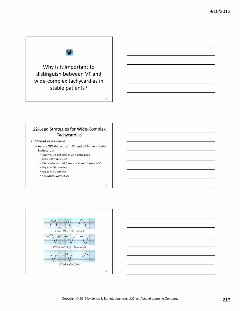
9/10/2012
213
Why is it important to distinguish between VT and wide‐complex tachycardias in
stable patients?
637
12‐Lead Strategies for Wide‐Complex Tachycardias
• 12‐lead assessment
– Assess QRS deflection in V1 and V6 for ventricular tachycardia
• Positive QRS deflections with single peak
• Taller left “rabbit ear”
• RS complex with fat R wave or slurred S wave in V1
• Negative QS complex
• Negative RS complex
• Any wide Q wave in V6
638
639
Copyright © 2013 by Jones & Bartlett Learning, LLC, an Ascend Learning Company

9/10/2012
214
12‐Lead Strategies for Wide‐Complex Tachycardias
• 12‐lead assessment
– Ventricular tachycardia indicators
• A negative QRS complex in lead I
• Positive QRS complex in leads II and III
• Negative QRS complex in V1 indicates – All precordial leads (V leads) are positive or negative (precordial concordance)
– RS interval is more than 0.10 second in any V lead (increased ventricular activation time)
640
641
642
Copyright © 2013 by Jones & Bartlett Learning, LLC, an Ascend Learning Company

9/10/2012
215
643
ST Segment and T Wave Changes
• When heart muscle is damaged, damaged area is unable to contract effectively
– Area remains in constant depolarized state
– Current flow between pathologically depolarized and normally repolarized areas can produce
• ST segment elevation
• Ischemic ST segment depression
• Normal or nondiagnostic changes in ST segment or T waves
644
645
Copyright © 2013 by Jones & Bartlett Learning, LLC, an Ascend Learning Company

9/10/2012
216
ST Segment and T Wave Changes
• Can classify patient into one of three groups
– ST‐segment Elevation MI (STEMI)
• ST‐segment elevation is characterized by ST‐segment elevation greater then 1 mm (0.1 mV) in two or more adjacent limb leads or any two contiguous chest leads
• STEMI also occurs with presumed new LBBB
646
647
ST Segment and T Wave Changes
• Can classify patient into one of three groups
– High‐Risk UA/non‐ST‐Elevation MI (NSTEMI)
• Ischemic ST‐segment depression 0.5 mm or more (0.05 mV) or dynamic T‐wave inversion with pain or discomfort
• Nonpersistent or transient ST‐segment elevation 0.5 mm or more (0.05 mV) for more than 20 minutes is also included
648
Copyright © 2013 by Jones & Bartlett Learning, LLC, an Ascend Learning Company

9/10/2012
217
ST Segment andT Wave Changes
• Can classify patient into one of three groups
– Normal or nondiagnostic changes in ST segment or T wave
• Findings are inconclusive
• Includes patients with normal ECGs and those with ST‐segment deviation of less than 0.5 mm (0.05 mV) or T‐wave inversion 0.2 mV or less
• Special cardiac studies and testing are needed for these patients
649
12‐Lead ECG to Assess Infarcts
• Early recognition and management of acute MI sometimes can salvage damaged myocardium (“time is muscle”)
• Five‐step analysis for infarct recognition:
– Step 1
• Identify rate and rhythm
• Manage any life‐threatening dysrhythmias
650
12‐Lead ECG to Assess Infarcts
• Five‐step analysis for infarct recognition
– Step 2
• Identify area of infarct
• ST segment elevation is most reliable indicator during first hours of infarction
• ST segment elevation can be present before permanent tissue damage has occurred
• If ST segment elevation is present in patient with chest pain, identify degree of elevation and visualize cardiac anatomy to predict which coronary artery is occluded
651
Copyright © 2013 by Jones & Bartlett Learning, LLC, an Ascend Learning Company

9/10/2012
218
12‐Lead ECG to Assess Infarcts
• Five‐step analysis for infarct recognition– Step 2 (cont’d)
• Use systematic approach for 12‐lead assessment
• One method is to begin by assessing inferior leads (II, III, aVF), followed by septal leads (V1, V2), anterior leads (V3, V4), and lateral leads (V5, V6, I, aVL)
• Evaluate each lead for ST segment elevation (most important sign of injury), deep symmetrically inverted T waves (a sign of ischemia), ST segment depression (a reciprocal change to ST elevation), and pathological Q waves
652
653
654
Copyright © 2013 by Jones & Bartlett Learning, LLC, an Ascend Learning Company

9/10/2012
219
12‐Lead ECG to Assess Infarcts
• Five‐step analysis for infarct recognition
– Step 2 (cont'd)
• In acute STEMI, reciprocal changes may be seen between leads that face acute injury and leads that face lateral boundary of injury (between ischemic and healthy tissue)
• Leads that face injury often show ST segment elevation
• Leads that face boundary often show ST segment depression
• Reciprocal changes not always visible on 12‐lead ECG during acute STEMI
• If visible, confirms diagnosis
655
12‐Lead ECG to Assess Infarcts
• Five‐step analysis for infarct recognition
– Step 2 (cont’d)
• At times, extent of infarction can be gauged by number of leads showing ST segment elevation
• Degree of ST segment elevation also important
• ST segment elevation or new or presumably new LBBB is suspicious for injury
656
12‐Lead ECG to Assess Infarcts
• Five‐step analysis for infarct recognition
– Step 3
• Consider other conditions that could be responsible for ST segment changes
• “Infarct impostors” also may be present in patient who is experiencing acute MI
• With exception of LBBB, left ventricular hypertrophy looks less like infarction
657
Copyright © 2013 by Jones & Bartlett Learning, LLC, an Ascend Learning Company

9/10/2012
220
12‐Lead ECG to Assess Infarcts
• Five‐step analysis for infarct recognition
– Step 3 (cont’d)
• Ventricular rhythms often produce Q waves and ST segment elevation
• Ventricular rhythms will not have reciprocal ST depression
• ECG changes with pericarditis are subtle
• Early repolarization produces no clinical symptoms
658
12‐Lead ECG to Assess Infarcts
• Five‐step analysis for infarct recognition
– Step 4
• Assess patient’s clinical presentation
• Assessment of clinical condition is just as crucial as ECG findings
• Obtaining thorough patient history and performing physical examination should be incorporated into ECG interpretation
• Not all patients with acute MI have classic signs and symptoms
659
12‐Lead ECG to Assess Infarcts
• Five‐step analysis for infarct recognition
– Step 4 (cont’d)
• Maintain high degree of suspicion in absence of pain, especially with diabetic patients, older adults, postmenopausal women
• As many as 50 percent of patients with acute MI have no early ECG changes
• Clinical picture is important
660
Copyright © 2013 by Jones & Bartlett Learning, LLC, an Ascend Learning Company

9/10/2012
221
12‐Lead ECG to Assess Infarcts
• Five‐step analysis for infarct recognition
– Step 5
• Recognize infarction and initiate care
• When all indications point to acute MI, speed process of data collection, physician evaluation, definitive care
• Clinical presentation and ECG findings that suggest acute MI must be confirmed by medical direction to determine appropriate care
661
15‐ and 18‐Lead Diagnostics
• Wall of right ventricle and posterior wall of left ventricle are areas of heart that are difficult to evaluate with 6 precordial leads
• ECG monitoring that includes 12 leads plus V4R, V8, and V9 leads (15‐lead electrocardiogram) increases sensitivity for MIs that occur in these areas (e.g., isolated posterior myocardial infarction)
662
15‐ and 18‐Lead Diagnostics
• For 15‐lead ECG monitoring, V4R lead is placed at fifth intercostal space in right anterior midclavicular line
• V8 lead is placed at posterior fifth intercostal space in right midscapular line
• V9 lead is placed between V8 lead and spinal column at posterior fifth intercostal space
663
Copyright © 2013 by Jones & Bartlett Learning, LLC, an Ascend Learning Company

9/10/2012
222
664
15‐ and 18‐Lead Diagnostics
• 18‐lead ECG monitoring (not routinely performed in prehospital setting) uses 15‐lead ECG along with V5R, V6R, and V7
– Leads are placed in same horizontal line as V4 to V6
– V7 is placed at posterior axillary line
– V8 is placed at posterior scapular line
665
666
Copyright © 2013 by Jones & Bartlett Learning, LLC, an Ascend Learning Company

9/10/2012
223
15‐ and 18‐Lead Diagnostics
• When obtaining 15‐lead ECG, new 12‐lead ECG printout must be labeled to identify 3 leads that have been moved– Notation at top of ECG printout should denote “Posterior Chest Leads”
– Lead V4 should be relabeled V4R
– V5 should be relabeled V8
– V6 should be relabeled V9
– The more leads that reveal acute changes in heart, the larger area of infarct is presumed to be
667
Lesson 22.8
Assessment
668
Learning Objective
• Outline the appropriate assessment of a patient who may be experiencing a cardiovascular disorder.
669
Copyright © 2013 by Jones & Bartlett Learning, LLC, an Ascend Learning Company

9/10/2012
224
Assessment
• Focused evaluation
– Chief complaint
– History of event
– Significant medical history
– Physical examination
• Aid in directing initial patient care and in anticipating potential problems during transport to medical facility
670
Chief Complaint
• Cardiovascular disease may cause variety of symptoms– Obtaining appropriate history of each symptom important to form diagnostic impression
– Common chief complaints • Chest pain or discomfort, including shoulder, arm, neck, or jaw pain or discomfort
• Dyspnea
• Syncope
• Abnormal heartbeat or palpitations
671
Chief Complaint
• In some patients, cardiovascular problems commonly have atypical symptoms
– Mental status changes
– Abdominal or gastrointestinal symptoms (including persistent heartburn)
– Vague complaints of being ill
672
Copyright © 2013 by Jones & Bartlett Learning, LLC, an Ascend Learning Company

9/10/2012
225
Chest Pain or Discomfort
• Chest pain or discomfort is most common chief complaint of patients with MI
– Many causes of chest pain are not related to cardiac disease
• History of chest pain is key
673
Chest Pain or Discomfort
• Assess chief complaint
• Use OPQRST method
– Onset/Origin
• Ask patient to describe the pain or discomfort
• What does it feel like?
• What were you doing when the pain began?
• Have you ever had this type of pain before?
• Is it the same or different than last time?
674
Chest Pain or Discomfort
• OPQRST method– Provokes
• Try to determine events surrounding patient’s symptoms
• What do you think might have caused this pain?
• Does anything you do make the pain better or worse?
• Does the pain go away when you rest?
• Have you taken nitroglycerin for the pain, and if so, did it help?
• Does the pain get worse when you exercise, walk, or when you eat certain foods?
675
Copyright © 2013 by Jones & Bartlett Learning, LLC, an Ascend Learning Company

9/10/2012
226
Chest Pain or Discomfort
• OPQRST method
– Quality
• Ask patient to describe the pain or discomfort using his or her own words
• Common descriptions for the quality of chest pain associated with a coronary event include sharp, tearing, burning, heavy, and squeezing
676
Chest Pain or Discomfort
• OPQRST method
– Region
• Ask patient to localize the pain
• With one finger, point to where the pain hurts most
• Does the pain move (radiate) to another area of the body, or does it stay in one place?
• If the pain moves, where does it move? (cardiac chest pain often radiates to arms, neck, jaw, and back)
677
Chest Pain or Discomfort
• OPQRST method
– Severity
• Ask patient to rate the pain or discomfort to establish baseline
• On a scale of 0 to 10, with 10 being the worst pain you have ever had, what number would you use to describe this pain?
• If you had pain like this before, is it worse than the last time, or not as bad as the last time?
678
Copyright © 2013 by Jones & Bartlett Learning, LLC, an Ascend Learning Company

9/10/2012
227
What factors may influence a person’s perception and description of pain?
679
Chest Pain or Discomfort
• OPQRST method
– Time
• Try to determine length of pain episode and document it
• How long have you had this pain?
• Is the pain better or worse than it was when you called for help?
• Is the pain constant, or does it come and go?
680
Think of a way to ask a patient a question about chest pain that does not invite a yes/no answer.
681
Copyright © 2013 by Jones & Bartlett Learning, LLC, an Ascend Learning Company

9/10/2012
228
Dyspnea
• Difficulty breathing
– Often associated with MI
– Main symptom of pulmonary congestion caused by heart failure
– Causes that may be unrelated to heart disease
• COPD
• Respiratory infection
• Pulmonary embolus
• Asthma
682
Dyspnea
• Historical factors important in differentiating breathing difficulties– Duration and circumstances of onset of dyspnea
– Anything that aggravates or relieves dyspnea, including medications
– Previous episodes
– Associated symptoms
– Orthopnea
– Prior cardiac problems
683
Syncope
• Brief loss of consciousness caused by a sudden decrease in oxygenated blood to brain
• Cardiac causes of syncope result from events that decrease cardiac output
– Most common cardiac disorders associated with syncope are dysrhythmias
684
Copyright © 2013 by Jones & Bartlett Learning, LLC, an Ascend Learning Company

9/10/2012
229
Syncope
• Other causes
– Stroke
– Drug or alcohol intoxication
– Aortic stenosis
– Pulmonary embolism
– Hypoglycemia
• In older patients, may be only symptom of cardiac problem
685
Syncope
• Young, healthy persons may have syncopalepisode
• May result from stimulation of the vagusnerve (vasovagal syncope) that can produce hypotension and bradycardia
686
Syncope
• History of syncopal event should include– Presyncope aura (nausea, weakness, light‐headedness)
– Circumstances of occurrence (e.g., patient’s position before the event, severe pain, or emotional stress)
– Duration of syncopal episode
– Symptoms before syncopal episode (palpitation, seizure, incontinence)
– Other associated symptoms
– Previous episodes of syncope
687
Copyright © 2013 by Jones & Bartlett Learning, LLC, an Ascend Learning Company

9/10/2012
230
Syncopal events often occur in public places, such as a church. How can you decrease the feelings of embarrassment that the patient may have during this
situation?
688
Abnormal Heartbeat and Palpitations
• Heartbeats
– Many patients are aware of own heartbeat, particularly if irregular (skipping beats) or rapid (fluttering)
– Abnormal heartbeats or palpitations (irregular or forceful beating of heart) sometimes are normal occurrence
• May indicate serious dysrhythmia
689
Abnormal Heartbeat and Palpitations
• Obtain information– Pulse rate
– Regular versus irregular rhythm
– Circumstances of occurrence
– Duration
– Associated symptoms (chest pain, diaphoresis, syncope, confusion, dyspnea)
– Previous episodes and frequency
– Medication (drug stimulant) or alcohol use
690
Copyright © 2013 by Jones & Bartlett Learning, LLC, an Ascend Learning Company

9/10/2012
231
Significant Medical History
• Medical history
– Is patient taking prescription medications, particularly cardiac medications?
– Common medications for possible coronary event
• Nitroglycerin
• Atenolol
• Metoprolol
• Other beta‐blockers
• Digoxin
• Furosemide and other diuretics
• Antihypertensives
• Antihyperlipidemic agents
691
Significant Medical History
• Medical history
– Ask patient about medication compliance
– Use of any nonprescription drugs
– Alcohol use or illicit drug use may be contributing factor in patient’s chief complaint
692
Significant Medical History
• Is patient being treated for any other illness?
– Medical history that increases likelihood of significant coronary event
• Angina pectoris
• Previous MI
• Coronary artery bypass
• Angioplasty
• Heart failure
• Valvular disease
• Renal disease
• Hypertension
• Aneurysms
• Diabetes
• Inflammatory cardiac disease
• Lung disease
693
Copyright © 2013 by Jones & Bartlett Learning, LLC, an Ascend Learning Company

9/10/2012
232
Significant Medical History
• Does patient have any allergies?
– Medication allergies may be important in course of patient’s care
– Document allergies and report medical direction
694
Significant Medical History
• Does patient have risk factors for heart attack?– Older age
– Tobacco use
– Diabetes
– Family history of heart disease
– Obesity
– Increased serum cholesterol level (hypercholesterolemia)
– Illicit drug use
695
Significant Medical History
• Does patient have implanted pacemaker or implantable cardioverter defibrillator?
– Presence indicates significant coronary history
696
Copyright © 2013 by Jones & Bartlett Learning, LLC, an Ascend Learning Company

9/10/2012
233
Physical Examination
• Classic presentation of MI is pain or discomfort beneath sternum that lasts more than 30 minutes– Pain often described as crushing, pressure, squeezing, or burning
• Associated signs and symptoms– Apprehension
– Diaphoresis
– Dyspnea
– Nausea and vomiting
– Sense of impending doom
697
Physical Examination
• At times, presentation is atypical
– Paramedic’s skill in gathering relevant medical history and performing focused physical examination will direct patient care
• Example: patients with myocardial ischemia may deny they have chest pain
• May need to be asked specifically about tightness or squeezing in chest
698
Physical Examination
• For patients with chest pain caused by heart problems, understand patient is frightened– Chest pain is associated with life‐threatening consequences
– Calm and reassure to decrease anxiety
• Primary survey for patient with possible coronary event should include more in‐depth evaluation of patient’s level of consciousness, respirations, pulse, and blood pressure
699
Copyright © 2013 by Jones & Bartlett Learning, LLC, an Ascend Learning Company

9/10/2012
234
Physical Examination
• Primary survey
– Change in level of consciousness may indicate decreased cerebral perfusion caused by poor cardiac output
• If possible, determine patient’s normal level of functioning by interviewing patient, family members, others who are familiar with patient
700
Physical Examination
• Primary survey
– Baseline information is important during reassessment to identify trending and guide patient care
• Vital signs
• Respiratory assessment
• Pulse rate and regularity
• Initial BP
701
Physical Examination
• Use look‐listen‐feel approach
• Look: skin
– Pale and diaphoretic skin may indicate peripheral vasoconstriction and sympathetic stimulation
– Cyanosis is indicator of poor oxygenation
– Use pulse oximetry to measure hemoglobin oxygenation
702
Copyright © 2013 by Jones & Bartlett Learning, LLC, an Ascend Learning Company

9/10/2012
235
Physical Examination
• Look: jugular veins
– Increase in central venous pressure from heart failure and cardiac tamponade can produce distention of internal jugular veins
– Jugular vein distention best evaluated with patient’s head elevated at 45 degrees
– Distention may be difficult to assess in obese patients
703
Physical Examination
• Look: peripheral and presacral edema
– Edema can result from chronic back pressure in systemic venous circulation
– May be related to right heart failure
– Most obvious in dependent areas
– Can be classified as nonpitting or pitting
704
Physical Examination
• Look: additional indicators of cardiac disease
– More subtle signs of cardiac disease that may be found on a visual inspection
• Midsternal scar from coronary surgery
• Nitroglycerin patch on skin
• Implanted pacemaker or implantable cardioverterdefibrillator in left upper chest or abdominal wall
• Medical alert identification necklaces or bracelets
705
Copyright © 2013 by Jones & Bartlett Learning, LLC, an Ascend Learning Company

9/10/2012
236
706
Physical Examination
• Listen: lung sounds
– Assess chest visually for accessory muscle use in breathing before listening to lung sounds
– Lung sounds should be clear and equal bilaterally
– Adventitious breath sounds may indicate pulmonary congestion or edema
707
Physical Examination
• Listen: heart sounds
– Abnormal heart sounds may indicate congestive heart failure in adult patients
– Best heard at point of maximum impulse
– Point of maximum impulse is location at which apical impulse is most readily visible or palpable
• Often in fifth intercostal space, just medial to left midclavicular line
708
Copyright © 2013 by Jones & Bartlett Learning, LLC, an Ascend Learning Company

9/10/2012
237
Physical Examination
• Although detection of abnormal heart sounds is difficult in prehospital setting, can be useful to confirm your field impression
– Abnormal heart sounds do not alter prehospitalcare
– Evaluation should never delay other patient care measures or transportation
709
Physical Examination
• Listen: carotid artery bruit
– Murmurs that indicate turbulent blood flow through vessel
• Most commonly from atherosclerosis
– Presence with cardiac disease is evaluated at carotid artery with stethoscope and should always be assessed before performing carotid sinus massage
– If carotid artery bruit is present, carotid sinus massage is contraindicated
• May dislodge plaque in artery and cause stroke
710
What may happen if you perform carotid sinus massage on a patient with bruits or
known carotid artery disease?
711
Copyright © 2013 by Jones & Bartlett Learning, LLC, an Ascend Learning Company

9/10/2012
238
Physical Examination
• Feel: skin, pulse
– Skin:
• Assess skin with the back of hand for diaphoresis or fever
• Normal skin is warm and dry
– Pulse:
• Assess pulse for rate, regularity, equality
• Pulse deficit is radial pulse that is less than ventricular rate
• Pulse deficit in peripheral and apical pulse sites may indicate rhythm disturbance or vascular disease
• Note any pulse deficit and report it to medical direction
712
Aside from blood pressure, how will you evaluate the mechanical activity of the
heart?
713
Physical Examination
• Feel: thorax and abdomen
– Check thorax and abdomen of patient with cardiac disease for chest wall tenderness and pulsating masses
• Chest wall tenderness not uncommon in patients with acute MI
• Pulsating mass or distention in abdomen or epigastricarea may indicate an abdominal aneurysm
714
Copyright © 2013 by Jones & Bartlett Learning, LLC, an Ascend Learning Company

9/10/2012
239
Lesson 22.9
Cardiovascular Disorders
Management
715
Learning Objective
• Describe prehospital assessment and management of patients with selected cardiovascular disorders based on knowledge of the pathophysiology of the illness.
716
Specific Cardiovascular Diseases• Cardiovascular emergencies
– Acute coronary syndromes
– Atherosclerosis
– Angina pectoris
– Myocardial infarction
– Left ventricular failure and pulmonary edema
– Right ventricular failure
717
Copyright © 2013 by Jones & Bartlett Learning, LLC, an Ascend Learning Company

9/10/2012
240
Specific Cardiovascular Diseases• Cardiovascular emergencies
– Cardiogenic shock
– Cardiac tamponade
– Thoracic and abdominal aneurysm
– Acute arterial occlusion
– Noncritical peripheral vascular disorders
– Hypertension
718
Acute Coronary Syndromes
• Acute myocardial infarction (AMI) and unstable angina (UA) are components
– Is most common cause of sudden cardiac death
– Pathophysiology of both conditions is ruptured or eroded atheromatous plaque
– ECG findings common to ACS include ST segment elevation, ST segment depression, and T wave abnormalities
719
Acute Coronary Syndromes
• Rapid transport indications
– Chest pain of cardiac origin
– Sense of urgency for reperfusion
– No relief of pain with medications
– Hypotension or hypoperfusion with CNS involvement
– Significant changes in patient’s ECG
720
Copyright © 2013 by Jones & Bartlett Learning, LLC, an Ascend Learning Company

9/10/2012
241
Acute Coronary Syndromes
• Primary goals of therapy
– Reduce amount of myocardial necrosis that occurs in patients with MI, preserving left ventricular function, preventing heart failure
– Prevent major adverse cardiac events
• Death
• Nonfatal MI
• Need for urgent revascularization
721
Acute Coronary Syndromes
• Primary goals of therapy
– Treat acute, life‐threatening complications
• Ventricular fibrillation
• Pulseless ventricular tachycardia
• Symptomatic bradycardias
• Unstable tachycardias
722
Atherosclerosis Pathophysiology• Atherosclerosis
– Disease process characterized by progressive narrowing of lumen of medium and large arteries
• Aorta and its branches
• Cerebral arteries
• Coronary arteries
723
Copyright © 2013 by Jones & Bartlett Learning, LLC, an Ascend Learning Company

9/10/2012
242
Atherosclerosis Pathophysiology• Atherosclerosis
– Process results in development of thick, hard atherosclerotic plaque
• Atheromata or atheromatous lesions
• Most often found in areas of turbulent blood flow
• Includes vessel bifurcations or in vessels with decreased lumen diameter
724
Atherosclerosis Pathophysiology
• Results from damage to endothelial cell from mechanical or chemical injury and perhaps excess inflammation– Response includes platelet adhesion and clotting
– Smooth muscle cells may move from middle muscle layer into lining of artery
– In the lining, muscle cells form atheroma• Over time, atheromata become fibrous and hardened
• Partially or fully obstruct opening of arteries
• Some collateral circulation develops to make up for narrowed vessels
725
Atherosclerosis Pathophysiology
• Major risk factor: middle‐aged and older persons
• Inherited, seen at younger age in men than in women
• Associated risk factors
– Age
– Family history of heart disease
– Diabetes
726
Copyright © 2013 by Jones & Bartlett Learning, LLC, an Ascend Learning Company

9/10/2012
243
Atherosclerosis Pathophysiology
• Risk factors can be reduced or eliminated
– Cigarette smoking
– Obesity
– Hypertension
– Hypercholesterolemia
727
Atherosclerosis Pathophysiology
• Effects on blood vessels– Disrupts innermost lining of vessels
• Causes loss of vessel elasticity and increase in formation of clots
– Reduces diameter of vessel lumen• Decreases blood supply to tissues
• Both effects result in insufficient supply of nutrients to tissue
• Especially true under conditions of increased tissue demand for nutrients and O2
728
Atherosclerosis Pathophysiology
• Effects
– Severity related to extent of narrowing (stenosis) of blocked artery
• Also depends on how long atheroma took to develop and patient’s ability to develop collateral circulation around obstruction
729
Copyright © 2013 by Jones & Bartlett Learning, LLC, an Ascend Learning Company

9/10/2012
244
Angina Pectoris
• Symptom of myocardial ischemia
• Literally means “choking pain in the chest”
730
Angina Pectoris
• Caused by imbalance between myocardial O2
supply and demand
– Result is buildup of lactic acid and CO2 in ischemic tissues of myocardium
– Metabolites irritate nerve endings that produce anginal pain
– Most common cause is atherosclerotic disease of coronary arteries
731
Angina Pectoris
• Prinzmetal’s angina
– Temporary occlusion caused by spasm of coronary artery with or without atherosclerosis
– Can cause angina
732
Copyright © 2013 by Jones & Bartlett Learning, LLC, an Ascend Learning Company

9/10/2012
245
Angina Pectoris
• Emotional stress and any activity that increases myocardial O2 demand may cause anginal pain, particularly in patients with atherosclerosis
• Myocardial ischemia puts patient at risk for cardiac dysrhythmias
733
Stable Angina
• Angina pectoris
– Stable angina
• Usually precipitated by physical exertion or emotional stress
• Pain usually lasts 1 to 5 minutes, may last as long as 15 minutes
• Relieved by rest, nitroglycerin, or O2
• Attacks usually are similar and are always relieved by same mode of therapy
734
Unstable Angina
• Unstable angina
– Preinfarction angina
– Denotes anginal pattern that has changed in its ease of onset, frequency, intensity, duration, or quality
– May occur during periods of light exercise or at rest
– Pain usually lasts 10 or more minutes
735
Copyright © 2013 by Jones & Bartlett Learning, LLC, an Ascend Learning Company

9/10/2012
246
Unstable Angina
• Pain is relieved less promptly with cessation of activity or nitroglycerin than with stable angina
• Mimics acute MI
– Difficult to differentiate in prehospital setting
• Patients are at increased risk of acute MI and sudden death
736
Unstable Angina
• Pain of angina usually is described as pressure, squeezing, heaviness, or tightness in chest– Although 30 percent of patients with angina feel pain only in chest, others describe pain as radiating to shoulders, arms, neck, and jaw and through chest to back
– Associated signs and symptoms• Anxiety• Shortness of breath• Nausea or vomiting• Diaphoresis
– Patient history often reveals previous attacks of angina
737
Unstable Angina
• Many times patient will have taken nitroglycerin before arrival of EMS
– If so, determine
• Age of nitroglycerin prescription (nitroglycerin is unstable and quickly loses its strength)
• Amount taken
• Effect
– If pain is not relieved by rest and medication, suspect MI
738
Copyright © 2013 by Jones & Bartlett Learning, LLC, an Ascend Learning Company

9/10/2012
247
Myocardial Ischemia Management
• All patients with chest pain and signs and symptoms of myocardial ischemia should be managed as though acute MI were evolving
– Goal of management is to increase coronary blood supply, decrease myocardial O2 demand, or both
739
Myocardial Ischemia Management
• Management guidelines
– Place patient at rest physically and emotionally
– Administer O2
– Administer aspirin (per protocol)
– Initiate IV therapy for any drugs that may be needed
740
Myocardial Ischemia Management
• Management guidelines
– If pain is present on arrival of EMS, use pharmacological therapy
• Sublingual or topical nitroglycerin followed by morphine or fentanyl
– Monitor ECG for dysrhythmias
• Whenever possible (and if scene time is not delayed), record a 3‐lead or 12‐lead ECG, or both, during pain
• Measure, record, and communicate any ST segment changes
– Transport patient for physician evaluation
741
Copyright © 2013 by Jones & Bartlett Learning, LLC, an Ascend Learning Company

9/10/2012
248
Myocardial Infarction
• Acute myocardial infarction
– Occurs with sudden and total blockage or near blockage of blood flowing through affected coronary artery to an area of heart muscle
• Blockage results in ischemia, injury, and necrosis to area of myocardium distal to occlusion
– Most often associated with atherosclerotic heart disease
742
MI Precipitating Events
• Process is complex– Begins with formation of atherosclerotic plaque involving intimal layer of coronary artery
– Plaque disrupts smooth arterial lining and results in uneven surface
• Creates turbulent blood flow• Plaque may rupture• If rupture occurs, injured tissue is exposed to circulating platelets
• Results in formation of thrombus that occludes artery• As thrombus enlarges, further reduces blood flow in coronary vessel
743
MI Precipitating Events
• Acute thrombotic occlusion generally accepted as cause of most MI
• Other factors– Coronary spasm
– Coronary embolism
– Severe hypoxia
– Hemorrhage into diseased arterial wall
– Reduced blood flow after any form of shock
– All factors may result in inadequate amount of blood reaching myocardium
744
Copyright © 2013 by Jones & Bartlett Learning, LLC, an Ascend Learning Company

9/10/2012
249
Infarct Types & Locations
• Myocardial cells beyond occluded artery die (infarct) from lack of O2
– Size of infarct is determined by needs of tissue supplied by occluded vessel, by presence of collateral circulation, and by time it takes to reestablish blood flow
745
Why is collateral circulation important?
746
Infarct Types and Locations
• Emergency care directed at
– Increasing O2 supply by administering supplemental O2
– Decreasing metabolic needs and providing collateral circulation
– Reestablishing perfusion to ischemic myocardium as quickly as possible after onset of symptoms
747
Copyright © 2013 by Jones & Bartlett Learning, LLC, an Ascend Learning Company

9/10/2012
250
Infarct Types and Locations
• Majority involve left ventricle or interventricular septum
– Areas are supplied by either of two major coronary arteries
– If occlusion is in left coronary artery, result is anterior, lateral, or septal wall infarction
• Inferior wall infarction (of inferior‐posterior wall of left ventricle) usually is result of right coronary artery occlusion
748
Infarct Types and Locations
• Infarction also can be classified into one of three ischemic syndromes based on rupture of unstable plaque in epicardial artery
– Unstable angina
– Non‐STEMI
– STEMI
749
Infarct Types and Locations
• Sudden cardiac death may occur with– Unstable angina
• Early thrombus has not obstructed coronary blood flow completely
• Partial occlusion produces symptoms of ischemia
• Blockage eventually may result in complete occlusion and produce non‐STEMI
• Fibrinolytic therapy effective in unstable angina
• Such therapy may accelerate occlusion
• Therapy with antiplatelet agents most effective at this time because thrombus is rich in platelets
750
Copyright © 2013 by Jones & Bartlett Learning, LLC, an Ascend Learning Company

9/10/2012
251
Infarct Types and Locations
• Sudden cardiac death may occur with
– Non‐STEMI occurs as microemboli from thrombus become lodged in coronary arteries
• This produces minimal damage to myocardium
• At highest risk for progression to MI
• Non‐STEMIs are evident only with ST segment depression or T wave abnormalities
751
Infarct Types and Locations
• Sudden cardiac death may occur with any of these syndromes
– STEMI occurs when thrombus occludes coronary vessel for prolonged period
• Diagnosed by development of elevated ST segments in two or more contiguous (adjacent) leads
• Clot is rich in thrombin
• Early management with fibrinolytics may help to limit size of infarct
752
753
Copyright © 2013 by Jones & Bartlett Learning, LLC, an Ascend Learning Company

9/10/2012
252
Myocardium Death
• When blood flow to myocardium ceases, cells switch from aerobic to anaerobic metabolism
– Results in release of lactic acid and an increase in tissue CO2 levels
• Contribute to ischemic pain (angina)
• As cells lose ability to maintain their electrochemical gradients, begin to swell and depolarize
754
Myocardium Death
• These initial changes are reversible
– Within a few hours, if collateral flow and reperfusion are inadequate, much muscle distal to occlusion dies
– Area surrounding necrotic tissue may survive because of collateral circulation
– Surviving tissue may become origin of dysrhythmias
755
756
Copyright © 2013 by Jones & Bartlett Learning, LLC, an Ascend Learning Company

9/10/2012
253
Myocardium Death
• Scar tissue replaces infarcted area in process that takes about 8 weeks– Starts with deposits of connective tissue on about 12th day
– Scar tissue is durable• Lacks elasticity
• Does not contract
• Conducts electrical impulses poorly in damaged area of myocardium
– Left ventricle can lose as much as 25 percent of its muscle and still function as effective pump
757
Myocardium Death
• Areas with poor perfusion after large MI may not develop strong scar tissue
– May result in aneurysm
• Can greatly decrease effective ventricular contractility
• May lead to development of serious dysrhythmias
758
Myocardium Death
• Damaged myocardium is most susceptible to rupture during first one to two weeks after MI because scar tissue has not reached adequate strength
– Patient activity is limited
– Prevention of hypertension and excitement during this period also is necessary
759
Copyright © 2013 by Jones & Bartlett Learning, LLC, an Ascend Learning Company

9/10/2012
254
Myocardium Death
• Length of hospitalization of patients with uncomplicated MI has decreased– Today, most patients resume activity within 2 to 3 days
– Most leave hospital within 7 to 10 days
– Many patients get stress test before they are discharged
• Determines patient’s exercise tolerance level and whether ischemia or dysrhythmias are present during exercise
• Result helps determine what activities patient may resume after discharge
760
Deaths Following MI
• Usually result from– Lethal dysrhythmias
• Ventricular tachycardia
• Ventricular fibrillation
• Cardiac standstill
– Pump failure• Cardiogenic shock
• Congestive heart failure
– Myocardial tissue rupture• Rupture of ventricle, septum, or papillary muscle
761
Death Following MI
• Fatal dysrhythmias are most common cause of death from MI
• Deaths that occur within first two hours after onset of illness or injury are sudden deaths
• Majority of patients who suffer sudden death have no immediate warning symptoms
762
Copyright © 2013 by Jones & Bartlett Learning, LLC, an Ascend Learning Company

9/10/2012
255
MI Signs and Symptoms
• Some patients with acute MI have only symptoms of dyspnea, syncope, or confusion– Diabetic patients
– Some women
– Older age groups
• Substernal chest pain is present in 70 to 90 percent of patients with acute MI– Pain generally has same characteristics and locations as anginal pain
– May radiate to arms, neck, jaw, or back
763
MI Signs and Symptoms
• Signs and symptoms that accompany pain, occasionally present in absence of pain (silent MI)
– Agitation
– Anxiety
– Cyanosis
– Diaphoresis
– Dyspnea
– Nausea and vomiting
– Palpitations
– Sense of impending doom
– Weakness
764
MI Signs and Symptoms
• Chest pain associated with acute MI often is constant
– Pain often not altered or alleviated by nitroglycerin or other cardiac medications, rest, changes in body position, or breathing patterns
– With angina pectoris, onset often occurs during periods of activity
– Onset of pain in more than half of all patients with acute MI occurs during rest
765
Copyright © 2013 by Jones & Bartlett Learning, LLC, an Ascend Learning Company

9/10/2012
256
MI Signs and Symptoms
• Most patients have had warning anginal pains (preinfarction angina) hours or days before attack– Many patients deny possibility of evolving MI
• Blame chest pain or discomfort to unrelated causes such as fatigue or indigestion
• Denial delays request for EMS assistance during most critical phase of illness
• According to AHA, 50+ percent of deaths from ischemic heart disease occur outside hospital within first 4 hours after onset of pain
766
Why do you think patients may deny that their signs and symptoms may be due to a
heart attack?
767
MI Signs and Symptoms
• Vital signs vary
– Depend on extent of damage to heart muscle and conduction system
– Depend on degree and type of autonomic nervous system response
• Inferior MIs often show mainly parasympathetic response
• Anterior MIs commonly show mainly sympathetic response
768
Copyright © 2013 by Jones & Bartlett Learning, LLC, an Ascend Learning Company

9/10/2012
257
MI Signs and Symptoms
• Pulse rate depends on presence or absence of dysrhythmias
– May be normal, tachycardic, bradycardic, regular, or irregular
• Respirations may be normal or increased
769
Common ECG Findings
• Sulfonylurea drugs may lessen magnitude of ST segment elevation in presence of infarct
– Diabetes drugs
• Glyburide
• Glipizide
• Obtaining diabetic history is crucial for all patients with cardiac event
770
Uncomplicated Acute MI Management
• All patients with anginal chest pain are assumed to have acute MI until proved otherwise
• Any patient with chest pain should be transported to medical facility for physician evaluation
– Regardless of apparent severity on EMS arrival, patient’s age, or associated complaints
771
Copyright © 2013 by Jones & Bartlett Learning, LLC, an Ascend Learning Company

9/10/2012
258
772
Uncomplicated Acute MI Management
• Primary goals of prehospital care– Identify patient with possible MI
– Relieve pain and apprehension
– Prevent development of serious dysrhythmias
– Limit size of infarct
• Obtain full patient history while conducting physical examination and during initial patient care– Time is important
773
Uncomplicated Acute MI Management
• Patient care high priority– Place patient at rest or in comfortable position
• Helps decrease anxiety and heart rate
• Will decrease O2 demand
– Administer low‐concentration O2 (4 L/min) via nasal cannula
• Patients with respiratory compromise need higher O2
concentration
– Initiate transport quickly• Do without audible or visual warning devices if patient is stable, will help decrease patient anxiety
774
Copyright © 2013 by Jones & Bartlett Learning, LLC, an Ascend Learning Company

9/10/2012
259
How can the prehospitalrecognition of AMI affect the hospital care of the patient?
775
What effect will the arrival of your ambulance likely have on the heart rate and blood pressure of a conscious,
alert patient?
776
Uncomplicated Acute MI Management
• Patient care high priority– Administer aspirin (per protocol)
– Measure pulse oximetry
– Establish IV line with normal saline or lactated Ringer’s solution to keep vein open or supply fluid boluses (if needed)
– Obtain baseline vital signs
– Repeat assessment often
– Vital sign assessment should include auscultation of lungs for heart failure indicators (presence of crackles)
777
Copyright © 2013 by Jones & Bartlett Learning, LLC, an Ascend Learning Company

9/10/2012
260
What breath sounds might you hear if the patient has congestive
heart failure or pulmonary edema?
778
Uncomplicated Acute MI Management
• Patient care high priority
– Attach ECG electrodes, obtain 12‐lead ECG document initial rhythm, monitor for dysrhythmias
– Administer medications (per protocol) for relief of pain and management of dysrhythmias
• Medications used for analgesia and to decrease preload and afterload include nitroglycerin, followed by morphine
• Medications used to manage the various dysrhythmiasinclude lidocaine, procainamide, atropine, verapamil, adenosine, magnesium, propranolol, amiodarone, and others (refer to appropriate treatment algorithm)
779
Fibrinolytic Therapy
• Acute intracoronary thrombus can be dissolved with salvage of ischemic myocardium if fibrinolytic agent is administered within 6 hours after onset of symptoms
– Restores blood flow to ischemic area
– Some EMS are authorized by medical direction to administer these agents in prehospital setting
780
Copyright © 2013 by Jones & Bartlett Learning, LLC, an Ascend Learning Company

9/10/2012
261
Fibrinolytic Therapy
• AHA recommends prehospital systems focus on early diagnosis
– Field administration of fibrinolytics should occur in special circumstances when physician is present or if transport time is delayed
• Onset of symptoms time to door time at hospital will be 1 or more hours
781
Fibrinolytic Therapy
• Common fibrinolytic agents
– Streptokinase
– Tissue plasminogen activator
– Tenecteplase
– Anistreplase
– Reteplase
782
Fibrinolytic Therapy
• Agents work through activation of plasma protein plasminogen to dissolve coronary thrombus
– Plasminogen is converted to plasmin (active form)
• Plasmin degrades fibrin, basic component of clot (thrombus)
– Aspirin and heparin are part of “fibrinolyticpackage”
783
Copyright © 2013 by Jones & Bartlett Learning, LLC, an Ascend Learning Company
9/10/2012
262
Fibrinolytic Therapy
• Can dissolve beneficial and pathological thrombi
– Administered selectively
– Most EMS systems using fibrinolytic agents establish inclusion–exclusion criteria
784
Congestive Heart Failure
• Condition in which heart is unable to pump blood at rate to meet metabolic needs of tissues
• Most often caused by
– Volume overload
– Pressure overload
– Loss of myocardial tissue
– Impaired contractility
785
Congestive Heart Failure
• Can impair left ventricular function
• Precipitating causes of heart failure
– MI
– Pulmonary embolism
– Enlarged heart (cardiomegaly)
786
Copyright © 2013 by Jones & Bartlett Learning, LLC, an Ascend Learning Company

9/10/2012
263
Left Ventricular Failure and Pulmonary Edema
• Left ventricular failure
– Occurs when left ventricle fails to work as effective forward pump
• Causes backpressure of blood into pulmonary circulation
– May be caused by forms of heart disease
• Ischemic
• Valvular
• Hypertensive heart disease
– If left unmanaged, significant left ventricular failure results in pulmonary edema
787
Left Ventricular Failure and Pulmonary Edema
• In left ventricular failure, blood is delivered to left ventricle
– Blood is not fully ejected from ventricle
– Increase in end‐diastolic blood volume increases left ventricular end‐diastolic pressure
• This pressure is transmitted to left atrium
• Then is transmitted to pulmonary veins and capillaries
788
Left Ventricular Failure and Pulmonary Edema
• As pulmonary capillary hydrostatic pressure increases, plasma portion of blood is forced into alveoli– There plasma mixes with air– Results in typical finding in pulmonary edema: foamy, blood‐tinged sputum
– If left unmanaged, progressive fluid buildup can result in death from hypoxia
– MI is common cause of left ventricular failure• All patients with pulmonary edema (particularly those with an abrupt onset) also should be suspected of having acute MI
789
Copyright © 2013 by Jones & Bartlett Learning, LLC, an Ascend Learning Company

9/10/2012
264
Left Ventricular Failure and Pulmonary Edema
• Left ventricular failure results in a reduction of stroke volume– Initiates several compensatory mechanisms that restore cardiac output and organ perfusion
• Tachycardia
• Vasoconstriction
• Activation of renin‐angiotensin‐aldosterone system
– Mechanisms often increase myocardial O2
demand• Further decrease ability of myocardium to contract
790
Pulmonary Edema Management• Pulmonary edema
– Acute and critical emergency
– May lead to death unless treated rapidly
– Emergency management directed at
• Decreasing venous return to heart
• Improving myocardial contractility
• Decreasing myocardial O2 demand
• Improving ventilation and oxygenation
• Rapid transport
791
792Continued
Copyright © 2013 by Jones & Bartlett Learning, LLC, an Ascend Learning Company

9/10/2012
265
793Continued
794
Pulmonary Edema Management• Emergency care
– Patient positioning
– Oxygenation
– Continuous positive airway pressure (CPAP)
– Ventilatory support as needed
– Pharmacological therapy
795
Copyright © 2013 by Jones & Bartlett Learning, LLC, an Ascend Learning Company

9/10/2012
266
Pulmonary Edema Management• Perform full but focused patient history and examination while initiating treatment
– No characteristic ECG changes are associated with pulmonary edema
– Obtain initial tracing
– Monitor patient’s rhythm continuously for evidence of myocardial irritability and dysrhythmias
796
Pulmonary Edema Management• Place patient in sitting position with legs dependent
– Increases lung volume and vital capacity
– Diminishes work of respiration
– Decreases venous return to heart as well
797
Pulmonary Edema Management• Administer high‐concentration O2 using well‐fitted face mask
– Mask should be a non‐rebreathing mask to optimize amount of inspired O2
– Some patients may require (and will tolerate) positive‐pressure assistance (including CPAP or biphasic positive airway pressure
798
Copyright © 2013 by Jones & Bartlett Learning, LLC, an Ascend Learning Company

9/10/2012
267
Pulmonary Edema Management• Positive pressure assistance
– Highest priorities
– Reduces need for high levels of inspired O2
– Pulse oximeter should be used to ensure arterial O2 saturation of at least 90 percent
– If cannot be achieved with 100 percent O2 or if signs of cerebral hypoxia or progressive hypercapnia, tracheal intubation and assisted ventilations may be indicated
799
Pulmonary Edema Management
• Medications used to decrease venous return, enhance contractile function of myocardium, and reduce dyspnea– Nitroglycerin
• Induction of peripheral vasodilation• Possible reduction of preload and afterload, reducing myocardial workload and improving cardiac function
– Morphine• Decrease of venous return by dilation of capacitance vessels of peripheral venous bed (reduces preload)
• Reduction of myocardial work• Reduction of anxiety
800
Right Ventricular Failure
• Most often results from left ventricular failure that produces elevated pressure in pulmonary vascular system– Pressure causes resistance to pulmonary blood flow
• Increases workload of right side of heart to overcome resistance
• Over time, right ventricle fails as effective forward pump
• Causes back‐pressure of blood into systemic venous circulation
801
Copyright © 2013 by Jones & Bartlett Learning, LLC, an Ascend Learning Company
9/10/2012
268
Right Ventricular Failure
• When pressure in systemic venous circulation becomes too high, plasma portion of blood is forced out into interstitial tissues of body
– Results in edema, particularly in dependent areas of body
802
Right Ventricular Failure
• Right ventricular failure can result from several diseases
– Chronic hypertension
• Left ventricular failure usually precedes right ventricular failure
– COPD
– Pulmonary embolism
– Valvular heart disease
– Infarction of right ventricle
803
Right Ventricular Failure
• Management
– Right ventricular failure is often chronic condition
– Usually not medical emergency in itself
– If right ventricular failure is associated with pulmonary edema or hypotension, may be medical emergency
– Be prepared to manage patient for either of these situations
804
Copyright © 2013 by Jones & Bartlett Learning, LLC, an Ascend Learning Company

9/10/2012
269
Right Ventricular Failure
• Management– Place patient at rest in sitting or semi‐Fowler position (head elevated)
– Administer high‐concentration O2
– Obtain baseline vital signs and an ECG tracing
– Initiate an IV line to keep vein open or to manage hypotension
– Monitor ECG and O2 saturation
– Manage symptoms of left ventricular failure, if present
805
Cardiogenic Shock
• Most extreme form of pump failure
– Occurs when left ventricular function is so compromised that heart cannot meet metabolic needs of body
– Result is significant decrease in stroke volume (resulting from ineffective myocardial contraction), cardiac output, and blood pressure
• All result in inadequate blood supply to organs
– Occurs in 5 to 10 percent of patients with acute MI
• May be result of acute left‐ or right‐sided heart failure
806
Cardiogenic Shock
• Present when shock persists after correction of existing dysrhythmias, volume deficit, or decreased vascular tone
• Usually caused by extensive MI (often involving more than 40 percent of the left ventricle) or by diffuse ischemia
• Even with aggressive therapy, has mortality rate of 70 percent or higher
807
Copyright © 2013 by Jones & Bartlett Learning, LLC, an Ascend Learning Company

9/10/2012
270
Cardiogenic Shock
• Patients show clinical evidence of hypoperfusion to vital organs and significant systemic hypotension similar to that found in other forms of shock– Acidosis
– Altered level of consciousness
– Cool, clammy, cyanotic, or ashen skin
– Hypoxemia
– Profound hypotension (systolic BP usually less than80 mm Hg)
– Pulmonary congestion (crackles)
– Sinus tachycardia or other dysrhythmias
– Tachypnea
808
Cardiogenic Shock
• In early stages of cardiogenic shock, patient’s heart tries to compensate
– Heart rate increases
– If possible, heart also increases contractility and cardiac output
– If condition managed inadequately, heart progresses toward hypodynamic failure with depressed contractility, reduced stroke volume, and subsequent hypoperfusion
809
Cardiogenic Shock
• Management
– Patients in cardiogenic shock are ill
– Need rapid transport
• Do not delay attempting field treatment
810
Copyright © 2013 by Jones & Bartlett Learning, LLC, an Ascend Learning Company

9/10/2012
271
What dose of each of these drugs should be given for this condition?
811
Cardiogenic Shock
• Management
– Prehospital care
• Airway management and ventilatory support with high‐concentration O2
• Placement in supine position (or semi‐Fowler position, if patient is dyspneic)
• Insertion of IV line with normal saline or lactated Ringer’s solution to keep vein open
812
Cardiogenic Shock
• Management
– Prehospital care
• ECG monitoring
• Correction of dysrhythmias
• Frequent evaluation of vital signs
• Patient in respiratory failure may require intubation and ventilatory support
813
Copyright © 2013 by Jones & Bartlett Learning, LLC, an Ascend Learning Company

9/10/2012
272
Why would large‐volume fluid resuscitation not be indicated
in this situation?
814
Cardiogenic Shock
• Management– Drug therapy may include drugs that strengthen force of contraction (inotropic agents) to improve cardiac output
• Dopamine
• Dobutamine
– Use of vasodilator drugs to reduce afterload generally is reserved for in‐hospital coronary care settings
• BP can be evaluated more accurately
– If left‐sided heart failure and pulmonary edema are present, they should be treated at same time
815
How should you respond to unstable patients with signs and symptoms indicating cardiogenic shock when they ask you, "Am I going to die?"
816
Copyright © 2013 by Jones & Bartlett Learning, LLC, an Ascend Learning Company
9/10/2012
273
Cardiac Tamponade
• Impaired diastolic filling of heart caused by increased intrapericardial pressure and volume
– As pressure of buildup in pericardial fluid compresses atria and ventricles, unable to fill adequately
• Results in decrease in ventricular filling, and stroke volume is decreased
817
Cardiac Tamponade
• May have gradual onset
– May result from cancerous growth or infection
• May be acute, resulting from trauma to chest, including CPR
• May result from renal disease or hypothyroidism
818
Cardiac Tamponade
• Signs and symptoms
– Chest pain
– Decreased systolic pressure (late sign)
– Ectopy
– ECG changes (usually inconclusive)
– Elevated venous pressure (early sign) with associated jugular vein distention
– Faint or muffled heart sounds
819
Copyright © 2013 by Jones & Bartlett Learning, LLC, an Ascend Learning Company

9/10/2012
274
Cardiac Tamponade
• Signs and symptoms
– Shortness of breath
– Low‐voltage QRS complexes and T waves
– Alternating amplitude and vector of P waves, QRS complexes, and T waves (electrical alternans)
– Pulsus paradoxus
– ST segment elevation or nonspecific T wave changes
– Tachycardia
820
Cardiac Tamponade
• Most important reliable signs
– Elevated venous pressure
– Hypotension
– Distant heart sounds (Beck’s triad)
821
Cardiac Tamponade
• Management
– Obtain thorough history to attempt to identify cause of cardiac tamponade
– Perform physical examination
– Prehospital care
• Ensure adequate airway and ventilatory support
• Rapid transport for physician evaluation and possible drainage of pericardial sac (pericardiocentesis)
822
Copyright © 2013 by Jones & Bartlett Learning, LLC, an Ascend Learning Company

9/10/2012
275
Why is pericardiocentesis not routinely done in the prehospital setting?
823
Cardiac Tamponade
• Management
– Fluid bolus may help support circulatory system temporarily if patient becomes hypotensive
– Definitive management requires drainage of pericardial sac
– Cardiac tamponade may result in death if condition is not relieved
824
Thoracic and Abdominal Aneurysms
• Aneurysm
– “Dilation of a vessel”
– May result from
• Atherosclerotic disease (most common)
• Infectious disease (primarily syphilis)
• Traumatic injury
• Certain genetic disorders (e.g., Marfan syndrome)
825
Copyright © 2013 by Jones & Bartlett Learning, LLC, an Ascend Learning Company

9/10/2012
276
826
Thoracic andAbdominal Aneurysms
• Most aneurysms develop at weak point in wall of artery– Results from degenerative changes in medial layer
– Weakening of supportive elements of vessel wall allows dilation
• Causes turbulence and increasing lateral pressure
• Tends to enlarge over time as lateral pressure increases in dilated segment
– Eventually aneurysm may rupture• May produce life‐threatening hemorrhage
827
Abdominal Aortic Aneurysm
• Affects about 2 percent of population
• Most common site: below renal arteries and above branching of common iliac arteries
• 10 times more common in men
828
Copyright © 2013 by Jones & Bartlett Learning, LLC, an Ascend Learning Company

9/10/2012
277
Abdominal Aortic Aneurysm
• Most prevalent between ages 60 and 70
• Usually asymptomatic as long as it is stable
• If begins to expand or leak, symptoms will indicate impending rupture
829
Abdominal Aortic Aneurysm
• Rupture may begin with small tear in intima
– Allows blood to leak into wall of aorta
– As process continues with increasing pressure, tear may extend through outer layer of vessel
– Tear may cause bleeding into retroperitoneal space
830
Abdominal Aortic Aneurysm
• If bleeding is tamponaded by retroperitoneal tissues, patient may be normotensive on arrival of EMS
– If rupture opens into peritoneal cavity, massive fatal hemorrhage may follow
– In either case, major blood loss results, and hypovolemic shock ensues
831
Copyright © 2013 by Jones & Bartlett Learning, LLC, an Ascend Learning Company

9/10/2012
278
Abdominal Aortic Aneurysm
• Patient with rupturing aneurysm will have syncope followed by hypotension with bradycardia despite large amount of blood loss– Reason for bradycardia is stimulation of vagus nerve
– Aorta has fibers of vagus nerve wrapped around it
– When aorta tears, tear stretches these fibers, which in turn produces bradycardia
– Bradycardia is present despite hemorrhagic shock condition, which usually produces hypotension and tachycardia in patient
832
What other condition has signs and symptoms similar to abdominal aortic aneurysm?
833
Abdominal Aortic Aneurysm
• Management
– Patients with leaking or ruptured abdominal aneurysm appear ill
• Usually need immediate surgery to repair vessel
• 20 percent of patients with abdominal aortic aneurysm rupture aneurysm before reaching hospital
• 80 percent of these patients die
• Early recognition and rapid transport can prevent death
834
Copyright © 2013 by Jones & Bartlett Learning, LLC, an Ascend Learning Company

9/10/2012
279
Abdominal Aortic Aneurysm
• Management
– Prehospital care should be limited to
• Gentle handling
• O2 administration
• Cardiac monitoring (MI may be associated with advanced aneurysms)
• Initiation of volume‐expanding IV fluids while en route to receiving hospital
• Alerting receiving facility to prepare for imminent surgery
835
Abdominal Aortic Aneurysm
• Management
– Pulsatile masses
• Fragile, membrane thin
• Avoid aggressive examination or deep palpation of mass
• Palpation may cause mass to rupture
• Examination can be made by auscultation
• May reveal sound similar to systolic murmur or bruit
836
Abdominal Aortic Aneurysm
• Management
– Management of hypotension varies and depends on whether aneurysm is leaking or ruptured
• Suspected leaking aneurysm can be maintained with mildly hypotensive BP to try to prevent rupture during transport
• Fluid resuscitation should be minimal and less aggressive than in patients who have ruptured aneurysm
837
Copyright © 2013 by Jones & Bartlett Learning, LLC, an Ascend Learning Company

9/10/2012
280
Abdominal Aortic Aneurysm
• Management
– If rupture occurred, hypotension, tachycardia, and loss of the pulsating mass may develop suddenly
• May become unresponsive
• Often followed by full cardiac and respiratory arrest
• Require rapid and aggressive resuscitation (intubation, ventilation, fluid replacement, rapid transport for surgery)
838
Acute Dissecting Aortic Aneurysm
• Separation of arterial wall
– Most common aortic catastrophe, affecting 5 to 10 persons/million population each year (3 times as many as ruptured abdominal aortic aneurysm)
839
Acute Dissecting Aortic Aneurysm
• Factors that can lead to development
– Systemic hypertension
– Atherosclerosis
– Congenital abnormalities that affect connective tissue (Marfan’s syndrome)
– Degenerative changes in connective tissue of aortic media (cystic medial necrosis)
– Trauma
– Pregnancy
840
Copyright © 2013 by Jones & Bartlett Learning, LLC, an Ascend Learning Company

9/10/2012
281
Acute Dissecting Aortic Aneurysm
• Results from small tear in intimal layer of vessel wall– After tear, process of dissection begins
– Tear in inner wall allows blood to move between inner and outer layers
• Creates false passage between layers of vessel wall.
• Blood that enters false passage results in formation of hematoma
• Results in rupture through outer wall (adventitia) at any time, usually into pericardial or pleural cavity
841
842
Acute Dissecting Aortic Aneurysm
• Any area of aorta may be involved– Ascending aorta mainly
– Once begun, aneurysm may extend distally or proximally to involve
• Thoracic and abdominal aorta and tributaries
• Coronary arteries
• Aortic valve
• Carotid and subclavian vessels
– Any vessels (including carotid and other aortic arch vessels) bypassed by dissection have their blood flow decreased
843
Copyright © 2013 by Jones & Bartlett Learning, LLC, an Ascend Learning Company

9/10/2012
282
Acute Dissecting Aortic Aneurysm
• May cause– Syncope
– Stroke
– Absent or reduced pulses
– Unequal BP readings (right vs. left)
– Heart failure resulting from sudden aortic valve regurgitation
– Pericardial tamponade
– Acute MI
844
Acute Dissecting Aortic Aneurysm
• Signs and symptoms
– Depend on site of intimal tear (ascending or descending aorta)
– Depend on extent of dissection
– More than 70 percent of patients complain of severe pain in back, epigastrium, abdomen, or extremities
845
Acute Dissecting Aortic Aneurysm
• Signs and symptoms– Often describe pain as most intense pain they have
ever experienced• Sudden onset
• Characterized by patient as “ripping,” “tearing,” or “sharp and cutting, like a knife”
• Often originates in back (between scapulae)
• Possibly extends down into legs
• May appear “shocky,” with pallor, sweating, and peripheral cyanosis (from impaired perfusion), even when BP is normal or elevated
• If patient is hypotensive, suspect cardiac tamponade or aortic rupture
846
Copyright © 2013 by Jones & Bartlett Learning, LLC, an Ascend Learning Company

9/10/2012
283
Acute Dissecting Aortic Aneurysm
• Signs and symptoms
– Severity of pain is maximal from onset (compared with crescendo pain characteristic of acute MI)
– Pain may migrate from anterior portion of chest or interscapular area downward as dissection progresses
– Significant differences in BP occur between left and right arm or between arms and legs
– Peripheral pulses are unequal
– Neurological deficits result from occlusion of a cerebral vessel
847
Acute Dissecting Aortic Aneurysm
• Management
– Prehospital setting
• Relief of pain
• Rapid transport (transport should not be delayed; analgesics should be administered en route)
• Be ready to initiate intubation
• Be ready to assist ventilation in case patient begins to decompensate
848
Acute Dissecting Aortic Aneurysm
• Management
– Other prehospital care measures
• Gently handle patient
• Decrease anxiety
• Administer high‐concentration O2
• Begin large‐bore IV line of crystalloid solution (fluids should be kept to minimum unless severe hypotension present)
• Giving analgesia (e.g., morphine or fentanyl) per medical direction if diagnosis is strongly suspected
849
Copyright © 2013 by Jones & Bartlett Learning, LLC, an Ascend Learning Company

9/10/2012
284
Acute Dissecting Aortic Aneurysm
• Management
– Definitive in‐hospital care:
• Reduce myocardial contractile force to stop progressive dissection (with antihypertensives and beta‐blockers)
• Monitor intraarterial pressure
• Possibly surgical repair
850
Acute Arterial Occlusion
• Sudden blockage of arterial flow– Most commonly caused by trauma, embolus, or thrombosis
– Severity of ischemic episode depends on site of occlusion
• Severity also depends on how much collateral circulation is around blockage
– Vascular occlusion caused by thrombosis is complication of atherosclerosis
– Occlusions caused by emboli may indicate abnormal cardiac rhythm, particularly atrial fibrillation
851
Acute Arterial Occlusion
• May follow blunt or penetrating trauma
– Often associated with long bone fractures
– Vary from injuries to lining of vessel to vessel being severed completely
– Usually evident because no signs of circulation in tissue or limb
852
Copyright © 2013 by Jones & Bartlett Learning, LLC, an Ascend Learning Company

9/10/2012
285
Acute Arterial Occlusion
• Embolism– Occurs when blood clot breaks away and enters arterial system
– Clot travels until it reaches narrow point in vessel• Often at branching site of artery
– 90 percent of peripheral emboli originate in heart
– History of cardiac disease favors diagnosis of embolic occlusion, particularly when patient has asymptomatic opposite extremity with normal pulses
853
Acute Arterial Occlusion
• Most common sites of embolic occlusion
– Abdominal aorta
– Common femoral artery
– Popliteal artery
– Carotid artery
– Brachial artery
– Mesenteric artery
854
855
Copyright © 2013 by Jones & Bartlett Learning, LLC, an Ascend Learning Company

9/10/2012
286
Acute Arterial Occlusion
• Thrombosis usually results from atherosclerotic disease
– Usually occurs at site of severe narrowing of vessel
– Usually develops over time
– As it gets larger, collateral blood supply also can become occluded, causing progressive ischemia
856
Acute Arterial Occlusion
• Location of ischemic pain often related to site of occlusion– Terminal portion of abdominal aorta: pain in both hips or lower limbs
– Iliac artery: pain in buttocks or hip on involved side– Femoral artery: claudication (cramp‐like pain) in calf of involved leg
– Mesenteric artery: severe abdominal pain– Severe ischemia persists, muscle necrosis occurs– Thrombotic occlusion seen most often in men, smokers, those over 60 years of age
857
858
Copyright © 2013 by Jones & Bartlett Learning, LLC, an Ascend Learning Company

9/10/2012
287
Acute Arterial Occlusion
• Signs and symptoms– Pain in extremity that may be severe and sudden in onset or absent as result of paresthesia
– Pallor (skin also may be mottled or cyanotic)– Lowered skin temperature distal to occlusion– Changes in sensory and motor function– Diminished or absent pulse distal to injury– Bruit over affected vessel– Slow capillary filling– Sometimes shock (particularly in mesenteric occlusion)
859
Acute Arterial Occlusion
• Management
– In extremity is serious and painful
• Occlusion may be limb threatening if blood flow is not reestablished within 4 to 8 hours
• Affected limb should be immobilized and protected
• Patient should be transported for physician evaluation
860
Acute Arterial Occlusion
• Management– Mesenteric occlusion is managed for shock with O2 and IV fluids
– Analgesics may be prescribed by medical direction to relieve pain
– In‐hospital, definitive care• Anticoagulant or fibrinolytic therapy
• Transluminal arterial dilation using balloon catheter
• Embolectomy
• Vascular reconstruction
861
Copyright © 2013 by Jones & Bartlett Learning, LLC, an Ascend Learning Company
9/10/2012
288
Noncritical Peripheral Vascular Conditions• Varicose veins
• Superficial thrombophlebitis
• Acute deep vein thrombosis
– Can cause life‐threatening pulmonary embolus
862
Noncritical Peripheral Vascular Conditions
• Predisposing factors to venous thrombosis
– Birth control pills
– Coagulopathies
– History of trauma
– Malignancy
– Obesity
– Pregnancy
– Recent immobilization
(e.g., leg fracture)
– Sepsis
– Smoking
– Stasis or inactivity (e.g., bedridden patients or long air flights)
– Varicose veins (usually benign condition)
863
Acute Deep Vein Thrombosis
• Serious, common problem
– Occlusion may involve any portion of deep venous system
– Much more common in lower extremities
864
Copyright © 2013 by Jones & Bartlett Learning, LLC, an Ascend Learning Company

9/10/2012
289
Acute Deep Vein Thrombosis
• Risk factors
– Recent lower extremity trauma
– Recent surgery
– Advanced age
– Recent MI
– Inactivity
– Confinement to bed
– Congestive heart failure
– Cancer
– Previous thrombosis
– Oral contraceptive therapy
– Sickle cell disease
– Obesity
865
Acute Deep Vein Thrombosis
• Signs and symptoms
– Pain
– Edema
– Warmth
– Erythema or bluish discoloration
– Tenderness
866
Acute Deep Vein Thrombosis
• Management
– Requires hospitalization
– Prehospital care
• Limited to immobilization
• Elevation of extremity
• Transport for physician evaluation
867
Copyright © 2013 by Jones & Bartlett Learning, LLC, an Ascend Learning Company

9/10/2012
290
Acute Deep Vein Thrombosis
• Management
– Deep vein thrombosis in calf of leg usually is much less serious than deep vein thrombosis of thigh
• Latter has higher incidence of associated pulmonary embolus
• Definitive care includes bed rest, administration of anticoagulants or occasionally fibrinolytic agents, rarely thrombectomy
868
Hypertension
• Common disorder– Afflicts about 23 percent of U.S. population
– Directly responsible for about 23,000 deaths/year
• Often defined by resting BP consistently greater than 140/90 mm Hg (hypertension, Stage 1)
• Several categories are based on level of BP, symptomatology, and urgency of need for intervention
869
Hypertension
• Categories
– Chronic hypertension
– Hypertensive emergencies
• Common cause is patient who has stopped taking medication or other therapy prescribed by physician
870
Copyright © 2013 by Jones & Bartlett Learning, LLC, an Ascend Learning Company

9/10/2012
291
What factors contribute to the failure of patients to take medicines prescribed for
hypertension?
871
Chronic Hypertension
• Has adverse effect on function of heart and blood vessels
– Requires heart to perform more work than normal
– Leads to hypertrophy of cardiac muscle and left ventricular failure
872
Chronic Hypertension
• Chronic hypertension increases rate at which atherosclerosis develops
– Increases probability of cardiovascular, cerebrovascular, and peripheral vascular disease and risk of aneurysm formation
– Conditions associated with chronic, uncontrolled hypertension
• CHF
• Cerebral hemorrhage and stroke
• MI
• Renal failure (caused by vascular changes in kidney)
• Development of thoracic or abdominal aortic aneurysm
873
Copyright © 2013 by Jones & Bartlett Learning, LLC, an Ascend Learning Company
9/10/2012
292
Chronic Hypertension
• Established hypertension has elevated peripheral resistance and elevated cardiac output (function of Starling’s law)– Results from increase in heart rate and stroke volume
– Heart responds to increased workload that results from high peripheral resistance by becoming enlarged
– Enlarged heart may be able to work fine for many years
– In time, heart will no longer be able to maintain adequate blood flow
• Will develop symptoms of pump failure
874
What condition will increase the afterload?
875
Chronic Hypertension
• Any hypertension‐related illness requires stabilization and prompt, appropriate management
– Associated hypertension often is result of primary problem
– Managing primary problem (e.g., toxemia) often makes it easier to control BP
876
Copyright © 2013 by Jones & Bartlett Learning, LLC, an Ascend Learning Company

9/10/2012
293
Chronic Hypertension
• Primary cause may not be easily correctable
– In situations such as dissecting aortic aneurysm, controlling BP also is key to managing primary problem
– Life‐threatening condition that develops from unmanaged or partially managed hypertension may lead to hypertensive emergency
877
Hypertensive Emergencies
• Conditions in which increase in BP leads to significant, irreversible damage to organs
– Damage can occur within hours if hypertension not treated
– Organs most likely to be at risk are brain, heart, kidneys
– Uncommon condition is experienced by 1 percent of all hypertensive patients whose illness is poorly controlled or unmanaged
878
Hypertensive Emergencies
• Diagnosis is based on organ function loss– Based on rate of rise in BP, not level of BP (although diastolic blood pressure usually greater than 100 mm Hg)
– All hypertensive emergencies (except hypertension in ischemic stroke) require a 5 to 20 percent reduction in BP within few hours of discovery to avoid permanent organ damage
– BP readings ranging from 220/120 to 240/140 mm Hg in hypertensive emergencies not uncommon
879
Copyright © 2013 by Jones & Bartlett Learning, LLC, an Ascend Learning Company

9/10/2012
294
Hypertensive Emergencies
• Include following clinical conditions
– Myocardial ischemia with hypertension
– Aortic dissection with hypertension
– Pulmonary edema with hypertension
– Hypertensive intracranial hemorrhage
– Toxemia
– Hypertensive encephalopathy
880
Hypertensive Emergencies
• Persistent hypertension produces brain damage (hypertensive encephalopathy)– Results in decrease in blood and O2 to brain (cerebral hypoperfusion)
– Damages tissues that make up blood‐brain barrier
– Results in fluid exudation into brain tissue
– May progress over several hours from initial symptoms of severe headache, nausea, vomiting, aphasia, transient blindness to seizures, stupor, coma, death
881
Hypertensive Emergencies
• True emergency
– Requires immediate transport
– Goal of therapy
• Controlled but rapid lowering of BP to normalize cerebral blood flow
• If BP is lowered too fast, infarction of end organs (heart, kidney, brain) may occur
882
Copyright © 2013 by Jones & Bartlett Learning, LLC, an Ascend Learning Company

9/10/2012
295
Hypertensive Emergencies
• Prehospital management
– Supportive care
– Calming patient
– O2 therapy
– Intravenous line to keep the vein open
– ECG monitoring
– Rapid transport
883
Hypertensive Emergencies
• In most cases, drug therapy not initiated in prehospital setting
– In severe cases of hypertensive encephalopathy or if transport is delayed, medical direction may recommend administration of antihypertensivessuch as nitroglycerin or labetalol
• Induce arteriolar vasodilatation and may cause BP to decrease
884
Lesson 22.10
Interventions and Pharmacology
885
Copyright © 2013 by Jones & Bartlett Learning, LLC, an Ascend Learning Company

9/10/2012
296
Learning Objectives
• Describe the cause and nature of selected congenital cardiovascular defects.
• List indications, contraindications, and prehospital considerations when using selected cardiac interventions, including basic life support, monitor‐defibrillators, defibrillation, implantable cardioverterdefibrillators, synchronized cardioversion, and transcutaneous cardiac pacing.
886
Learning Objectives
• List indications, contraindications, dose, and mechanism of action for pharmacological agents used to manage cardiovascular disorders.
• Identify appropriate actions to take in the prehospital setting to terminate resuscitation.
887
Specific Heart Disease
• Other heart diseases
– Valvular heart disease
– Infectious heart disease
– Congenital heart disease
888
Copyright © 2013 by Jones & Bartlett Learning, LLC, an Ascend Learning Company

9/10/2012
297
Valvular Heart Disease
• Any disease process that affects one or more valves of heart
– Mitral
– Aortic
– Tricuspid
– Pulmonary
889
Valvular Heart Disease
• Mitral and tricuspid valves
– Control flow of blood between atria and ventricles (upper and lower chambers of heart)
• Pulmonary valve
– Controls flow of blood from heart to lungs
– Aortic valve governs blood flow between heart and aorta, and blood vessels to rest of body
890
Valvular Heart Disease
• When one or more valves become narrowed, hardened, or thickened (stenotic), valves do not open or close completely– As result, blood does not flow with proper force or direction
• Stenotic valve forces blood back up into adjacent chamber of heart
• Valve that is unable to close properly allows blood to “leak” back (regurgitate) into previous chamber
– Defects in pumping action of valves can cause heart to enlarge and thicken
891
Copyright © 2013 by Jones & Bartlett Learning, LLC, an Ascend Learning Company

9/10/2012
298
892
Valvular Heart Disease
• Can be congenital, develop slowly or may be acute
• Depending on course of disease, signs and symptoms may be similar to those seen in congestive heart failure– Palpitations with or without chest pain
– Fatigue
– Dizziness or syncope
– Weight gain
893
Valvular Heart Disease
• If cause is bacterial infection, fever may be present
• Following physician evaluation, treatment may include– Antibiotics to manage infection
– Anticoagulants to prevent clot formation
– Balloon dilation to widen stenotic valve
– Surgical valve replacement
– Prehospital care primarily supportive:• O2 administration
• ECG monitoring
• IV fluids
• Transport for physician evaluation
894
Copyright © 2013 by Jones & Bartlett Learning, LLC, an Ascend Learning Company

9/10/2012
299
Infectious Heart Disease
• Caused by intravascular contamination by pathogens
– Infections can damage muscles and valves of heart
• May also lead to emboli formations that can travel to brain, kidneys, lungs, or abdomen
• Three common forms
– Endocarditis
– Pericarditis
– Myocarditis
895
Infectious Heart Disease
• Most patients also have underlying heart disease or problems with heart valves
• Prehospital care is primarily supportive
896
Endocarditis
• Infection of endocardium (inner layer of heart)
– Usually results from bacterium that enters bloodstream (bacterial or infective endocarditis)
897
Copyright © 2013 by Jones & Bartlett Learning, LLC, an Ascend Learning Company

9/10/2012
300
Endocarditis
• Risk factors
– Injection drug use
– Permanent central venous access lines
– Prior valve surgery
– Recent dental surgery
– Weakened heart valves
– Valvular heart disease history
– Rheumatic fever
898
Endocarditis
• Complications– Atrial fibrillation
– Blood clots
– Brain abscess
– CNS changes
– Congestive heart failure
– Glomerulonephritis
– Jaundice
– Severe heart valve damage
– Stroke
899
Endocarditis
• Signs and symptoms– Abnormal urine color
– Chills (common)
– Excessive sweating (common)
– Fatigue
– Fever (common)
– Joint pain
– Muscle aches and pains
– Night sweats
900
Copyright © 2013 by Jones & Bartlett Learning, LLC, an Ascend Learning Company

9/10/2012
301
Endocarditis
• Signs and symptoms– Nail abnormalities (splinter hemorrhages under nails)
– Paleness
– Red, painless skin spots on palms and soles (Janeway lesions)
– Red, painful nodes in pads of fingers and toes (Osler's nodes)
– Shortness of breath with activity
– Swelling of feet, legs, abdomen
– Weakness
– Weight loss
901
Endocarditis
• Treatment
– Blood cultures to identify bacterium causing disease
– Short‐term IV antibiotics
– Several weeks of oral antibiotic therapy
– Valve replacement surgery may be indicated in some cases
902
Pericarditis
• Inflammation of pericardium (fibrous sac surrounding heart)
– Usually complication of viral infection, most commonly echovirus, adenovirus, or Coxsackie virus
– Less frequently, caused by influenza or HIV infection
– Most common in men 20 to 50 years of age and in children following respiratory infection
903
Copyright © 2013 by Jones & Bartlett Learning, LLC, an Ascend Learning Company

9/10/2012
302
Pericarditis
• Associated with
– Autoimmune disorders
– Cancer (including leukemia)
– HIV infection and AIDS
– Hypothyroidism
– Kidney failure
– Rheumatic fever
– Tuberculosis
904
Pericarditis
• Cause often unknown (idiopathic pericarditis)
– Possible causes
• MI (post‐MI pericarditis)
• Injury, including surgery or trauma to chest, esophagus, or heart
• Medications that suppress immune system
• Myocarditis
• Radiation therapy to chest
905
Pericarditis
• Signs and symptoms
– Ankle, feet, and leg swelling (occasionally)
– Anxiety
– Breathing difficulty when lying down
• Crackles
• Decreased breath sounds
906
Copyright © 2013 by Jones & Bartlett Learning, LLC, an Ascend Learning Company

9/10/2012
303
Pericarditis
• Signs and symptoms
– Chest pain, caused by the inflamed pericardium rubbing against the heart
• May radiate to the neck, shoulder, back, or abdomen
• Often increases with deep breathing and lying flat; may increase with coughing and swallowing
• Pleuritic chest pain (often relieved by sitting up and leaning forward)
907
Pericarditis
• Signs and symptoms
– Pericardial friction rub
– Dry cough
– Fatigue
– Fever
908
Pericarditis
• Assessed with diagnostic imaging (chest x‐ray, MRI, CT scan), blood and fluid cultures, other laboratory tests– ECG and echocardiogram may be useful to rule out MI and heart enlargement
– Managed with analgesics, antibiotics, NSAIDs, corticosteroids, and diuretics
– With decreased cardiac function or cardiac tamponade, pericardiocentesis may be needed
– Most patients completely recover within 2 to 3 months – May recur
909
Copyright © 2013 by Jones & Bartlett Learning, LLC, an Ascend Learning Company

9/10/2012
304
Myocarditis
• Inflammation of heart muscle– Uncommon disorder caused by viral, bacterial, or fungal infection that reaches heart
– Can also be caused by chemical exposure, allergic reactions, and inflammatory disease such as rheumatoid arthritis and sarcoidosis
– Immune response can damage heart muscle• Can cause heart to become thick, swollen, and weak, leading to symptoms of heart failure
• Some patients will be asymptomatic
910
Myocarditis
• Signs and symptoms
– Abnormal heartbeat, sometimes leading to syncope
– Chest pain that may be severe
– Fever and other signs of infection (headache, muscle aches, sore throat, diarrhea, rashes)
– Joint pain or swelling
– Leg swelling
– Shortness of breath
– Decreased urine output
911
Myocarditis
• Diagnosed and managed similarly to pericarditis– If damaged heart muscle, may need to treated for heart failure
– Dysrhythmias amanges with• Antidysrhythmics
– Insertion of pacemaker or implantable cardioverter‐defibrillator
– If blood clot has formed in heart chamber, anticoagulants may be prescribed.
– Depending on severity of heart damage, may completely recover or have permanent heart failure
912
Copyright © 2013 by Jones & Bartlett Learning, LLC, an Ascend Learning Company

9/10/2012
305
Cardiomyopathy
• Weakening of heart muscle or change in heart muscle structure
– Often associated with inadequate heart pumping or other heart function problems
913
Cardiomyopathy
• Common causes
– Alcoholism and cocaine use
– Chemotherapy drugs
– Pregnancy
– Genetic defects
– Amyloidosis
– End‐stage kidney disease
– Viral infection
– Long‐term hypertension
– Nutritional deficiencies
– Lupus
914
Cardiomyopathy
• Dilated cardiomyopathy (most common)
– Heart becomes weakened and enlarged
– Cannot pump blood efficiently
– Many different causes
• Coronary artery disease
• Rheumatoid arthritis
• Muscular dystrophy
• HIV infection
915
Copyright © 2013 by Jones & Bartlett Learning, LLC, an Ascend Learning Company

9/10/2012
306
Cardiomyopathy
• Restrictive cardiomyopathy
– Heart chambers unable to properly fill with blood because of stiffness in heart
– Most common causes
• Amyloidosis (deposits of abnormal protein in heart tissue)
• Scarring of heart muscle
• Frequently occurs after heart transplant
916
Cardiomyopathy
• Hypertrophic cardiomyopathy (HCM)
– Parts of heart muscle becomes thicker than other parts of heart muscle
– Thickening makes it harder for blood to leave heart and forces heart to work harder to pump blood
– Inherited
– First symptom among many young patients is sudden collapse and possible death, caused by dysrhythmias
– Major cause of death in young athletes who seem completely healthy but die during heavy exercise
917
918
Copyright © 2013 by Jones & Bartlett Learning, LLC, an Ascend Learning Company

9/10/2012
307
Cardiomyopathy
• Signs and symptoms– Some patients have no signs and symptoms in early stage of disease
– As disease progresses, signs and symptom usually appear
• Breathlessness with exertion or even at rest
• Swelling of legs, ankles, and feet
• Bloating (distention) of abdomen with fluid
• Fatigue
• Irregular heartbeats that feel rapid, pounding, or fluttering
• Dizziness, lightheadedness, and fainting
919
Cardiomyopathy
• Treatment is based on patient’s age, general health, and specific type and severity of disease
– Drugs are often prescribed to improve heart function, and to prevent clot formation and fluid retention
• Vasodilators
• Digitalis
• ACE inhibitors
• Anticoagulants
• Diuretics
920
Cardiomyopathy
• Dilated cardiomyopathies usually respond well (at least initially) to medication
– May not be effective cardiomyopathies that result from viral infections
• Therapy for restrictive cardiomyopathy may be particularly limited
– If end‐stage heart failure develops, heart transplant may be necessary
921
Copyright © 2013 by Jones & Bartlett Learning, LLC, an Ascend Learning Company

9/10/2012
308
Basic Cardiac Life Support
• Provides circulation and respiration of victim of cardiac arrest until advanced cardiac life support (ACLS) is available
• According to AHA, “the highest hospital discharge rate—a measure of resuscitation success—is achieved in patients for whom CPR is initiated within 4 minutes of the time of the arrest and who, in addition, are provided with ACLS management within 8 minutes of their arrest"
922
Basic Cardiac Life Support
• Victim whose heart and breathing have stopped for less than 4 minutes has excellent chance for recovery if CPR is administered immediately
– After 4 to 6 minutes without circulation, brain damage may occur
– After 6 minutes, brain damage will almost always occur
923
Basic Cardiac Life Support
• Cardiac arrest– Most often associated with cardiovascular disease
– Precipitated by ventricular fibrillation or ventricular asystole
– May result from noncardiac causes• Poisonings
• Drug overdose
• Toxic inhalation
• Trauma
• Foreign body airway obstruction
924
Copyright © 2013 by Jones & Bartlett Learning, LLC, an Ascend Learning Company

9/10/2012
309
Circulation via External Chest Compression
• Mechanisms responsible for blood flow during CPR
– Direct compression of heart between sternum and spine
• Increases pressure within ventricles enough to provide blood flow to lungs and body organs
925
Circulation via External Chest Compression • Mechanisms responsible for blood flow during CPR– Provide blood flow is increased pressure in chest cavity that gets transmitted to vessels in chest
• Causes forward blood flow through heart
• Which mechanism contributes more to blood flow is unknown
• Other mechanisms not currently known may be involved as well
• Artificial circulation generates only about 20 to 30 percent of normal output of heart
926
Circulation via External Chest Compression • Ways to improve CPR
– Simultaneous chest compressions and ventilation
– Abdominal compression with synchronized ventilation
– CPR augmented by pneumatic antishock garments
– Interposed abdominal compression
– Continuous abdominal binding
– Plunger mechanisms for chest compression that cause active compression and active expansion
– No alternative method been shown to improve survival or circulation unequivocally
927
Copyright © 2013 by Jones & Bartlett Learning, LLC, an Ascend Learning Company

9/10/2012
310
928
Mechanical CPR Devices
• Devices to produce external chest compressions
– Most operate to decrease intrathoracic pressure during decompression of chest, thereby improving venous return to heart
– Some devices provide chest compression and synchronized ventilation in patient with cardiac arrest
929
930
Copyright © 2013 by Jones & Bartlett Learning, LLC, an Ascend Learning Company

9/10/2012
311
Mechanical CPR Devices
• Devices help– Standardize CPR technique– Minimize need to interrupt chest compressions– Eliminate rescuer fatigue– Free other rescuers to participate in advanced cardiac life support procedures
– Ensure adequate compression during patient transport
– Permit acceptable ECG recordings during compressions
– Defibrillation without interruption of CPR
931
Mechanical CPR Devices
• AHA recommends that use of mechanical devices be limited to adult patients
– Use requires special training and authorization from medical direction
– Follow directions supplied with equipment
932
Monitor‐Defibrillators
• Cardiac monitor‐defibrillators
– Classified as manual or automated external defibrillators
• Latter may be semiautomated or fully automated
– Be familiar with monitor‐defibrillators used in local EMS system or community settings
933
Copyright © 2013 by Jones & Bartlett Learning, LLC, an Ascend Learning Company
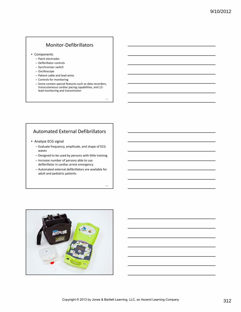
9/10/2012
312
Monitor‐Defibrillators
• Components– Patch electrodes
– Defibrillator controls
– Synchronizer switch
– Oscilloscope
– Patient cable and lead wires
– Controls for monitoring
– Some contain special features such as data recorders, transcutaneous cardiac pacing capabilities, and 12‐lead monitoring and transmission
934
Automated External Defibrillators
• Analyze ECG signal
– Evaluate frequency, amplitude, and shape of ECG waves
– Designed to be used by persons with little training
– Increase number of persons able to use defibrillator in cardiac arrest emergency
– Automated external defibrillators are available for adult and pediatric patients
935
936
Copyright © 2013 by Jones & Bartlett Learning, LLC, an Ascend Learning Company

9/10/2012
313
Automated External Defibrillators
• Attached to patient by two adhesive monitor‐defibrillator pads (electrodes) and connecting cables
• Have variety of features and controls
• Most units provide programmable modules, data recorders, and voice messages to operator
– All users should become familiar with automated external defibrillator device used in their system
– Follow recommendations of manufacturer
937
Automated External Defibrillators
• Fully automated defibrillator
– Requires only that operator attach defibrillation pads and turn on device
– Rhythm is analyzed in internal circuitry of automated external defibrillator
– If shockable rhythm detected, automated external defibrillator charges capacitors and delivers shock
938
Automated External Defibrillators
• Semiautomated defibrillator
– Requires operator to press an “analyze” button to interpret rhythm and “shock” button to deliver shock
– Operator presses shock control only when automated external defibrillator identifies shockable rhythm and “advises” operator to press shock button
939
Copyright © 2013 by Jones & Bartlett Learning, LLC, an Ascend Learning Company

9/10/2012
314
Automated External Defibrillators
• Automated external defibrillators have four safety features– Can analyze ECG waves
– Have built‐in filters that check for QRS‐like signals, radio transmission waves, 60‐cycle interference, and loose or poor electrode contact
– Most are programmed to detect spontaneous patient movements, continued heartbeat and blood flow, and movement of patient by others
– Make multiple evaluations of rhythm before making shock advisory or delivering shock
940
What type of patients will have these devices?
941
Biphasic Technology
• In past, defibrillation has used monophasic waveforms, in which current travels in only one direction, from positive pad to negative pad
– Require high energy to defibrillate patient effectively
– In fact, may deliver more energy than is needed for some patients
– These machines also require large batteries, energy storage capacitors, inductors, and large, high‐voltage mechanical devices
942
Copyright © 2013 by Jones & Bartlett Learning, LLC, an Ascend Learning Company

9/10/2012
315
Biphasic Technology
• Most newer automated external defibrillators and implantable defibrillators use biphasic waveform technology
– Predicts patient’s energy requirements by determining chest wall impedance
– Shock is delivered by current that travels in one direction, is stopped, then is reversed to travel in opposite direction
943
Biphasic Technology
• Allows for effective defibrillation to occur with lower energy for most patients
– Biphasic defibrillation of 115 and 130 J appears to be as effective as 200 and 360 J delivered with monophasic shocks
– Biphasic waveforms are more effective at lower energy than monophasic waveforms
– Thus automated external defibrillators (using smaller batteries) have become smaller, lighter, more durable
– Less expensive to manufacture
944
Defibrillation
• Delivery of electrical current through chest wall
– Purpose is to terminate ventricular fibrillation and pulseless ventricular tachycardia
– Shock depolarizes large mass of myocardial cells at once
– If about 75 percent of these cells are in resting state (depolarized) after shock is delivered, normal pacemaker may resume discharging
945
Copyright © 2013 by Jones & Bartlett Learning, LLC, an Ascend Learning Company

9/10/2012
316
Defibrillation
• Early defibrillation supported by following rationale– Most frequent initial rhythm in sudden cardiac arrest is ventricular fibrillation
– Most effective management for ventricular fibrillation is electrical defibrillation
– Probability of successful defibrillation decreases rapidly over time
– Ventricular fibrillation tends to convert to asystolewithin few minutes
946
Defibrillation
• Modern defibrillator
– Designed to deliver electrical shock via patches, or pads to patient’s chest
– Accepts electrical charge from battery source
• Stores charge in capacitor
• Releases current into patient in short, controlled burst (within 5 to 30 milliseconds)
947
Patch Electrodes
• Should be placed so heart (mainly ventricles) is in path of current, and distance between electrodes and heart is minimized
– Helps ensure adequate delivery of current through heart
– Bone is not good conductor
• For that reason, patches should not be placed over sternum
948
Copyright © 2013 by Jones & Bartlett Learning, LLC, an Ascend Learning Company

9/10/2012
317
949
Patch Electrodes
• AHA recommends one patch should be placed to right of upper sternum below right clavicle, and other to left of nipple in midaxillary line
– Anterior‐posterior position also acceptable
– Most manufacturers have adult and pediatric patches available
– Adult patches are usually 10 to 13 cm in diameter
– Pediatric patches are used for children less than 1 year of age, are 4.5 cm in diameter
950
Patch Electrodes
• Impedance
– Resistance to current by chest wall
– Determined by body size, bone structure, skin properties, underlying health conditions, other variables
– The greater resistance, the less current delivered
– Dry, unprepared skin has high impedance
• To reduce resistance, electrode patches are gelled
951
Copyright © 2013 by Jones & Bartlett Learning, LLC, an Ascend Learning Company

9/10/2012
318
Patch Electrodes
• Must prevent contact (bridging) between two conductive areas on chest wall
– If contact is made, superficial burns of skin may result
– Effective current also may bypass heart
– Even with proper technique and equipment, minor skin damage may still occur
952
Stored and Delivered Energy
• Electrical energy is commonly measured in joules (watt seconds)– 1 joule of electrical energy is product of 1 V (potential) x 1 A (current) x 1 second
– Delivered energy is about 80 percent of stored energy because of losses within circuitry of defibrillator and resistance to flow of current across chest wall
– As rule, 80 percent of stored energy approximates number of joules delivered to patient
953
Stored and Delivered Energy
• AHA currently recommends one initial defibrillation be attempted at 360 J monophasic or equivalent biphasic energy and delivered in succession
• Pediatric initial defibrillation generally is 2 J/kg– Followed by 4 J/kg if needed
• Biphasic defibrillator users: follow manufacturer recommendations to select defibrillation energy– Typically will be 120 J or 200 J
954
Copyright © 2013 by Jones & Bartlett Learning, LLC, an Ascend Learning Company

9/10/2012
319
Defibrillation Procedure
• Defibrillation procedure as recommended by AHA– Turn on defibrillator
– Select energy level at 360 J for monophasic defibrillators (or clinically equivalent biphasic energy)
– Set lead I, II, or III if monitor leads are used
– Position electrode patches on patient’s chest (sternum‐apex)
– Visually check monitor display and assess rhythm• Subsequent steps assume ventricular tachycardia/ventricular fibrillation is present
955
Defibrillation Procedure
• Defibrillation procedure as recommended by AHA (cont'd)– Announce to team members, “Charging defibrillator"
– Press “charge” button on defibrillator controls
– Continue CPR while defibrillator is charging
– When defibrillator is fully charged, state quickly in forceful voice following chant (or some suitable equivalent) before each shock
956
Defibrillation Procedure
• Defibrillation procedure as recommended by AHA (cont'd)
– “I am going to shock on three. One, I’m clear.”
• Check to make sure you are clear of contact with patient, stretcher, and equipment
957
Copyright © 2013 by Jones & Bartlett Learning, LLC, an Ascend Learning Company

9/10/2012
320
Defibrillation Procedure• Defibrillation procedure as recommended by AHA (cont'd)– “Two, you’re clear.”
• Make visual check to ensure that no one continues to touch patient or stretcher
• In particular, do not forget about person providing ventilations
• That person’s hands should not be touching ventilatoryadjuncts, including tracheal tube
• Turn O2 off or direct flow away from patient’s chest
– “Three, everybody’s clear.” • Check yourself one more time before pressing “shock” buttons
958
Defibrillation Procedure
• Defibrillation procedure as recommended by AHA (cont'd)
– Perform five cycles of CPR
– Check monitor
• If ventricular fibrillation/ventricular tachycardia remains, recharge defibrillator at once
– Shock at 360 J for monophasic defibrillators (or clinically equivalent biphasic energy level), repeating same verbal statements above
959
Operator and Personnel Safety
• Guidelines for safe defibrillator use– Make certain that all personnel are clear of patient, bed, and defibrillator before making defibrillation attempt
– Do not make contact with patient during discharge
– Do not discharge current over pacemaker or implantable cardioverter defibrillator generator or nitroglycerin paste
• Remove nitroglycerin patches before defibrillation
960
Copyright © 2013 by Jones & Bartlett Learning, LLC, an Ascend Learning Company

9/10/2012
321
Operator and Personnel Safety
• Guidelines for safe defibrillator use– Do not “open air” discharge defibrillator to cancel an unwanted charge
• Turn defibrillator off to “dump” charge
• In most models, changing energy setting dumps charge
– Treat equipment with respect, it is safe when used properly
– Routinely check defibrillator (including batteries) to make sure equipment is functioning properly
• Follow recommendations of manufacturer
961
What safety measure is still the responsibility of the
AED operator?
962
Special Environment Defibrillator Use• On occasion, patient requires defibrillation in special environment (e.g., in inclement weather)
– Operator guidelines and personnel safety always apply
– Additional precautions are taken in special situations
• In wet conditions, patient’s chest should be kept dry between defibrillator electrode sites
• Operator’s hands should be kept as dry as possible
• In rainstorm, safest to find shelter
963
Copyright © 2013 by Jones & Bartlett Learning, LLC, an Ascend Learning Company

9/10/2012
322
Special Environment Defibrillator Use• Depending on defibrillator and its equipment specifications, device may not be guaranteed to work properly in nonpressurized aircraft
– Some electrical interference may occur between radio equipment in aircraft and monitor‐defibrillator or vice versa
• Affected by distance and angle between defibrillator and radio equipment
964
Special Environment Defibrillator Use• Defibrillation with current equipment would be expected to be safe in all types of rotary aircraft used for emergency medical transport
– Medical crew should always inform pilot(s) when electrical therapy is being used
– Consult with pilot(s) to make sure flight instruments are well shielded from electromagnetic interference
965
Implantable Cardioverter Defibrillators
• Implantable cardioverter defibrillators (ICDs)
– Commonly used in patients at risk for recurrent, sustain ventricular tachycardia or fibrillation
– During implantation, various leads of ICD are fed through vessel (usually subclavian vein) into right ventricle or placed on epicardium
966
Copyright © 2013 by Jones & Bartlett Learning, LLC, an Ascend Learning Company

9/10/2012
323
967
Implantable Cardioverter Defibrillators
• Leads are tunneled to pulse generator in biphasic defibrillator device
– Device is placed surgically in left upper quadrant of abdomen
• Outline of generator usually can be felt or seen under patient’s skin
– Leads are used to
• Deliver shocks
• Monitor cardiac rhythm
• Sometimes pace the heart as needed if bradycardia occurs
968
Implantable CardioverterDefibrillators
• Work by monitoring patient’s cardiac rhythm, rate, and QRS complex morphology
– When monitored ventricular rate exceeds preprogrammed rate, implantable cardioverterdefibrillator delivers shock of about 6 to 30 J through patches to restore normal sinus rhythm
– Device requires 10 to 30 seconds to sense ventricular tachycardia or ventricular fibrillation and to charge capacitor before delivering shock
969
Copyright © 2013 by Jones & Bartlett Learning, LLC, an Ascend Learning Company

9/10/2012
324
Implantable Cardioverter Defibrillators
• If defibrillation does not restore normal sinus rhythm, implantable cardioverter defibrillator will charge again– Then it will deliver up to four shocks– Complete sequence of five shocks, if required, may take up to 2 minutes
– If tachycardia or fibrillation persists after five shocks, no further shocks are delivered
• Once slower rhythm is restored (i.e., sinus or idioventricular) for at least 35 seconds, device can deliver another series of up to five shocks if ventricular tachycardia or ventricular fibrillation recurs
970
Implantable Cardioverter Defibrillators
• Manage patients with implantable cardioverter defibrillators as if they did not have device
– Follow standard advanced cardiac life support protocols if patient is in cardiac arrest or in any other way medically unstable
971
Implantable Cardioverter Defibrillators
• AHA recommends guidelines when caring for patient with implantable cardioverterdefibrillator– If implantable cardioverter defibrillator discharges while rescuer is touching victim, rescuer may feel shock
• Shock will not be dangerous
• Personnel shocked by implantable cardioverterdefibrillators report sensations similar to contact with electrical current
972
Copyright © 2013 by Jones & Bartlett Learning, LLC, an Ascend Learning Company

9/10/2012
325
Implantable Cardioverter Defibrillators
• AHA recommends guidelines when caring for patient with implantable cardioverterdefibrillator
– Implantable cardioverter defibrillators are protected against damage from traditional transchest defibrillation shocks
• Require implantable cardioverter defibrillator readiness check after external defibrillation occurs
973
Implantable Cardioverter Defibrillators
• AHA recommends guidelines when caring for patient with implantable cardioverter defibrillator
– If ventricular fibrillation or ventricular tachycardia is present despite implantable cardioverter defibrillator, external shock should be given immediately
• Implantable cardioverter defibrillator likely has failed to defibrillate heart
• After initial series of shocks, implantable cardioverterdefibrillator will become operative again only if period of nonfibrillatory rhythm occurs to reset unit
974
Implantable Cardioverter Defibrillators
• AHA recommends guidelines when caring for patient with implantable cardioverter defibrillator
– Older implantable cardioverter defibrillator units use patch electrodes instead of leads
• If transthoracic shocks of up to 360 J fail to defibrillate patient with implantable cardioverter defibrillator, chest electrode positions should be changed immediately (e.g., anterior‐apex to anteroposterior)
• Electrodes cover portion of epicardial surface
• May reduce amount of current delivered to heart from transthoracic shocks
975
Copyright © 2013 by Jones & Bartlett Learning, LLC, an Ascend Learning Company

9/10/2012
326
Implantable Cardioverter Defibrillators
• AHA recommends guidelines when caring for patient with implantable cardioverterdefibrillator
– Older implantable cardioverter defibrillator units use patch electrodes instead of leads
• Transthoracic shocks should be repeated
• Different electrode positions could increase transthoracic current flow
• May facilitate defibrillation
976
Implantable Cardioverter Defibrillators
• Implantable cardioverter defibrillator can be deactivated and activated with magnet
– Patients with defibrillators should be kept away from strong magnets
• Will prevent accidental deactivation or reactivation of device
• Ability to use magnet to deactivate and reactivate many of these devices can be useful when unit is not working properly
• Use of handheld magnet to turn unit off or back on should be considered only with advice and under direction of physician
977
Left Ventricular Assist Device
• Battery‐operated pump that is sometimes implanted during heart surgery for people who are waiting for heart transplant (“bridge to transplant”)
– Used in some patients to allow weakened heart to recover
– Common left ventricular assist device (LVAD) is implanted in patient’s abdomen
978
Copyright © 2013 by Jones & Bartlett Learning, LLC, an Ascend Learning Company

9/10/2012
327
Left Ventricular Assist Device
• Catheter in pump pulls blood from weakened left ventricle and directs blood into aorta
– Takes stress off left ventricle
– Second catheter is brought out of abdominal wall to outside of body where it can be attached to pump's battery and control system
979
Left Ventricular Assist Device
• Modern LVADs are portable and are often used for weeks to months while waiting for donor heart to become available– Prescribed for patients who are terminally ill and ineligible for heart
• Can improve quality of life (“destination therapy”)
– Possible complications• Infection
• Internal bleeding
• Heart failure
• Mechanical malfunction
980
981
Copyright © 2013 by Jones & Bartlett Learning, LLC, an Ascend Learning Company

9/10/2012
328
Special Care Considerations
• Because LVAD assumes pumping function of left ventricle, patient may have no palpable pulse or measurable BP– Depends on pulsatility of specific device
– Doppler may be required for BP readings
– Patient and family are usually quite knowledgeable about LVAD and often are valuable resource
– Many patients with LVADs advise emergency service agencies so that personnel can be made aware of their special equipment needs
982
Special Care Considerations
• Due to location of LVAD and tubing connecting it to heart, device can be dislodged during CPR and cause severe bleeding
– Chest compressions to manage cardiac arrest are contraindicated in these patients
– Procedure for defibrillation and cardioversion will be specific to LVAD and manufacturer recommendations
983
Synchronized Cardioversion
• Used to terminate dysrhythmias other than ventricular fibrillation and pulseless ventricular tachycardia– Defibrillation (unsynchronized cardioversion) delivers shock on
operator’s command and with no regard as to where shock occurs in cardiac cycle
– In contrast, synchronized cardioversion is designed to deliver shock about 10 milliseconds after peak of R wave of cardiac cycle
• Avoids vulnerable relative refractory period of ventricles
• Synchronization may reduce energy required to end dysrhythmia
• May decrease chance of causing another dysrhythmia
984
Copyright © 2013 by Jones & Bartlett Learning, LLC, an Ascend Learning Company

9/10/2012
329
Synchronized Cardioversion
• When defibrillator is placed in synchronized mode, ECG displayed on oscilloscope shows marker denoting where in cardiac cycle energy will be discharged
– Marker should appear on R wave
• If it does not, select another lead
– Adjustment of ECG size may be needed if marker does not appear
985
Synchronized Cardioversion
• Procedure
– Consider sedation
– Turn on defibrillator (monophasic or biphasic)
– Attach monitor leads to patient (“white to right, red to ribs, what’s left over to the left shoulder”) and ensure proper display of patient’s rhythm
– Engage synchronization mode by pressing “sync” control button
– Look for markers on R waves indicating sync mode
986
Synchronized Cardioversion
• Procedure (cont'd)
– If necessary, adjust monitor gain until sync markers occur with each R wave
– Select appropriate energy level
– Position electrode patches on patient (sternum‐apex)
– Announce to team members: “Charging defibrillator”
– Press “charge” button on defibrillator
987
Copyright © 2013 by Jones & Bartlett Learning, LLC, an Ascend Learning Company

9/10/2012
330
Synchronized Cardioversion
• Procedure (cont'd)– When defibrillator is charged, begin final clearing chant
• State firmly in forceful voice following chant before each shock
– “I am going to shock on three. One, I’m clea.r”
• Check to make sure you are clear of contact with patient, stretcher, and equipment
– “Two, you’re clear.”
• Make visual check to ensure that no one continues to touch patient or stretcher
• In particular, do not forget about person providing ventilations
988
Synchronized Cardioversion
• Procedure (cont'd)
– When defibrillator is charged, begin final clearing chant
• That person’s hands should not be touching ventilatoryadjuncts, including tracheal tube
• Turn O2 off or direct flow away from patient’s chest– “Three, everybody’s clear.”
• Check yourself one more time before pressing “shock” button
989
Synchronized Cardioversion
• Procedure (cont'd)– Press “discharge” button
– Check monitor• If tachycardia persists, increase joules according to electrical cardioversion algorithm
– Reset sync mode after each synchronized cardioversion because most defibrillators default back to unsynchronized mode
• Default allows immediate shock if cardioversionproduces ventricular fibrillation
990
Copyright © 2013 by Jones & Bartlett Learning, LLC, an Ascend Learning Company

9/10/2012
331
Trancutaneous Cardiac Pacing
• Also known as external cardiac pacing
– Is effective emergency therapy for
• Bradycardia
• Complete heart block
• Asystole
• Suppression of some malignant ventricular tachydysrhythmias
– Have been recognized by AHA
– Used to treat bradycardia
991
Artificial Pacing
• Artificial pacemakers
– Deliver repetitive electrical currents to heart
– Act as substitute for natural pacemaker
• May have become blocked or dysfunctional
• Patient with severe sinus bradycardia, heart block, or idioventricular rhythm
– If capable of generating pulse with cardiac contractions, may respond to external pacing device and produce perfusing pulse
992
Artificial Pacing
• Sinus bradycardia also may be paced
– Responds well to atropine
• Most patients in cardiac arrest do not respond to pacing because heart does not receive adequate perfusion, cannot achieve effective contractions
993
Copyright © 2013 by Jones & Bartlett Learning, LLC, an Ascend Learning Company

9/10/2012
332
994
Artificial Pacing
• Two modes
– Transcutaneous cardiac pacing are nondemand(asynchronous) pacing and demand pacing
– Most devices provide both modes
– Asynchronous pacemaker delivers timed electrical stimuli at selected rate
• Occurs regardless of patient’s own cardiac activity
995
Artificial Pacing
• Asynchronous pacing
– Used less often than demand pacers
– May discharge during vulnerable period of cardiac cycle (producing R‐on‐T phenomenon)
– Generally used only as last resort, usually in asystole
– Can be used when artifact on ECG interferes with ability of machine to sense patient’s own heart beat
996
Copyright © 2013 by Jones & Bartlett Learning, LLC, an Ascend Learning Company

9/10/2012
333
Artificial Pacing
• Asynchronous pacing
– Used in overriding high heart rates in tachydysrhythmias (e.g., torsades de pointes)
• Should be attempted only if other means of controlling dysrhythmia have failed
• May be limited by design of machine
997
Artificial Pacing
• Demand pacing
– Senses patient’s QRS complex
– Pacemaker delivers electrical stimuli only when needed
– Much safer to apply than nondemand mode
– When pacemaker senses intrinsic beat, pacemaker is inhibited
– If no beats are sensed, pacemaker delivers pacing stimuli at selected rate
998
Artificial Pacing
• Demand pacing
– Device usually is set to discharge at rate between 70 to 80 beats/minute beginning with 50 mA
– Charge then is increased in increments beginning with 0 mA of electricity, until electrical and mechanical capture is achieved
– Patient’s clinical condition (BP, level of consciousness, skin color, temperature) improves at this point
999
Copyright © 2013 by Jones & Bartlett Learning, LLC, an Ascend Learning Company

9/10/2012
334
Artificial Pacing
• Ensure that each pacemaker spike on oscilloscope is followed by QRS complex
– If not, current should be increased gradually until there is consistent capture
– Motion artifact often makes ECG confirmation of electrical capture difficult
– Only accurate method of monitoring mechanical function of heart produced by pacing device is presence of pulse with each QRS complex
1000
Artificial Pacing
• Must constantly monitor patient’s pulse
– Assess patient’s pulse rate and BP on patient’s right side
– Will help to minimize interference from muscle artifact
1001
Transcutaneous Pacing
• Procedure
– Consider sedation
– Gather required equipment
– Explain procedure to patient
– Connect patient to cardiac monitor and obtain rhythm strip
– Obtain baseline vital signs
1002
Copyright © 2013 by Jones & Bartlett Learning, LLC, an Ascend Learning Company

9/10/2012
335
What should you tell the patient before initiating TCP?
1003
Transcutaneous Pacing
• Procedure
– Attach limb leads and apply pacing electrodes
• Often defibrillation position is used when same pads can be used for defibrillation and pacing
• Pads can also be placed in anterior‐posterior position (left of lower sternum and just below left scapula)
– Select pacing mode
– Select pacing rate (usually 80 beats/minute)
• Set current (begin with 0 mA and then increase current until ventricular capture is obtained)
1004
Transcutaneous Pacing
• Procedure
– Activate pacemaker, observing patient and ECG
– Obtain rhythm strips as appropriate
– Continue monitoring patient and anticipate further therapy
1005
Copyright © 2013 by Jones & Bartlett Learning, LLC, an Ascend Learning Company

9/10/2012
336
TCP Indications and Contraindications
• Indications in prehospital setting are
– Symptomatic bradycardia
– Heart block associated with reduced cardiac output that is unresponsive to atropine
– Pacemaker failure
1006
TCP Indications and Contraindications
• Contraindications
– Rarely is effective in cardiac arrest
– Ineffective in pulseless electrical activity unless underlying cause of pulseless electrical activity is corrected
– Not advised in patients with open wounds or burns to chest or for patients in wet environment
1007
Why should patient movement be minimized
during TCP?
1008
Copyright © 2013 by Jones & Bartlett Learning, LLC, an Ascend Learning Company

9/10/2012
337
Electrode Placement
• Proper electrode placement is key in providing effective external pacing
– Defibrillation position often used when same pads can be used for defibrillation and pacing
– Can apply negative (anterior) electrode to left of sternum
• Should be centered as close as possible to point of maximal cardiac impulse
1009
Electrode Placement
• Positive (posterior) electrode is placed directly behind anterior electrode, just below left scapula– In rare cases, posterior placement cannot be used
– Positive electrode can then be placed in line with patient’s left nipple at midaxillary line
– Anterior‐anterior placement may produce pronounced chest muscle twitching
– Electrodes should be applied to clean, dry skin without localized trauma or infection
1010
Why should alcohol or benzoin not be used under
defibrillator pads?
1011
Copyright © 2013 by Jones & Bartlett Learning, LLC, an Ascend Learning Company

9/10/2012
338
Electrode Placement
• Conscious patient most likely will experience some pain and discomfort during transcutaneous cardiac pacing
– Related directly to intensity of muscle contractions and amount of applied current
– Analgesia or sedation of patient should be provided
1012
Cardiac Arrest and Sudden Death
• Patients who cannot be resuscitated in prehospital setting rarely survive
– Even if resuscitated temporarily in emergency department
– Patient’s best chance for survival is to have rapid and appropriate treatment in field
1013
Cardiac Arrest and Sudden Death
• If procedures needed for appropriate care cannot be completed quickly, maintain ventilation and compressions and rapid transport
– Initial defibrillation should always be attempted as soon as possible
• Prolonged field resuscitation in face of difficulties with IV/IO access and intubation almost always is destined to fail
1014
Copyright © 2013 by Jones & Bartlett Learning, LLC, an Ascend Learning Company

9/10/2012
339
Patient Care After Spontaneous Circulation Return
• Some patients will survive cardiac arrest and will have return of spontaneous circulation (ROSC)
– Principal objective of postresuscitation care is reestablishment of effective perfusion of organs and tissues
– Ideally, patient will be alert and awake
1015
Patient Care After Spontaneous Circulation Return
• May be comatose but have full potential for recovery with good neurologic outcome
– Research is underway to improve survival rate of patients with ROSC who remain comatose after resuscitation
– One method being studied through clinical trials is temperature regulation
1016
Induced Hypothermia
• In 2005, AHA issued recommendations and guidelines for inducing mild hypothermia in comatose survivors of cardiac arrest
– When patient is resuscitated, reperfusion sets off series of chemical reactions that can continue for up to 24 hours, possibly causing significant inflammation in brain
1017
Copyright © 2013 by Jones & Bartlett Learning, LLC, an Ascend Learning Company

9/10/2012
340
Induced Hypothermia
• Inducing mild hypothermia decreases– Intracranial pressure
– Cerebral metabolic rate
– Brain's demand for O2 consumption
• Suppresses many chemical reactions associated with reperfusion injury– Free radical production
– Excitatory amino acid release
– Calcium shifts
1018
Induced Hypothermia
• Inducing hypothermia in unconscious patient after ROSC is practiced by some EMS agencies
– More often initiated in emergency department by way of two cooling methods: internal cooling and external cooling
1019
Induced Hypothermia
• With internal cooling, cooling catheter is inserted into femoral vein and positioned in inferior vena cava or into central vein into superior vena cava
– Once in place, cool or warm fluid circulates inside catheter, cooling or warming patient’s blood to maintain mild hypothermia
1020
Copyright © 2013 by Jones & Bartlett Learning, LLC, an Ascend Learning Company

9/10/2012
341
Induced Hypothermia
• With external cooling, special pads are placed directly on patient’s skin around chest and thighs– Temperature‐controlled water is then distributed through pads via electronic monitoring device
– When core body temperature of 91 to 92ºF (28 to 32ºC) has been established, patient transferred to intensive care
– After 12 to 24 hours, patient is slowly rewarmed to normal core body temperature
1021
Induced Hypothermia
• Current recommendations– Therapeutic hypothermia induced in comatose patient within 30
to 60 minutes following resuscitation from cardiac arrest when initial rhythm was ventricular fibrillation
– Other arrest rhythms may benefit from cooling as well
– Associated with improved functional recovery and neurological outcome
– Other patient groups who may benefit from induced hypothermia are being studied
– These groups include victims of severe stoke and adults with head injury
1022
Resuscitation Termination
• According to AHA, health care professionals are expected to provide basic life support and advanced cardiac life support as part of professional duty to respond, with following exceptions:– When person lies dead, with obvious clinical signs of irreversible death
– When attempts to perform CPR would place rescuer at risk of physical injury
– When patient or surrogate has indicated that resuscitation is not desired
1023
Copyright © 2013 by Jones & Bartlett Learning, LLC, an Ascend Learning Company

9/10/2012
342
Resuscitation Termination
• AHA has advised that termination of resuscitative efforts in prehospital setting follow rules established by local EMS system
– Established by medical direction
– Rules should include consideration for advance directives and no CPR orders
– If at any time paramedics are presented with an advance directive that indicates patient should not be resuscitated, they should follow established protocol or immediately consult with medical direction
1024
Resuscitation Termination
• Resuscitation (in some cases) may be stopped appropriately if asystole persists
– Termination of resuscitative efforts should occur only after reviewing quality of resuscitation attempt
1025
Resuscitation Termination
• Questions about resuscitation attempt to be considered
– Has there been an adequate trial of basic life support and advanced cardiac life support?
– Has patient’s trachea been secured?
– Has effective ventilation been provided?
– If ventricular fibrillation was present, was patient defibrillated?
– Was IV/IO access obtained?
1026
Copyright © 2013 by Jones & Bartlett Learning, LLC, an Ascend Learning Company

9/10/2012
343
Resuscitation Termination
• Questions about resuscitation attempt to be considered – Were epinephrine and atropine administered?
– Have reversible causes been ruled out?
– Has asystole been documented continuously for more than 5 to 10 minutes after all of foregoing have been accomplished?
– If capnography is available, is capnography reading less than 10 mm Hg in spite of good compressions?
1027
Resuscitation Termination
• Presence of unusual clinical features may be indicators that continued resuscitation is appropriate– Drowning or profound hypothermia
– Young age
– Toxins or electrolyte abnormalities
– Drug overdose
• Termination of resuscitation in prehospitalsetting should be guided by medical direction
1028
Resuscitation Termination
• Process for terminating resuscitation in field varies by protocol– Follow guidelines set by medical direction
– When termination is considered to be appropriate, contact medical direction to convey following information
• Medical condition of patient
• Known causes of arrest
• Any treatment provided
• Family’s appraisal of situation and any resistance or uncertainty
1029
Copyright © 2013 by Jones & Bartlett Learning, LLC, an Ascend Learning Company

9/10/2012
344
Resuscitation Termination
• While gathering and giving this information to medical direction, maintain ongoing documentation of event
– This should include continuous ECG monitoring
– Documentation will help in review of call
– Review usually is performed for quality assurance in most EMS systems
1030
What benefit is there to the community, and to the patient’s family, when resuscitation is terminated in the
field following all appropriate guidelines?
1031
Special Considerations
• In addition to needs of patient, grief support for family must be considered
– Support services vary by agency
– Often paramedic (or other EMS personnel) will be assigned to stay with family for period of time
– At times, community agency referral will be arranged
1032
Copyright © 2013 by Jones & Bartlett Learning, LLC, an Ascend Learning Company

9/10/2012
345
What resources can you contact to help the family after you have terminated resuscitation in the home?
1033
Special Considerations
• Law enforcement officers may have additional duties at scene as part of their professional role
– May include on‐scene determination that patient be assigned to medical examiner
– May occur when death or event is suspicious, or when patient’s private physician refuses or hesitates to sign death certificate
– Be familiar with local and state laws related to reporting and disposition of patient remains after out‐of‐hospital death
1034
Summary
• Persons at high risk for cardiovascular disease include males, older adults and those with diabetes, hypertension, a family history of premature cardiovascular disease, and prior myocardial infarction
• Prevention strategies include community educational programs in nutrition, cessation of smoking (smoking prevention for children), and screening for hypertension and high cholesterol
1035
Copyright © 2013 by Jones & Bartlett Learning, LLC, an Ascend Learning Company

9/10/2012
346
Summary
• Left coronary artery carries about 85 percent of blood supply to myocardium– Right coronary artery carries the rest– Pumping action of heart is product of rhythmic, alternate contraction and relaxation of atria and ventricles
– Stroke volume is amount of blood ejected from each ventricle with one contraction
• Depends on preload, afterload, and myocardial contractility
– Cardiac output is amount of blood pumped by each ventricle per minute
1036
Summary
• In addition to intrinsic control of body in regulating heart, extrinsic control by parasympathetic and sympathetic nerves of autonomic nervous system is a major factor influencing heart rate, conductivity, and contractility
– Sympathetic impulses cause adrenal medulla to secrete epinephrine and norepinephrine into blood
1037
Summary
• Major electrolytes that influence cardiac function are calcium, potassium, sodium, and magnesium– Electrical charge (potential difference) between inside and outside
of cells is expressed in millivolts
– When cell is in resting state, electrical charge difference is referred to as resting membrane potential
– Specialized sodium–potassium exchange pump actively pumps sodium ions out of cell, also pumps potassium ions into cell
– Cell membrane appears to have individual protein‐lined channels, which allow for passage of specific ion or group of ions
1038
Copyright © 2013 by Jones & Bartlett Learning, LLC, an Ascend Learning Company

9/10/2012
347
Summary
• Nerve and muscle cells are capable of producing action potentials
– Property is known as excitability
– Action potential at any point on cell membrane stimulates excitation process
• Process is spread down length of cell and is conducted across synapses from cell to cell
1039
Summary
• Contraction of cardiac and skeletal muscle is believed to be activated by calcium ions, resulting in binding between myosin and actin myofilaments
• Conduction system of heart is composed of two nodes and conducting bundle
– Sinoatrial node
– Atrioventricular node
1040
Summary
• Parasympathetic stimulation by vagus nerve affects primarily SA and AV node causing heart to slow
– Sympathetic stimulation increases heart rate and contractility
• ECG represents electrical activity of heart
– ECG is generated by depolarization and repolarization of atria and ventricles
1041
Copyright © 2013 by Jones & Bartlett Learning, LLC, an Ascend Learning Company

9/10/2012
348
Summary
• Routine monitoring of cardiac rhythm in prehospital setting usually is obtained in lead II or V1
– Best leads to monitor for dysrhythmias because they allow visualization of P waves
– Paper used to record ECGs is standardized
• Allows comparative analysis of an ECG wave
1042
Summary
• Normal ECG consists of P wave, QRS complex, and T wave– P wave is first positive deflection on ECG and represents atrial depolarization
– P‐R interval is time it takes for electrical impulse to be conducted through atria and AV node up to instant of ventricular depolarization
– QRS complex represents ventricular depolarization
– ST segment represents early part of repolarizationof right and left ventricles
1043
Summary
• Normal ECG consists of P wave, QRS complex, and T wave• T wave represents repolarization of ventricular myocardial cells
– Repolarization occurs during last part of ventricular systole
• Q‐T interval is period from beginning of ventricular depolarization (onset of QRS complex) until end of ventricular repolarization or end of T wave
1044
Copyright © 2013 by Jones & Bartlett Learning, LLC, an Ascend Learning Company

9/10/2012
349
Summary
• Steps in ECG analysis include analyzing QRS complex, P waves, rate, rhythm, and P‐R interval
• Dysrhythmias originating in SA node include sinus bradycardia, sinus tachycardia, sinus dysrhythmia, and sinus arrest
– Most sinus dysrhythmias are result of increases or decreases in vagal tone
1045
Summary
• Dysrhythmias originating in atria include wandering pacemaker, premature atrial complexes, paroxysmal supraventricular tachycardia, atrial flutter, and atrial fibrillation
– Common causes of atrial dysrhythmias are ischemia, hypoxia, and atrial dilation caused by congestive heart failure or mitral valve abnormalities
1046
Summary
• When SA node and atria cannot generate electrical impulses needed to begin depolarization because of factors such as hypoxia, ischemia, myocardial infarction, and drug toxicity, the AV node or area surrounding AV node may assume role of secondary pacemaker
– Dysrhythmias originating in AV junction include premature junctional contractions, junctional escape complexes or rhythms, and accelerated junctional rhythm
1047
Copyright © 2013 by Jones & Bartlett Learning, LLC, an Ascend Learning Company

9/10/2012
350
Summary
• Ventricular dysrhythmias pose threat to life
– Ventricular rhythm disturbances generally result from failure of atria, atrioventricular junction, or both to initiate electrical impulse
– May result from enhanced automaticity or reentry phenomena in ventricles
– Dysrhythmias originating in ventricles include ventricular escape complexes or rhythms, premature ventricular complexes, ventricular tachycardia, ventricular fibrillation, asystole, and artificial pacemaker rhythm
1048
Summary
• 12‐lead ECG can be used to help identify changes relative to myocardial ischemia, injury, and infarction; distinguish ventricular tachycardia from supraventricular tachycardia; determine electrical axis and presence of fascicular blocks; and determine presence of bundle branch blocks
1049
Summary
• Partial delays or full interruptions in cardiac electrical conduction are called heart blocks– Causes include AV junctional ischemia, AV junctionalnecrosis, degenerative disease of the conduction system, and drug toxicity
– Dysrhythmias that are disorders of conduction are first‐degree AV block, type I second‐degree AV block (Wenckebach), type II second‐degree AV block, third‐degree AV block, disturbances of ventricular conduction, pulseless electrical activity, and preexcitation (Wolff‐Parkinson‐White) syndrome
1050
Copyright © 2013 by Jones & Bartlett Learning, LLC, an Ascend Learning Company

9/10/2012
351
Summary
• Common chief complaints of patient with cardiovascular disease include chest pain or discomfort, including shoulder, arm, neck, or jaw pain or discomfort; dyspnea; syncope; and abnormal heartbeat or palpitations– Paramedics should ask patients suspected of having cardiovascular disorder whether they take prescription medications, especially cardiac drugs
– Paramedics should ask whether patients are being treated for any serious illness as well
1051
Summary
• Common chief complaints of patient with cardiovascular disease include chest pain or discomfort, including shoulder, arm, neck, or jaw pain or discomfort; dyspnea; syncope; and abnormal heartbeat or palpitations– Paramedics should ask whether patients have a history of myocardial infarction, angina, heart failure, hypertension, diabetes, or chronic lung disease
– In addition, paramedics should ask whether patients have any allergies or have other risk factors for heart disease
1052
Summary
• After performing initial assessment of patient with cardiovascular disease, paramedic should look for skin color, jugular venous distention, and the presence of edema or other signs of heart disease
– Paramedic should listen for lung sounds, heart sounds, and carotid artery bruit
– Paramedic should feel for edema, pulses, skin temperature, and moisture
1053
Copyright © 2013 by Jones & Bartlett Learning, LLC, an Ascend Learning Company

9/10/2012
352
Summary
• Atherosclerosis is disease process characterized by progressive narrowing of lumen of medium and large arteries
– Atherosclerosis has two major effects on blood vessels
– First, disease disrupts the intimal surface
• Causes loss of vessel elasticity and increase in thrombogenesis
– Second, atheroma reduces diameter of the vessel lumen
• Decreases blood supply to tissues
1054
Summary
• Angina pectoris is symptom of myocardial ischemi– Angina is caused by imbalance between myocardial oxygen supply and demand
– Prehospital management includes placing patient at rest, administering oxygen, initiating intravenous therapy, administering nitroglycerin and possibly morphine, monitoring the patient for dysrhythmias, and transporting patient for physician evaluation
1055
Summary
• Acute myocardial infarction occurs when a coronary artery is blocked and blood does not reach an area of heart muscle– Results in ischemia, injury, and necrosis to the area of myocardium supplied by the affected artery
– Death caused by myocardial infarction usually results from lethal dysrhythmias (ventricular tachycardia, ventricular fibrillation, and cardiac standstill), pump failure (cardiogenic shock and congestive heart failure), or myocardial tissue rupture (rupture of the ventricle, septum, or papillary muscle)
1056
Copyright © 2013 by Jones & Bartlett Learning, LLC, an Ascend Learning Company

9/10/2012
353
Summary
• Acute MI– Some patients, particularly those in older age groups, have only symptoms of dyspnea, syncope, or confusion
• Substernal chest pain is usually present in patients with acute MI (70 to 90 percent of patients)
– ST segment elevation 1 mV or more in at least two side‐by‐side ECG leads indicates an acute MI
• Some patients infarct without ST segment elevation changes
– Other conditions also can produce ST segment elevation
1057
Summary
• Acute MI– Prehospital management should include placing patient at rest; administering oxygen at 4 L per minute via nasal cannula; frequently assessing vital signs and breath sounds; initiating an intravenous line with normal saline or lactated Ringer’s solution to keep the vein open; monitoring for dysrhythmias; administering medications such as nitroglycerin, morphine, and aspirin; and screening for risk factors for fibrinolytic therapy
1058
Summary
• Left ventricular failure occurs when left ventricle fails to function as effective forward pump
– Causes back‐pressure of blood into pulmonary circulation
• May lead to pulmonary edema
– Emergency management is directed at decreasing venous return to heart, improving myocardial contractility, decreasing myocardial oxygen demand, improving ventilation and oxygenation, and rapidly transporting patient to medical facility
1059
Copyright © 2013 by Jones & Bartlett Learning, LLC, an Ascend Learning Company

9/10/2012
354
Summary
• Right ventricular failure occurs when right ventricle fails as a pump
– Causes back‐pressure of blood into systemic venous circulation
– Right ventricular failure is not usually medical emergency in itself unless it is associated with pulmonary edema or hypotension
1060
Summary
• Cardiogenic shock is most extreme form of pump failure
– Usually caused by extensive MI
– Even with aggressive therapy, cardiogenic shock has mortality rate of 70 percent or higher
– Patients in cardiogenic shock need rapid transport to medical facility
1061
Summary
• Cardiac tamponade is defined as impaired filling of heart caused by increased pressure in pericardial sac
• Abdominal aortic aneurysms are usually asymptomatic– Signs and symptoms will signal impending or active rupture
– If vessel tears, bleeding initially may be stopped by retroperitoneal tissues
– Patient may be normotensive on arrival of EMS
– If rupture opens into peritoneal cavity, massive fatal hemorrhage may follow
1062
Copyright © 2013 by Jones & Bartlett Learning, LLC, an Ascend Learning Company

9/10/2012
355
Summary• Acute dissection is most common aortic catastrophe– Any area of the aorta may be involved– In 60 to 70 percent of cases, site of dissecting aneurysm is in ascending aorta, just beyond takeoff of left subclavian artery
– Signs and symptoms depend on site of intimal tear• Also depend on extent of dissection
– Goals of managing suspected aortic dissection in prehospital setting are relief of pain and immediate transport to medical facility
1063
Summary
• Acute arterial occlusion is sudden blockage of arterial flow– Occlusion most commonly is caused by trauma, embolus, or thrombosis
– Most common sites of embolic occlusion are abdominal aorta, common femoral artery, popliteal artery, carotid artery, brachial artery, mesenteric artery
– Location of ischemic pain is related to site of occlusion
1064
Summary
• Noncritical peripheral vascular conditions include varicose veins, superficial thrombophlebitis, and acute deep vein thrombosis
– Of these conditions, deep vein thrombosis is only one that can cause a life‐threatening problem
– This problem is pulmonary embolus
1065
Copyright © 2013 by Jones & Bartlett Learning, LLC, an Ascend Learning Company

9/10/2012
356
Summary
• Hypertension often is defined by resting BP that is consistently greater than 140/90 mm Hg
– Chronic hypertension has adverse effect on heart and blood vessels
– Requires heart to perform more work than normal
• Leads to hypertrophy of cardiac muscle and left ventricular failure
– Conditions associated with chronic, uncontrolled hypertension are cerebral hemorrhage and stroke, myocardial infarction, and renal failure
1066
Summary
• Hypertensive emergencies are conditions in which BP increase leads to significant, irreversible end‐organ damage within hours if not treated
– Organs most likely to be at risk are brain, heart, and kidneys
– As a rule, diagnosis is based on altered end‐organ function and rate of rise in BP, not on level of BP
1067
Summary
• Valvular heart disease may occur as result of infection or be related to heart disease– When one or more of valves become narrowed, hardened, or thickened (stenotic), valves do not open or close completely
• As a result, blood does not flow with proper force or direction
• Infectious heart disease includes endocarditis, pericarditis and myocarditis– Complications can be severe and may include heart failure
1068
Copyright © 2013 by Jones & Bartlett Learning, LLC, an Ascend Learning Company

9/10/2012
357
Summary• Cardiomyopathy is alteration or weakness of heart muscle and
can cause heart failure or sudden death
• Congenital heart abnormalities are defects in structure of heart present at birth
– Causes include heredity and maternal ingestion of alcohol/drugs
– Defects vary widely and can include abnormal structure of the heart, holes in the heart, transposed or narrowed blood vessels
– Signs and symptoms vary widely
– Some of conditions are fatal, others treated with surgery
1069
Summary• Basic cardiac life support helps to maintain circulation and respiration
of victim of cardiac arrest
– Continued until advanced cardiac life support is available
– Two mechanisms responsible for blood flow during CPR
• Direct compression of heart between sternum and spine, which increases pressure within ventricles to provide small, but critical amount of blood flow to lungs and body organs.
• Increased intrathoracic pressure transmitted to all intrathoracic vascular structures, which creates an intrathoracic‐to‐extrathoracic pressure gradient, causing blood to flow out of thorax
• Number of mechanical devices provide external chest compression, others provide chest compression with ventilation in cardiac arrest patient
1070
Summary
• Cardiac monitor‐defibrillators are classified as manual or automated external defibrillators
– Defibrillation is delivery of electrical current through chest wall
• Purpose is to terminate ventricular fibrillation and certain other nonperfusing rhythms
1071
Copyright © 2013 by Jones & Bartlett Learning, LLC, an Ascend Learning Company

9/10/2012
358
Summary
• Implantable cardioverter defibrillators work by monitoring patient’s cardiac rhythm
– When monitored ventricular rate exceeds preprogrammed rate, implantable cardioverterdefibrillator delivers shock of about 6 to 30 J through patches
• Is attempt to restore normal sinus rhythm
1072
Summary
• Synchronized cardioversion is designed to deliver shock about 10 milliseconds after peak of R wave of cardiac cycle
– Device avoids relative refractory period
– Synchronization may reduce amount of energy needed to end dysrhythmia
– May decrease chances of causing another dysrhythmia
1073
Summary
• Transcutaneous cardiac pacing is effective emergency therapy for bradycardia, complete heart block, and suppression of some malignant ventricular dysrhythmias
– Proper electrode placement is important for effective external pacing
1074
Copyright © 2013 by Jones & Bartlett Learning, LLC, an Ascend Learning Company

9/10/2012
359
Summary
• Patients who cannot be resuscitated in prehospital setting rarely survive
– Is the case even if they are resuscitated temporarily in emergency department
– Cessation of resuscitative efforts in prehospitalsetting should follow system‐specific criteria established by medical direction
1075
Questions?
1076
Copyright © 2013 by Jones & Bartlett Learning, LLC, an Ascend Learning Company