Embed Size (px)
Citation preview
Chapter 15
Injuries to the Thigh, Leg, and Knee
In Your Notebooks:
• Based on what you already know about the bony anatomy of the lower leg, write down 2 of the 4 bones that make up the knee joint.

Anatomy Review
Bones of the Region
Femur
Patella
Tibia
Fibula
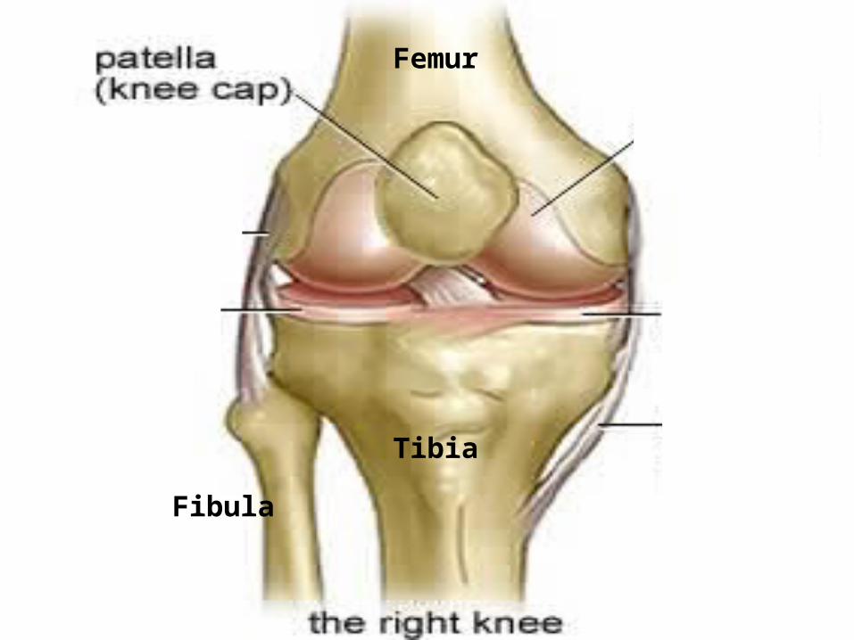
Femur
Fibula
Tibia
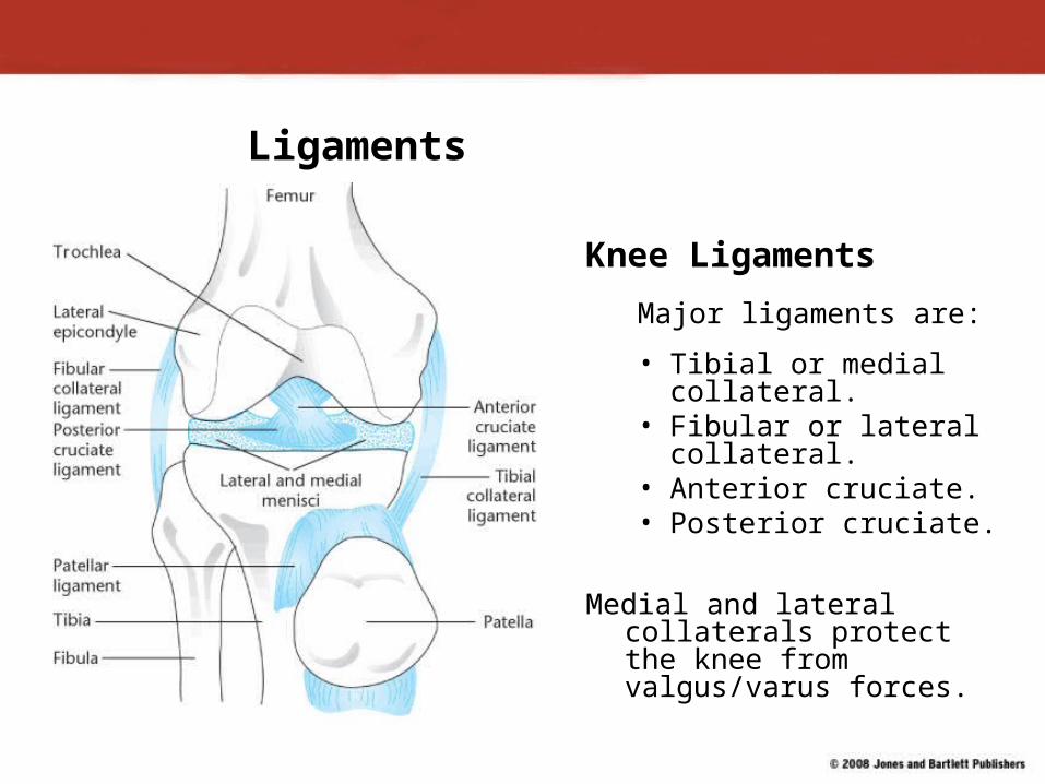
Ligaments
Knee Ligaments
Major ligaments are:
• Tibial or medial collateral.
• Fibular or lateral collateral.
• Anterior cruciate.• Posterior cruciate.
Medial and lateral collaterals protect the knee from valgus/varus forces.
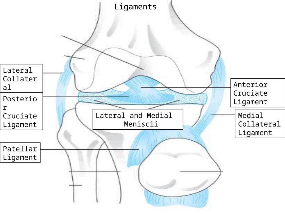
Lateral Collateral Ligament
Medial Collateral Ligament
Ligaments
Posterior Cruciate Ligament
Patellar Ligament
Anterior Cruciate Ligament
Lateral and Medial Meniscii

With a partner define and draw a picture of the following terms on a clean sheet of
paper:
• Knee Flexion:• Knee Extension:• Thigh Adduction:• Thigh Abduction:

In Your Notebooks:
• Explain the difference between thigh abduction and adduction.
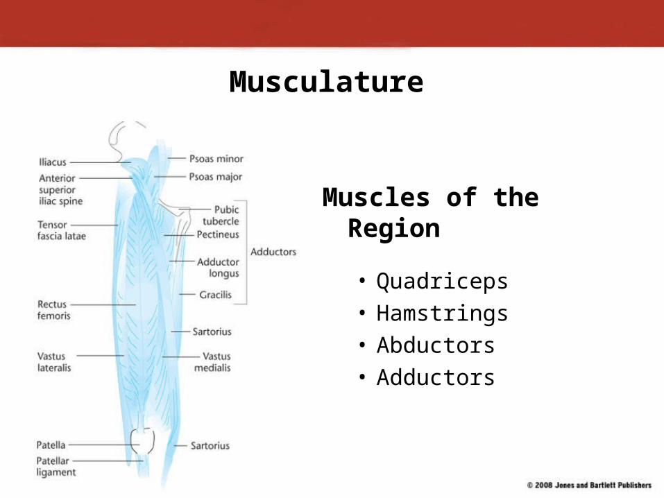
Musculature
Muscles of the Region
• Quadriceps• Hamstrings• Abductors• Adductors
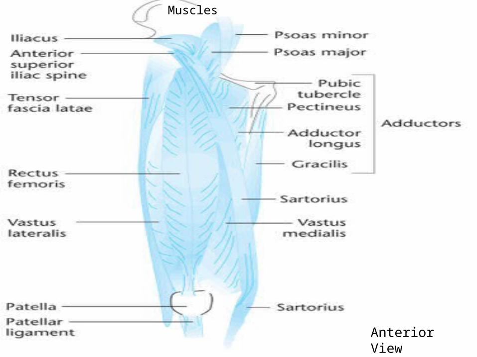
Muscles
Anterior View
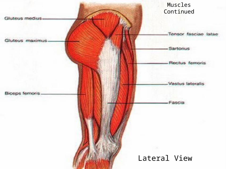
Muscles Continued
Lateral View

Muscles Continued
Posterior View
In Your Notebooks…
• List the four individual Quadricep Muscles

View of Leg Muscles
• Picture of View of Leg Muscles
1.
View of Leg Muscles
• Picture of View of Leg Muscles
• Label Drawn Muscles
2.
View of Leg Muscles
• Picture of View of Leg Muscles
• Label Drawn Muscles
• List the ACTION of each muscle
3.
View of Leg Muscles
• Picture of View of Leg Muscles
• Label Drawn Muscles
• List the ACTION of each muscle
• Look for Mistakes & Make Poster Perfect!
4.

On a clean sheet of paper:
1.The 4 bones that make up the knee joint
2.The 5 ligaments of the Knee ( do not abbreviate)
3.The 4 muscles that make up the quadriceps
4.The action of the quadriceps
5.The 3 muscles that make up the hamstrings
6.The action of the hamstrings
7.The 2 adductors muscles I wanted you to know
8.The 2 abductors muscles I wanted you to know
9.Where the quadriceps are located
10.Where the hamstrings are located
11.Where the adductors are located
12.Where the abductors are located
13.What are the disc like structures inside the knee joint called?

In Your Notebooks:• What is the function of the Meniscii?
( 4 things)

Meniscus
• There are two semicircular fibrocartilaginous disks in the knee known as the menisci.
• These disks are located in the space between the tibia and femur.
• Responsible for lubrication and nourishment of the knee joint, weight distribution, and assistance with joint biomechanics.
Common Sports Injuries
Fractures of the Femur and/or Patella
• Femoral fractures result from an extremely traumatic event.
• These injuries may also be in the form of a stress fracture, especially in the femoral neck region.
• Patellar fractures almost always occur as a result of a traumatic event.

Fractures of the Femur and/or Patella
• In the adolescent, femoral fractures may include slipped capital epiphysis injuries.
• In the adult, fractures of the femoral neck may result in avascular necrosis of the femoral head.• This injury results from disrupted blood
supply to the articular cartilage on the femoral head.
Fractures of the Femur and/or Patella (cont.)
Signs and symptoms include:
• Pain at the injury site.• Difficulty walking on the affected leg.• Swelling and/or deformity. Athlete’s report of having suffered
a traumatic event.• Athlete may report a pop or snap at time of injury.
The injury needs to be evaluated by a physician. Avascular necrosis is a serious complication.
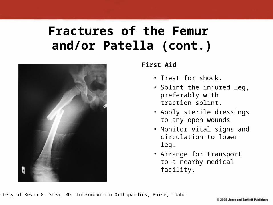
Fractures of the Femur and/or Patella (cont.)
First Aid
• Treat for shock.• Splint the injured leg,
preferably with traction splint.
• Apply sterile dressings to any open wounds.
• Monitor vital signs and circulation to lower leg.
• Arrange for transport to a nearby medical facility.
Courtesy of Kevin G. Shea, MD, Intermountain Orthopaedics, Boise, Idaho
Dislocation of the Knee or Tibiofemoral Joint
Dislocation of the knee or the tibiofemoral joint can compromise blood flow to the lower leg.
Signs and symptoms include:• Extreme pain.• Dislocation of the joint.
First Aid• The injury must be splinted.• Refer athlete to the nearest medical facility.

Soft Tissue Injuries to the Thigh
• These injuries usually result from direct contact with an opponent or self-inflicted muscle strain.
• Myositis ossificans traumatica may develop.
Signs and symptoms of a muscle contusion include:• History of forceful impact to the area and a
feeling of tightness.• Swelling may occur in affected area.• Inability to forcibly contract the muscle.• Difficulty walking with affected leg.
Muscular Strains to the Thigh
Hamstrings and adductor muscles are most likely to sustain strains.
• Strains to adductor muscles are called “groin pulls.” • Hamstrings usually are weaker and more
susceptible to strains than quadriceps.• Groin injuries take a long time to heal. • Stretching is a part of recovery program.
Muscular Strains to the Thigh (cont.)
Signs and symptoms include:
• A sharp pain in the affected muscle.• Swelling and redness in the immediate area.• Muscle weakness.• Inability to contract the muscle forcibly.• Discoloration of the area.• A defect is visible in severe cases.
Muscular Strains to the Thigh (cont.)
First Aid
• Apply ice and compression.• Athlete should rest and, if necessary, use crutches.• Obtain a medical evaluation of the injury.
Patellofemoral Joint Injuries
Acute and chronic injuries can affect patellofemoral joint. Such injuries can be debilitating and must be treated.
Osteochondritis dissecans (OCD) or “joint mice”• Condition occurs when small pieces of bone are
dislodged from joint and float within capsule.• A bone fragment can block or lock a joint’s
motion.• Damage to joint surface can occur.
Patellofemoral Joint Injuries (cont.)
Signs and symptoms of OCD include:• Chronic knee pain with exertion.• Chronic swelling.• Knee may lock; quadriceps may atrophy.• One or more femoral condyles may be
tender when palpated.
First Aid• Application of ice and compression.• If necessary, crutches for walking.• Refer athlete to physician.
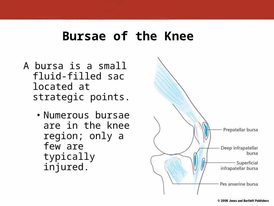
Bursae of the Knee
A bursa is a small fluid-filled sac located at strategic points.
• Numerous bursae are in the knee region; only a few are typically injured.
Bursae of the Knee (cont.)
Inflammation can be caused by:
• Trauma.• Infection.• Overuse.
The prepatellar bursa is susceptible to direct trauma.

Bursae of the Knee (cont.)
Signs and symptoms include:
• Swelling and tenderness at site.
• Pain when increased external pressure is applied.
• Athlete may report direct trauma to knee.
Courtesy of Brent Mangus

Bursa of the Knee (cont.)
First Aid
• Application of ice and compression.• Reduced activity for a short time. • In chronic cases, anti-inflammatory agents
may be helpful.

Patellar Dislocation/Subluxatio
n
• Injury may be caused by a quick cutting motion that generates a great deal of abnormal force within the knee.
• Instead of moving normally, the patella moves laterally and may dislocate.

Patellar Dislocation/Subluxation (Cont.)
Signs and symptoms include:
• Severe pain and abnormal movement of the patella when injury occurred.
• Swelling.• Patella may be obviously out-of-place.• Extreme pain along the medial aspect of
the patella.

Patellar Dislocation/Subluxation
(cont.)
First Aid
• Apply ice and compression.
• Elevate.
• Splint the entire leg.
• Transport to a medical facility.
Osgood-Schlatter Disease and Jumper’s
KneeOsgood-Schlatter and “jumper’s knee” usually
involve irritation of the patellar tendon complex.
Signs and symptoms of Osgood-Schlatter include:• Pain and tenderness about the patellar tendon
complex.• Swelling in the area.• Decreased ability to use the quadriceps.• If inflammation continues, area over tibial
tuberosity may become solid when palpated.
Osgood-Schlatter Disease
First Aid
• Apply ice and compression.
• Refer to physician for specific diagnosis.
• Until inflammation subsides, rest is important.
Jumper’s Knee
Signs and symptoms of jumper’s knee include:• Pain and tenderness around the patellar tendon complex
that may spread to tibial tuberosity. • Decreased ability to use quadriceps for running or
jumping.• Symptoms that worsen with activity.
First Aid• Apply ice and compression.• Refer to physician for possible anti-inflammatory
medications• Rest will be helpful.
GOOD MORNING!I would like you to start the morning off by writingdown the following information on the sheet ofpaper on your desk:• Based on what we have learned about
Anatomical Terms, what do you think the term “Patellofemoral” means?
• Think back to the anatomy of the knee; what are the menisci?
• What are the actual names given to the four ligaments of the knee ( ACL,PCL,MCL,LCL)
• What is a VALGUS and VARUS force?• How do you think injuries to the ligaments of the
knee could be prevented?

Now…
• Turn to your neighbor and discuss your answers to the previous questions!

In your Notebooks:
• What is the Q-angle?

Patellofemoral ConditionsSome conditions of the patella
may be related to the Q angle.• The Q angle is the
difference between a straight line drawn from the anterior superior iliac spine and the center of the patella and a line drawn from the center of the patella through the center of the tibial tuberosity.
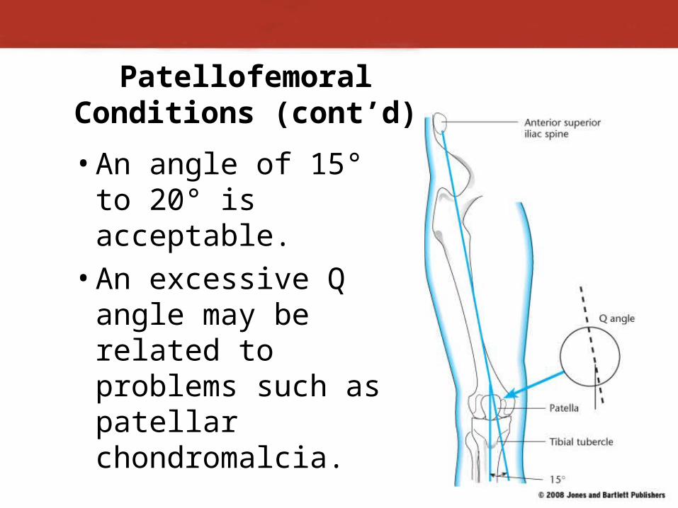
Patellofemoral Conditions (cont’d)
• An angle of 15° to 20° is acceptable.
• An excessive Q angle may be related to problems such as patellar chondromalcia.

Demonstration Time!
Volunteer Please!
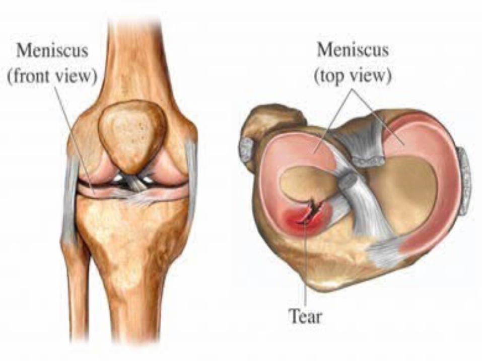
Meniscus Injuries
Menisci are typically damaged by quick, sharp, cutting movements.
• Injury is more likely to occur if the foot is planted firmly on the playing surface.
There are many different types of tears, and they affect each athlete differently.
• In some cases, a torn flap of meniscus will get caught in the joint, causing it to lock.
Demonstration:
Where’s my athletes?!
I need a quick, sharp cutting motion!

Meniscus Injuries (cont.)
Signs and symptoms include:
• Pop or snap when the knee was injured.• May not see any significant swelling.• May not be painful.• Loss of ROM.• Athlete may be able to continue
participating.• A feeling the knee is “giving out”
periodically.

Meniscus Injuries (cont.)
First Aid
• Apply ice and compression.
• Have athlete use crutches.
• Refer athlete to a physician.

Knee Ligament Injuries
Injury may occur to the MCL, LCL, ACL, or PCL.
Common mechanisms include cutting maneuvers when running and direct blows to the joint.
© Alessandro Bianchi/Reuters/Landov

Knee Ligament Injuries (cont.)
Sprain to MCL is a common sports injury.
• Occurs as a result of valgus stress.
• Varus stress can cause a sprain of the LCL.
Both types of sprains render knee unstable in side-to-side movements.
And yet again….
Another Demonstration!
A Volunteer please!
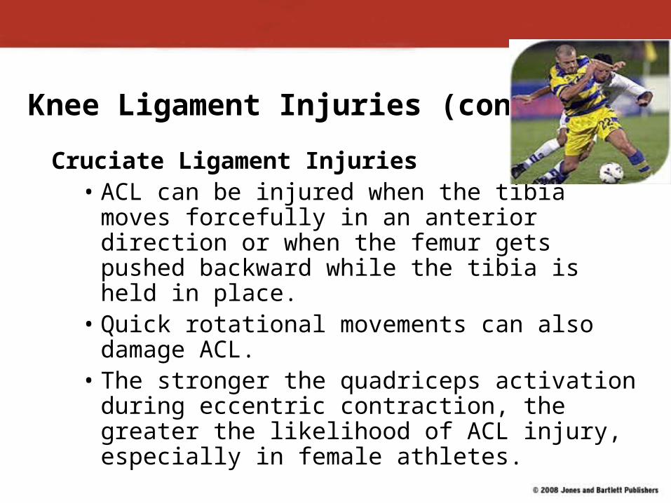
Knee Ligament Injuries (cont.)
Cruciate Ligament Injuries• ACL can be injured when the tibia moves
forcefully in an anterior direction or when the femur gets pushed backward while the tibia is held in place.
• Quick rotational movements can also damage ACL.
• The stronger the quadriceps activation during eccentric contraction, the greater the likelihood of ACL injury, especially in female athletes.

Cruciate Ligament Injuries
Signs and symptoms include:• Athlete reports the knee was forced
beyond its normal ROM.• Pain at the site of the injury.• Swelling around the knee.• Athlete indicates the knee feels unstable.• Athlete reports having a snapping or
popping sensation at the time of injury.

Cruciate Ligament Injuries (cont.)
First Aid
• Immediately apply ice and compression.
• Have athlete walk on crutches.
• Refer to a physician for medical evaluation.

In Your Notebooks:
• Where is the Tibial Tuberosity located?

Prevention
• Research is continuing to outline techniques that will hopefully prevent various injuries.
• Proper warm-up and stretching is important.• Protective bracing should be the athlete’s
choice.• Jump and landing training programs may
reduce the chance of an ACL tear, especially females.
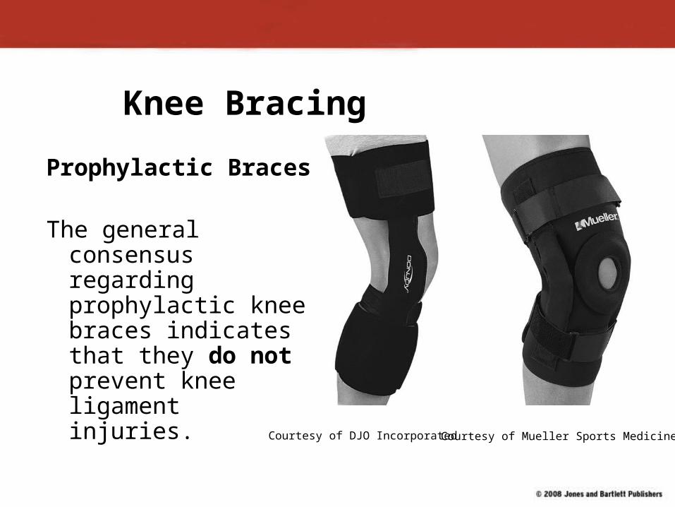
Knee Bracing
Prophylactic Braces
The general consensus regarding prophylactic knee braces indicates that they do not prevent knee ligament injuries. Courtesy of DJO Incorporated Courtesy of Mueller Sports Medicine
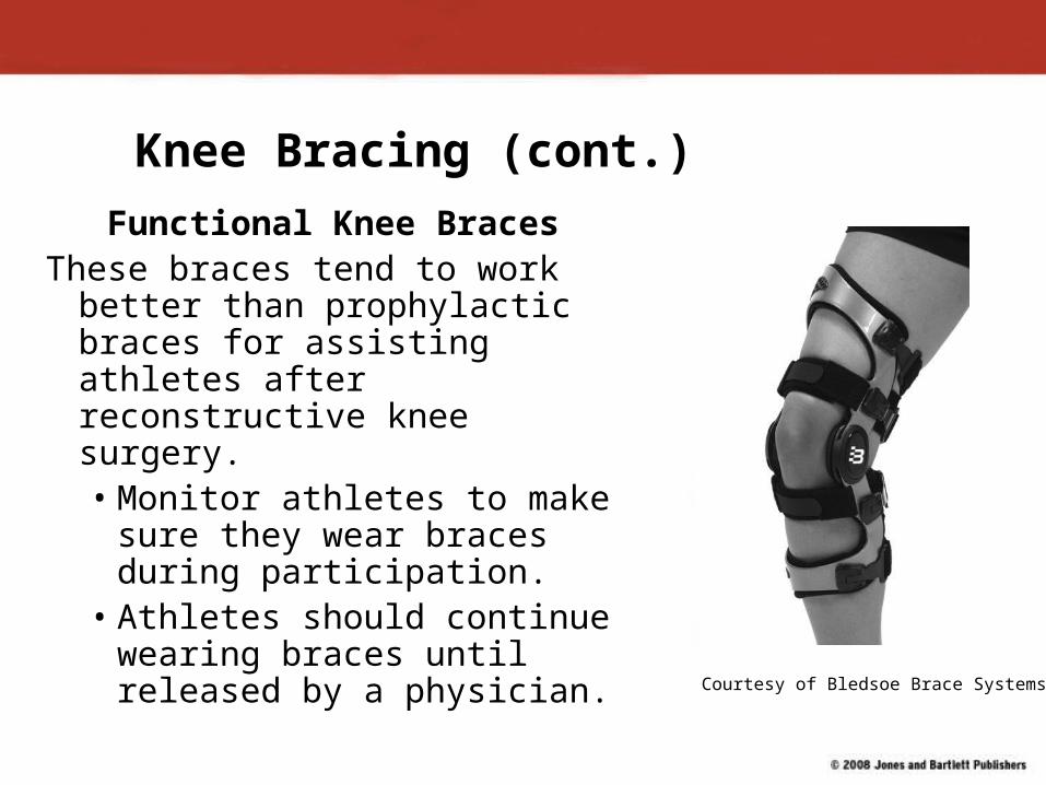
Knee Bracing (cont.)
Functional Knee BracesThese braces tend to work better
than prophylactic braces for assisting athletes after reconstructive knee surgery. • Monitor athletes to make
sure they wear braces during participation.
• Athletes should continue wearing braces until released by a physician. Courtesy of Bledsoe Brace Systems