Embed Size (px)
Citation preview

3
Biology of Cancer
julia eggert, Phd, aPrn-BC, gnP, aoCn®
CHAPTER
Introduction
Many in the lay public describe a diagnosis of “cancer” as if it is one disease. In reality, it encompasses more than 200 diseases that will occur at different ages with different rates of growth, differentiation, abilities to be detected, invasiveness, capacities to spread or metasta-size, treatment responses, and prognoses. How-ever, at the cellular and molecular levels, can-cer is beginning to be viewed as a few diseases caused by genetic alterations and defective cell function that are actually very similar (Ward, Brueggemeier, Caligiuri, Gahbauer, & Kraut, 2005). These alterations can be associated with “nature,” such as inherited cancer syndromes like hereditary breast and ovarian syndrome (HBOS) or immune deficiencies. Or, the ge-netic alterations can be caused by “nurture,” which includes obesity, poor diet, and social habits, such as smoking. A malignant growth is the result of changes in DNA, gene transcrip-tion, or translation. The resultant defective protein or proteins lead to transformation of normal cell components into uncontrolled pro-liferation, spread, and/or metastasis (Ward et al.). This chapter focuses on a description of the malignant changes of a cell that will pro-vide nurses new to oncology with a foundation for understanding the growth of cancer and its treatment, along with the basis to provide edu-cation to patients and their families.
Models of Cancer Development
Two models are commonly used to describe how a cancer develops. The first is the Stochas-tic Model, also known as Knudson’s random “two hit” model (see Figure 1-1). This mod-el suggests that each cancer cell has the abil-ity to multiply and form new tumors. Using one set of chromosomes within the nucleus of the cell as an example, the two chromosomes each have an identical allele, or gene, control-ling the growth of a cell. When the initial gene on the first chromosome is damaged, the un-damaged gene on the second chromosome is still able to take over the normal functions as-sociated with that gene and its protein product. When the gene on the second chromosome be-comes damaged, a normal gene message is no longer present, thus allowing malignant trans-formation. The malignant cells have a selective advantage over their normal neighbors and be-gin to proliferate rapidly, accumulating genet-ic damage with each generation. As the dam-age collects, the most aggressive characteristics promote immortalized growth and the forma-tion of a tumor (Hanahan & Weinberg, 2000; Knudson, 1971).
The individual cancer stem cell (CSC) is the focus of the second model. This model states that many different types of cancer cells exist, demonstrating heterogeneity. With prolifera-tion, cell division occurs, where all of these cells
1

4 ■ Cancer Basics
have the ability to multiply, but only one cell type—the CSC—has the ability to become a new tumor (Reya, Morrison, Clarke, & Weiss-man, 2001). This is becoming the model most supported by cancer researchers. See Figure 1-2 for an illustration of CSC tumor develop-ment. Once the new tumor is established, the heterogeneous cells begin to proliferate, al-lowing the tumor to enlarge, and the CSC moves into the resting phase (G0) of the cell
cycle. It is well known that cells in this phase are resistant to treatment and would remain as one surviving cancer cell while treatment destroys the other rapidly dividing non-CSCs (Wicha, Liu, & Dontu, 2006). Thus, months or years later, the “resting” CSC could move into the active phases of the cell cycle, pro-liferate, and cause exacerbation of the once-dormant cancer that was thought to be de-stroyed (Reya et al.).
Figure 1-1. Knudson “Two Hit” Theory of Cancer Development
Theory: Two genes within a cell, one on each of a pair of chromosomes, each receive damage-causing hits prior to the development of cancer. The malignant cells are heterogeneous and all have the capability of developing a tumor.
Note. Based on information from Hanahan & Weinberg, 2000; Knudson, 1971; Reya et al., 2001; Wicha et al., 2006.

Chapter 1. Biology of Cancer ■ 5
Structure and Function of DNA and Chromosomes
The human genome consists of 23 pairs of chromosomes. Each chromosome is a single double-helix DNA molecule with millions of base pairs connected in a long, unbroken string that intricately coils back upon itself and is scat-tered with proteins, called histones (see Figure 1-3).
Similar to sewing bobbins, histones con-trol the long threads of DNA, wrapping them into a tight coil so that the DNA is able to fit in-side the nucleus of a cell. The coiling is neces-sary because the strands of DNA in a person’s body would stretch almost six feet but would be only fifty-trillionths of an inch wide. This long but thin physical structure would be extremely fragile, hence the need for tight packaging to keep the DNA message intact. Once it is coiled
Figure 1-2. Cancer Stem Cell Theory of Cancer Development
Theory: Cancer stem cells are the only type of heterogeneous cancer cells to develop into tumors.
Note. Based on information from Reya et al., 2001; Wicha et al., 2006.

6 ■ Cancer Basics
Figure 1-3. DNA Packaging
Note. From Chromosome, by National Human Genome Research Institute, n.d. Retrieved November 9, 2009, from http://www.genome.gov/Pages/Hyperion/DIR/VIP/Glossary/Illustration/Pdf/chromosome.pdf.

Chapter 1. Biology of Cancer ■ 7
around the histones, DNA continues twisting back upon itself (much like the continued twist-ing of a rope) until it is tightly wound, forming the chromatid seen in Figure 1-3. These chro-matids enable the chromosomes to be visual-ized for karyotyping during the metaphase of cell division (Klug & Cummings, 2003).
Within the nucleus of each normal human cell, 23 pairs of chromosomes are present. These consist of 22 pairs of nonsex chromo-somes (autosomes) and one pair of sex chro-mosomes (XX for female, XY for male). A person inherits one chromosome of the pair from the father and the other from the moth-er. A chromosome has a short arm (“p” for “petite”) and a long arm (“q” because it fol-lows “p” in the alphabet) with a unique band-ing pattern that identifies specific regions. These regions are numbered from the cen-tromere to the end of each arm (Genetics Home Reference, 2009a). For example, the breast cancer gene (BRCA1) is found on chro-mosome 12q, with a band position of 12.1 on the long arm of chromosome 12 (see Figure 1-4). If the position of a gene is uncertain, a range might be noted, such as 17q21–24 (Ge-netics Home Reference, 2009b).
The central dogma of molecular biology states that DNA (adenine [A], cytosine [C], guanine [G], and thymine [T]) is transcribed to RNA (adenine [A], cytosine [C], guanine [G], and uracil [U] instead of thymine) and then translated into proteins (Klug & Cum-mings, 2003) (see Figure 1-5). For example, the DNA (ACTGTC) would be transcribed as RNA (ACUGUC) and then to messenger RNA (mRNA), where it is divided into codons (three nucleotides used to specify an amino acid) (UGA CAG) for translation from amino acid to protein. This is important to remember be-cause any changes in the codon (mRNA trip-let) “spelling” could change the protein out-come. Some amino acids have multiple codon “spellings.” One example is leucine, which has six (Algorithmic Arts, 2007). These would al-
Figure 1-4. Breast Cancer 1 (BRCA1) Gene
Note. From Ideogram: Breast Cancer I (BRCA1), by NCBI Map Viewer, n.d. Retrieved December 7, 2009, from http://www.ncbi.nlm.nih.gov/projects/mapview/maps.cgi?TAXID=9606&CHR=17&MAPS=genes-r%2Cpheno%2Cmorbid%2Cgenec&QUERY=BRCA1&BEG=17q21.1&END=17q21.1&thmb=on
Figure 1-5. Central Dogma of Molecular Biology
Note. From “Central Dogma of Molecular Biology,” by F. Crick, 1970, Nature, 227(5258), p. 562. Copyright 1970 by Nature Publishing Group. Adapted with permission.
low several mistakes without creating a prob-lem protein. However, tryptophan has one spelling (Algorithmic Arts, 2007). Any error in this codon spelling would cause the assembly of

8 ■ Cancer Basics
a dysfunctional or nonfunctional protein (Na-tional Cancer Institute [NCI], 2006).
Changes in the DNA nucleotides can be ei-ther a mutation or a polymorphism. This de-pends on the frequency with which the change occurs in the general population. If it occurs in at least 1% of the population, it is called a poly-morphism. If it occurs in less than 1%, it is la-beled as a mutation (NCI, n.d.). To further ex-plain, a normal length of DNA is similar to a recipe for the most common type of cake—for example, a chocolate cake. Although this is the most common, other types of cakes exist, in-cluding strawberry, white, spice, pineapple up-side-down, and lemon. These would be poly-morphisms. They are good-tasting cakes but are not the most common. Sometimes the rec-ipe is misread, and the cake comes out of the oven as a pudding. This is uncommon and not the desired outcome. This is a mutation (NCI, n.d.) (see Figure 1-6).
Several types of mutations exist. The most common type is the point mutation, in which only one nucleotide base is altered. A nonsense mutation occurs when there is a premature ter-mination of the protein. This happens when the stop codon, which signals termination of the length of amino acids, has been “spelled” incorrectly and gives an early or late signal to end the compilation of amino acids into a pro-tein (NCI, 2006).
Mutations that occur within any of the cells of the body are labeled as somatic. They accu-mulate over a lifetime and are believed to cause sporadic cancers, which typically occur after an individual has reached 50 years of age. Muta-tions that are present in the ova or sperm are labeled as germ line and are associated with in-herited cancers, which typically occur in people at ages younger than 50 (see Figure 1-7). The results of this genomic instability affect all fu-ture generations, depending on the pattern of inheritance (NCI, 2006).
Most patterns of inheritance follow the dom-inant or recessive model developed by Gregor
Mendel (Klug & Cummings, 2003). Each in-dividual normally has two sets of chromo-somes. On each chromosome is a gene, or al-lele, for a particular characteristic. Although an allele may have a collection of many dif-ferent traits (such as blue, green, hazel, or brown eyes), each chromosome can exhibit only one of these. So, one chromosome may have the blue-eyes gene (called an allele), and the second chromosome could have the brown-eyes gene (or allele). All of the other eye colors are still allelic options but are not displayed by this set of chromosomes. Each allele is either a dominant type or a recessive type. It is well known that the brown-eyes al-lele is dominant over the recessive blue-eyes allele. If the dominant allele is inactivated or lost, then the recessive allele will become ac-tive (Klug & Cummings).
Sometimes an individual will have the dom-inant allele without it being expressed. This is known as incomplete penetrance. The gene is there, but the phenotype (the observable phys-ical trait) is not expressed. An example that il-lustrates this is a house in a fog; the house is there but is not visible because of the dense-ness of the low-lying cloud cover. Age, modi-fier genes, carcinogens, repair enzymes, and hormonal or reproductive factors affect pene-trance (Klug & Cummings, 2003).
Much of the scientific evidence about the development of cancer and its progression suggests that genomic instability is a precur-sor to changes associated with transformation of a cell into malignancy. Of question in this hypothesis is how the instability circumvents the careful security provided within the cell to monitor and guarantee genomic stability and purity for continued survival of the human cell. These protective teams include DNA monitor-ing and repair enzymes. Checkpoint gatekeep-ers function at significant points in the active phases of the cell cycle prior to DNA synthe-sis (S phase) and mitosis (M phase) to guaran-tee the accuracy of the genome and cell cycle

Chapter 1. Biology of Cancer ■ 9
processes. If an error is present, the P53 tumor suppressor protein causes cell cycle arrest for repair or apoptosis (programmed cell death) if too much damage has occurred (Hanahan & Weinberg, 2000; Kastan & Bartek, 2004; Zhi-votovsky & Orrenius, 2006). Much research has confirmed that most human cancers have loss of function in the P53 tumor suppressor pathway. Other genes involved in targeting and repairing DNA damage also have been found to have loss of function in multiple can-cers (American Cancer Society, 2005; Narter et al., 2009). Acquiring genomic damage per-mits evolving populations of precancerous cells to gain functional capabilities associat-
ed with malignant transformation. These in-clude (a) self-sufficiency in growth signals, (b) insensitivity to antigrowth signals, (c) evasion of apoptosis, (d) sustained angiogenesis, (e) tissue invasion and metastasis, and (f) limitless replicative potential (Hanahan & Weinberg).
Self-Sufficiency in Growth Signals
Growth FactorsThe behavior of cells is controlled by cir-
culating growth factors (ligands) that have
Figure 1-6. Polymorphisms Versus Mutations
The wild-type genotype is transcribed to a codon that correctly spells a protein (e.g., recipe for a chocolate cake). When there is a polymorphism in the codon spelling, it can still spell a correct amino acid (e.g., a cake, just a dif-ferent flavor). When the amino acid spelling is rare and uncommon, it is a mutation and creates an undesirable outcome (e.g., cookie, pudding, or another undesired result).
10 ■ Cancer Basics
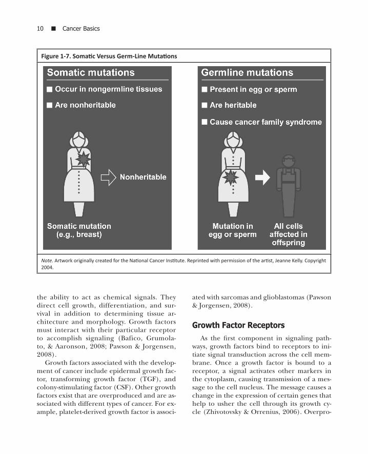
the ability to act as chemical signals. They direct cell growth, differentiation, and sur-vival in addition to determining tissue ar-chitecture and morphology. Growth factors must interact with their particular receptor to accomplish signaling (Bafico, Grumola-to, & Aaronson, 2008; Pawson & Jorgensen, 2008).
Growth factors associated with the develop-ment of cancer include epidermal growth fac-tor, transforming growth factor (TGF), and colony-stimulating factor (CSF). Other growth factors exist that are overproduced and are as-sociated with different types of cancer. For ex-ample, platelet-derived growth factor is associ-
ated with sarcomas and glioblastomas (Pawson & Jorgensen, 2008).
Growth Factor ReceptorsAs the first component in signaling path-
ways, growth factors bind to receptors to ini-tiate signal transduction across the cell mem-brane. Once a growth factor is bound to a receptor, a signal activates other markers in the cytoplasm, causing transmission of a mes-sage to the cell nucleus. The message causes a change in the expression of certain genes that help to usher the cell through its growth cy-cle (Zhivotovsky & Orrenius, 2006). Overpro-
Figure 1-7. Somatic Versus Germ-Line Mutations
Note. Artwork originally created for the National Cancer Institute. Reprinted with permission of the artist, Jeanne Kelly. Copyright 2004.

Chapter 1. Biology of Cancer ■ 11
duction of some growth factors causes altered cellular communication and is associated with cancers. One of these, vascular endothelial growth factor (VEGF), has an important role in tumor neoangiogenesis—that is, the new growth of vessels on a tumor.
Tumor cells induce hypoxia. This lack of oxygen leads to transcription of the VEGF-al-pha protein by binding to its designated cell surface receptors. The binding trips a signal indicating the need for increased blood ves-sel permeability, resulting in angiogenesis with even more proliferation of cells (Nguy-en, Tran, Lipkin, & Fruehauf, 2006). VEGF is overexpressed in metastases of breast and col-orectal cancers.
Growth Factor Receptors and Tyrosine Kinase Activity
Many cancer-related growth factor receptors are stationed on the surface of the cell. Once they are bound by a ligand that causes activa-tion, proliferative signals are sent into the cy-toplasm. Most growth factor receptors possess tyrosine kinase (TK) activity, which leads to reactions that stimulate mitotic cell division, thereby allowing rapid growth of the malignant cell (Chan & Feng, 2007; Zhivotovsky & Orre-nius, 2006).
Examples of growth factor receptors that are cancer-causing (oncogenic) when overex-pressed are epidermal growth factor receptor (EGFR), human epidermal growth factor recep-tor 2 (HER2/neu), and transforming growth factor-beta (TGF-β). A variety of cancers ex-press EGFR, including non-small cell lung can-cer and breast, ovarian, and colorectal cancers. Approximately 80%–100% of head and neck cancers overexpress EGFR, which is also associ-ated with lower survival. Increased HER2/neu expression corresponds with more aggressive cancers, including ovarian and breast cancers. When EGFR and TGF-β are both expressed, it is a prognostic marker for tumor relapse and
decreased survival (Bafico et al., 2008; Pawson & Jorgensen, 2008).
Nonreceptor Tyrosine KinasesSome oncogenes do not require a recep-
tor to initiate TK activity at the cell membrane. One example is the SRC gene family. The pro-tein from this gene initiates TK activity at the C-terminus of the DNA where biosynthesis is sup-posed to end. Because there is no endpoint, the protein function persists, allowing continued signaling to the cell nucleus and causing per-sistent cell growth. Such SRC-initiated activity is increased in colon cancer and other malignan-cies such as neuroblastoma, small cell lung can-cer, breast adenocarcinomas, and rhabdomyo-sarcoma (GeneCards, 2009c; Okutani, Lodyga, Han, & Liu, 2006).
Intercellular Signaling Enzymes Oncoproteins with certain enzyme activity
are important for sending signals within cells and are called intracellular signaling enzymes. A common example is the enzymatic pro-tein produced by the RAF1 gene (GeneCards, 2009a). In the cytoplasm, TK activates the RAF1 enzyme. Once activated, the enzyme acts as a mediator between the RAS (associated with the RAS oncogene) receptor on the cell membrane and processes occurring in the cell nucleus by activating a series of other kinases, including mitogen-activated protein (referred to as MAP) kinases. These kinases are critical for regulat-ing the onset of cell division, apoptosis, cell dif-ferentiation, and cell migration (Bafico et al., 2008; Chan & Feng, 2007; GeneCards, 2009a; Zhivotovsky & Orrenius, 2006).
Membrane-Associated G Proteins The guanine nucleotide-binding proteins (G
proteins) are products of a family of genes, the ras proto-oncogenes, which normally act as “on-

12 ■ Cancer Basics
off switches” for cell-surface growth factor recep-tors. Instead of being transmitted inside the cell membrane, they transform adjacent G protein subunits below the membrane surface, which then begin the signaling cascade inside the cell (Bafico et al., 2008; Bos, Rehmann, & Witting-hofer, 2007; Pawson & Jorgensen, 2008).
When the RAS gene mutates and becomes a cancer-causing gene (oncogene), the changes interrupt a cascade of normally occurring sig-nals that take place in the cell cytoplasm. Nor-mal RAS genes wait for prompting to send stim-ulatory signals from growth factor receptors to other proteins. Mutant RAS genes activate sig-naling pathways, even when unprompted. Mu-tant RAS is found in virtually all types of human cancer and occurs in approximately two-thirds of all malignant tumors (Bos et al., 2007). G proteins act at the cell membrane to cause ma-lignant transformation (Bafico et al., 2008; Paw-son & Jorgensen, 2008).
Transcription FactorsProteins that bind to DNA and cause chang-
es in gene expression are called transcription factors. These proteins have structures that can recognize specific DNA sequences (genes) in-volved in growth and survival. Mutation of the transcription factors that bind to genes involved in cell growth and survival allows for the ma-lignant transformation found in many tumors. Examples of cancers caused by this mechanism include Ewing sarcoma, clear cell sarcoma, al-veolar rhabdomyosarcoma, and many kinds of leukemia. Many of the transcription factor– induced cancers are characterized by transloca-tion of chromosomes (Aplan, 2006) (see Chro-mosomal Abnormalities later in this chapter). One of the tumor suppressor genes, TP53, also acts as a transcription factor. In this role, TP53 “senses” DNA damage and halts cell division by controlling the expression of other genes that directly regulate the cell cycle (Ozanne, Spen-ce, McGarry, & Hennigan, 2007).
Tumor Suppressor Genes
Tumor suppressor genes (also called anti- oncogenes) normally suppress or negative-ly regulate cell proliferation by encoding pro-teins that block the action of growth-promot-ing proteins. Using the example of a car, with cell growth caused by an accelerator, the tumor suppressor genes are the brakes, which can pre-vent cellular poliferation or suppress malignant transformation. At the cellular level, mutations in the cell cause tumor suppressor genes to lose function of both alleles. In other words, the loss of function or mutations of both copies of the gene are required for uncontrolled cell growth leading to tumorigenesis (National Center for Biotechnology Information, 2008).
Loss of HeterozygosityHomozygosity refers to alleles that are identi-
cal. If there is an inherited mutation of a tu-mor suppressor gene, it is termed heterozygous because the alleles are different. Because there is a normal allele, the function of the gene and its protein product is maintained. Once the remaining allele is mutated, the gene and its product will lose normal functioning. The heterozygosity has been further altered and is now labeled as loss of heterozygosity (LOH). Cells can experience LOH with the loss of an entire chromosome, translocation of a piece of the chromosome, reduplication of a piece of chro-mosome that already has an abnormal gene, or the development of a point mutation in the sec-ond functioning allele. LOH is associated with cancer susceptibility genes such as oncogenes and tumor suppressor genes, such as TP53. Ba-sic research is identifying an increasing number of tumor suppressor genes that, when mutat-ed, are closely associated with the development and progressions of human cancers (Ameri-can Cancer Society, 2005; Levine, Hu, & Feng, 2008; NCI, 2006; National Center for Biotech-nology Information, 2008).

Chapter 1. Biology of Cancer ■ 13
The tumor suppressor gene TP53 (located on 17p13) commonly has deletions and muta-tions associated with a wide variety of cancers, including lung, breast, esophageal, liver, blad-der, and ovarian carcinomas; brain tumors; sarcomas; lymphomas; and leukemias. It is believed to contribute to half of all sporadic hu-man cancers, making TP53 the most common genetic target for mutations leading to cancers (Levine et al., 2008). When TP53 is inherited in the germ line as a mutation, it is transmitted in an autosomal dominant fashion, a hallmark of Li-Fraumeni syndrome. This is a rare disor-der causing multiple types of cancers, includ-ing soft tissue sarcomas, osteosarcomas, breast cancers, and different types of leukemias (Ge-netics Home Reference, 2007; Levine et al.).
Specific Functions of Tumor Suppressor Genes
Tumor suppressor gene products have spe-cific functions in the cell nucleus and cyto-plasm. If deregulation of the cell cycle occurs, which results in excess cell proliferation, the normal TP53 gene can halt cell division and induce programmed cell death, or apoptosis (Levine et al., 2008) (see Apoptosis later in this chapter).
Tumor suppressor genes also can encode for proteins in the cytoplasm. The NF1 (neu-rofibromatosis) gene encodes a protein that is similar to the proteins that modulate the ras oncogene function. Loss of NF1 may keep ras activated and prolong the signal for cell prolif-eration. Loss of other tumor suppressor genes, such as NF2 and APC (adenomatous polypos-is coli), may cause cellular disorganization that leads to abnormal cell proliferation (Schindel-er & Little, 2008).
Insensitivity to Anti-Growth Signals In the normal cell, anti-growth signals move
the cell out of the cell cycle growth phases into
the resting from proliferation, or G0, phase. The TGF-β, referred to as both tumor or trans-forming growth factor, pathway is the best ex-ample of a signaling mechanism that causes the inhibition of cell growth and proliferation. This occurs in two ways in the normal path-way. First, it prevents inactivation of the retin-oblastoma protein (pRb), a tumor suppressor protein and synthesis of the tumor suppressor genes p15INK4α and p21. Second, synthesis of these genes will block cyclin, a protein that al-lows cells to move into the cell cycle. In cancer, the TGF-α pathway causes the loss and or mu-tation of the TGF-α receptor function (Dhara, 2008). The tumor suppressor function of one protein, SMAD4, is eliminated because of mu-tation of its gene (GeneCards, 2009b). The p15 protein is not synthesized, so cyclin is not blocked, thereby allowing cells to continuous-ly move into the active cell cycle with growth and proliferation. Finally, pRb tumor suppres-sor function is lost. Any of these interfering mechanisms, alone or in combination, allow the cell continued growth and proliferation (Dhara).
Evasion of ApoptosisApoptosis is “programmed” cell death.
With this process, cell death is a controlled, deliberate, and distinct series of biochemi-cal and cellular changes that allows an organ-ism to remove old, dead, or unwanted cells. There is no inflammation, and some of the cellular materials are ingested by neighbor-ing cells and reused. Apoptosis is a normal process that occurs when there is severe or irreparable damage to the DNA in order to prevent duplication of inaccurate messag-es (Zhivotovsky & Orrenius, 2006). The ABL and BCR genes are associated with this pro-cess (see Figure 1-8). A translocation of ABL on chromosome 9 to the BCR locus on chro-mosome 22 becomes the Philadelphia chro-mosome, prevents apoptosis, and is associated

14 ■ Cancer Basics
with chronic myeloid leukemia (Zhivotovsky & Orrenius).
Cells that lose their ability to signal apop-tosis contribute to early tumorigenesis be-cause they are unable to repair problems in the DNA in order to eliminate genetical-ly damaged cells. Without repair, survival of damaged cells ultimately leads to tumorigen-esis (Zhivotovsky & Orrenius, 2006). Inacti-vation of the TP53 gene leads to decreased apoptosis and rapid tumor progression. The loss of TP53 function may indirectly contrib-ute to tumor development by permitting the
proliferation of mutated cells (Levine et al., 2008).
Follicular lymphoma, a type of indolent (slow-growing) non-Hodgkin lymphoma, is an exam-ple of the loss of apoptosis. This slow-growing lymphoma accounts for approximately 20% of all non-Hodgkin lymphomas and commonly has a rearrangement of the BCL2 gene. The overex-pression of the bcl2 protein inhibits apoptosis, al-lowing continued cellular proliferation and mak-ing it difficult to destroy this lymphoma (NCI, 2009). It is hypothesized that restoration of apop-tosis may provide an approach to cancer therapy.
Figure 1-8. Translocation of the ABL Proto-Oncogene on Chromosome 9
Diagrammatic and karyotypic comparisons of the translocation of ABL on chromosome 9 to the BCR locus on chromosome 22, which forms the Philadelphia chromosome. This chromosome is activated by the BCR-ABL fusion oncogene.
Note. From Targeting BCR-ABL, by Novartis Pharmaceuticals Corporation. Retrieved October 31, 2009, from http://www.cmlalliance .com/health-care-professional/targeting-bcr-abl.jsp. Copyright 2010 by Novartis Pharmaceuticals Corporation. Reprinted with permission.

Chapter 1. Biology of Cancer ■ 15
Sustained Angiogenesis
Tumor cells have limitations in oxygen sup-ply that cause areas of hypoxia. This boosts the need for glucose uptake and glycolysis to gen-erate energy, resulting in lactate production. Although this supports the use of positron-emission tomography in nuclear medicine to identify increased metabolism, it also can result in decreased adenosine triphosphate produc-tion and ultimately contribute to the fatigue ex-perienced by patients with a malignancy (Stasi, Abriani, Beccaglia, Terzoli, & Amadori, 2003). This lack of oxygen also curtails the prolifera-tion of the malignant cells (Hanahan & Folk-man, 1996). For tumors to grow to a larger size, they need to develop a microcirculatory system, through the process of angiogenesis (Hanahan & Weinberg, 2000).
VEGF causes the growth of new vessels, forming a microcirculatory system in a tumor (i.e., angiogenesis). Tumor cells induce hypox-ia, leading to the transcription of VEGF-alpha, which binds to cell surface receptors and ulti-mately causes increased blood vessel perme-ability, angiogenesis, and the proliferation of cells (Nguyen et al., 2006). Multiple malignan-cies, including metastatic breast and colorectal cancers, overexpress VEGF.
Tissue Invasion and Metastasis
Altered Cytoskeletal ControlCells have a skeleton with interior and ex-
terior functions. The shape and the cell’s abil-ity to move are included in the external func-tion of the cytoskeleton. The internal function of the cytoskeleton permits substances to move within the cell. On the exterior membrane, mi-crotubules evoke a rigidity to add strength to the membrane surface. Internally, they pro-mote movement of organelles within the cyto-plasm. During mitosis, the microtubules are ar-
ranged in a centriole as nine bundles of three microtubules each. These form the spindle fi-bers, which are responsible for the separation of the chromosomes prior to the actual split-ting of the cell (McCance, 2010). With a malig-nancy, the cell loses external cytoskeleton con-trol. This enables the cell to lose rigidity and be more amenable to continued cellular division. In addition, the cytoskeleton is needed for spin-dle microtubule formation, mitosis, and cellu-lar growth. Multiple protein types participate in these changes associated with malignant trans-formation (Liaw, Chang, & Kavallaris, 2007).
Altered Mobility of Membrane Components
Proteins, glycoproteins, and glycolipids are known to have altered mobility on the mem-brane of a malignant cell. One outcome of this change enables the cancer to avoid immune surveillance. Other outcomes could promote spread and metastases (Hanahan & Weinberg, 2000; Wallach, 1968).
Modified Contact Adhesion and Inhibition of Movement
After replication, normal cells contact the ad-jacent cell membrane and are inhibited to grow. Malignant cells lose this characteristic and con-tinue to proliferate even though they are touch-ing the cell next to them. This contributes to the lack of control in malignant cells (Hanahan & Weinberg, 2000; McCance, 2010).
Altered Surface Charge DensityThe electrical potential of the malignant
cell membrane has a lower level than that of a normal cell. This is because of the increased amounts of negatively charged phospholipids in the cell membrane (Dobrzynska, Szachowicz-Petelska, Sulkowski, & Figaszewski, 2005). Pos-itively charged sodium and calcium channels

16 ■ Cancer Basics
contribute to apoptosis (Williams & Djamgoz, 2004). Changes in the charge of the cell mem-brane inhibit apoptosis and contribute to the longevity of the malignant cell.
Increased Lectin Agglutinability Alterations in lectin binding enable leuko-
cytes to adhere to and cover malignant cells. This change allows the malignant cell to escape sur-veillance and travel to distant sites in the body as a bolus of normal and abnormal cells (Hanahan & Weinberg, 2000; McCance, 2006).
Limitless Replication PotentialOne factor allowing the potential for lim-
itless replication is the expression of telom-erase. This enzyme prevents the destruction of the telomere. Because telomeres protect chro-mosomes, cells with increased telomerase are known to be associated with longer telomeres and longevity of cell life. Short telomeres are associated with a shorter life span. Cancer stem cells are known to have increased levels of te-lomerase and thus have an extended life en-hanced by the protected telomeres at the ends of the chromosome. This also protects the cell from apoptosis (NCI, 2010).
Conclusion
The curricula in undergraduate nursing pro-grams do not typically include molecular biol-ogy. With completion of the sequencing of the human genome in 2003, many diagnostics and treatments have been developed that require a level of understanding of certain characteristics of the cell, the central dogma, and how cell sig-naling and communication occur. For nurses in oncology, this knowledge is important to under-stand and helps them to anticipate the symp-toms of cancer in their patients, to be aware of how the treatments work, and to have a basic
foundation to use when developing teaching plans related to individualized treatment plans for their patients. This chapter has provided a description of how malignancies develop and some of the molecular biology of the cell that is used for diagnostics and treatment in oncolo-gy. These include self-sufficiency in growth sig-nals; insensitivity to antigrowth signals; evasion of apoptosis; sustained angiogenesis; tissue in-vasion and metastasis; and limitless replicative potential (Hanahan & Weinberg, 2000). For people new to oncology or for those who sim-ply want to review and close some gaps in their knowledge, an understanding of the biology of cancer will assist them as they care for patients and their families.
ReferencesAlgorithmic Arts. (2007). Amino acid codon table. Retrieved
July 6, 2009, from http://algoart.com/aatable.htmAmerican Cancer Society. (2005). Oncogenes and tumor suppres-
sor genes. Retrieved May 25, 2009, from http://www.cancer.org/docroot/ETO/content/ETO_1_4x_oncogenes _and_tumor_suppressor_genes.asp
Aplan, P.D. (2006). Causes of oncogenic chromosomal translocation. Trends in Genetics, 22(1), 46–55.
Bafico, A., Grumolato, L., & Aaronson, S. (2008). Onco-genes and signal transduction. In J. Mendelsohn, P. Howley, M. Israel, J. Gray, & C. Thompson (Eds.), The molecular basis of cancer (3rd ed., pp. 17–30). Philadel-phia: Elsevier Saunders.
Bos, J.L., Rehmann, H., & Wittinghofer, A. (2007). GEFs and GAPs: Critical elements in the control of small G proteins. Cell, 129(5), 865–877.
Chan, R.J., & Feng, G.S. (2007). PTPN11 is the first iden-tified proto-oncogene that encodes a tyrosine phos-phatase. Blood, 109(3), 862–867.
Dhara, S. (2008). Tumor biology. In M.G. Pomper & J.G. Gelovani (Eds.), Molecular imaging in oncology (pp. 1–28). New York: Informa Healthcare.
Dobrzynska, I., Szachowicz-Petelska, B., Sulkowski, S., & Figaszewski, Z. (2005). Changes in electric charge and phospholipids composition in human colorectal can-cer cells. Molecular and Cellular Biochemistry, 276(1–2), 113–119.
GeneCards. (2009a). RAF1. Retrieved June 22, 2009, from http://www.genecards.org/cgi-bin/carddisp.pl?gene= Raf1