Embed Size (px)
Citation preview

Chaperones, protein folding
and unexpected accidents
Maria Grazia Ortore
Dipartimento di Scienze della Vita e dell’Ambiente
Colloqui interdisciplinari sulla biologia,Università di Roma Tor Vergata, 15 marzo 2017

• Protein Folding: why it is so appealing for physicists
• Misfolding and Amyloid fibrils
• Chaperons and Chaperonins
• Chaperonins toward amyoid fibril aggregation
OUTLOOK

Native structure is compact and stabilized by multiple hydrophobic contacts
and hydrogen bonds
In a folding reaction, the native state has the lowest free energy
protein folding is spontaneous in principle
in practice, impractically slow
Folding intermediates are flexible, less compact, with exposed hydrophobicity
PROTEIN FOLDING
Model of folding reactionDaggett and Fersht
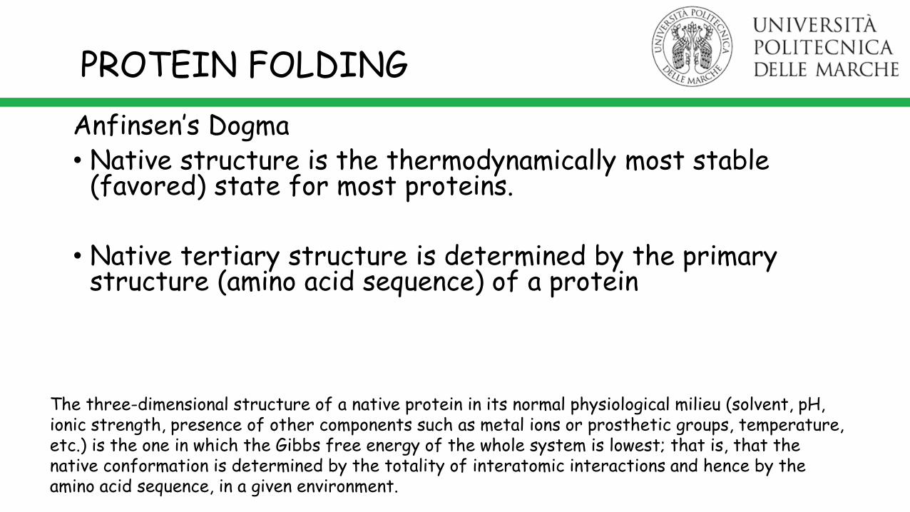
Anfinsen’s Dogma• Native structure is the thermodynamically most stable
(favored) state for most proteins.
• Native tertiary structure is determined by the primary structure (amino acid sequence) of a protein
PROTEIN FOLDING
The three-dimensional structure of a native protein in its normal physiological milieu (solvent, pH, ionic strength, presence of other components such as metal ions or prosthetic groups, temperature, etc.) is the one in which the Gibbs free energy of the whole system is lowest; that is, that the native conformation is determined by the totality of interatomic interactions and hence by the amino acid sequence, in a given environment.
PROTEIN FOLDING
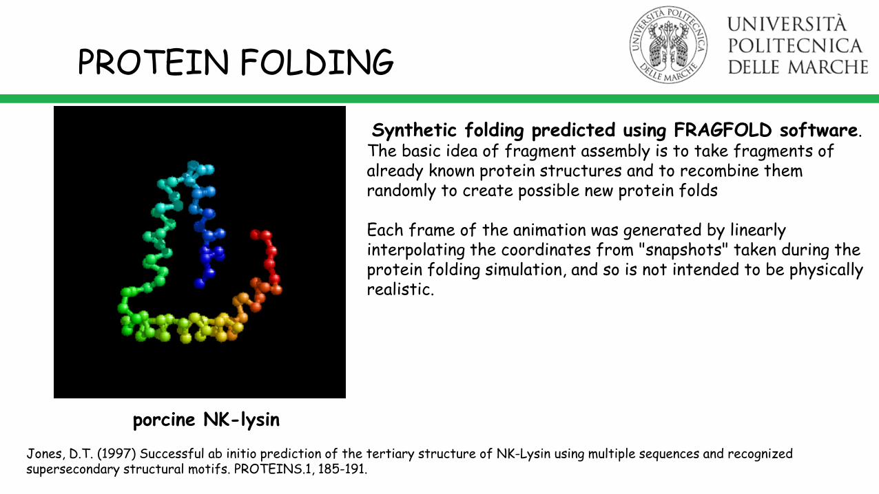
Synthetic folding predicted using FRAGFOLD software.The basic idea of fragment assembly is to take fragments of already known protein structures and to recombine them randomly to create possible new protein folds
Each frame of the animation was generated by linearly interpolating the coordinates from "snapshots" taken during the protein folding simulation, and so is not intended to be physically realistic.
porcine NK-lysin
Jones, D.T. (1997) Successful ab initio prediction of the tertiary structure of NK-Lysin using multiple sequences and recognized supersecondary structural motifs. PROTEINS.1, 185-191.
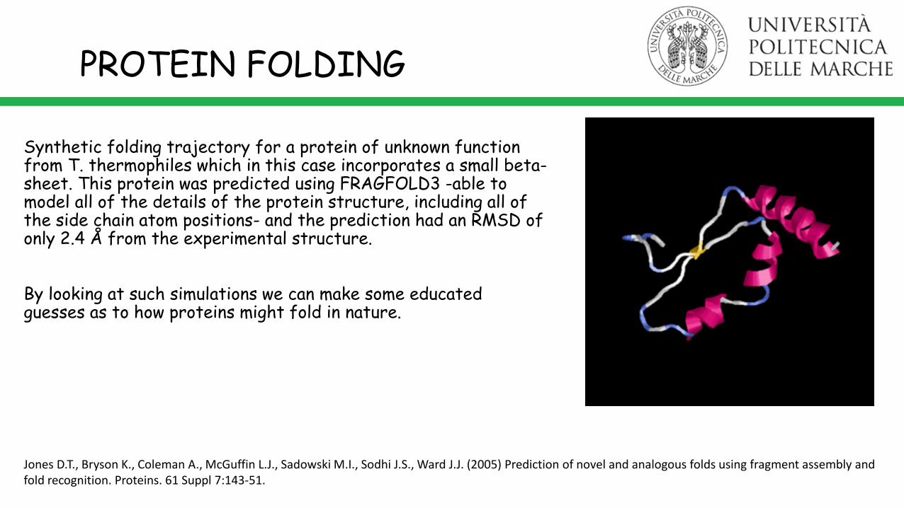
Synthetic folding trajectory for a protein of unknown function from T. thermophiles which in this case incorporates a small beta-sheet. This protein was predicted using FRAGFOLD3 -able to model all of the details of the protein structure, including all of the side chain atom positions- and the prediction had an RMSD of only 2.4 Å from the experimental structure.
By looking at such simulations we can make some educated guesses as to how proteins might fold in nature.
PROTEIN FOLDING
Jones D.T., Bryson K., Coleman A., McGuffin L.J., Sadowski M.I., Sodhi J.S., Ward J.J. (2005) Prediction of novel and analogous folds using fragment assembly and fold recognition. Proteins. 61 Suppl 7:143-51.

PROTEIN FOLDING
A protein’s native structure is determined solely bythe protein’s amino acid sequence components and theenvironmental conditions in which the folding occurs,such that the native structure is a unique, stable, andkinetically accessible state corresponding to a Gibbsfree energy minimum.
Dobson (2003) Nature 426, 884-890
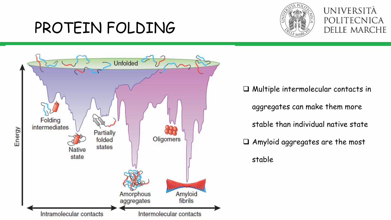
Many different unfolded and partially folded statesdifferent folding pathways lead to one native stateintermediates can persist in local free energy minima“kinetically trapped”, requires energy to escapeminimum
PROTEIN FOLDING
Multiple intermolecular contacts in
aggregates can make them more
stable than individual native state
Amyloid aggregates are the most
stable

AMYLOID AGGREGATION
“The amyloid state is more like the default state of a protein, and in the absence of specific protective mechanisms, many of our proteins could fall into it. “
Christopher Dobson, Annu. Rev. Biochemistry, 2006
“effectively all complex proteins have these short segments that, if exposed and flexible enough, are capable of triggering amyloidformation”
David Eisenberg, PNAS, 2010

*From “Course on SAXS from Biological Material” at SYBILS beamline, Lawrence Berkeley Laboratory
Small Angle X-ray Scattering (SAXS)
AMYLOID AGGREGATION
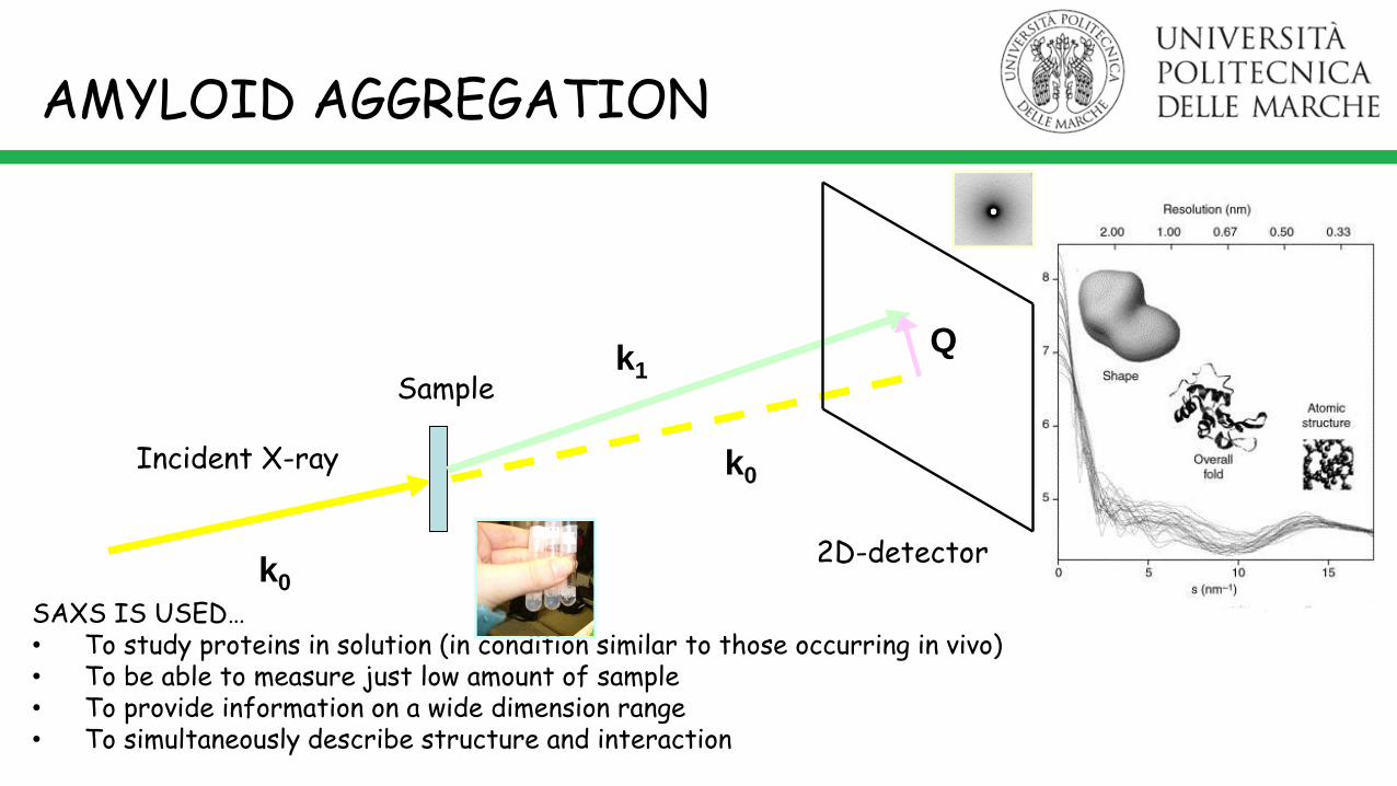
SAXS IS USED…• To study proteins in solution (in condition similar to those occurring in vivo)• To be able to measure just low amount of sample• To provide information on a wide dimension range• To simultaneously describe structure and interaction
AMYLOID AGGREGATION
k1
k0
k0
Incident X-ray
Sample
Q
2D-detector
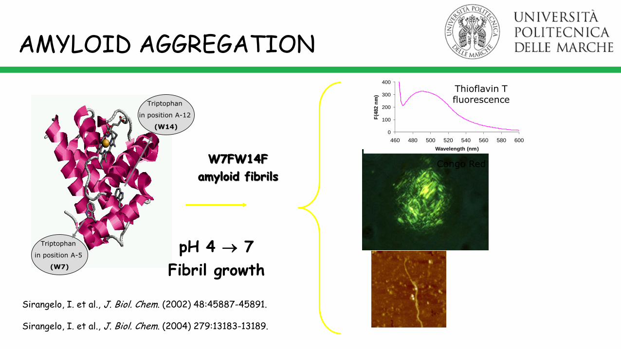
W7FW14F
amyloid fibrilsCongo Red
Triptophan
in position A-12
(W14)
Triptophan
in position A-5
(W7)
Sirangelo, I. et al., J. Biol. Chem. (2002) 48:45887-45891.
Sirangelo, I. et al., J. Biol. Chem. (2004) 279:13183-13189.
0
100
200
300
400
460 480 500 520 540 560 580 600
Wavelength (nm)
F(4
82
nm
)
Thioflavin T fluorescence
AMYLOID AGGREGATION
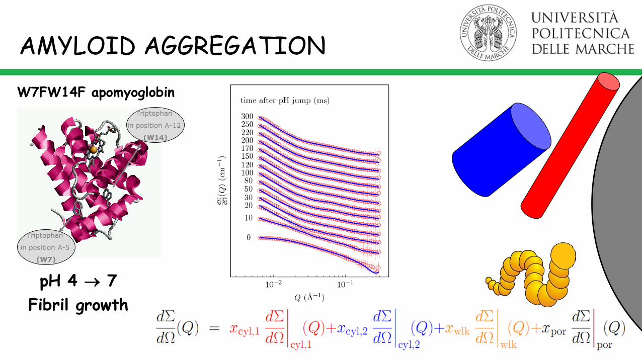
pH 4 7
Fibril growth

pH jumpfrom 4.0 to 7.0
W7FW14F4 g l-1
pH 4.0
W7FW14F2 g l-1
pH 7.0
As time goes on…Buffer SolutionpH 7.0
AMYLOID AGGREGATION
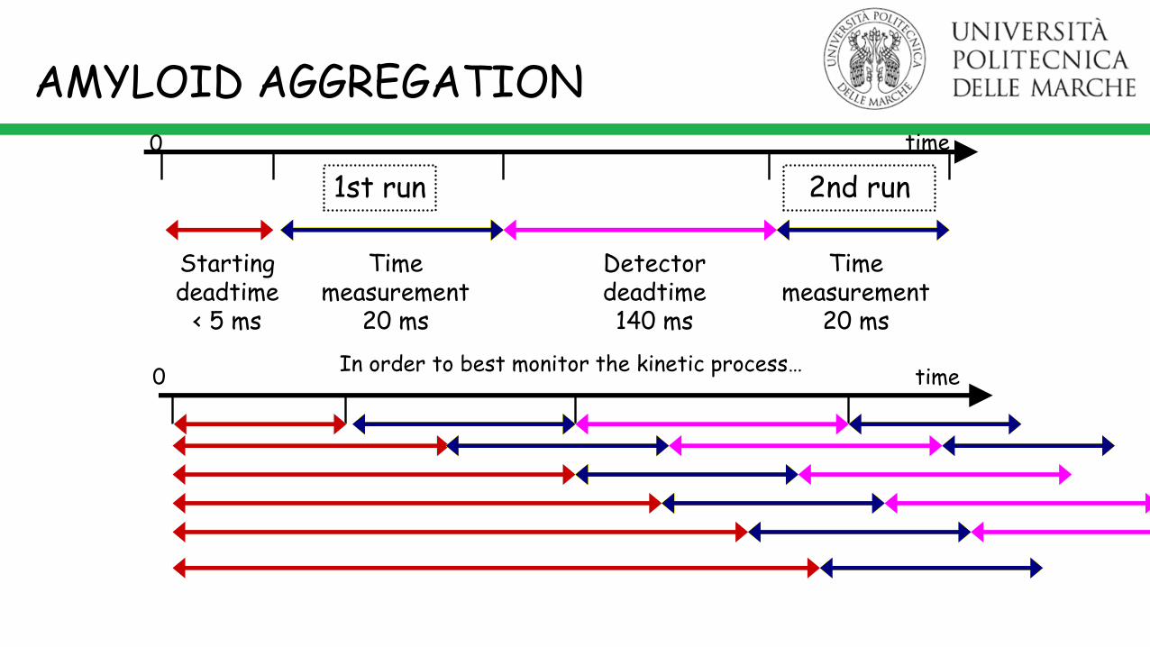
time0
Starting deadtime
< 5 ms
Time measurement
20 ms
Detector deadtime140 ms
Time measurement
20 ms
1st run 2nd run
In order to best monitor the kinetic process…time0
AMYLOID AGGREGATION
pH 4 7
Fibril growth
Triptophan
in position A-12
(W14)
Triptophan
in position A-5
(W7)
W7FW14F apomyoglobin
AMYLOID AGGREGATION

Amount of monomers in the particles
1. After a few ten of ms, ≈ 50% ofW7FW14F are in the largeaggregates, whose concentrationremains rather costant with time.
2. The first aggregates arecylindrically shaped
3. A large amount of monomeric, worm-like protein is still present insolution.
large aggregatesworm-like monomerscylindrical particles
AMYLOID AGGREGATION

MG. Ortore et al. Phys Rev E 84, 061904 (2011)
AMYLOID AGGREGATION

Complex nucleation-elongation mechanism: particles grow
instantaneously, then the process stops, resets, and restarts.
The initial acceleration is probably due to the addition of monomers on the seeds rapidly formed after the pH jump.
The polymerization reaction gives rise to two different aggregates (at least!), the protofibrils, rather compact and fast growing, and the soluble oligomers, loosely packed and rather unstable.
MG. Ortore et al. Phys Rev E 84, 061904 (2011)
There is a long lag phasebefore fibrils appear.
My name is ThT.Fibrils are homogeneous.
My name is AFM.
AMYLOID AGGREGATION

Septins build 'cages' around bacterial pathogens, immobilizing the harmful microbes and preventing them from invading other healthy cells.
Mostowy, S. & Cossart, P. Autophagy 7, 780–782 (2011).
Septins are GTP binding proteins and are involved in leukaemia, colon cancer,Parkinson’s disease and Alzheimer’s disease.
Human Septin 3-GCForm factors of SEPT3-GC1 monomers (red curves) and SEPT3-GC2 dimers (cyan curves) corresponding to 100 conformations ofeach species obtained by using the 3SOP crystallographic structure as template, including the construct primary amino-acids sequence (59–350 aa), the HisTag linked with the N-terminal domain and other missing residues.Three representative conformations (blue, green, and red) of monomers and dimers are shown.
AMYLOID AGGREGATION
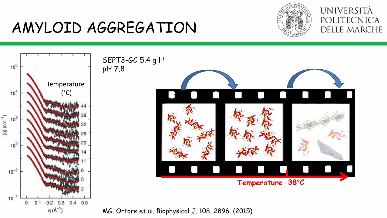
Temperature 38°C
MG. Ortore et al. Biophysical J. 108, 2896. (2015)
SEPT3-GC 5.4 g l-1
pH 7.8
Temperature (°C)
AMYLOID AGGREGATION

AMYLOID AGGREGATION
Amyloid β peptide (Aβ42 ) experiment performed at ID2, ESRF, Grenoble, France
AMYLOID AGGREGATION
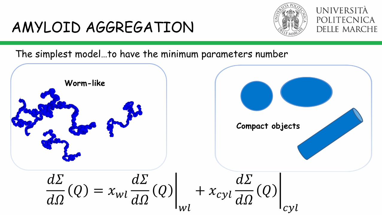
The simplest model…to have the minimum parameters number
Worm-like
Compact objects
𝑑𝛴
𝑑𝛺𝑄 = 𝑥𝑤𝑙
𝑑𝛴
𝑑𝛺𝑄
𝑤𝑙
+ 𝑥𝑐𝑦𝑙 𝑑𝛴
𝑑𝛺𝑄
𝑐𝑦𝑙

AMYLOID AGGREGATION
+*S.L. Bernestein et al. 2009, 10.1038/NCHEM.247
#S. Lesné et al. Nature 2006, 440:352-357§MN Reed et alNeurobiol Aging 2009
*
*
Paranucleus
Toxic species#§
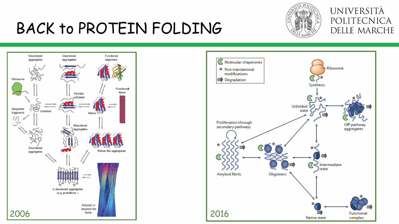
BACK to PROTEIN FOLDING
2006 2016
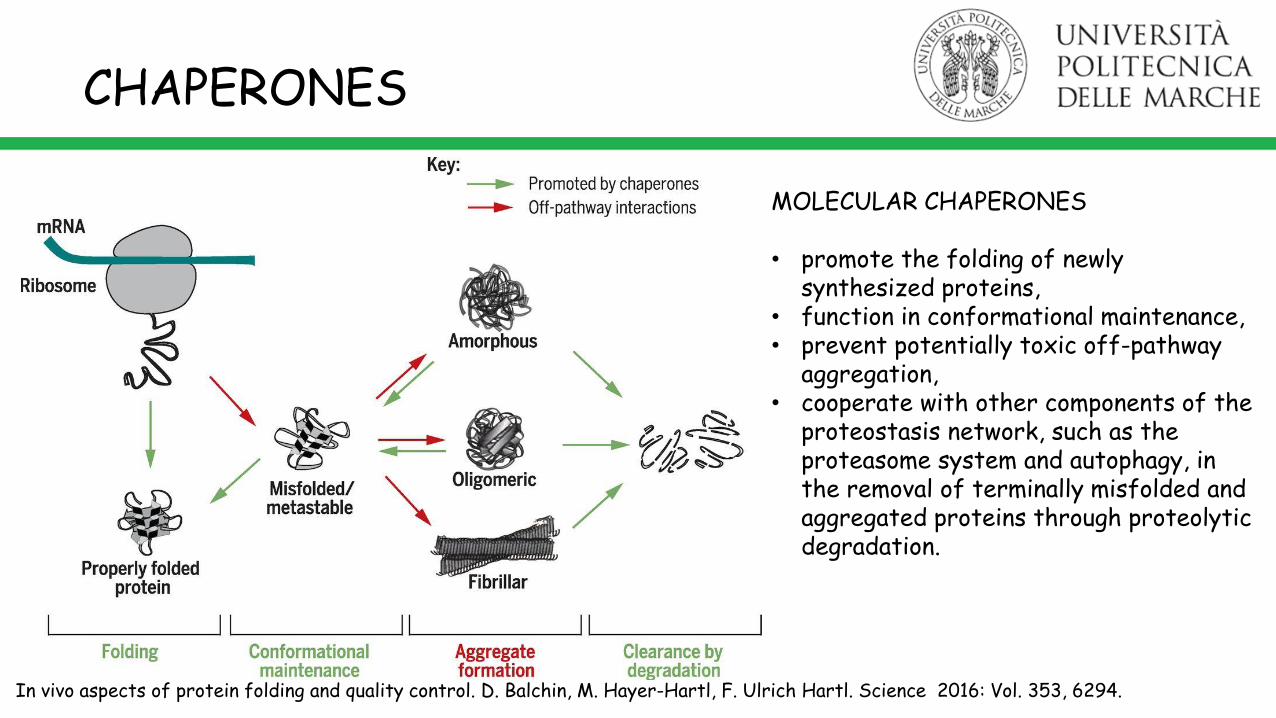
CHAPERONES
MOLECULAR CHAPERONES
• promote the folding of newly synthesized proteins,
• function in conformational maintenance, • prevent potentially toxic off-pathway
aggregation,• cooperate with other components of the
proteostasis network, such as the proteasome system and autophagy, in the removal of terminally misfolded and aggregated proteins through proteolytic degradation.
In vivo aspects of protein folding and quality control. D. Balchin, M. Hayer-Hartl, F. Ulrich Hartl. Science 2016: Vol. 353, 6294.
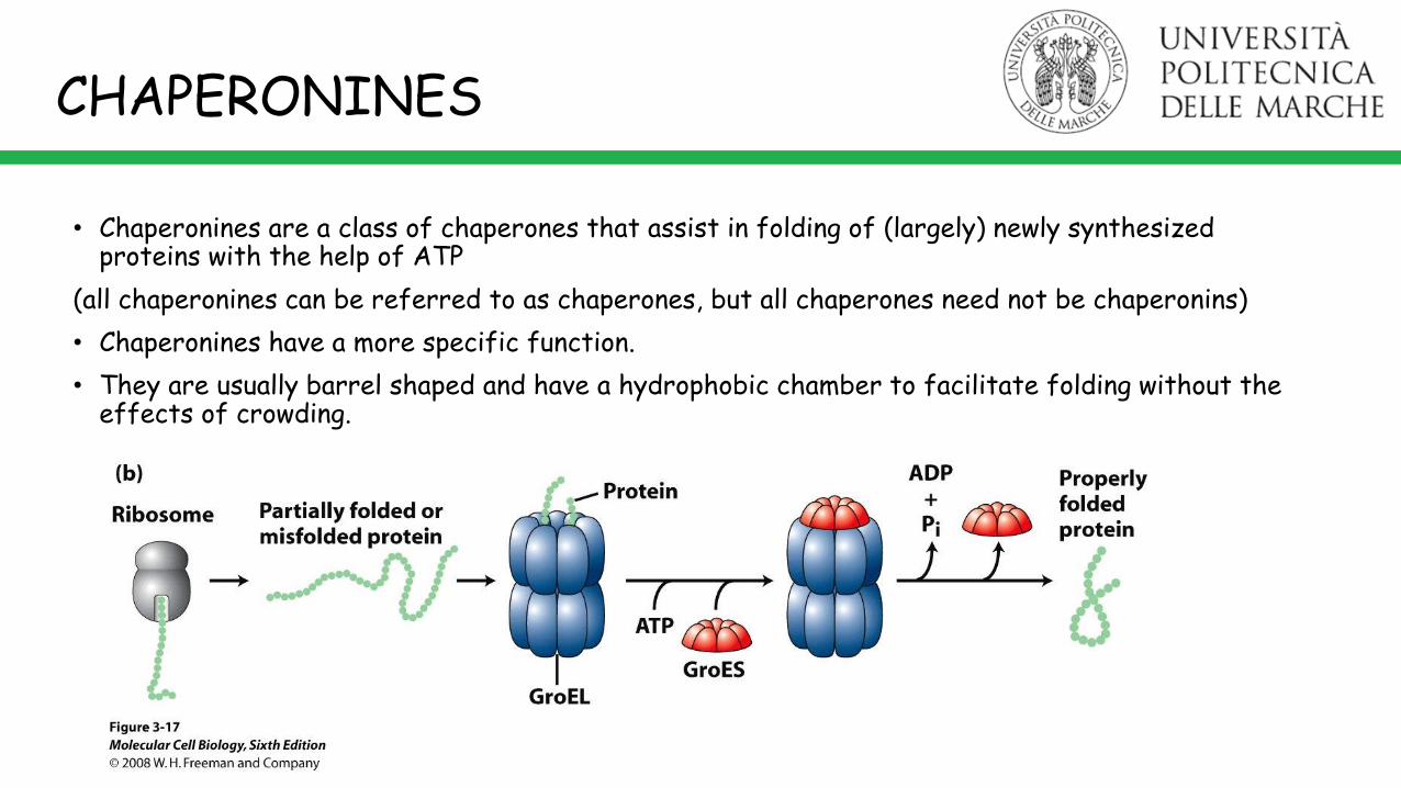
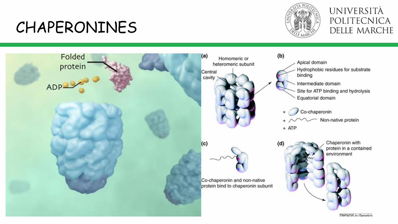
• Chaperonines are a class of chaperones that assist in folding of (largely) newly synthesized proteins with the help of ATP
(all chaperonines can be referred to as chaperones, but all chaperones need not be chaperonins)
• Chaperonines have a more specific function.
• They are usually barrel shaped and have a hydrophobic chamber to facilitate folding without the effects of crowding.
CHAPERONINES
CHAPERONINES
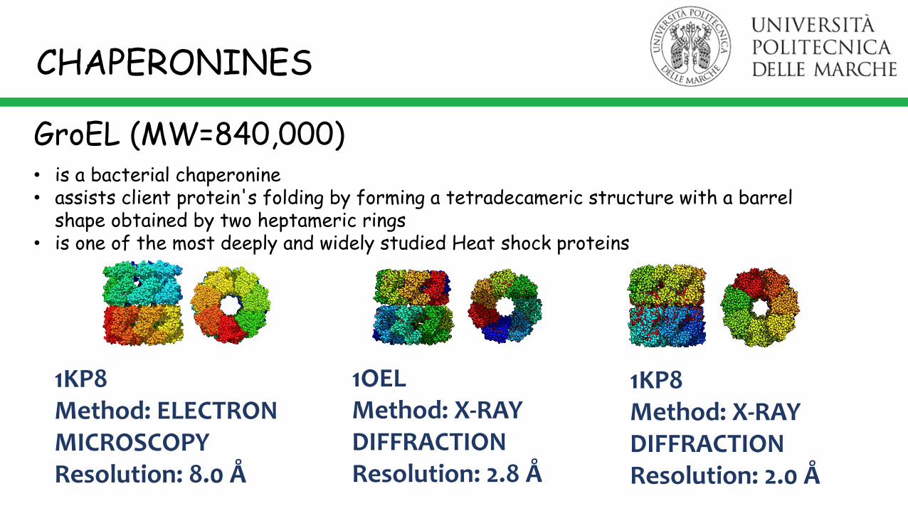
1KP8Method: X-RAY DIFFRACTIONResolution: 2.0 Å
1KP8Method: ELECTRON MICROSCOPYResolution: 8.0 Å
1OELMethod: X-RAY DIFFRACTIONResolution: 2.8 Å
• is a bacterial chaperonine• assists client protein's folding by forming a tetradecameric structure with a barrel
shape obtained by two heptameric rings• is one of the most deeply and widely studied Heat shock proteins
GroEL (MW=840,000)
CHAPERONINES
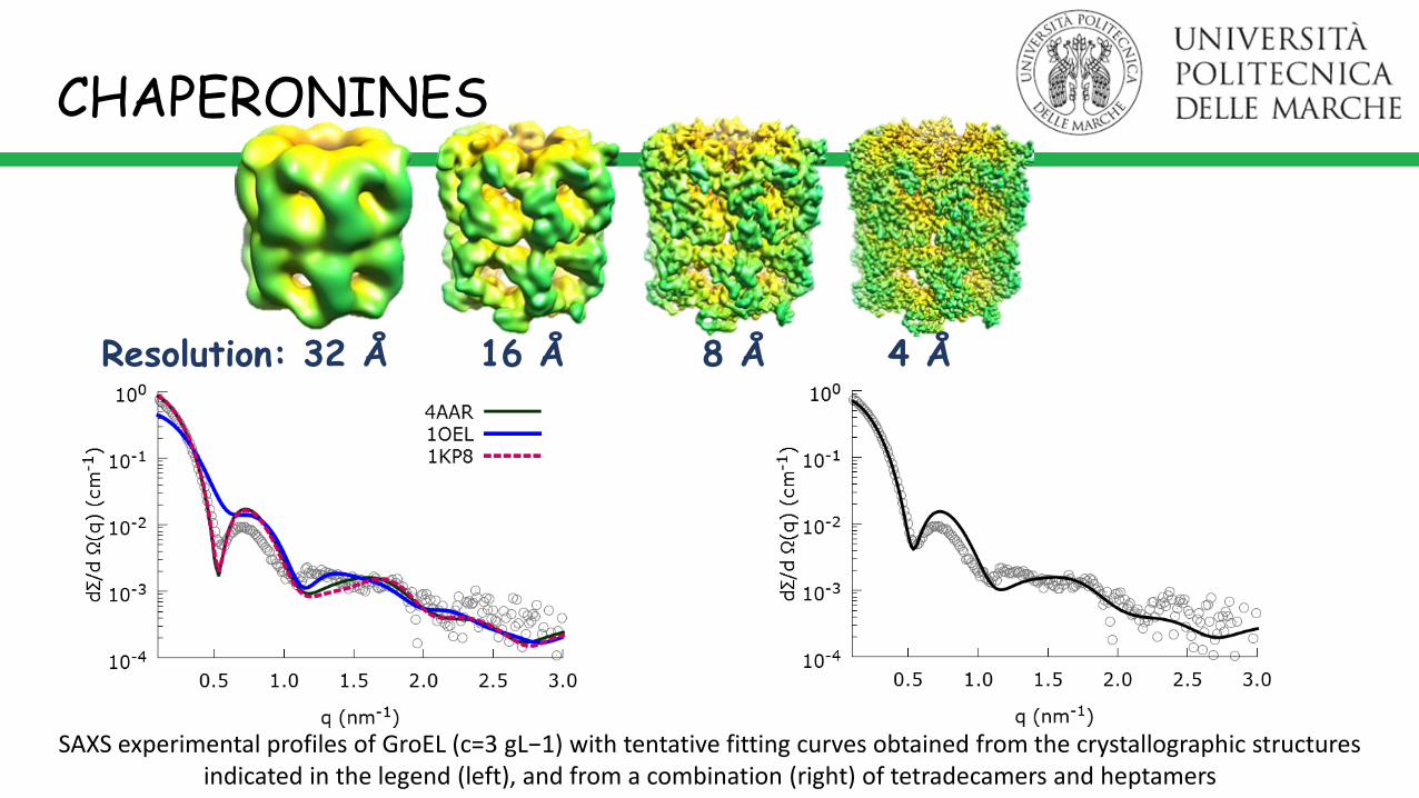
Resolution: 32 Å 16 Å 8 Å 4 Å
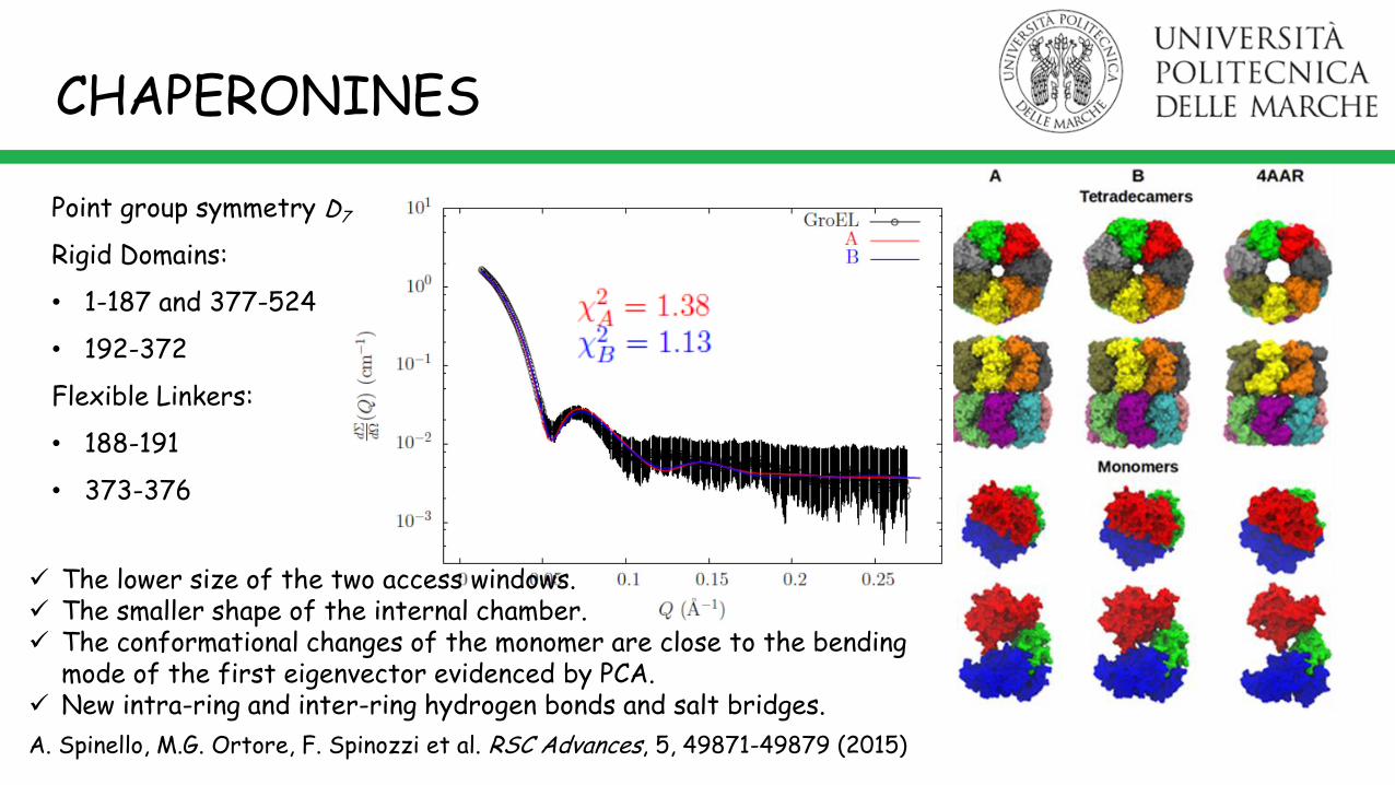
SAXS experimental profiles of GroEL (c=3 gL−1) with tentative fitting curves obtained from the crystallographic structures indicated in the legend (left), and from a combination (right) of tetradecamers and heptamers
CHAPERONINES
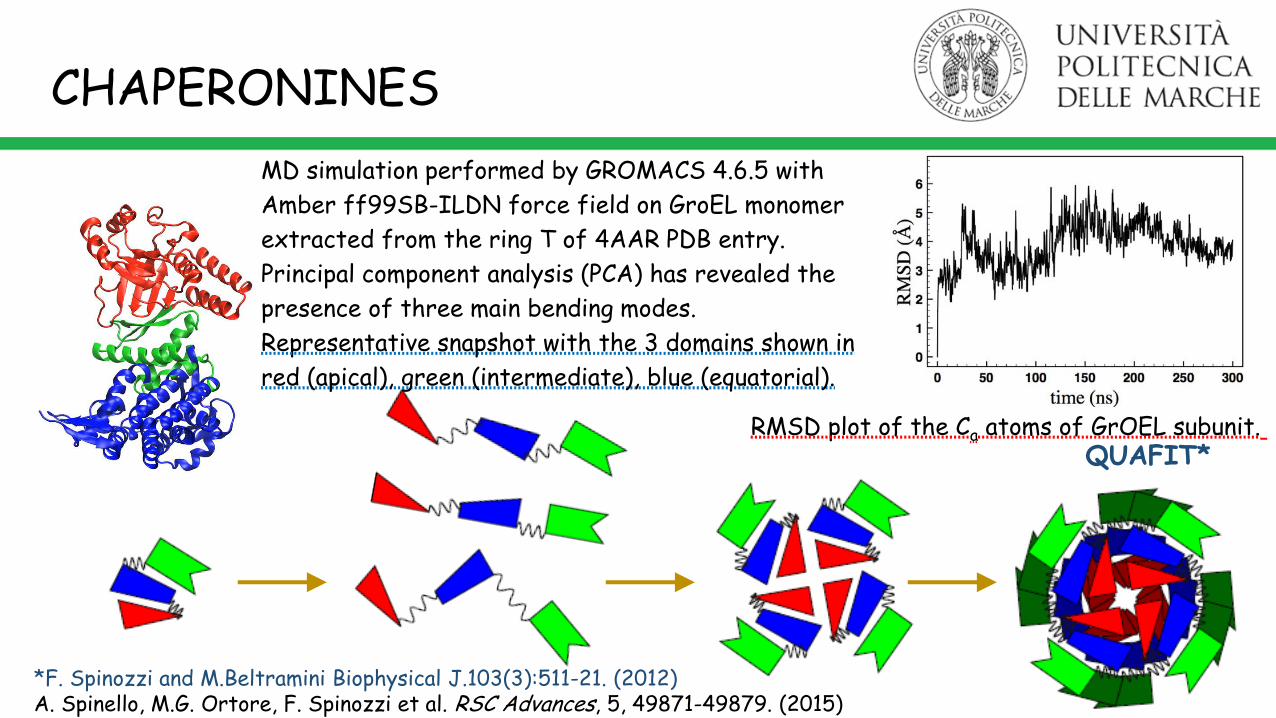
MD simulation performed by GROMACS 4.6.5 with
Amber ff99SB-ILDN force field on GroEL monomer
extracted from the ring T of 4AAR PDB entry.
Principal component analysis (PCA) has revealed the
presence of three main bending modes.
Representative snapshot with the 3 domains shown in
red (apical), green (intermediate), blue (equatorial).
*F. Spinozzi and M.Beltramini Biophysical J.103(3):511-21. (2012)A. Spinello, M.G. Ortore, F. Spinozzi et al. RSC Advances, 5, 49871-49879. (2015)
QUAFIT*
CHAPERONINES
RMSD plot of the Ca atoms of GrOEL subunit.
A. Spinello, M.G. Ortore, F. Spinozzi et al. RSC Advances, 5, 49871-49879 (2015)
Point group symmetry D7
Rigid Domains:
• 1-187 and 377-524
• 192-372
Flexible Linkers:
• 188-191
• 373-376
CHAPERONINES
The lower size of the two access windows. The smaller shape of the internal chamber. The conformational changes of the monomer are close to the bending
mode of the first eigenvector evidenced by PCA. New intra-ring and inter-ring hydrogen bonds and salt bridges.
*S. Vilasi et al. PLoS One 9(5): e97657 (2014)A. Spinello, M.G. Ortore, F. Spinozzi et al. RSC Advances, 5, 49871-49879 (2015)
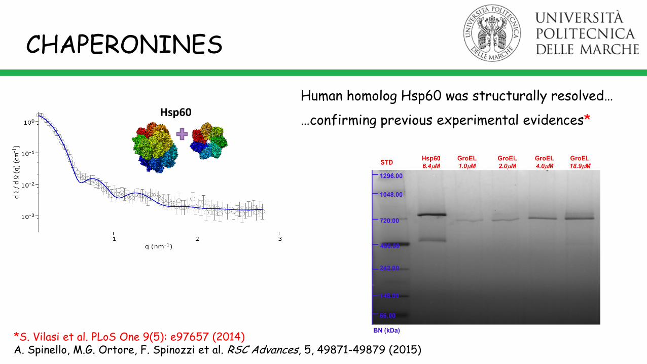
Hsp60
Human homolog Hsp60 was structurally resolved…
…confirming previous experimental evidences*
CHAPERONINES
CHAPERONINES and AMYLOIDS
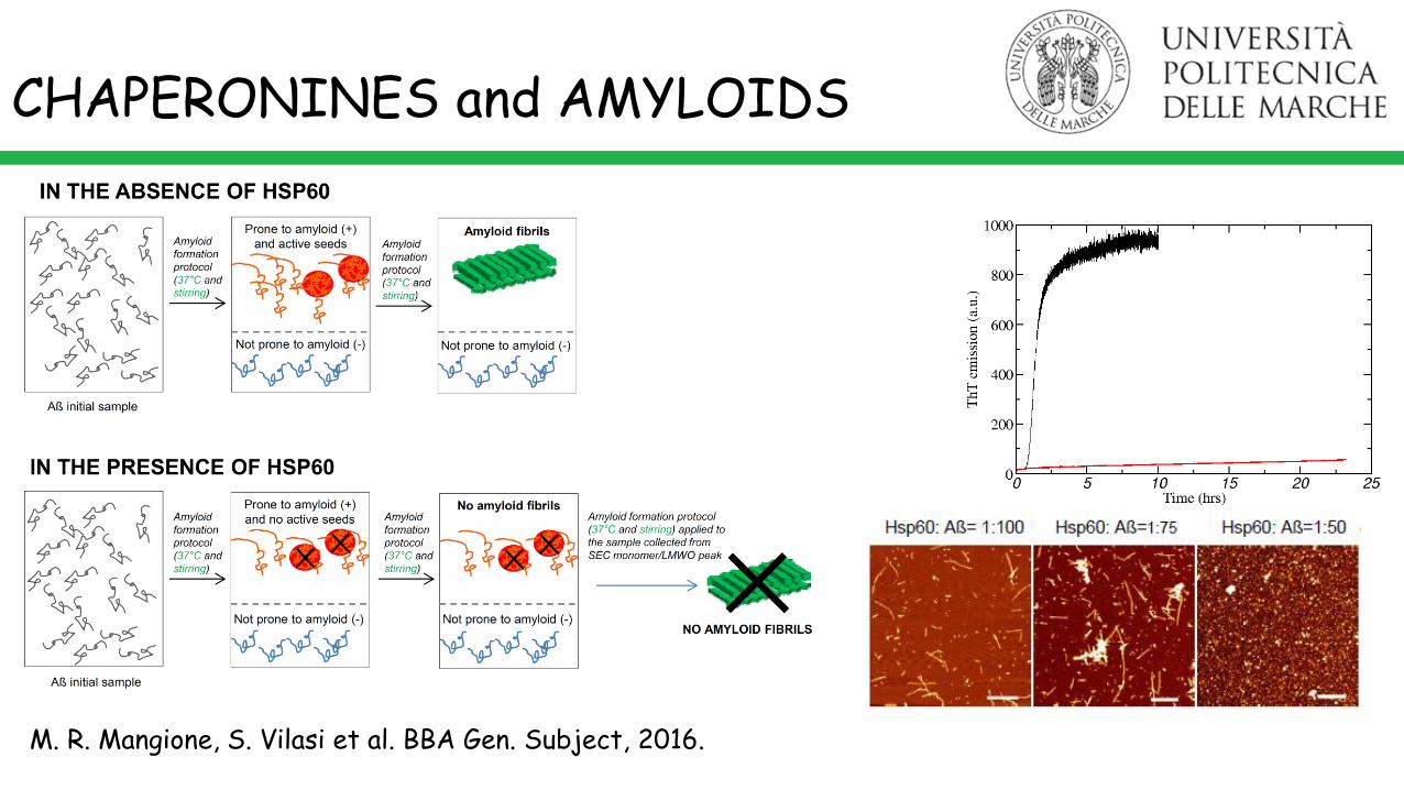
M. R. Mangione, S. Vilasi et al. BBA Gen. Subject, 2016.
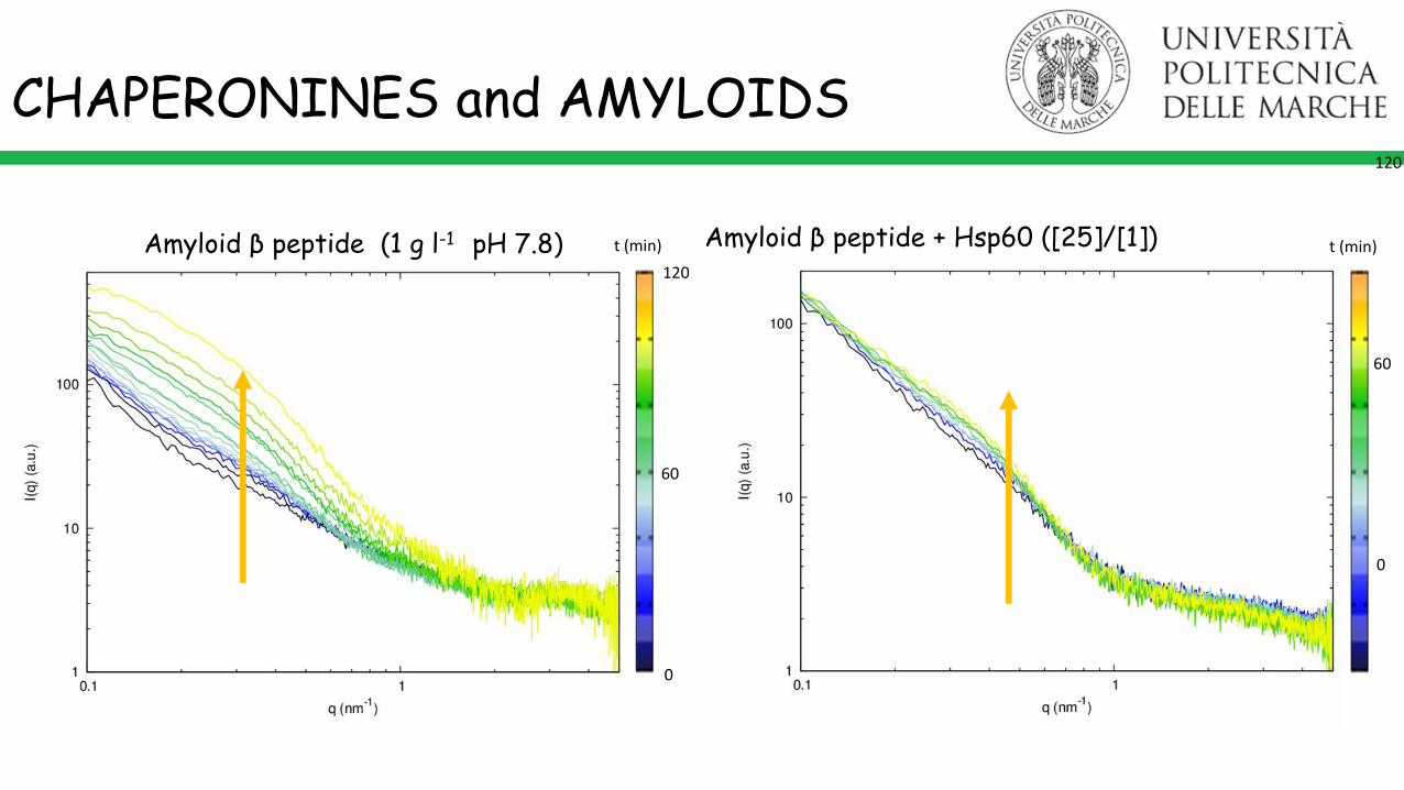
Amyloid β peptide (1 g l-1 pH 7.8) Amyloid β peptide + Hsp60 ([25]/[1])t (min) t (min)
0
120
60
0
120
60
CHAPERONINES and AMYLOIDS

1.390
1.385
1.380
1.375
1.370
6050403020100
404
402
400
398
396
394
392
390
388
x1
03
290
288
286
284
x1
03
cor_len integ_int invariant
1.39
1.38
1.37
1.36
1.35
1.34
6050403020100
7300
7200
7100
7000
6900
6800
6700
5200
5100
5000
4900
4800
cor_len integ_int invariant
Aβ42
Aβ42+Hsp60
Aβ Aβ + HSP60 [25]:[1]
Invariant
IntegratedIntensity
Correlation lengths
CHAPERONINES and AMYLOIDS
Kratky plots evidences that bothwith and without Hsp60 the finalproduct of aggregation is mainlycompact.The average dimension of the finalaggregation is ≈30% greater inabsence of Hsp60
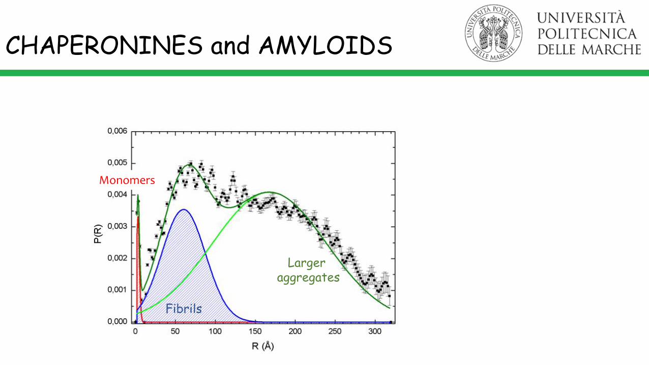
Monomers
Fibrils
Largeraggregates
CHAPERONINES and AMYLOIDS

MONOMERS
FIBRILS
Aβ42 200 μM
Aβ42 200 μM + Hsp60 8 μM
Hsp60 seems to interact with monomers, seizing them and inhibiting their aggregation, slowing the fibril formation.
CHAPERONINES and AMYLOIDS

CHAPERONINES and AMYLOIDS
Hsp70 + α-synuclein
F. A. Aprile, P. Sormanni, M. Vendruscolo. Biochemistry 2015

IN PROGRESS…
SEVERAL PARAMETERS NEED TO BE FURTHER INVESTIGATED
• Crowding
• Co-aggregation phenomena
• Ionic strenght and salts specificity
• Specific elements influence (Cu!)
AND MANY OTHERS
BioMed Research International 2014(1):495091 DOI: 10.1155/2014/495091 ·
The Scientist must set in order.Science is built up with facts, asa house is with stones. But acollection of facts is no more ascience than a heap of stones isa house.Henri Poincaré

Thank you for paying attention!
P. Mariani F. Spinozzi S. Vilasi
J. Teixeira
R. Itri
D. RussoC. Ferrero
C. Ricci
R. Sinibaldi
H. Amenitsch
P. Moretti