Embed Size (px)
Citation preview
Hindawi Publishing CorporationInterdisciplinary Perspectives on Infectious DiseasesVolume 2011, Article ID 462767, 10 pagesdoi:10.1155/2011/462767
Research Article
Changes Related to Age in Natural and Acquired Systemic Self-IgGResponses in Malaria
Romuald Dasse,1, 2 Didier Lefranc,1 Sylvain Dubucquoi,1
Patricia Dussart,1 Virginie Dutoit-Lefevre,1 Boualem Sendid,3
Francois Sombo Mambo,2 Patrick Vermersch,4 and Lionel Prin1
1 Laboratoire d’Immunologie EA 2686, IMPRT-IFR 114, Faculte de Medecine Pole Recherche, Universite Lille 2, 1 Place de Verdun,59045 Lille Cedex, France
2 Laboratoire d’Immunologie et Hematologie du CHU-Cocody, Abidjan, Cote D’Ivoire3 Laboratoire de Parasitologie et de Mycologie, Institute de Biologie et Pathologie, CHRU de Lille 59037 Lille, France4 Service de Neurologie D, Hopital Roger Salengro, 59037 Lille Cedex, France
Correspondence should be addressed to Romuald Dasse, [email protected]
Received 6 July 2011; Accepted 23 September 2011
Academic Editor: Jose G. Estrada-Franco
Copyright © 2011 Romuald Dasse et al. This is an open access article distributed under the Creative Commons AttributionLicense, which permits unrestricted use, distribution, and reproduction in any medium, provided the original work is properlycited.
Background. Absence of acquired protective immunity in endemic areas children leads to higher susceptibility to severe malaria. Toinvestigate the involvement of regulatory process related to self-reactivity, we evaluated potent changes in auto-antibody reactivityprofiles in children and older subjects living in malaria-endemic zones comparatively to none-exposed healthy controls. Methods.Analysis of IgG self-reactive footprints was performed using Western blotting against healthy brain antigens. Plasmas of 102malaria exposed individuals (MEIs) from endemic zone, with or without cerebral malaria (CM) were compared to plasmas fromnon-endemic controls (NECs). Using linear discriminant and principal component analysis, immune footprints were comparedby counting the number, the presence or absence of reactive bands. We identified the most discriminant bands with respectto age and clinical status. Results. A higher number of bands were recognized by IgG auto-antibodies in MEI than in NEC.Characteristic changes in systemic self-IgG-reactive repertoire were found with antigenic bands that discriminate Plasmodiumfalciparum infections with or without CM according to age. 8 antigenic bands distributed in MEI compared with NEC wereidentified while 6 other antigenic bands were distributed within MEI according to the age and clinical status. Such distortionmight be due to evolutionary processes leading to pathogenic/protective events.
1. Introduction
Plasmodium falciparum infection remains a major causeof death in most of developing countries, particularly insub-Saharan Africa [1]. It generates different clinical man-ifestations with either few or no symptoms [2] or severesigns such as impaired neurological function termed cerebralmalaria (CM) [3, 4]. These variable courses of the diseasehave been related to different factors such as the levels ofparasites transmitted, the age of the infected subjects, thegenetic background of the population studied [3, 4], and,above all, the immune status of affected individuals [2].Thus, unprotected children living in endemic zones or
unprotected adults coming from nonendemic zones have ahigh susceptibility to severe malaria [5].
It is postulated that partial protective immunity can beinduced after iterative infection through triggering the actionof the immune response, particularly the humoral response[6, 7]. Recent studies have highlighted the influence of theautoantibody response [8, 9]. Therefore, appropriate analysisof the serum self-IgG repertoire could contribute to a betterunderstanding of the immuneregulation processes involvedduring the course of the disease [10].
In healthy subjects, despite of interindividual differences,the human serum self-IgG response is thought to be well con-served and restricted to the recognition of a few self-antigens
2 Interdisciplinary Perspectives on Infectious Diseases
in autologous tissues [11]. In contrast, durable distortionof these immune profiles has been found in our laboratoryamong patients with multiple sclerosis (MS) or otherautoimmune diseases with predominant neurological signssuch as neuropsychiatric systemic lupus erythematosus [12,13]. When we induced experimental autoimmune enceph-alomyelitis, dynamic changes in immune profiles related topathogenic or protective events were also identified [14,15]. Despite the predominant neurological symptoms inclinical and experimental situations, discriminant self-IgGreactivity involves mostly ubiquitous antigens rather thanspecific targets in nervous system tissue [16]. Although thesefootprints have allowed the identification of new usefulbiomarkers [12, 13], their pathophysiological significanceremains to be defined.
In the present study, we aimed to evaluate the impact ofthe environment and self-reactive natural and acquired anti-body repertoires on humoral immune profiles. The findingsof numerous epidemiological and clinical studies suggest thatthe risk of allergic and autoimmune diseases is related tothe hygiene hypothesis [17]. Parasitic infections, especiallymalaria, may influence the development, or the course ofautoimmune disease such as MS [18]. In contrast some self-reactive antibody responses might also influence the courseof malaria leading to protective [8] or pathogenic events [19].
To further evaluate the relationships between environ-mental factors, autoimmune profiles, and the clinical statusof malaria, the natural and acquired self-IgG antibodyresponses were analyzed in subjects of different age bracketsliving in endemic zones of parasitic transmission. Immuneprofiles were compared between malaria patients with (cere-bral malaria) and without (including uncomplicated disease,asymptomatic Plasmodium falciparum carriers) neurologicalsymptoms. Nonimmune individuals living in countries freeof malaria were tested as controls. Our data revealed thepresence of antigenic bands specifically targeted by plasmaIgG collected in patients of a well-defined clinical status andage range. The pathophysiological significance of such newbiomarkers is discussed.
2. Methods
2.1. Population Studied: Clinical Criteria. Plasma self-IgGreactivity against brain tissue antigens was evaluated in 119subjects. Blood samples were collected from subjects exposedto malaria parasite, termed malaria-exposed individuals([MEI] n = 102, mean age ± SD: 21.07 ± 20.2), andfrom healthy subjects living in European nonendemic areas,termed nonendemic controls ([NEC] n = 17, mean age ±SD: 39 ± 4.5).
The MEI group was divided into two subgroups. Onesubgroup consisted of patients with neurological symptoms,usually termed cerebral malaria ([CM] n = 28, mean age ±SD: 16.2 ± 21.4). The other subgroup was termed MEIwithout neurological symptoms (n = 74, mean age ± SD:21.1 ± 21.3) and was comprised of parasite carriers withclassical symptoms of malaria but without other compli-cations and asymptomatic carriers (without any detectablesymptoms). MEIs with symptoms (neurological and clas-
sical) were recruited from the Emergency Department ofthe University Hospital of Cocody-Abidjan (Cote d’Ivoire).Using available data bases, plasma was classified on the basisof World Health Organization (WHO) guidelines. Thus,MEI with neurological symptoms included patients witha Blantyre Coma scale ≤2 (concerning the children), amodified Glasgow coma scale ≤9 (concerning the adult),the occurrence of at least one convulsive episode during the24 h before admission to the emergency department, andPlasmodium falciparum-positive thin blood smears. As ex-pected, all of these patients were cured after quinine therapy.
Blood samples of asymptomatic parasites carriers wereobtained from the National Blood Transfusion Centre ofAbidjan, from the Mother-Infant Department of the DistrictHospital, and from some volunteer students. All subjectswere included after informed consent from themselves orfrom their parents. All the procedures were reviewed andapproved by the National Health Office Ethics Committee ofCote d’Ivoire.
2.2. Population Studied: Biological Criteria. For the NECgroup, blood samples were obtained from healthy vol-unteers following approval by the “Centre de RessourcesBiologiques-Centre d’Investigation Clinique” of CHRU-Lille. Isolated plasmas were then stored at −40◦C in ourlaboratory (EA 2686, Universite Lille-Nord de France).
For the MEI group, the samples were taken on the day ofadmission (in the emergency department before any therapyconcerning those who were ill, the Mother-Infant Depart-ment, the National Centre of Blood Transfusion, or in theschool for those who were asymptomatic parasite carriers).Venous blood was collected into EDTA sterile vacutainers(5 mL). Plasma was isolated after centrifugation at 500×gfor 15 minutes and stored at −40◦C at the “Laboratoired’Immunologie of CHU Cocody-Abidjan” before being sentthe EA 2686 Research Group (Lille-France).
Parasitemia was assessed at the department of “Para-sitologie-Mycologie, Faculte de Medecine d’Abidjan-Co-cody-Cote d’Ivoire” as follows.
Giemsa-stained thick and thin blood films were preparedfor detection and identification of Plasmodium falciparumand for parasite counts. Parasitemia is expressed as thepercentage of infected red blood cells.
Anti-falciparum total antibodies detection and quantifi-cation were performed at the “Laboratoire de ParasitologieMycologie-Centre de Biologie, de Pathologie-CHRU de Lille-France.” Acetone-fixed Plasmodium falciparum strain 3D7was used as antigen using indirect immunofluorescence.Briefly, sera were diluted 1/100, 1/200, 1/400, 1/800, 1/1600,and 1/3200 in phosphate-buffered saline (Ref 77511 Biom-erieux). Diluted serum was incubated with acetone-fixedPlasmodium falciparum at 37◦C for 30 mn. The secondaryantibody used was fluorescein-conjugated goat anti-humanIgG, IgM, IgA, Heavy and Light chains-H, and L-(Ref 74511Diagnostic Pasteur) in Bleu Evans solution (Ref 75491Biomerieux). Any fluorescence in Fluopep (Ref 75521 Biom-erieux) obtained at a dilution 1/100 was considered negative.Quantification was expressed by the titre of total antifalci-parum antibodies.
Interdisciplinary Perspectives on Infectious Diseases 3
2.3. Brain Samples. Brain samples were obtained as previ-ously described [20, 21]. Briefly, cerebral tissue was extractedby autopsy from the frontal lobe in Broadman’s area 10,from healthy subjects with no history of neurological disease(Department of Neuropathology, Centre Hospitalier del’Universite de Lille, and Institut National de la Sante, de laRecherche Medicale, Unite 422, Lille, France). Autopsies wereperformed within the framework of a tissue collection pro-gram that had been approved by the local ethics committee.
2.4. Sodium Dodecyl Sulfate—Polyacrylamide Gel Electropho-resis (SDS-PAGE). The brain samples were homogenized ina Tris buffer containing 5% SDS at a final concentrationof 10 mg/mL and heated at 95◦C for 10 min. 100 μL of thislysate was loaded onto a 10–20% gradient polyacrylamide gel(Navix Tris-Glycine Gels-Invitrogen), beside a well of 15 μLof molecular mass marker (Amersham Pharmacia Biotech).Just before 1-dimension electrophoresis (1D), the homoge-nates were reduced for 10 min with 10 mM DTT (Sigma-Aldrich) at 100◦C. Then electrophoresis was run in Laemmlibuffer using the following conditions: run time 2 h atconstant 125 V and 30–40 mA/gel (start); 8–12 mA/gel (end).
2.5. Western-Blot Analysis. Proteins were transferred ontoECL nitrocellulose membranes (Amersham PharmaciaBiotech) at 0.8 mA/cm2 and later saturated with 5% fat-free dried milk. Nitrocellulose membrane was cut into 15–20strips, 3 to 4 mm wide. Western blotting was conducted withtotal sera, diluted 1/100 in TNT (100 mM Tris—pH 8.3-,0.3 M NaCl containing 0.5% Tween) and 5% fat-free driedmilk. Reaction IgG antibodies were revealed with an anti-human Fcγ HRP-conjugated antibody (1/10000; Sigma-Aldrich) after overnight incubation at 4◦C. Fluorogramswere prepared using an enhanced chemiluminescence kit(Amersham Pharmacia Biotech Europe). Immune profileswere analyzed when 2 independent assays had producedidentical patterns. Image analysis was performed on aMolecular Imager GS-800 Calibrated Densitometer (Bio-Rad) to localize and compare the IgG immune profilepatterns. Band detection, alignment, and matching of theantibody reactivities were performed using Phoretix 1D and1D Pro software version 10 (Totallab-Nonlinear Dynamics)and validated by two operators. We performed comparativeanalyses using detection-band parameters that allowed usto consider each band intensity that was >13% higher thanthe global background intensity of the gel to be significant.To calibrate and more accurately define the alignmentof antibody reactivities, molecular weight marker proteinstandards (StdMW-Benchmark Pre-Stained Protein Ladder,Invitrogen) as well as internal reference standards were used.The level to which each molecular standard migrates is calledrelative front of migration (RF). A function curve defined byRF values on the X axis and standard molecular weight on theY axis allowed the molecular weight of each antigenic bandto be determined by extrapolation because the reactive bandalign on its RF.
2.6. Statistical Analysis. Data were expressed in binary mode(0 = absence of an antigenic band and 1 = presence of an
antigenic band) for submission of IgG antibody patterns foranalysis using either the chi-square test or Fisher’s exact test.This approach allowed us to select the most relevant antigensthat qualitatively supported different types of immune recog-nition. We then used linear discriminant analysis (LDA) tobalance the discriminating antigens between the populationsof individuals, as described previously [22, 23]. Using astepwise logistic regression model and supported by the LDAmethod, we isolated a subgroup of brain antigens accordingto their strength of discrimination between the differentpopulations involved in the study. Using 2 parameters (forthe presence—1—or absence—0—of each selected antigen)and the coefficient previously defined by the LDA, a scorewas calculated for each subject as a representative value ofthe individual immune profile, using the following formula:
Score = Ag1coef1× [0(absent) or 1(present
)]
+ Ag2coef2× [0(absent) or 1(present
)]+ Ag3coef3,
(1)
where Ag represents antigen and coef represents coefficient.The calculated scores were then represented graphically.
A threshold value was determined using a receiver operatingcharacteristic curve, and the sensitivity and specificity ofthis approach were evaluated. When the number of patientswas too small to apply LDA, chi-square and Fisher’s exacttests were performed to retain the most discriminant band.To compare the number of bands between the differentgroups of subjects, we used the chi-square test. The frequencyof each discriminant self-reactive band has been calculateddetermining its percentage of its presence within the groupof subjects.
Using principal component analysis (PCA) and nonpara-metric test of Kruskal-Wallis, we evaluated the relationshipbetween the discriminant band and the clinical status.
3. Results
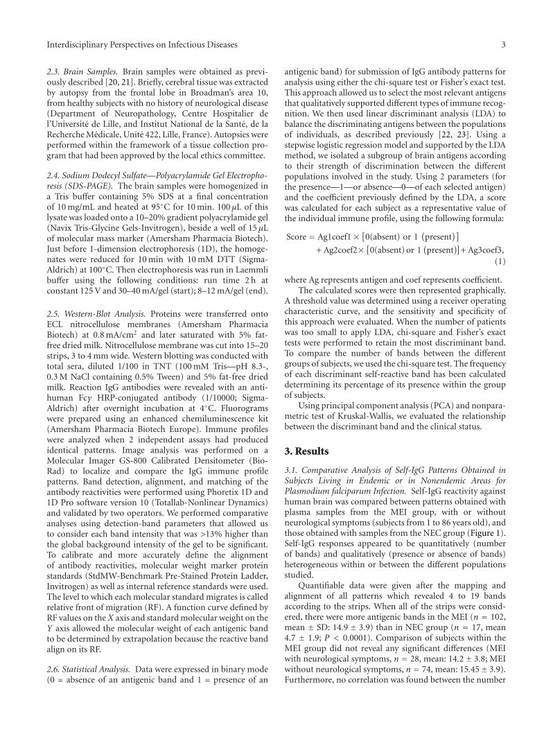
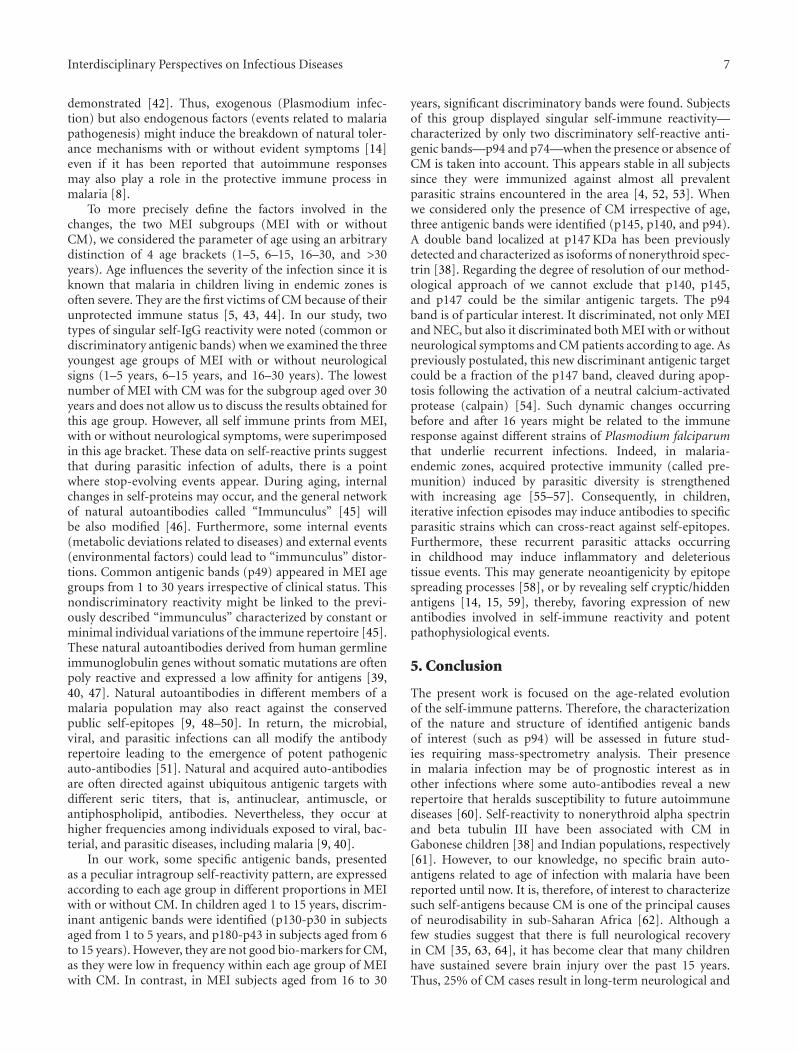
3.1. Comparative Analysis of Self-IgG Patterns Obtained inSubjects Living in Endemic or in Nonendemic Areas forPlasmodium falciparum Infection. Self-IgG reactivity againsthuman brain was compared between patterns obtained withplasma samples from the MEI group, with or withoutneurological symptoms (subjects from 1 to 86 years old), andthose obtained with samples from the NEC group (Figure 1).Self-IgG responses appeared to be quantitatively (numberof bands) and qualitatively (presence or absence of bands)heterogeneous within or between the different populationsstudied.
Quantifiable data were given after the mapping andalignment of all patterns which revealed 4 to 19 bandsaccording to the strips. When all of the strips were consid-ered, there were more antigenic bands in the MEI (n = 102,mean ± SD: 14.9 ± 3.9) than in NEC group (n = 17, mean4.7 ± 1.9; P < 0.0001). Comparison of subjects within theMEI group did not reveal any significant differences (MEIwith neurological symptoms, n = 28, mean: 14.2± 3.8; MEIwithout neurological symptoms, n = 74, mean: 15.45± 3.9).Furthermore, no correlation was found between the number
4 Interdisciplinary Perspectives on Infectious Diseases
Non-endemic controls (NECs)
1 111 1 11 1 1 1
112 3 4 5
2 23 4 5
6 7 8 9 1 2 3 4 5 6 7 8 91 3 4 5 6 7 8 90 0
p180p115
p82
p64
p49p37p26
p15p19
p94
p74
p6
p43
Stan
dard
mol
ecu
lar
wei
ght
(kD
a)Without neurological
manifestationsWith neurological
manifestations
Malaria-exposed individuals (MEIs)
Common intergroup bands
Common intragroup bands
Figure 1: Representative IgG autoreactive patterns obtained with plasma from subjects living in malaria endemic area. The tested plasmaswere obtained from malaria-exposed individuals (MEIs) with or without neurological manifestations, of all ages (from 1 to 86 years old).In parallel nonexposed healthy subjects called nonendemic controls (NECs) were also evaluated. For these western blot analyses, proteinextracts of the central nervous system (CNS) tissue collected in one subject with no history of neurological disease were used as antigenictargets. Despite the heterogeneous band patterns, comparison of the strips allowed identification of some singular antigenic bands. Somebands were shared by different groups of subjects (vertical arrows). In contrast, other bands were specifically found in some groups ofsubjects (diagonal arrows). As illustrated, p94 and p43 protein bands were specifically found in either MEI with neurological symptoms orin MEI without neurological symptoms.
of protein bands within MEI and the titre of antifalciparumantibodies evaluated by the procedure described in materialsand methods (parametric test of Pearson used for MEI withneurological symptoms, P = 0.625 and in MEI withoutneurological symptoms, P = 0.974).
For qualitative analysis, the presence or the absence ofeach protein band for all bands identified in the 119 strips(n = 42) was evaluated on each strip. Despite interindividualdifferences, some conserved sets of protein bands werefound. As indicated in Figure 1, common responses werenoted either in the same group (MEI with neurologicalmanifestations) or between distinct groups (MEI with neu-rological symptoms, MEI without neurological symptoms,and NEC).
3.2. Identification of Discriminant Antigenic Bands
Targeted by Self-IgG
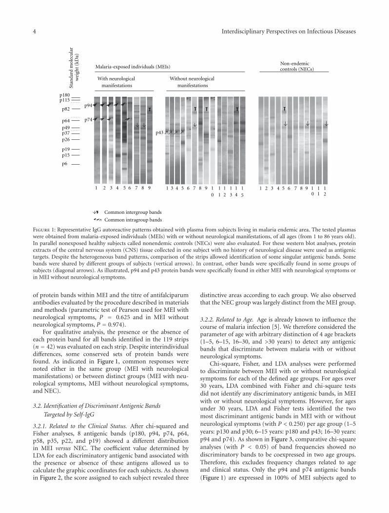
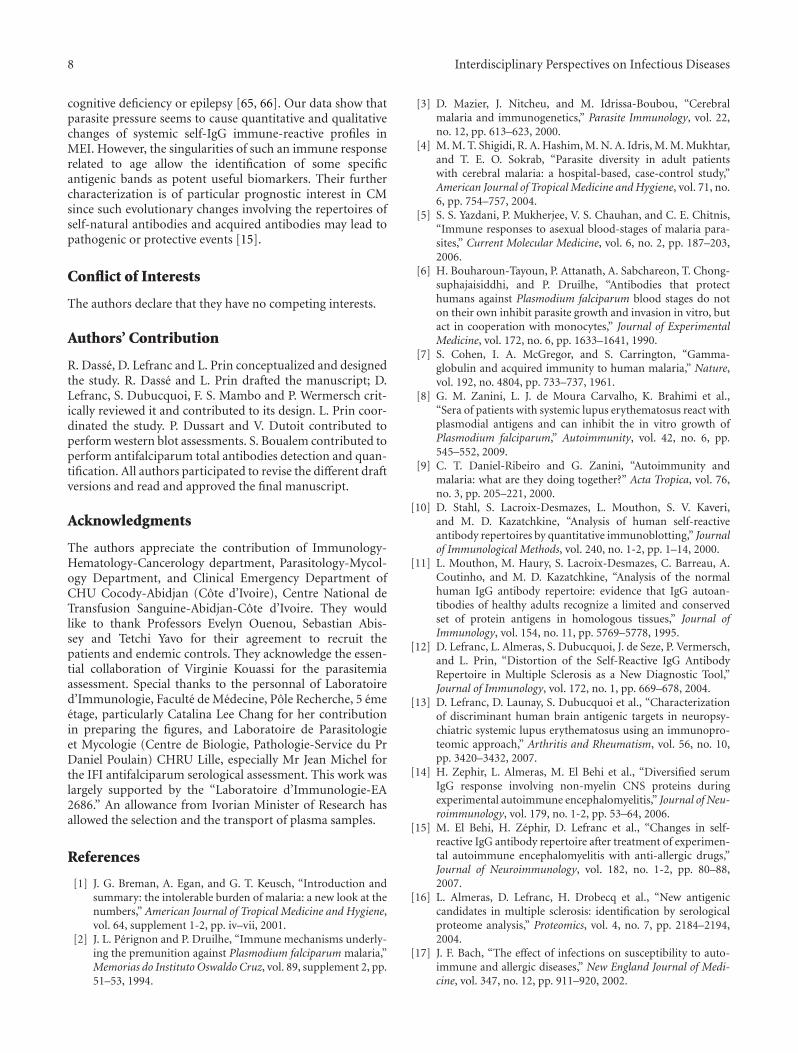
3.2.1. Related to the Clinical Status. After chi-squared andFisher analyses, 8 antigenic bands (p180, p94, p74, p64,p58, p35, p22, and p19) showed a different distributionin MEI versus NEC. The coefficient value determined byLDA for each discriminatory antigenic band associated withthe presence or absence of these antigens allowed us tocalculate the graphic coordinates for each subjects. As shownin Figure 2, the score assigned to each subject revealed three
distinctive areas according to each group. We also observedthat the NEC group was largely distinct from the MEI group.
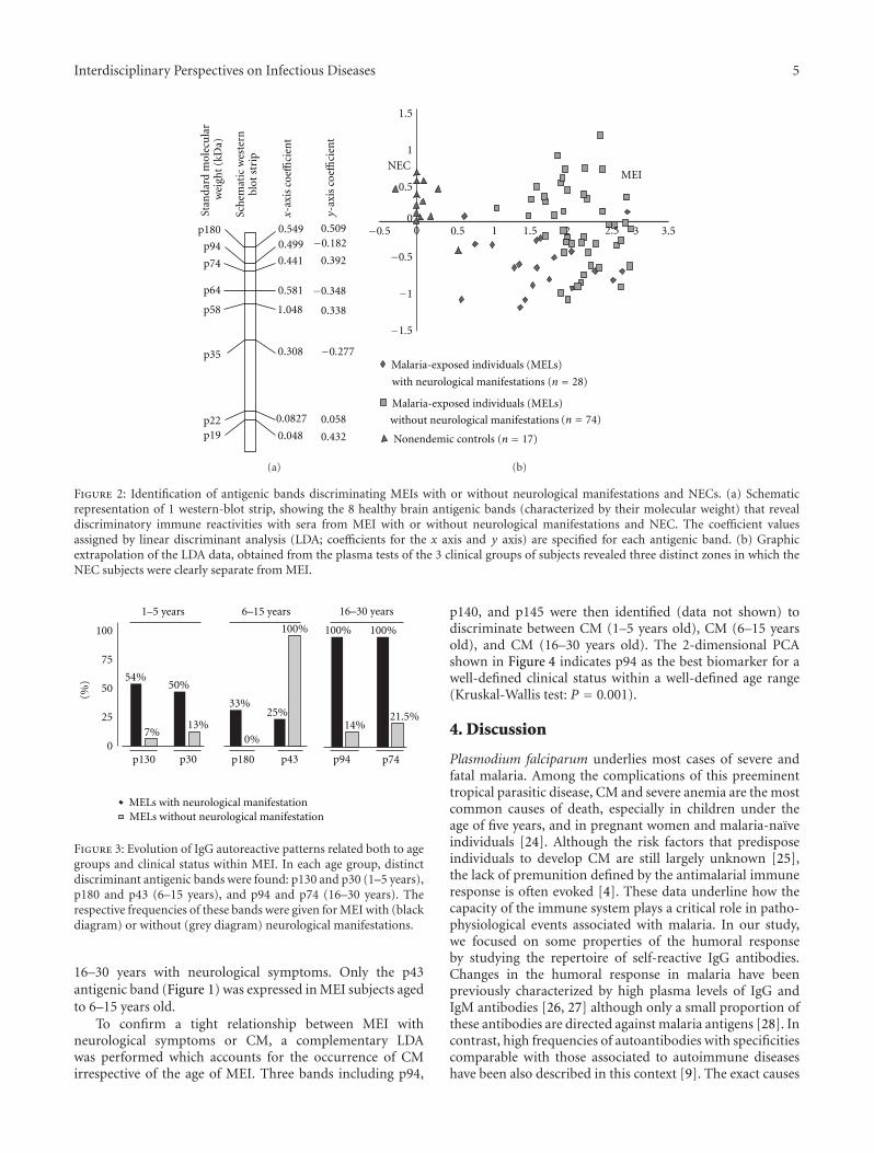
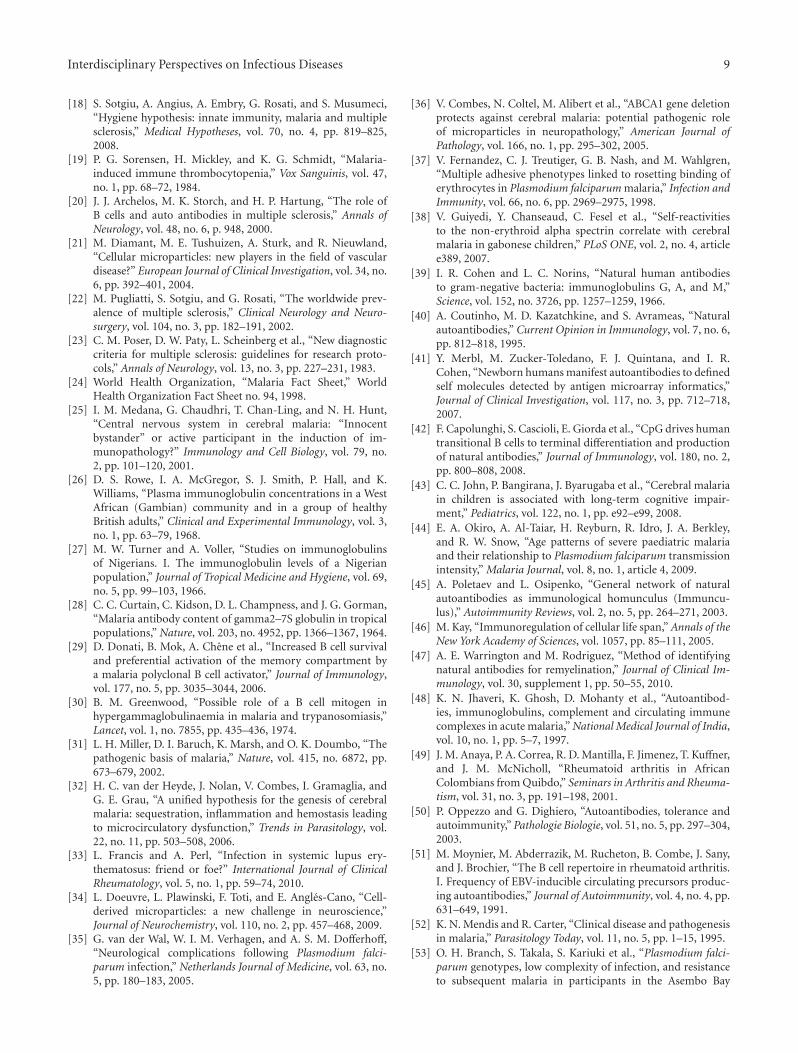
3.2.2. Related to Age. Age is already known to influence thecourse of malaria infection [5]. We therefore considered theparameter of age with arbitrary distinction of 4 age brackets(1–5, 6–15, 16–30, and >30 years) to detect any antigenicbands that discriminate between malaria with or withoutneurological symptoms.
Chi-square, Fisher, and LDA analyses were performedto discriminate between MEI with or without neurologicalsymptoms for each of the defined age groups. For ages over30 years, LDA combined with Fisher and chi-square testsdid not identify any discriminatory antigenic bands, in MEIwith or without neurological symptoms. However, for agesunder 30 years, LDA and Fisher tests identified the twomost discriminant antigenic bands in MEI with or withoutneurological symptoms (with P < 0.250) per age group (1–5years: p130 and p30; 6–15 years: p180 and p43; 16–30 years:p94 and p74). As shown in Figure 3, comparative chi-squareanalyses (with P < 0.05) of band frequencies showed nodiscriminatory bands to be coexpressed in two age groups.Therefore, this excludes frequency changes related to ageand clinical status. Only the p94 and p74 antigenic bands(Figure 1) are expressed in 100% of MEI subjects aged to
Interdisciplinary Perspectives on Infectious Diseases 5
p180
p94
p74
p64
p58
p35
p22p19 0.048
0.0827
0.308
1.048
0.581
0.441
0.4990.549 0.509
−0.182
0.392
0.338
−0.277
0.058
0.432
Sch
emat
icw
este
rnbl
otst
rip
−0.348
Stan
dard
mol
ecu
lar
wei
ght
(kD
a)
x-ax
is
y-ax
is c
oeffi
cien
t
coeffi
cien
t
(a)
−0.5
0 0.5 1 1.5 2 2.5 3 3.5
1.5
1
0.5
0−0.5
−1
−1.5
with neurological manifestations (n = 28)
(n = 74)
NECMEI
without neurological manifestations
Nonendemic controls (n = 17)
Malaria-exposed individuals (MELs)
Malaria-exposed individuals (MELs)
(b)
Figure 2: Identification of antigenic bands discriminating MEIs with or without neurological manifestations and NECs. (a) Schematicrepresentation of 1 western-blot strip, showing the 8 healthy brain antigenic bands (characterized by their molecular weight) that revealdiscriminatory immune reactivities with sera from MEI with or without neurological manifestations and NEC. The coefficient valuesassigned by linear discriminant analysis (LDA; coefficients for the x axis and y axis) are specified for each antigenic band. (b) Graphicextrapolation of the LDA data, obtained from the plasma tests of the 3 clinical groups of subjects revealed three distinct zones in which theNEC subjects were clearly separate from MEI.
0
25
50
75
100
54%
7%
50%
13%
33%
0%
25%
100% 100%100%
14%
p130 p30 p180 p43 p94 p74
1–5 years 6–15 years 16–30 years
21.5%
(%)
MELs with neurological manifestationMELs without neurological manifestation
Figure 3: Evolution of IgG autoreactive patterns related both to agegroups and clinical status within MEI. In each age group, distinctdiscriminant antigenic bands were found: p130 and p30 (1–5 years),p180 and p43 (6–15 years), and p94 and p74 (16–30 years). Therespective frequencies of these bands were given for MEI with (blackdiagram) or without (grey diagram) neurological manifestations.
16–30 years with neurological symptoms. Only the p43antigenic band (Figure 1) was expressed in MEI subjects agedto 6–15 years old.
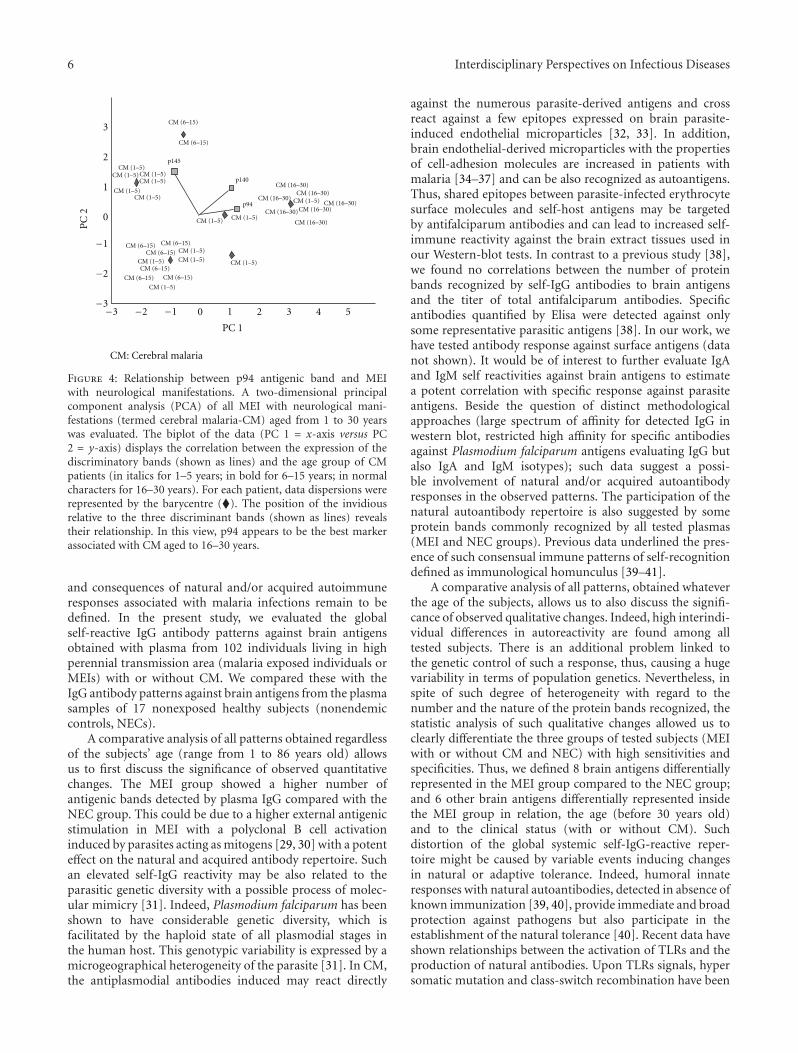
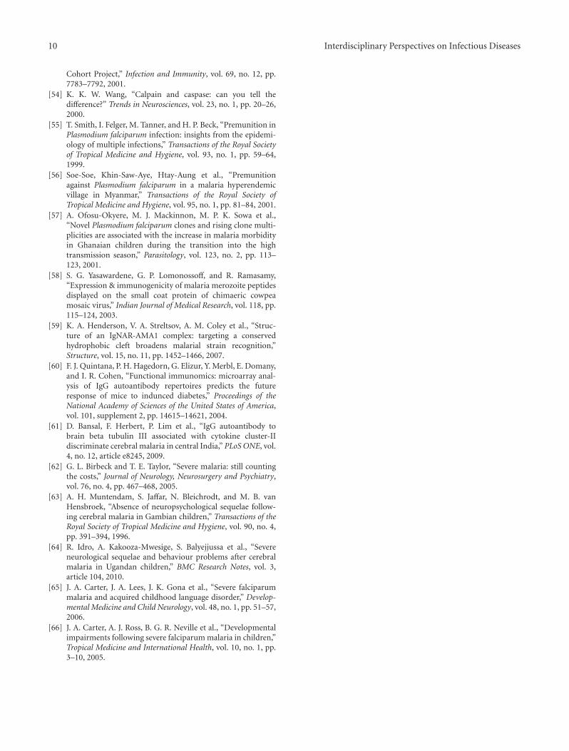
To confirm a tight relationship between MEI withneurological symptoms or CM, a complementary LDAwas performed which accounts for the occurrence of CMirrespective of the age of MEI. Three bands including p94,
p140, and p145 were then identified (data not shown) todiscriminate between CM (1–5 years old), CM (6–15 yearsold), and CM (16–30 years old). The 2-dimensional PCAshown in Figure 4 indicates p94 as the best biomarker for awell-defined clinical status within a well-defined age range(Kruskal-Wallis test: P = 0.001).
4. Discussion
Plasmodium falciparum underlies most cases of severe andfatal malaria. Among the complications of this preeminenttropical parasitic disease, CM and severe anemia are the mostcommon causes of death, especially in children under theage of five years, and in pregnant women and malaria-naıveindividuals [24]. Although the risk factors that predisposeindividuals to develop CM are still largely unknown [25],the lack of premunition defined by the antimalarial immuneresponse is often evoked [4]. These data underline how thecapacity of the immune system plays a critical role in patho-physiological events associated with malaria. In our study,we focused on some properties of the humoral responseby studying the repertoire of self-reactive IgG antibodies.Changes in the humoral response in malaria have beenpreviously characterized by high plasma levels of IgG andIgM antibodies [26, 27] although only a small proportion ofthese antibodies are directed against malaria antigens [28]. Incontrast, high frequencies of autoantibodies with specificitiescomparable with those associated to autoimmune diseaseshave been also described in this context [9]. The exact causes
6 Interdisciplinary Perspectives on Infectious Diseases
−3 −2 −1 0 1 2 3 4 5
PC
2
PC 1
−3
−2
−1
0
1
2
3
p145
p140
p94
CM (6–15)
CM (6–15)
CM (6–15)
CM (6–15)
CM (6–15)CM (6–15)
CM (6–15)CM (6–15)
CM (1–5)CM (1–5)CM (1–5)
CM (1–5)
CM (1–5)
CM (1–5)
CM (1–5)CM (1–5) CM (1–5)
CM (1–5) CM (1–5)
CM (1–5)
CM (1–5)
CM (1–5)
CM (16–30)
CM (16–30)
CM (16–30)CM (16–30)
CM (16–30)CM (16–30)
CM (16–30)
CM: Cerebral malaria
Figure 4: Relationship between p94 antigenic band and MEIwith neurological manifestations. A two-dimensional principalcomponent analysis (PCA) of all MEI with neurological mani-festations (termed cerebral malaria-CM) aged from 1 to 30 yearswas evaluated. The biplot of the data (PC 1 = x-axis versus PC2 = y-axis) displays the correlation between the expression of thediscriminatory bands (shown as lines) and the age group of CMpatients (in italics for 1–5 years; in bold for 6–15 years; in normalcharacters for 16–30 years). For each patient, data dispersions wererepresented by the barycentre (�). The position of the invidiousrelative to the three discriminant bands (shown as lines) revealstheir relationship. In this view, p94 appears to be the best markerassociated with CM aged to 16–30 years.
and consequences of natural and/or acquired autoimmuneresponses associated with malaria infections remain to bedefined. In the present study, we evaluated the globalself-reactive IgG antibody patterns against brain antigensobtained with plasma from 102 individuals living in highperennial transmission area (malaria exposed individuals orMEIs) with or without CM. We compared these with theIgG antibody patterns against brain antigens from the plasmasamples of 17 nonexposed healthy subjects (nonendemiccontrols, NECs).
A comparative analysis of all patterns obtained regardlessof the subjects’ age (range from 1 to 86 years old) allowsus to first discuss the significance of observed quantitativechanges. The MEI group showed a higher number ofantigenic bands detected by plasma IgG compared with theNEC group. This could be due to a higher external antigenicstimulation in MEI with a polyclonal B cell activationinduced by parasites acting as mitogens [29, 30] with a potenteffect on the natural and acquired antibody repertoire. Suchan elevated self-IgG reactivity may be also related to theparasitic genetic diversity with a possible process of molec-ular mimicry [31]. Indeed, Plasmodium falciparum has beenshown to have considerable genetic diversity, which isfacilitated by the haploid state of all plasmodial stages inthe human host. This genotypic variability is expressed by amicrogeographical heterogeneity of the parasite [31]. In CM,the antiplasmodial antibodies induced may react directly
against the numerous parasite-derived antigens and crossreact against a few epitopes expressed on brain parasite-induced endothelial microparticles [32, 33]. In addition,brain endothelial-derived microparticles with the propertiesof cell-adhesion molecules are increased in patients withmalaria [34–37] and can be also recognized as autoantigens.Thus, shared epitopes between parasite-infected erythrocytesurface molecules and self-host antigens may be targetedby antifalciparum antibodies and can lead to increased self-immune reactivity against the brain extract tissues used inour Western-blot tests. In contrast to a previous study [38],we found no correlations between the number of proteinbands recognized by self-IgG antibodies to brain antigensand the titer of total antifalciparum antibodies. Specificantibodies quantified by Elisa were detected against onlysome representative parasitic antigens [38]. In our work, wehave tested antibody response against surface antigens (datanot shown). It would be of interest to further evaluate IgAand IgM self reactivities against brain antigens to estimatea potent correlation with specific response against parasiteantigens. Beside the question of distinct methodologicalapproaches (large spectrum of affinity for detected IgG inwestern blot, restricted high affinity for specific antibodiesagainst Plasmodium falciparum antigens evaluating IgG butalso IgA and IgM isotypes); such data suggest a possi-ble involvement of natural and/or acquired autoantibodyresponses in the observed patterns. The participation of thenatural autoantibody repertoire is also suggested by someprotein bands commonly recognized by all tested plasmas(MEI and NEC groups). Previous data underlined the pres-ence of such consensual immune patterns of self-recognitiondefined as immunological homunculus [39–41].
A comparative analysis of all patterns, obtained whateverthe age of the subjects, allows us to also discuss the signifi-cance of observed qualitative changes. Indeed, high interindi-vidual differences in autoreactivity are found among alltested subjects. There is an additional problem linked tothe genetic control of such a response, thus, causing a hugevariability in terms of population genetics. Nevertheless, inspite of such degree of heterogeneity with regard to thenumber and the nature of the protein bands recognized, thestatistic analysis of such qualitative changes allowed us toclearly differentiate the three groups of tested subjects (MEIwith or without CM and NEC) with high sensitivities andspecificities. Thus, we defined 8 brain antigens differentiallyrepresented in the MEI group compared to the NEC group;and 6 other brain antigens differentially represented insidethe MEI group in relation, the age (before 30 years old)and to the clinical status (with or without CM). Suchdistortion of the global systemic self-IgG-reactive reper-toire might be caused by variable events inducing changesin natural or adaptive tolerance. Indeed, humoral innateresponses with natural autoantibodies, detected in absence ofknown immunization [39, 40], provide immediate and broadprotection against pathogens but also participate in theestablishment of the natural tolerance [40]. Recent data haveshown relationships between the activation of TLRs and theproduction of natural antibodies. Upon TLRs signals, hypersomatic mutation and class-switch recombination have been
Interdisciplinary Perspectives on Infectious Diseases 7
demonstrated [42]. Thus, exogenous (Plasmodium infec-tion) but also endogenous factors (events related to malariapathogenesis) might induce the breakdown of natural toler-ance mechanisms with or without evident symptoms [14]even if it has been reported that autoimmune responsesmay also play a role in the protective immune process inmalaria [8].
To more precisely define the factors involved in thechanges, the two MEI subgroups (MEI with or withoutCM), we considered the parameter of age using an arbitrarydistinction of 4 age brackets (1–5, 6–15, 16–30, and >30years). Age influences the severity of the infection since it isknown that malaria in children living in endemic zones isoften severe. They are the first victims of CM because of theirunprotected immune status [5, 43, 44]. In our study, twotypes of singular self-IgG reactivity were noted (common ordiscriminatory antigenic bands) when we examined the threeyoungest age groups of MEI with or without neurologicalsigns (1–5 years, 6–15 years, and 16–30 years). The lowestnumber of MEI with CM was for the subgroup aged over 30years and does not allow us to discuss the results obtained forthis age group. However, all self immune prints from MEI,with or without neurological symptoms, were superimposedin this age bracket. These data on self-reactive prints suggestthat during parasitic infection of adults, there is a pointwhere stop-evolving events appear. During aging, internalchanges in self-proteins may occur, and the general networkof natural autoantibodies called “Immunculus” [45] willbe also modified [46]. Furthermore, some internal events(metabolic deviations related to diseases) and external events(environmental factors) could lead to “immunculus” distor-tions. Common antigenic bands (p49) appeared in MEI agegroups from 1 to 30 years irrespective of clinical status. Thisnondiscriminatory reactivity might be linked to the previ-ously described “immunculus” characterized by constant orminimal individual variations of the immune repertoire [45].These natural autoantibodies derived from human germlineimmunoglobulin genes without somatic mutations are oftenpoly reactive and expressed a low affinity for antigens [39,40, 47]. Natural autoantibodies in different members of amalaria population may also react against the conservedpublic self-epitopes [9, 48–50]. In return, the microbial,viral, and parasitic infections can all modify the antibodyrepertoire leading to the emergence of potent pathogenicauto-antibodies [51]. Natural and acquired auto-antibodiesare often directed against ubiquitous antigenic targets withdifferent seric titers, that is, antinuclear, antimuscle, orantiphospholipid, antibodies. Nevertheless, they occur athigher frequencies among individuals exposed to viral, bac-terial, and parasitic diseases, including malaria [9, 40].
In our work, some specific antigenic bands, presentedas a peculiar intragroup self-reactivity pattern, are expressedaccording to each age group in different proportions in MEIwith or without CM. In children aged 1 to 15 years, discrim-inant antigenic bands were identified (p130-p30 in subjectsaged from 1 to 5 years, and p180-p43 in subjects aged from 6to 15 years). However, they are not good bio-markers for CM,as they were low in frequency within each age group of MEIwith CM. In contrast, in MEI subjects aged from 16 to 30
years, significant discriminatory bands were found. Subjectsof this group displayed singular self-immune reactivity—characterized by only two discriminatory self-reactive anti-genic bands—p94 and p74—when the presence or absence ofCM is taken into account. This appears stable in all subjectssince they were immunized against almost all prevalentparasitic strains encountered in the area [4, 52, 53]. Whenwe considered only the presence of CM irrespective of age,three antigenic bands were identified (p145, p140, and p94).A double band localized at p147 KDa has been previouslydetected and characterized as isoforms of nonerythroid spec-trin [38]. Regarding the degree of resolution of our method-ological approach of we cannot exclude that p140, p145,and p147 could be the similar antigenic targets. The p94band is of particular interest. It discriminated, not only MEIand NEC, but also it discriminated both MEI with or withoutneurological symptoms and CM patients according to age. Aspreviously postulated, this new discriminant antigenic targetcould be a fraction of the p147 band, cleaved during apop-tosis following the activation of a neutral calcium-activatedprotease (calpain) [54]. Such dynamic changes occurringbefore and after 16 years might be related to the immuneresponse against different strains of Plasmodium falciparumthat underlie recurrent infections. Indeed, in malaria-endemic zones, acquired protective immunity (called pre-munition) induced by parasitic diversity is strengthenedwith increasing age [55–57]. Consequently, in children,iterative infection episodes may induce antibodies to specificparasitic strains which can cross-react against self-epitopes.Furthermore, these recurrent parasitic attacks occurringin childhood may induce inflammatory and deleterioustissue events. This may generate neoantigenicity by epitopespreading processes [58], or by revealing self cryptic/hiddenantigens [14, 15, 59], thereby, favoring expression of newantibodies involved in self-immune reactivity and potentpathophysiological events.
5. Conclusion
The present work is focused on the age-related evolutionof the self-immune patterns. Therefore, the characterizationof the nature and structure of identified antigenic bandsof interest (such as p94) will be assessed in future stud-ies requiring mass-spectrometry analysis. Their presencein malaria infection may be of prognostic interest as inother infections where some auto-antibodies reveal a newrepertoire that heralds susceptibility to future autoimmunediseases [60]. Self-reactivity to nonerythroid alpha spectrinand beta tubulin III have been associated with CM inGabonese children [38] and Indian populations, respectively[61]. However, to our knowledge, no specific brain auto-antigens related to age of infection with malaria have beenreported until now. It is, therefore, of interest to characterizesuch self-antigens because CM is one of the principal causesof neurodisability in sub-Saharan Africa [62]. Although afew studies suggest that there is full neurological recoveryin CM [35, 63, 64], it has become clear that many childrenhave sustained severe brain injury over the past 15 years.Thus, 25% of CM cases result in long-term neurological and
8 Interdisciplinary Perspectives on Infectious Diseases
cognitive deficiency or epilepsy [65, 66]. Our data show thatparasite pressure seems to cause quantitative and qualitativechanges of systemic self-IgG immune-reactive profiles inMEI. However, the singularities of such an immune responserelated to age allow the identification of some specificantigenic bands as potent useful biomarkers. Their furthercharacterization is of particular prognostic interest in CMsince such evolutionary changes involving the repertoires ofself-natural antibodies and acquired antibodies may lead topathogenic or protective events [15].
Conflict of Interests
The authors declare that they have no competing interests.
Authors’ Contribution
R. Dasse, D. Lefranc and L. Prin conceptualized and designedthe study. R. Dasse and L. Prin drafted the manuscript; D.Lefranc, S. Dubucquoi, F. S. Mambo and P. Wermersch crit-ically reviewed it and contributed to its design. L. Prin coor-dinated the study. P. Dussart and V. Dutoit contributed toperform western blot assessments. S. Boualem contributed toperform antifalciparum total antibodies detection and quan-tification. All authors participated to revise the different draftversions and read and approved the final manuscript.
Acknowledgments
The authors appreciate the contribution of Immunology-Hematology-Cancerology department, Parasitology-Mycol-ogy Department, and Clinical Emergency Department ofCHU Cocody-Abidjan (Cote d’Ivoire), Centre National deTransfusion Sanguine-Abidjan-Cote d’Ivoire. They wouldlike to thank Professors Evelyn Ouenou, Sebastian Abis-sey and Tetchi Yavo for their agreement to recruit thepatients and endemic controls. They acknowledge the essen-tial collaboration of Virginie Kouassi for the parasitemiaassessment. Special thanks to the personnal of Laboratoired’Immunologie, Faculte de Medecine, Pole Recherche, 5 emeetage, particularly Catalina Lee Chang for her contributionin preparing the figures, and Laboratoire de Parasitologieet Mycologie (Centre de Biologie, Pathologie-Service du PrDaniel Poulain) CHRU Lille, especially Mr Jean Michel forthe IFI antifalciparum serological assessment. This work waslargely supported by the “Laboratoire d’Immunologie-EA2686.” An allowance from Ivorian Minister of Research hasallowed the selection and the transport of plasma samples.
References
[1] J. G. Breman, A. Egan, and G. T. Keusch, “Introduction andsummary: the intolerable burden of malaria: a new look at thenumbers,” American Journal of Tropical Medicine and Hygiene,vol. 64, supplement 1-2, pp. iv–vii, 2001.
[2] J. L. Perignon and P. Druilhe, “Immune mechanisms underly-ing the premunition against Plasmodium falciparum malaria,”Memorias do Instituto Oswaldo Cruz, vol. 89, supplement 2, pp.51–53, 1994.
[3] D. Mazier, J. Nitcheu, and M. Idrissa-Boubou, “Cerebralmalaria and immunogenetics,” Parasite Immunology, vol. 22,no. 12, pp. 613–623, 2000.
[4] M. M. T. Shigidi, R. A. Hashim, M. N. A. Idris, M. M. Mukhtar,and T. E. O. Sokrab, “Parasite diversity in adult patientswith cerebral malaria: a hospital-based, case-control study,”American Journal of Tropical Medicine and Hygiene, vol. 71, no.6, pp. 754–757, 2004.
[5] S. S. Yazdani, P. Mukherjee, V. S. Chauhan, and C. E. Chitnis,“Immune responses to asexual blood-stages of malaria para-sites,” Current Molecular Medicine, vol. 6, no. 2, pp. 187–203,2006.
[6] H. Bouharoun-Tayoun, P. Attanath, A. Sabchareon, T. Chong-suphajaisiddhi, and P. Druilhe, “Antibodies that protecthumans against Plasmodium falciparum blood stages do noton their own inhibit parasite growth and invasion in vitro, butact in cooperation with monocytes,” Journal of ExperimentalMedicine, vol. 172, no. 6, pp. 1633–1641, 1990.
[7] S. Cohen, I. A. McGregor, and S. Carrington, “Gamma-globulin and acquired immunity to human malaria,” Nature,vol. 192, no. 4804, pp. 733–737, 1961.
[8] G. M. Zanini, L. J. de Moura Carvalho, K. Brahimi et al.,“Sera of patients with systemic lupus erythematosus react withplasmodial antigens and can inhibit the in vitro growth ofPlasmodium falciparum,” Autoimmunity, vol. 42, no. 6, pp.545–552, 2009.
[9] C. T. Daniel-Ribeiro and G. Zanini, “Autoimmunity andmalaria: what are they doing together?” Acta Tropica, vol. 76,no. 3, pp. 205–221, 2000.
[10] D. Stahl, S. Lacroix-Desmazes, L. Mouthon, S. V. Kaveri,and M. D. Kazatchkine, “Analysis of human self-reactiveantibody repertoires by quantitative immunoblotting,” Journalof Immunological Methods, vol. 240, no. 1-2, pp. 1–14, 2000.
[11] L. Mouthon, M. Haury, S. Lacroix-Desmazes, C. Barreau, A.Coutinho, and M. D. Kazatchkine, “Analysis of the normalhuman IgG antibody repertoire: evidence that IgG autoan-tibodies of healthy adults recognize a limited and conservedset of protein antigens in homologous tissues,” Journal ofImmunology, vol. 154, no. 11, pp. 5769–5778, 1995.
[12] D. Lefranc, L. Almeras, S. Dubucquoi, J. de Seze, P. Vermersch,and L. Prin, “Distortion of the Self-Reactive IgG AntibodyRepertoire in Multiple Sclerosis as a New Diagnostic Tool,”Journal of Immunology, vol. 172, no. 1, pp. 669–678, 2004.
[13] D. Lefranc, D. Launay, S. Dubucquoi et al., “Characterizationof discriminant human brain antigenic targets in neuropsy-chiatric systemic lupus erythematosus using an immunopro-teomic approach,” Arthritis and Rheumatism, vol. 56, no. 10,pp. 3420–3432, 2007.
[14] H. Zephir, L. Almeras, M. El Behi et al., “Diversified serumIgG response involving non-myelin CNS proteins duringexperimental autoimmune encephalomyelitis,” Journal of Neu-roimmunology, vol. 179, no. 1-2, pp. 53–64, 2006.
[15] M. El Behi, H. Zephir, D. Lefranc et al., “Changes in self-reactive IgG antibody repertoire after treatment of experimen-tal autoimmune encephalomyelitis with anti-allergic drugs,”Journal of Neuroimmunology, vol. 182, no. 1-2, pp. 80–88,2007.
[16] L. Almeras, D. Lefranc, H. Drobecq et al., “New antigeniccandidates in multiple sclerosis: identification by serologicalproteome analysis,” Proteomics, vol. 4, no. 7, pp. 2184–2194,2004.
[17] J. F. Bach, “The effect of infections on susceptibility to auto-immune and allergic diseases,” New England Journal of Medi-cine, vol. 347, no. 12, pp. 911–920, 2002.
Interdisciplinary Perspectives on Infectious Diseases 9
[18] S. Sotgiu, A. Angius, A. Embry, G. Rosati, and S. Musumeci,“Hygiene hypothesis: innate immunity, malaria and multiplesclerosis,” Medical Hypotheses, vol. 70, no. 4, pp. 819–825,2008.
[19] P. G. Sorensen, H. Mickley, and K. G. Schmidt, “Malaria-induced immune thrombocytopenia,” Vox Sanguinis, vol. 47,no. 1, pp. 68–72, 1984.
[20] J. J. Archelos, M. K. Storch, and H. P. Hartung, “The role ofB cells and auto antibodies in multiple sclerosis,” Annals ofNeurology, vol. 48, no. 6, p. 948, 2000.
[21] M. Diamant, M. E. Tushuizen, A. Sturk, and R. Nieuwland,“Cellular microparticles: new players in the field of vasculardisease?” European Journal of Clinical Investigation, vol. 34, no.6, pp. 392–401, 2004.
[22] M. Pugliatti, S. Sotgiu, and G. Rosati, “The worldwide prev-alence of multiple sclerosis,” Clinical Neurology and Neuro-surgery, vol. 104, no. 3, pp. 182–191, 2002.
[23] C. M. Poser, D. W. Paty, L. Scheinberg et al., “New diagnosticcriteria for multiple sclerosis: guidelines for research proto-cols,” Annals of Neurology, vol. 13, no. 3, pp. 227–231, 1983.
[24] World Health Organization, “Malaria Fact Sheet,” WorldHealth Organization Fact Sheet no. 94, 1998.
[25] I. M. Medana, G. Chaudhri, T. Chan-Ling, and N. H. Hunt,“Central nervous system in cerebral malaria: “Innocentbystander” or active participant in the induction of im-munopathology?” Immunology and Cell Biology, vol. 79, no.2, pp. 101–120, 2001.
[26] D. S. Rowe, I. A. McGregor, S. J. Smith, P. Hall, and K.Williams, “Plasma immunoglobulin concentrations in a WestAfrican (Gambian) community and in a group of healthyBritish adults,” Clinical and Experimental Immunology, vol. 3,no. 1, pp. 63–79, 1968.
[27] M. W. Turner and A. Voller, “Studies on immunoglobulinsof Nigerians. I. The immunoglobulin levels of a Nigerianpopulation,” Journal of Tropical Medicine and Hygiene, vol. 69,no. 5, pp. 99–103, 1966.
[28] C. C. Curtain, C. Kidson, D. L. Champness, and J. G. Gorman,“Malaria antibody content of gamma2–7S globulin in tropicalpopulations,” Nature, vol. 203, no. 4952, pp. 1366–1367, 1964.
[29] D. Donati, B. Mok, A. Chene et al., “Increased B cell survivaland preferential activation of the memory compartment bya malaria polyclonal B cell activator,” Journal of Immunology,vol. 177, no. 5, pp. 3035–3044, 2006.
[30] B. M. Greenwood, “Possible role of a B cell mitogen inhypergammaglobulinaemia in malaria and trypanosomiasis,”Lancet, vol. 1, no. 7855, pp. 435–436, 1974.
[31] L. H. Miller, D. I. Baruch, K. Marsh, and O. K. Doumbo, “Thepathogenic basis of malaria,” Nature, vol. 415, no. 6872, pp.673–679, 2002.
[32] H. C. van der Heyde, J. Nolan, V. Combes, I. Gramaglia, andG. E. Grau, “A unified hypothesis for the genesis of cerebralmalaria: sequestration, inflammation and hemostasis leadingto microcirculatory dysfunction,” Trends in Parasitology, vol.22, no. 11, pp. 503–508, 2006.
[33] L. Francis and A. Perl, “Infection in systemic lupus ery-thematosus: friend or foe?” International Journal of ClinicalRheumatology, vol. 5, no. 1, pp. 59–74, 2010.
[34] L. Doeuvre, L. Plawinski, F. Toti, and E. Angles-Cano, “Cell-derived microparticles: a new challenge in neuroscience,”Journal of Neurochemistry, vol. 110, no. 2, pp. 457–468, 2009.
[35] G. van der Wal, W. I. M. Verhagen, and A. S. M. Dofferhoff,“Neurological complications following Plasmodium falci-parum infection,” Netherlands Journal of Medicine, vol. 63, no.5, pp. 180–183, 2005.
[36] V. Combes, N. Coltel, M. Alibert et al., “ABCA1 gene deletionprotects against cerebral malaria: potential pathogenic roleof microparticles in neuropathology,” American Journal ofPathology, vol. 166, no. 1, pp. 295–302, 2005.
[37] V. Fernandez, C. J. Treutiger, G. B. Nash, and M. Wahlgren,“Multiple adhesive phenotypes linked to rosetting binding oferythrocytes in Plasmodium falciparum malaria,” Infection andImmunity, vol. 66, no. 6, pp. 2969–2975, 1998.
[38] V. Guiyedi, Y. Chanseaud, C. Fesel et al., “Self-reactivitiesto the non-erythroid alpha spectrin correlate with cerebralmalaria in gabonese children,” PLoS ONE, vol. 2, no. 4, articlee389, 2007.
[39] I. R. Cohen and L. C. Norins, “Natural human antibodiesto gram-negative bacteria: immunoglobulins G, A, and M,”Science, vol. 152, no. 3726, pp. 1257–1259, 1966.
[40] A. Coutinho, M. D. Kazatchkine, and S. Avrameas, “Naturalautoantibodies,” Current Opinion in Immunology, vol. 7, no. 6,pp. 812–818, 1995.
[41] Y. Merbl, M. Zucker-Toledano, F. J. Quintana, and I. R.Cohen, “Newborn humans manifest autoantibodies to definedself molecules detected by antigen microarray informatics,”Journal of Clinical Investigation, vol. 117, no. 3, pp. 712–718,2007.
[42] F. Capolunghi, S. Cascioli, E. Giorda et al., “CpG drives humantransitional B cells to terminal differentiation and productionof natural antibodies,” Journal of Immunology, vol. 180, no. 2,pp. 800–808, 2008.
[43] C. C. John, P. Bangirana, J. Byarugaba et al., “Cerebral malariain children is associated with long-term cognitive impair-ment,” Pediatrics, vol. 122, no. 1, pp. e92–e99, 2008.
[44] E. A. Okiro, A. Al-Taiar, H. Reyburn, R. Idro, J. A. Berkley,and R. W. Snow, “Age patterns of severe paediatric malariaand their relationship to Plasmodium falciparum transmissionintensity,” Malaria Journal, vol. 8, no. 1, article 4, 2009.
[45] A. Poletaev and L. Osipenko, “General network of naturalautoantibodies as immunological homunculus (Immuncu-lus),” Autoimmunity Reviews, vol. 2, no. 5, pp. 264–271, 2003.
[46] M. Kay, “Immunoregulation of cellular life span,” Annals of theNew York Academy of Sciences, vol. 1057, pp. 85–111, 2005.
[47] A. E. Warrington and M. Rodriguez, “Method of identifyingnatural antibodies for remyelination,” Journal of Clinical Im-munology, vol. 30, supplement 1, pp. 50–55, 2010.
[48] K. N. Jhaveri, K. Ghosh, D. Mohanty et al., “Autoantibod-ies, immunoglobulins, complement and circulating immunecomplexes in acute malaria,” National Medical Journal of India,vol. 10, no. 1, pp. 5–7, 1997.
[49] J. M. Anaya, P. A. Correa, R. D. Mantilla, F. Jimenez, T. Kuffner,and J. M. McNicholl, “Rheumatoid arthritis in AfricanColombians from Quibdo,” Seminars in Arthritis and Rheuma-tism, vol. 31, no. 3, pp. 191–198, 2001.
[50] P. Oppezzo and G. Dighiero, “Autoantibodies, tolerance andautoimmunity,” Pathologie Biologie, vol. 51, no. 5, pp. 297–304,2003.
[51] M. Moynier, M. Abderrazik, M. Rucheton, B. Combe, J. Sany,and J. Brochier, “The B cell repertoire in rheumatoid arthritis.I. Frequency of EBV-inducible circulating precursors produc-ing autoantibodies,” Journal of Autoimmunity, vol. 4, no. 4, pp.631–649, 1991.
[52] K. N. Mendis and R. Carter, “Clinical disease and pathogenesisin malaria,” Parasitology Today, vol. 11, no. 5, pp. 1–15, 1995.
[53] O. H. Branch, S. Takala, S. Kariuki et al., “Plasmodium falci-parum genotypes, low complexity of infection, and resistanceto subsequent malaria in participants in the Asembo Bay
10 Interdisciplinary Perspectives on Infectious Diseases
Cohort Project,” Infection and Immunity, vol. 69, no. 12, pp.7783–7792, 2001.
[54] K. K. W. Wang, “Calpain and caspase: can you tell thedifference?” Trends in Neurosciences, vol. 23, no. 1, pp. 20–26,2000.
[55] T. Smith, I. Felger, M. Tanner, and H. P. Beck, “Premunition inPlasmodium falciparum infection: insights from the epidemi-ology of multiple infections,” Transactions of the Royal Societyof Tropical Medicine and Hygiene, vol. 93, no. 1, pp. 59–64,1999.
[56] Soe-Soe, Khin-Saw-Aye, Htay-Aung et al., “Premunitionagainst Plasmodium falciparum in a malaria hyperendemicvillage in Myanmar,” Transactions of the Royal Society ofTropical Medicine and Hygiene, vol. 95, no. 1, pp. 81–84, 2001.
[57] A. Ofosu-Okyere, M. J. Mackinnon, M. P. K. Sowa et al.,“Novel Plasmodium falciparum clones and rising clone multi-plicities are associated with the increase in malaria morbidityin Ghanaian children during the transition into the hightransmission season,” Parasitology, vol. 123, no. 2, pp. 113–123, 2001.
[58] S. G. Yasawardene, G. P. Lomonossoff, and R. Ramasamy,“Expression & immunogenicity of malaria merozoite peptidesdisplayed on the small coat protein of chimaeric cowpeamosaic virus,” Indian Journal of Medical Research, vol. 118, pp.115–124, 2003.
[59] K. A. Henderson, V. A. Streltsov, A. M. Coley et al., “Struc-ture of an IgNAR-AMA1 complex: targeting a conservedhydrophobic cleft broadens malarial strain recognition,”Structure, vol. 15, no. 11, pp. 1452–1466, 2007.
[60] F. J. Quintana, P. H. Hagedorn, G. Elizur, Y. Merbl, E. Domany,and I. R. Cohen, “Functional immunomics: microarray anal-ysis of IgG autoantibody repertoires predicts the futureresponse of mice to indunced diabetes,” Proceedings of theNational Academy of Sciences of the United States of America,vol. 101, supplement 2, pp. 14615–14621, 2004.
[61] D. Bansal, F. Herbert, P. Lim et al., “IgG autoantibody tobrain beta tubulin III associated with cytokine cluster-IIdiscriminate cerebral malaria in central India,” PLoS ONE, vol.4, no. 12, article e8245, 2009.
[62] G. L. Birbeck and T. E. Taylor, “Severe malaria: still countingthe costs,” Journal of Neurology, Neurosurgery and Psychiatry,vol. 76, no. 4, pp. 467–468, 2005.
[63] A. H. Muntendam, S. Jaffar, N. Bleichrodt, and M. B. vanHensbroek, “Absence of neuropsychological sequelae follow-ing cerebral malaria in Gambian children,” Transactions of theRoyal Society of Tropical Medicine and Hygiene, vol. 90, no. 4,pp. 391–394, 1996.
[64] R. Idro, A. Kakooza-Mwesige, S. Balyejjussa et al., “Severeneurological sequelae and behaviour problems after cerebralmalaria in Ugandan children,” BMC Research Notes, vol. 3,article 104, 2010.
[65] J. A. Carter, J. A. Lees, J. K. Gona et al., “Severe falciparummalaria and acquired childhood language disorder,” Develop-mental Medicine and Child Neurology, vol. 48, no. 1, pp. 51–57,2006.
[66] J. A. Carter, A. J. Ross, B. G. R. Neville et al., “Developmentalimpairments following severe falciparum malaria in children,”Tropical Medicine and International Health, vol. 10, no. 1, pp.3–10, 2005.

Submit your manuscripts athttp://www.hindawi.com
Stem CellsInternational
Hindawi Publishing Corporationhttp://www.hindawi.com Volume 2014
Hindawi Publishing Corporationhttp://www.hindawi.com Volume 2014
MEDIATORSINFLAMMATION
of
Hindawi Publishing Corporationhttp://www.hindawi.com Volume 2014
Behavioural Neurology
EndocrinologyInternational Journal of
Hindawi Publishing Corporationhttp://www.hindawi.com Volume 2014
Hindawi Publishing Corporationhttp://www.hindawi.com Volume 2014
Disease Markers
Hindawi Publishing Corporationhttp://www.hindawi.com Volume 2014
BioMed Research International
OncologyJournal of
Hindawi Publishing Corporationhttp://www.hindawi.com Volume 2014
Hindawi Publishing Corporationhttp://www.hindawi.com Volume 2014
Oxidative Medicine and Cellular Longevity
Hindawi Publishing Corporationhttp://www.hindawi.com Volume 2014
PPAR Research
The Scientific World JournalHindawi Publishing Corporation http://www.hindawi.com Volume 2014
Immunology ResearchHindawi Publishing Corporationhttp://www.hindawi.com Volume 2014
Journal of
ObesityJournal of
Hindawi Publishing Corporationhttp://www.hindawi.com Volume 2014
Hindawi Publishing Corporationhttp://www.hindawi.com Volume 2014
Computational and Mathematical Methods in Medicine
OphthalmologyJournal of
Hindawi Publishing Corporationhttp://www.hindawi.com Volume 2014
Diabetes ResearchJournal of
Hindawi Publishing Corporationhttp://www.hindawi.com Volume 2014
Hindawi Publishing Corporationhttp://www.hindawi.com Volume 2014
Research and TreatmentAIDS
Hindawi Publishing Corporationhttp://www.hindawi.com Volume 2014
Gastroenterology Research and Practice
Hindawi Publishing Corporationhttp://www.hindawi.com Volume 2014
Parkinson’s Disease
Evidence-Based Complementary and Alternative Medicine
Volume 2014Hindawi Publishing Corporationhttp://www.hindawi.com