Embed Size (px)
Citation preview
Journal of Neurology, Neurosurgery, and Psychiatry 1987;50:159-166
Central motor conduction is abnormal in motorneuron diseaseD A INGRAM, M SWASH
From The London Hospital, London, UK
SUMMARY Conduction in the central motor pathways of the brain and spinal cord was studied in12 patients with motor neuron disease. Six healthy volunteers served as controls. Transcutaneouselectrical stimulation of the cortex, cervical cord, thoracic cord and conus medullaris was used todetermine motor latencies to the biceps brachii, thenar eminence and tibialis anterior muscles.Prominent, and often asymmetrical, slowing of central motor conduction was demonstrated in sevenof the 12 patients; these findings were most marked in the spinal cord and in most cases correlatedwith clinical features of corticospinal involvement. In general it was more difficult to excite motorpathways in the central nervous system in the patients with motor neuron disease than in controlsubjects. Evidence of subclinical involvement of central motor pathways was found in five patients.The central lesion in motor neuron disease may thus contribute more significantly to the clinicaldeficit than has been realised, since the clinical signs of the upper motor neuron lesion are oftenmasked by the more obvious lower motor neuron features.
In motor neuron disease there is degeneration of ante-rior horn cells, and thus of the lower motor neuron,causing weakness, wasting and fasciculation ofsomatic muscles. In most cases this is associated withsigns of involvement of central motor pathways, caus-ing hyperreflexia, spasticity and extensor plantarresponses. At necropsy degeneration and loss of fibresin the corticospinal pathways in the brain and spinalcord is found, which is particularly marked in thecaudal parts of these descending pathways."2 Thecombination of muscle wasting with degeneration ofthe corticospinal tracts led Charcot3 to use the termamyotrophic lateral sclerosis to describe the disease.
Hitherto the extent of corticospinal involvementduring life has been assessed by clinical findings, butthese are imprecise measures in a disease in whichmuscular weakness and wasting may be the dominantfeatures. The development of a technique for trans-cutaneous electrical stimulation of central motorpathways4-6 has enabled the fastest conductingmotor fibres to be assessed by a simple direct methodwhich uses motor latencies derived from cortical andneck stimulation.5 6 In addition, stimulation at theneck and lower spinal levels enables the fastest motor
Address for reprint requests: Dr M Swash, The London Hospital,London El IBB, UK.
Received 29 November 1985 and in revised form 11 April 1986.Accepted 17 April 1986
conduction velocity in the spinal cord to be calculated.This measure is thought to represent conduction inlarge diameter fibres in the corticospinal tracts7 8 10and might therefore be reduced in degenerative condi-tions, such as motor neuron disease, in which prefer-ential loss of large diameter myelinated fibresoccurs.12 We have therefore used these new methodsto investigate conduction in the central motor path-ways in a group of patients with motor neuron diseaseof varying severity. The electrophysiological findingsare compared with the clinical features ofupper motorneuron disturbance.
Patients and methods
PatientsTwelve patients, 10 ofwhom were men, aged between 38 and75 (mean 57) years with motor neuron disease were studied(table 1). In each of these patients the typical clinical featuresof the disease were present.9 Symptoms had been present for4 to 48 (mean 15) months. Four patients showed features ofbulbar involvement. All had muscular weakness and wastingof varying degree, with fasciculation at rest in several sepa-rate myotomes in two or more limbs. Definite clinical signsof corticospinal tract involvement were present in sixpatients (table 1). The patients were studied shortly after thediagnosis had been established by clinical examination andby investigation in hospital, including EMG, muscle biopsy,CSF and radiological tests as clinically indicated. Allpatients gave their informed consent to the investigation,which was approved by the Ethical Committee of the Lon-don Hospital.
159
Protected by copyright.
on 7 April 2019 by guest.
http://jnnp.bmj.com
/J N
eurol Neurosurg P
sychiatry: first published as 10.1136/jnnp.50.2.159 on 1 February 1987. D
ownloaded from
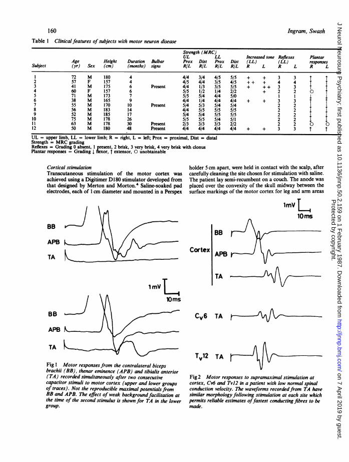
Table 1 Clinicalfeatures of subjects with motor neuron disease
Strength (MRC)UL LL Increased tone Reflexes Plantar
Age Height Duration Bulbar Prox Dist Prox Dist (LL) (LL) responsesSubject (yr) Sex (cm) (months) signs R/L R/L RIL R/L R L R L R L
1 72 M 180 4 4/4 3/4 4/5 5/5 + + 3 3 t t2 57 F 157 4 4/5 4/4 3/5 4/5 + + + 4 4 t t3 41 M 175 6 Present 4/4 1/3 3/5 5/5 + + + 3 3 t t4 60 F 157 6 5/5 1/2 1/4 2/2 + 2 2 0 t5 71 M 173 7 5/5 5/4 4/4 5/0 1 1 l l6 38 M 165 9 4/4 1/4 4/4 4/4 + + 3 3 t7 55 M 170 10 Present 5/4 5/3 5/4 5/4 2 2 18 56 M 183 14 4/4 5/5 5/5 5/5 2 2 49 52 M 185 17 5/4 5/4 5/5 5/5 2 2 410 75 M 178 26 5/5 5/5 5/4 5/l 2 2 l l11 63 M 178 30 Present 2/3 3/3 3/3 2/2 2 2 0 012 50 M 180 48 Present 4/4 4/4 4/4 4/4 + + 3 3 1 t
UL = upper limb, LL = lower limb; R = right, L = left; Prox = proximal, Dist = distalStrength = MRC gradingReflexes = Grading 0 absent, I present, 2 brisk, 3 very brisk, 4 very brisk with clonusPlantar responses = Grading 4 flexor, I extensor, 0 unobtainable
Cortical stimulation holder 5 cm apart, were held in contact with the scalp, afterTranscutaneous stimulation of the motor cortex was carefully cleaning the site chosen for stimulation with saline.achieved using a Digitimer D180 stimulator developed from The patient lay semi-recumbent on a couch. The anode wasthat designed by Merton and Morton.4 Saline-soaked pad placed over the convexity of the skull midway between theelectrodes, each of 1 cm diameter and mounted in a Perspex surface markings of the motor cortex for leg and arm areas
1mVjlOms
APB
TA
1mV
lOms
BB
APB
TA
Cortex
BB
APB
TA
CI6TA
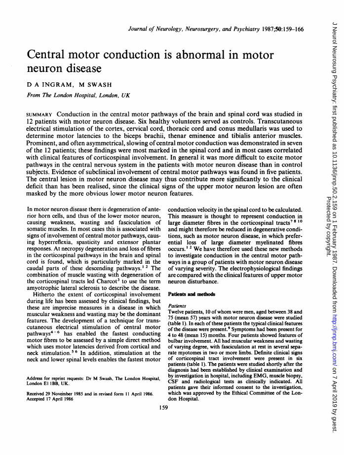
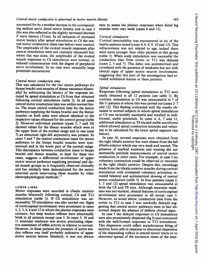
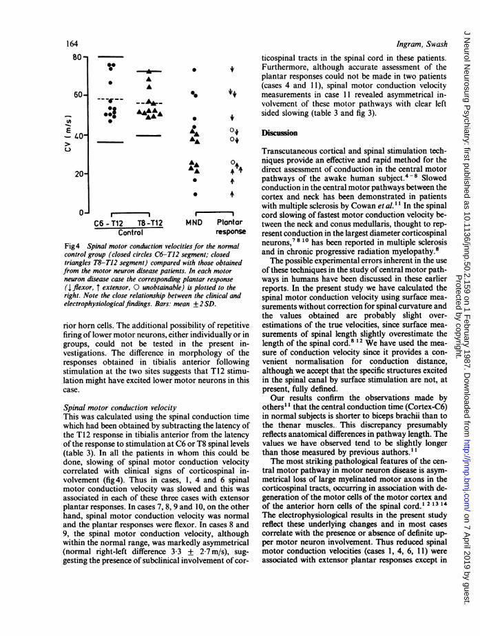
TV12 TAFig I Motor responses from the contralateral biceps Vbrachii (BB), thenar eminence (APB) and tibialis anterior(TA) recorded simultaneously after two consecutive Fig 2 Motor responses to supramaximal stimulation atcapacitor stimuli to motor cortex (upper and lower groups cortex, Cv6 and Tv12 in a patient with low normal spinalof traces). Not the reproducible maximal potentialsfrom conduction velocity. The waveforms recordedfrom TA haveBB and APB. The effect of weak backgroundfacilitation at similar morphology following stimulation at each site whichthe time of the second stimulus is shown for TA in the lower permits reliable estimates offastest conducting fibres to begroup. made.
BB
Ingram, Swash160
Protected by copyright.
on 7 April 2019 by guest.
http://jnnp.bmj.com
/J N
eurol Neurosurg P
sychiatry: first published as 10.1136/jnnp.50.2.159 on 1 February 1987. D
ownloaded from
Central motor conduction is abnormal in motor neuron disease
1mVj20ms
Right FC,6
Left
1 mV10 Ms
Right
T 12
Left
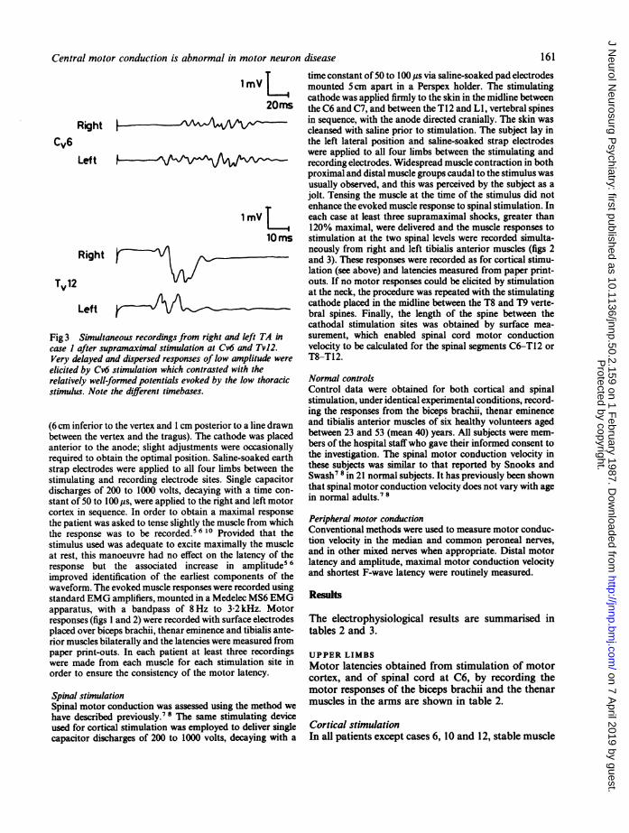
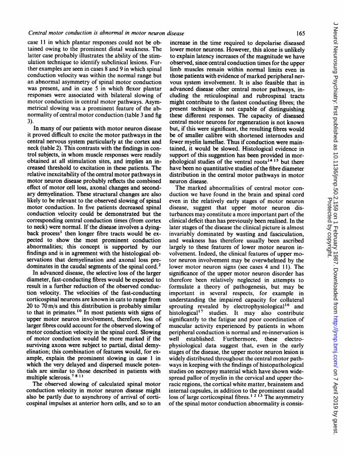
Fig 3 Simultaneous recordings from right and left TA incase I after supramaximal stimulation at Cv6 and Tv12.Very delayed and dispersed responses of low amplitude wereelicited by Cv6 stimulation which contrasted with therelatively well-formed potentials evoked by the low thoracicstimulus. Note the different timebases.
(6 cm inferior to the vertex and 1 cm posterior to a line drawnbetween the vertex and the tragus). The cathode was placedanterior to the anode; slight adjustments were occasionallyrequired to obtain the optimal position. Saline-soaked earthstrap electrodes were applied to all four limbs between thestimulating and recording electrode sites. Single capacitordischarges of 200 to 1000 volts, decaying with a time con-stant of 50 to 100 us, were applied to the right and left motorcortex in sequence. In order to obtain a maximal responsethe patient was asked to tense slightly the muscle from whichthe response was to be recorded.56 10 Provided that thestimulus used was adequate to excite maximally the muscleat rest, this manoeuvre had no effect on the latency of theresponse but the associated increase in amplitude5 6improved identification of the earliest components of thewaveform. The evoked muscle responses were recorded usingstandard EMG amplifiers, mounted in a Medelec MS6 EMGapparatus, with a bandpass of 8Hz to 3-2 kHz. Motorresponses (figs 1 and 2) were recorded with surface electrodesplaced over biceps brachii, thenar eminence and tibialis ante-rior muscles bilaterally and the latencies were measured frompaper print-outs. In each patient at least three recordingswere made from each muscle for each stimulation site inorder to ensure the consistency of the motor latency.
Spinal stimulationSpinal motor conduction was assessed using the method wehave described previously.' 8 The same stimulating deviceused for cortical stimulation was employed to deliver singlecapacitor discharges of 200 to 1000 volts, decaying with a
time constant of 50 to 100 ps via saline-soaked pad electrodesmounted 5cm apart in a Perspex holder. The stimulatingcathode was applied firmly to the skin in the midline betweenthe C6 and C7, and between the T12 and LI, vertebral spinesin sequence, with the anode directed cranially. The skin wascleansed with saline prior to stimulation. The subject lay inthe left lateral position and saline-soaked strap electrodeswere applied to all four limbs between the stimulating andrecording electrodes. Widespread muscle contraction in bothproximal and distal muscle groups caudal to the stimulus wasusually observed, and this was perceived by the subject as ajolt. Tensing the muscle at the time of the stimulus did notenhance the evoked muscle response to spinal stimulation. Ineach case at least three supramaximal shocks, greater than120% maximal, were delivered and the muscle responses tostimulation at the two spinal levels were recorded simulta-neously from right and left tibialis anterior muscles (figs 2and 3). These responses were recorded as for cortical stimu-lation (see above) and latencies measured from paper print-outs. If no motor responses could be elicited by stimulationat the neck, the procedure was repeated with the stimulatingcathode placed in the midline between the T8 and T9 verte-bral spines. Finally, the length of the spine between thecathodal stimulation sites was obtained by surface mea-surement, which enabled spinal cord motor conductionvelocity to be calculated for the spinal segments C6-T12 orT8-T12.
Normal controlsControl data were obtained for both cortical and spinalstimulation, under identical experimental conditions, record-ing the responses from the biceps brachii, thenar eminenceand tibialis anterior muscles of six healthy volunteers agedbetween 23 and 53 (mean 40) years. All subjects were mem-bers of the hospital staffwho gave their informed consent tothe investigation. The spinal motor conduction velocity inthese subjects was similar to that reported by Snooks andSwash7 8 in 21 normal subjects. It has previously been shownthat spinal motor conduction velocity does not vary with agein normal adults.7'8
Peripheral motor conductionConventional methods were used to measure motor conduc-tion velocity in the median and common peroneal nerves,and in other mixed nerves when appropriate. Distal motorlatency and amplitude, maximal motor conduction velocityand shortest F-wave latency were routinely measured.
Results
The electrophysiological results are summarised intables 2 and 3.
UPPER LIMBSMotor latencies obtained from stimulation of motorcortex, and of spinal cord at C6, by recording themotor responses of the biceps brachii and the thenarmuscles in the arms are shown in table 2.
Cortical stimulationIn all patients except cases 6, 10 and 12, stable muscle
161
Protected by copyright.
on 7 April 2019 by guest.
http://jnnp.bmj.com
/J N
eurol Neurosurg P
sychiatry: first published as 10.1136/jnnp.50.2.159 on 1 February 1987. D
ownloaded from
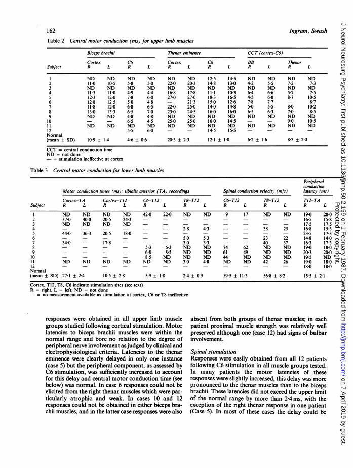
Table 2 Central motor conduction (ms) for upper limb muscles
Biceps brachii Thenar eminence CCT (cortex-C6)
Cortex C6 Cortex C6 BB ThenarSubject R L R L R L R L R L R L
I ND ND ND ND ND ND 12-5 14-5 ND ND ND ND2 11-0 105 58 50 22-0 20-3 148 13-0 4-2 55 7-2 7-33 ND ND ND ND ND ND ND ND ND ND ND ND4 11-3 11-0 4-9 4-4 168 178 111 103 6-4 6-6 57 755 12-3 12-0 7-8 6-0 27-0 27-0 183 165 45 60 8-7 1056 12-8 12-5 5-0 4-8 - 21-3 150 12-6 7-8 7-7 - 8-77 11-8 120 6-8 65 22-0 25-0 14-0 14-8 5-0 5-5 8-0 1028 13-0 133 65 7-0 23-0 245 16-0 160 65 63 70 859 ND ND 4-8 48 ND ND ND ND ND ND ND ND10 - - 6-5 4-5 250 250 16-0 14-5 - - 9-0 10-511 ND ND ND ND ND ND ND ND ND ND ND ND12 - - 55 6-0 - - 14-5 15-5 - - - -Nornal(mean + SD) 10-9 + 1-4 46 + 0-6 20-3 + 2-3 12-1 + 1-0 6-2 + 1-6 8-3 + 2-0
CCT = central conduction timeND = not done- = stimulation ineffective at cortex
Table 3 Central motor conduction for lower limb muscles
Peripheralconduction
Motor conduction times (ms): tibialis anterior (TA) recordings Spinal conduction velocity (mls) latency (ms)
Cortex-TA Cortex-Tl2 C6-TI2 T8-T12 C6-TI2 T8-T12 T12-TASubject R L R L R L R L R L R L R L
1 ND ND ND ND 42-0 22-0 ND ND 9 17 ND ND 190 20-02 37-0 400 20-5 24-3 - - - - 16-5 15-83 ND ND ND ND - - - - - - - - 175 17-54 - - - - - - 2-8 4-3 - - 38 25 16-8 15-35 440 36-3 20-5 18-0 - - - - - - - - 235 1736 - - - - - - 50 53 - - 23 22 14-8 1407 34-0 - 17-8 - - - 3-0 3-3 - - 40 37 16-3 17-38 - - - - 53 6-3 ND ND 74 62 ND ND 190 18-09 - - - - 6-8 85 ND ND 61 49 ND ND 20-3 20-010 - - - - 85 ND ND ND 44 ND ND ND 19-5 NDII ND ND ND ND ND ND 30 4-8 ND ND 42 26 190 18-012 - - - - - - - - - - - - 18-0 18-0Normal(mean + SD) 27-1 + 2-4 105 + 2-8 59 + 18 2-4 + 09 595 + 11-3 568 + 8-2 15-5 + 2-1
Cortex, T12, T8, C6 indicate stimulation sites (see text)R = right, L = left; ND = not done-= no measurement available as stimulation at cortex, C6 or T8 ineffective
responses were obtained in all upper limb muscle absent from both groups of thenar muscles; in eachgroups studied following cortical stimulation. Motor patient proximal muscle strength was relatively welllatencies to biceps brachii muscles were within the preserved although one (case 12) had signs of bulbarnormal range and bore no relation to the degree of involvement.peripheral nerve involvement asjudged by clinical andelectrophysiological criteria. Latencies to the thenar Spinal stimulationeminence were clearly delayed in only one instance Responses were easily obtained from all 12 patients(case 5) but the peripheral component, as assessed by following C6 stimulation in all muscle groups tested.C6 stimulation, was sufficiently increased to account In many patients the motor latencies of thesefor this delay and central motor conduction time (see responses were slightly increased; this delay was morebelow) was normal. In case 6 responses could not be pronounced to the thenar muscles than to the bicepselicited from the right thenar muscles which were par- brachii. These latencies did not exceed the upper limitticularly atrophic and weak. In cases 10 and 12 of the normal range by more than 2-4ms, with theresponses could not be obtained in either biceps bra- exception of the right thenar response in one patientchii muscles, and in the latter case responses were also (Case 5). In most of these cases the delay could be
162 Ingram, Swash
Protected by copyright.
on 7 April 2019 by guest.
http://jnnp.bmj.com
/J N
eurol Neurosurg P
sychiatry: first published as 10.1136/jnnp.50.2.159 on 1 February 1987. D
ownloaded from
Central motor conduction is abnormal in motor neuron diseaseaccounted for by a modest increase in the correspond-ing median nerve distal motor latency and in case 5this was also reflected in the slightly increased shortestF wave latency (32 ms). In all instances of increasedmotor latency after spinal stimulation at C6 the cen-tral motor conduction times (see below) were normal.The amplitudes of the evoked muscle responses aftercentral stimulation were not routinely measured but,where this was done, the amplitudes of the evokedmuscle responses to C6 stimulation were normal, orreduced commensurate with the degree of peripheralnerve involvement. In no case were unusually largepotentials encountered.
Central motor conduction timeThis was calculated for the two motor pathways (tobiceps brachii and muscles of thenar eminence bilater-ally) by subtracting the latency of the response ini-tiated by spinal stimulation at C6 from that obtainedfollowing cortical stimulation (table 2). In all casescentral motor conduction time was within normal lim-its. The mean central conduction time for the motorpathways supplying the' biceps brachii and the thenarmuscles on both sides were almost identical to therespective values obtained for the control group (table2). However individual patients (cases 5, 7 and 10)showed values for the thenar muscles which were atthe upper limit of the normal range and in one (case7) an abnormal right-left asymmetry was present. Incases 5 and 7 the central conduction times for motorpathways to the biceps brachii muscles were sym-metrical and in the lower part of the normal range.This discrepancy between conduction times to bicepsbrachii and thenar muscles, at least for these twocases, suggests a differential involvement of uppermotor neuron pathways supplying proximal and dis-tal muscle groups as is frequently observed clinicallyand has similarly been demonstrated for the motorneuronal pools innervating these muscles by otherelectrophysiological methods."7
LOWER LIMBSMotor responses were recorded in tibialis anteriormuscles bilaterally following cortical, C6 and T12stimulation (table 3). If C6 stimulation was un-successful, T8 stimulation was also carried out. Signsof corticospinal involvement were prominent in cases1, 2, 3, 4, 6 and 12 in whom the plantar responses wereextensor, but deep tendon reflexes were abnormallybrisk in all patients except case 5. In cases 5, 10 and11 muscular weakness was severe, precluding objec-tive assessment of reflex activity in relation to normal.However, in-these patients the presence of active ten-don reflexes was itself probably indicative of uppermotor neuron lesions. Similarly, it was not always
easy to assess the plantar responses when distal legmuscles were very weak (cases 4 and 11).
Cortical stimulationCortical inexcitability was encountered in six of thetwelve patients tested (cases 4, 6, 8, 9, 10 and 12). Thisrefractoriness was not related to age; indeed therewere more younger than older patients in this group(table 3). When scalp stimulation was successful theconduction time from cortex to T12 was delayed(cases 2, 5 and 7). This delay was asymmetrical andcorrelated with the presence of weakness but not withclinical signs of upper motor neuron involvement,suggesting that this part of the investigation had re-vealed subclinical lesions in these patients.
Spinal stimulationResponses following spinal stimulation at T12 wereeasily obtained in all 12 patients (see table 1). Bycontrast, stimulation at C6 was unsuccessful in 7 ofthe 11 patients in whom this was carried out (cases 2-7and 12). This finding contrasted with the results ob-tained in normal subjects in whom spinal stimulationat C6 was invariably successful and resulted in well-formed, stable potentials. In cases 4, 6, 7 and 11,additional stimulation at T8 elicited stable waveformswhich allowed spinal conduction latencies and veloci-ties to be calculated for the lower spinal segment (seebelow).
In case 10, normal responses were obtained fromthe right tibialis anterior but were absent from the lefttibialis anterior which was very weak and wasted. Thepresence of marked weakness and wasting did notnecessarily preclude measurements of central motorconduction in other cases. For example, in case 5 novoluntary contraction could be observed or recordedin the right tibialis anterior. Despite this, recordingsmade from the tibialis anterior muscles during corticalstimulation with attempted voluntary activation re-vealed bilateral and asymmetrical slowing of centralmotor conduction (table 3). In four patients (cases 2,3, 5 and 12) spinal stimulation was unsuccessful atboth the C6 and T8 sites. Although muscular weak-ness was not marked, clinical features of corticospinalinvolvement were prominent in all except case 5.However, as noted above, conduction time from thecortex to T12 in case 5 was markedly delayed sug-gesting that central motor pathways were in fact ab-normal despite the absence of definite clinical signs.
In case I the delayed responses to C6 stimulationwere also prominently dispersed (fig 3) and contrastedwith the well-formed responses to T12 stimulation.This dispersion could reflect asynchronous firing ofanterior horn cells in response to abnormal dispersionof the descending volleys in central motor tracts or toabnormal spread of the excitation times of the ante-
163
Protected by copyright.
on 7 April 2019 by guest.
http://jnnp.bmj.com
/J N
eurol Neurosurg P
sychiatry: first published as 10.1136/jnnp.50.2.159 on 1 February 1987. D
ownloaded from
A
A
--Ar
Ingram, Swash
ticospinal tracts in the spinal cord in these patients.a , Furthermore, although accurate assessment of the
plantar responses could not be made in two patients(cases 4 and 11), spinal motor conduction velocity
* 4'+ measurements in case 11 revealed asymmetrical in-volvement of these motor pathways with clear left
+ sided slowing (table 3 and fig 3).0 ofAAALA 04
U I
C6 - T12 T8-T12Control
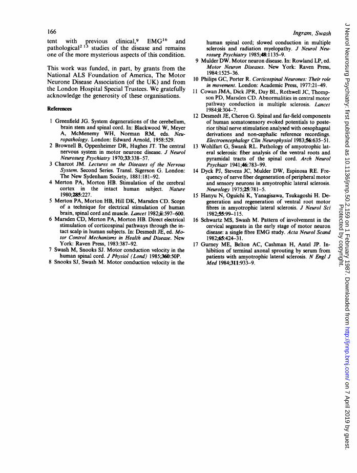
Fig 4 Spinal motor conduction velociticontrol group (closed circles C6-T12 sttriangles T8-T12 segment) compared ufrom the motor neuron disease patients.neuron disease case the corresponding;(4 flexor, T extensor, 0 unobtainable)right. Note the close relationship betweielectrophysiological findings. Bars: mea
rior horn cells. The additional possfiring of lower motor neurons, eithegroups, could not be tested investigations. The difference in mresponses obtained in tibialis astimulation at the two sites suggeslation might have excited lower mocase.
Spinal motor conduction velocityThis was calculated using the spinawhich had been obtained by subtracthe T12 response in tibialis anterioof the response to stimulation at C6(table 3). In all the patients in wldone, slowing of spinal motor cccorrelated with clinical signs ofvolvement (fig4). Thus in cases,motor conduction velocity was sloassociated in each of these three c;plantar responses. In cases 7, 8, 9 arhand, spinal motor conduction veland the plantar responses were flex9, the spinal motor conduction iwithin the normal range, was mark(normal right-left difference 3-3gesting the presence of subclinical ir
Discussion
Transcutaneous cortical and spinal stimulation tech-AA o niques provide an effective and rapid method for the
,4++ direct assessment of conduction in the central motor+ pathways of the awake human subject.4`8 Slowed
conduction in the central motor pathways between the* ' cortex and neck has been demonstrated in patients
with multiple sclerosis by Cowan et al. " In the spinalcord slowing of fastest motor conduction velocity be-
MND Plantor tween the neck and conus medullaris, thought to rep-
response resent conduction in the largest diameter corticospinalies for the normal neurons,7 8 10 has been reported in multiple sclerosisegment; closed and in chronic progressive radiation myelopathy.vith those obtained The possible experimental errors inherent in the useIn each motor of these techniques in the study of central motor path-'lantar response ways in humans have been discussed in these earlieris plotted to the reports. In the present study we have calculated theen the clinical and spinal motor conduction velocity using surface mea-
n + 2 SD. surements without correction for spinal curvature and
the values obtained are probably slight over-
,ibility of repetitive estimations of the true velocities, since surface mea-
r individually or in surements of spinal length slightly overestimate thethe present in- length of the spinal cord.8 12 We have used the mea-
orphology of the sure of conduction velocity since it provides a con-
.nterior following venient normalisation for conduction distance,ts that T12 stimu- although we accept that the specific structures excitedttor neurons in this in the spinal canal by surface stimulation are not, at
present, fully defined.Our results confirm the observations made by
others"I that the central conduction time (Cortex-C6)Ll conduction time in normal subjects is shorter to biceps brachii than tocting the latency of the thenar muscles. This discrepancy presumablyir from the latency reflects anatomical differences in pathway length. Theor T8 spinal levels values we have observed tend to be slightly longeriom this could be than those measured by previous authors."nduction velocity The most striking pathological features of the cen-
corticospinal in- tral motor pathway in motor neuron disease is asym-1, 4 and 6 spinal metrical loss of large myelinated motor axons in the)wed and this was corticospinal tracts, occurring in association with de-ases with extensor generation of the motor cells of the motor cortex andad 10, on the other of the anterior horn cells of the spinal cord.' 213 14locity was normal The electrophysiological results in the present studycor. In cases 8 and reflect these underlying changes and in most cases
velocity, although correlate with the presence or absence of definite up-;edly asymmetrical per motor neuron involvement. Thus reduced spinal+ 2-7 m/s), sug- motor conduction velocities (cases 1, 4, 6, 11) wereivolvement of cor- associated with extensor plantar responses except in
@00
0*I
0.
_
164807
60-
UA
40-C-0
20-
0-
Protected by copyright.
on 7 April 2019 by guest.
http://jnnp.bmj.com
/J N
eurol Neurosurg P
sychiatry: first published as 10.1136/jnnp.50.2.159 on 1 February 1987. D
ownloaded from
Central motor conduction is abnormal in motor neuron diseasecase 11 in which plantar responses could not be ob-tained owing to the prominent distal weakness. Thelatter case probably illustrates the ability of the stim-ulation technique to identify subclinical lesions. Fur-ther examples are seen in cases 8 and 9 in which spinalconduction velocity was within the normal range butan abnormal asymmetry of spinal motor conductionwas present, and in case 5 in which flexor plantarresponses were associated with bilateral slowing ofmotor conduction in central motor pathways. Asym-metrical slowing was a prominent feature of the ab-normality of central motor conduction (table 3 and fig3).
In many of our patients with motor neuron diseaseit proved difficult to excite the motor pathways in thecentral nervous system particularly at the cortex andneck (table 2). This contrasts with the findings in con-trol subjects, in whom muscle responses were readilyobtained at all stimulation sites, and implies an in-creased threshold to excitation in these patients. Therelative inexcitability of the central motor pathways inmotor neuron disease probably reflects the combinedeffect of motor cell loss, axonal changes and second-ary demyelination. These structural changes are alsolikely to be relevant to the observed slowing of spinalmotor conduction. In five patients decreased spinalconduction velocity could be demonstrated but thecorresponding central conduction times (from cortexto neck) were normal. If the disease involves a dying-back process1 then longer fibre tracts would be ex-pected to show the most prominent conductionabnormalities; this concept is supported by ourfindings and is in agreement with the histological ob-servations that demyelination and axonal loss pre-dominates in the caudal segments of the spinal cord.2
In advanced disease, the selective loss of the largerdiameter, fast-conducting fibres would be expected toresult in a further reduction of the observed conduc-tion velocity. The velocities of the fast-conductingcorticospinal neurons are known in cats to range from20 to 70 m/s and this distribution is probably similarto that in primates.'0 In most patients with signs ofupper motor neuron involvement, therefore, loss oflarger fibres could account for the observed slowing ofmotor conduction velocity in the spinal cord. Slowingof motor conduction would be more marked if thesurviving axons were subject to partial, distal demy-elination; this combination of features would, for ex-ample, explain the prominent slowing in case 1 inwhich the very delayed and dispersed muscle poten-tials are similar to those described in patients withmultiple sclerosis.7 8 11The observed slowing of calculated spinal motor
conduction velocity in motor neuron disease mightalso be partly due to asynchrony of arrival of corti-cospinal impulses at anterior horn cells, and so to an
increase in the time required to depolarise diseasedlower motor neurons. However, this alone is unlikelyto explain latency increases of the magnitude we haveobserved, since central conduction times for the upperlimb muscles remain within normal limits even inthose patients with evidence ofmarked peripheral ner-vous system involvement. It is also feasible that inadvanced disease other central motor pathways, in-cluding the reticulospinal and rubrospinal tractsmight contribute to the fastest conducting fibres; thepresent technique is not capable of distinguishingthese different responses. The capacity of diseasedcentral motor neurons for regeneration is not knownbut, if this were significant, the resulting fibres wouldbe of smaller calibre with shortened internodes andfewer myelin lamellae. Thus if conduction were main-tained, it would be slowed. Histological evidence insupport of this suggestion has been provided in mor-phological studies of the ventral roots14 15 but therehave been no quantitative studies of the fibre diameterdistribution in the central motor pathways in motorneuron disease.The marked abnormalities of central motor con-
duction we have found in the brain and spinal cordeven in the relatively early stages of motor neurondisease, suggest that upper motor neuron dis-turbances may constitute a more important part of theclinical deficit than has previously been realised. In thelater stages of the disease the clinical picture is almostinvariably dominated by wasting and fasciculation,and weakness has therefore usually been ascribedlargely to these features of lower motor neuron in-volvement. Indeed, the clinical features of upper mo-tor neuron involvement may be overwhelmed by thelower motor neuron signs (see cases 4 and 11). Thesignificance of the upper motor neuron disorder hastherefore been relatively neglected in attempts toformulate a theory of pathogenesis, but may beimportant in several respects, for example inunderstanding the impaired capacity for collateralsprouting revealed by electrophysiological'6 andhistological17 studies. It may also contributesignificantly to the fatigue and poor coordination ofmuscular activity experienced by patients in whomperipheral conduction is normal and re-innervation iswell established. Furthermore, these electro-physiological data suggest that, even in the earlystages of the disease, the upper motor neuron lesion iswidely distributed throughout the central motor path-ways in keeping with the findings of histopathologicalstudies on necropsy material which have shown wide-spread pallor of myelin in the cervical and upper tho-racic regions, the cortical white matter, brainstem andinternal capsules, in addition to the prominent caudalloss of large corticospinal fibres. 1 2 13 The asymmetryof the spinal motor conduction abnormality is consis-
165
Protected by copyright.
on 7 April 2019 by guest.
http://jnnp.bmj.com
/J N
eurol Neurosurg P
sychiatry: first published as 10.1136/jnnp.50.2.159 on 1 February 1987. D
ownloaded from
166tent with previous clinical,9 EMG16 andpathological2 13 studies of the disease and remainsone of the more mysterious aspects of this condition.
This work was funded, in part, by grants from theNational ALS Foundation of America, The MotorNeurone Disease Association (of the UK) and fromthe London Hospital Special Trustees. We gratefullyacknowledge the generosity of these organisations.
References
1 Greenfield JG. System degenerations of the cerebellum,brain stem and spinal cord. In: Blackwood W, MeyerA, McMenemy WH, Norman RM, eds. Neu-ropathology. London: Edward Arnold, 1958:529.
2 Brownell B, Oppenheimer DR, Hughes JT. The centralnervous system in motor neurone disease. J NeurolNeurosurg Psychiatry 1970;33:338-57.
3 Charcot JM. Lectures on the Diseases of the NervousSystem. Second Series. Transl. Sigerson G. London:The New Sydenham Society, 1881:181-92.
4 Merton PA, Morton HB. Stimulation of the cerebralcortex in the intact human subject. Nature1980;285:227.
5 Merton PA, Morton HB, Hill DK, Marsden CD. Scopeof a technique for electrical stimulation of humanbrain, spinal cord and muscle. Lancet 1982;ii:597-600.
6 Marsden CD, Merton PA, Morton HB. Direct electricalstimulation of corticospinal pathways through the in-tact scalp in human subjects. In: Desmedt JE, ed. Mo-tor Control Mechanisms in Health and Disease. NewYork: Raven Press, 1983:387-92.
7 Swash M, Snooks SJ. Motor conduction velocity in thehuman spinal cord. J Physiol (Lond) 1985;360:50P.
8 Snooks SJ, Swash M. Motor conduction velocity in the
Ingram, Swashhuman spinal cord; slowed conduction in multiplesclerosis and radiation myelopathy. J Neurol Neu-rosurg Psychiatry 1985;48: 1135-9.
9 Mulder DW. Motor neuron disease. In: Rowland LP, ed.Motor Neuron Diseases. New York: Raven Press,1984:1525-36.
10 Philips GC, Porter R. Corticospinal Neurones: Their rolein movement. London: Academic Press, 1977:21-49.
11 Cowan JMA, Dick JPR, Day BL, Rothwell JC, Thomp-son PD, Marsden CD. Abnormalities in central motorpathway conduction in multiple sclerosis. Lancet1984;ii:304-7.
12 Desmedt JE, Cheron G. Spinal and far-field componentsof human somatosensory evoked potentials to poste-rior tibial nerve stimulation analysed with oesophagealderivations and non-cephalic reference recordings.Electroencephalogr Clin Neurophysiol 1983;56:635-51.
13 Wohlfart G, Swank RL. Pathology of amyotrophic lat-eral sclerosis: fiber analysis of the ventral roots andpyramidal tracts of the spinal cord. Arch NeurolPsychiatr 1941;46:783-99.
14 Dyck PJ, Stevens JC, Mulder DW, Espinosa RE. Fre-quency ofnerve fiber degeneration of peripheral motorand sensory neurons in amyotrophic lateral sclerosis.Neurology 1975;25:781-5.
15 Hanyu N, Oguichi K, Yanagisawa, Tsukagoshi H. De-generation and regeneration of ventral root motorfibres in amyotrophic lateral sclerosis. J Neurol Sci1982;55:99-115.
16 Schwartz MS, Swash M. Pattern of involvement in thecervical segments in the early stage of motor neurondisease: a single fibre EMG study. Acta Neurol Scand1982;65:424-31.
17 Gurney ME, Belton AC, Cashman H, Antel JP. In-hibition of terminal axonal sprouting by serum frompatients with amyotrophic lateral sclerosis. N Engl JMed 1984;311:933-9.
Protected by copyright.
on 7 April 2019 by guest.
http://jnnp.bmj.com
/J N
eurol Neurosurg P
sychiatry: first published as 10.1136/jnnp.50.2.159 on 1 February 1987. D
ownloaded from