Embed Size (px)
Citation preview

From thUniversity
SubmitNo benReprint
Center of911-1, Mo
© 20110883-5doi:10.1
The Journal of Arthroplasty Vol. 26 No. 2 2011
Cementless Total Hip Arthroplasty WithAlumina-on-Highly Cross-Linked Polyethylene
Bearing in Young Patients With FemoralHead Osteonecrosis
Young-Hoo Kim, MD, Yoowang Choi, MD, and Jun-Shik Kim, MD
Abstract:We asked whether total hip arthroplasties (THAs) using alumina-on-highly cross-linkedpolyethylene bearing would improve functional activity and reduce aseptic loosening, poly-ethylene wear, and osteolysis. Consecutive primary THAs were performed in 71 patients (73 hips)who were younger than 50 years (mean age, 45.5 years) with femoral head osteonecrosis. Therewere 48 men (50 hips) and 23 women (23 hips). Osteolysis was evaluated using radiographs andcomputed tomographic scanning. The average follow-up was 8.5 years (range, 7-9 years). Themean preoperative Harris hip score was 50.6 points, which improved to 96 points at the finalfollow-up. Preoperative functional activity was improved significantly at the latest follow-up. Themean polyethylene linear penetration was 0.05 ± 0.02 mm/y and no hip had aseptic loosening orosteolysis. Keywords: femoral head osteonecrosis, total hip arthroplasty, alumina-on-highlycross-linked polyethylene, osteolysis.© 2011 Elsevier Inc. All rights reserved.
With early generation of cementless acetabular andfemoral components of total hip arthroplasty (THA),durability of fixation has been excellent in the patientsyounger than 50 years out to 15 and 20 years [1-4].Wear-induced osteolysis, however, is the dominantproblem. Alternative bearing couples such as aluminaceramic-on-highly cross-linked polyethylene is attrac-tive because of the potential for reduced wear andanticipated reduced osteolysis and loosening.Ceramic bearings offer the advantage of improved
lubrication, smoother surface finish, and improvedresistance to scratching and are biologically inertcompounds. Ceramic femoral heads therefore havesubstantial tribologic advantages over metal femoralheads and result in much lower wear and osteolysis rates[5]. Also, highly cross-linked ultrahigh-molecular-weight polyethylene has been shown to markedlyreduce wear in clinical studies [6,7]. Furthermore,
e The Joint Replacement Center of Korea, Ewha Woman'sSchool of Medicine, Seoul, South Korea.ted October 21, 2009; accepted March 17, 2010.efits or funds were received in support of the study.requests: Young-Hoo Kim, MD, The Joint ReplacementKorea at Ewha Woman's University MokDong Hospital,kDong, YangCheon-Gu, Seoul 158-710, South Korea.Elsevier Inc. All rights reserved.
403/2602-0009$36.00/0016/j.arth.2010.03.010
218
alumina-on-highly cross-linked polyethylene bearingcouple has potential for further reduction of polyethyl-ene wear and osteolysis.The purpose of the current study was to evaluate the
clinical and radiographic outcomes of THAs usingalumina-on-highly cross-linked polyethylene bearingcouple in 71 patients younger than 50 with femoralhead osteonecrosis. In addition, we determined theincidence of polyethylene wear and osteolysis usingradiographs and computed tomographic scans.
Materials and MethodsDemographicsFrom February 2000 to May 2002, the senior author
performed 79 consecutive cementless THAs usingalumina-on-highly cross-linked polyethylene (Mara-thon; DePuy, Warsaw, Ind) in 76 patients (3 patientshad bilateral THAs). The study was approved by ourinstitutional review board, and all patients providedinformed consent. Five patients were lost to follow-up(before 1 year), leaving 71 patients (73 hips), whocomprise the series of this study.There were 48 men (50 hips) and 23 women (23 hips).
The average age at the time of the index arthroplastywas 45.5 years (range, 20-50 years). The average weightof the patients was 67.5 kg (range of 47 to 87 kg). Theaverage height was 164.5 cm (range, 147-183 cm), andthe average body mass index was 24.8 kg/m2 (range,

Cementless THA With Alumina-on-Highly Cross-Linked PE Bearing � Kim et al 219
18.8-30.5 kg/m2). All hips with osteonecrosis of femoralhead had Ficat and Arlet stage III or IV [8]. Thepresumed cause of osteonecrosis was ethanol abuse in50 patients (70.4%), idiopathic in 18 patients (25.4%),and steroid use in 3 patients (4.2%). The average follow-up was 8.5 years (range, 7-9 years).
ProsthesisA cementless Duraloc 100 or 1200 series acetabular
component (DePuy, Warsaw, Ind) with a highly cross-linked polyethylene liner of inner diameter of 28 mmwas used in all hips. All patients received an ImmediatePostoperative Stability (DePuy, Leeds, United Kingdom)cementless femoral component with a 28-mm aluminaforte femoral head. The Immediate PostoperativeStability femoral component is an anatomical metaphy-seal fitting titanium stem with a polished and tapereddistal stem, designed to provide fixation in the meta-hysis only, thereby, avoiding metal-to-bone contactbelow this point. The proximal 30% of stem was porouscoated with sintered titanium beads, with a mean poresize of 250 μm to which a hydroxyapatite coating wasapplied to a thickness of 30 μm.
Surgical ProcedureAll operations were performed through a posterolat-
eral approach. The femoral component was insertedwith a press-fit technique. The largest broach that wouldfill the metaphysis and leave little cancellous boneremaining was used. The acetabular component wasfixed with a press-fit only without using a screw in the65 hips, and 1 or 2 screws were inserted for additionalfixation in the remaining 8 hips.The patients were allowed to stand on the second
postoperative day, and they progressed to full weightbearing with crutches as tolerated.
Clinical EvaluationClinical follow-up was performed at 3 months and 1
year and yearly thereafter. Harris hip scores weredetermined before surgery and at each follow-upexamination [9]. Patients subjectively evaluated thighpain on a 10-point visual analog scale (0 = no pain; 10 =severe pain). The level of activity of the patients after theTHA was assessed with the activity score of Tegner andLysholm [10]. The activity grading scale, with whichwork and sports activities are graded numerically, wasused as a complement to the functional score. Thepatients were given a score, according to the activities inwhich they engaged in daily life, ranging from 0 pointfor a hip-related disability to 10 points for participationin competitive sports at a national level.
Radiographic EvaluationRadiographic follow-up was performed at 3 months
and 1 year and yearly thereafter. A supine anteropos-terior radiograph of the pelvis with both hips in neutralrotation and 0° of abduction was made for every patient.
Consistent patient positioning was ensured with the useof x-ray frame. This frame is constructed so that it can beplaced at the end of a standard x-ray table. Plasticpolypropylene orthoses are secured to a plastic back-board through a vertical slot. A wing nut allowsadjustment for various limb lengths. Rotation andabduction remain constant. Cross-table lateral radio-graphs were also made of each hip.Femoral bone type was determined in preoperative
radiographs using the classification by Dorr [11]. Theadequacy of the intramedullary fill by the stem wasrecorded as satisfactory when the stem filled more than80% of the proximal part of the canal in the coronalplane and more than 70% in the sagittal plane,according to a previously described method [12]. Thecomponent was considered to be undersized if less of thecanal was filled in either or both planes.Definite loosening of the femoral component was
defined if there was a progressive axial subsidence ofmore than 2 mm or varus or valgus shift more than 2°[13]. A femoral component was considered to bepossibly loose when there was a complete radiolucentline surrounding the entire porous-coated surface onboth the anteroposterior and lateral radiographs [13].Anteversion of the acetabular component was mea-
sured on the true lateral radiographs of the hip as theangle between a horizontal line and a second linemarking the plane of the socket. To measure cupabduction, a line that joined the inferior margins of the2 acetabular teardrops was drawn on the anteroposteriorpelvic radiograph. The intersection of that line markingthe plane of opening of the socket determined the angleof abduction.Definite loosening of the acetabular component was
diagnosed when there was a change in the position ofthe component (N2 mm vertically and/or medially orlaterally) or a continuous radiolucent line wider than 2mm on both the anteroposterior and the lateralradiograph [13].A vertical change in the position of the cup was
measured between the inferior margin of the cup andthe inferior margin of the ipsilateral teardrop [14], and ahorizontal change was measured between the Köhlerline (ilioischial line) and the center of the outer shell ofthe acetabular component [15].
Radiographic Evaluation of Polyethylene WearPenetration of the polyethylene liner was measured,
with use of a software program (Auto CAD, Release 13;Autodesk, Sausalito, Calif [16]), by on observer who wasblinded to the clinical results. The observer made 3measurements in each radiograph, and the intraobser-ver error was ±0.021 mm. A scan-Maker 9600 X L flat-bed imaging scanner (Microtek, Carson, Calif) digitizedthe anteroposterior radiograph of the pelvis as 2-dimensional gray scale arrays of 12-bit (256 gray level)

220 The Journal of Arthroplasty Vol. 26 No. 2 February 2011
integers. The scanning resolution was 600 psi (pixels persquare inch). Penetration of the head into the liner wasdetermined at annual intervals from anteroposteriorpelvic radiographs. The amount of penetration onradiographs made 3 months postoperatively was con-sidered to be the “zero position.”
Radiographic Evaluation of OsteolysisThe presence and locations of areas of osteolysis in the
acetabulum were recorded in the anteroposterior andlateral radiographs according to the system of DeLee andCharnley [17], and those in the femur were recordedalso in the anteroposterior and lateral radiographsaccording to the system of Gruen et al [18]. The lengthand width of osteolytic lesions were measured, and thearea was expressed in square centimeters.
Computed Tomographic Evaluation of OsteolysisAlthough radiologic evaluation of osteolysis is a direct
measurement, the current method is insensitive andsubject to operator error. A more sensitive computerizedtomographic image sets provide 3-dimensional data, butthe beam hardening artifacts from the prosthesis itselfmake these images difficult to interpret. To address thebeam hardening artifacts, as well as to measure thevolume of lytic lesions, we developed an algorithm todiminish the effect of beam hardening artifacts. We thendeveloped a segmentation algorithm to segment the lyticlesions from image data and to measure their volumes.Computed tomographic images were acquired usingSiemens AG (Munich, Germany) with 1 mm collima-tion, a pitch of 1.5, and a 14-cm to 22-cm field of view.The raw data were reconstructed for 1-mm slices. Thearea within 5 cm from the prosthesis-bone interface inall directions was evaluated. The volume of osteolysiswas calculated by Virtual Scopics (Rochester, NY).
Statistical MethodsSurvivorship analysis [19] was performed with the
Kaplan-Meier method with revision for any reason asone end point and revision due to mechanical failure(clinical and radiographic evidence of aseptic loosening)at the time of follow-up as the other end point. Wedetermined differences in continuous variances (Harriship score, range of motion, body mass index) betweenpreoperative and postoperative results using Studentpaired t test, and in categorical variances (details offunctional evaluation and deformity according to theHarris hip score) and limb length between preoperativeand postoperative evaluations using χ2 test. Univariateregression analysis was used to evaluate the relation-ship, if any between osteolysis and the variables of age,sex, weight, diagnosis, the duration of follow-up, andacetabular inclination and anteversion. The level ofsignificance was set at P b .05.
ResultsClinical OutcomePreoperative Harris hip score was improved signifi-
cantly (P = .001) after the operation. The meanpreoperative Harris hip score was 50.6 points (range,27-55 points). The mean hip score at the final follow-upwas 96 points (range, 85-100 points).Preoperative functional activity was improved signif-
icantly (P = .001) at the latest follow-up. The ability toput on footwear, and to cut toenails and to use stairs andpublic transportation was improved markedly at thelatest follow-up.Activity level of patients was improved very much
after the operation. Many patients were quite activedespite our admonitions to avoid activities involvinghigh impact after the THA. All but 3 patients had anactivity score of 5 or 6 points [10] after the THA,indicating participation in strenuous farm work (a scoreof 5 points) or playing recreational sports such as tennis(a scores of 6 points). The 3 patients with low back painhad a score of 3 points.
Radiographic ResultsNo hip had aseptic loosening of any acetabular or
femoral component. All stems had a satisfactory canalfill on radiographs, and all hips had Dorr type A ortype B bones. The mean inclination of and anteversionof acetabular component was 42.3° (range, 39°-45°)and 22° (range, 19°-24°), respectively. All acetabularand femoral components were fixed by bone in-growth. Calcar rounding off was observed in all hips,but no hip had stress shielding-related proximalfemoral bone resorption.No hip had femoral head ceramic fracture.
Radiographic Measurement of Penetrationof PolyethyleneThe mean amount of highly cross-linked polyethylene
linear penetration was 0.05 ± 0.02 mm per year (range,0.02-0.08 mm/y). At the latest follow-up examination,no hip was an outlier for the so-called osteolysisthreshold of 0.10 mm/y [20,21] with all liners havinga penetration rate at or below this level. With thenumbers available, univariate regression analysis did notdemonstrate that age, sex, weight, activity, cup inclina-tion, or cup anteversion had any influence on polyeth-ylene liner penetration.
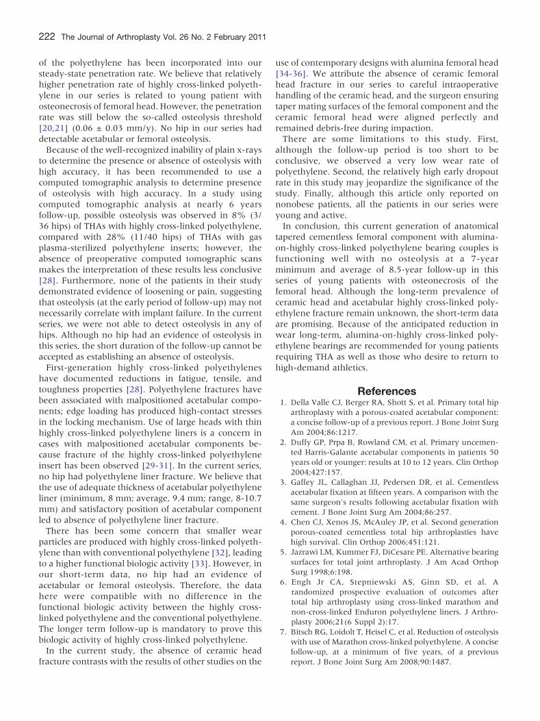
Radiographic and Computed Tomographic Resultsof OsteolysisRadiograph and computed tomographic scans (Fig. 1)
demonstrated that no acetabular or femoral osteolysiswas detected in any hip at the latest follow-up.
ComplicationsOne hip (1%) was dislocated 5 days after the
operation and was treated successfully with a closed

Fig. 1. Radiographic and computed tomographic scanningevaluation of osteolysis of right hip of a 43-year-old man withosteonecrosis of right hip. (A) An anteroposterior and lateralradiographs of right hip made 7 years after surgery demon-strated that Duraloc 1200 series cementless acetabular com-ponent is fixed in a satisfactory position by bone ingrowth.There is no radiolucent line or osteolysis around the acetabularor femoral components in both hips. Grade 3 bone loss isobserved in the calcar region of right hip. (B) Computedtomographic scanning of right hip taken 7 years after thesurgery reveals no evidence of osteolysis around the acetabularor femoral components in right hip.
Cementless THA With Alumina-on-Highly Cross-Linked PE Bearing � Kim et al 221
reduction and an abduction brace for 3 months. Nofurther dislocation was observed in this hip until thefinal follow-up examination.
Revisions and SurvivorshipNo hip had a revision or aseptic loosening of acetabular
and/or femoral component. Kaplan-Meier survivalanalysis, with revision as the end point for failure,showed that the rates of survival of both acetabularfemoral components at 8 years was 100% (95%confidence interval, 98-100).
DiscussionAt an 8.5-year follow-up, the young patients in this
series have performed well clinically and radiographi-cally. These results are consistent with those in otherstudies of ceramic-on-highly cross-linked or convention-al polyethylene [22,23]. In one report on 56 THAs inyoung, active patient population, no patient had radio-graphic evidence of osteolysis and no patient had beenrevised formechanical loosening orwear at an average of30 months [22]. In another report on 100 patientsyounger than 50 years, no patient had radiographicevidence of osteolysis and no patient had been revised formechanical loosening or wear at an average of 5.6 years[23]. In the other report on 64 total hip prostheses in 56patients who had ceramic-on-conventional polyethyl-ene bearing, 5 patients (8%) had revision, but no patienthad radiographic evidence of osteolysis at an average of18 years [24]. They suggest that the 8% revision rate ofan average of 18 years in their study is relatively low,despite the use of what is now considered an inferiorstem design (Charnley-Müller), first-generation cement-ing technique, and a head size (32 mm) associated withgreater volumetric wear.Data on the outcome of highly cross-linked polyeth-
ylene and its effect on the prevention of osteolysis arelimited. Results of studies out to 5 years have beenreported for many of the commonly used highly cross-linked polyethylenes. In one study with a minimum 5-year follow-up using manual radiographic techniques tomeasure wear, the highly cross-linked polyethylenelinear wear rate was 0.029 vs 0.065 mm/y forconventional polyethylene [25]. Using edge-detectiontechniques at 4 years, the rate for highly cross-linkedpolyethylene linear wear was 0.007 vs 0.174 mm/y forconventional polyethylene [26,27]. Bitsch et al [7]reported after a mean duration of follow-up of 5.8years (range, 5.0-7.7 years) that the average femoralhead penetration was 0.031 mm/y (range, 0.04-0.196mm/y) in the hips with a highly cross-linked polyeth-ylene liner and 0.104 mm/y (range, 0.04-0.196 mm/y)in the hips with an Enduron polyethylene liner (DePuy).The wear rate (femoral head penetration) in the highlycross-linked polyethylene group was 71% lower thanthat in the Enduron group (P = .003). Osteolysis was notobserved in any of hips with a highly cross-linkedpolyethylene liner. Engh et al [6] reported a reduction inthe mean wear rate of 95% for highly cross-linkedpolyethylene liners compared with Enduron liners(0.01 ± 0.12 mm/y) compared with 0.19 ± 0.12 mm/y.Our penetration data for highly cross-linked polyeth-
ylene liners show an approximately 70% reduction inlinear penetration when compared with the penetrationrates for conventional polyethylene liners as reported instudies by several authors [14,16,21,27]. In our series,the penetration rate of highly cross-linked polyethylenewas high compared to other series. A bedding-in period
222 The Journal of Arthroplasty Vol. 26 No. 2 February 2011
of the polyethylene has been incorporated into oursteady-state penetration rate. We believe that relativelyhigher penetration rate of highly cross-linked polyeth-ylene in our series is related to young patient withosteonecrosis of femoral head. However, the penetrationrate was still below the so-called osteolysis threshold[20,21] (0.06 ± 0.03 mm/y). No hip in our series haddetectable acetabular or femoral osteolysis.Because of the well-recognized inability of plain x-rays
to determine the presence or absence of osteolysis withhigh accuracy, it has been recommended to use acomputed tomographic analysis to determine presenceof osteolysis with high accuracy. In a study usingcomputed tomographic analysis at nearly 6 yearsfollow-up, possible osteolysis was observed in 8% (3/36 hips) of THAs with highly cross-linked polyethylene,compared with 28% (11/40 hips) of THAs with gasplasma-sterilized polyethylene inserts; however, theabsence of preoperative computed tomographic scansmakes the interpretation of these results less conclusive[28]. Furthermore, none of the patients in their studydemonstrated evidence of loosening or pain, suggestingthat osteolysis (at the early period of follow-up) may notnecessarily correlate with implant failure. In the currentseries, we were not able to detect osteolysis in any ofhips. Although no hip had an evidence of osteolysis inthis series, the short duration of the follow-up cannot beaccepted as establishing an absence of osteolysis.First-generation highly cross-linked polyethylenes
have documented reductions in fatigue, tensile, andtoughness properties [28]. Polyethylene fractures havebeen associated with malpositioned acetabular compo-nents; edge loading has produced high-contact stressesin the locking mechanism. Use of large heads with thinhighly cross-linked polyethylene liners is a concern incases with malpositioned acetabular components be-cause fracture of the highly cross-linked polyethyleneinsert has been observed [29-31]. In the current series,no hip had polyethylene liner fracture. We believe thatthe use of adequate thickness of acetabular polyethyleneliner (minimum, 8 mm; average, 9.4 mm; range, 8-10.7mm) and satisfactory position of acetabular componentled to absence of polyethylene liner fracture.There has been some concern that smaller wear
particles are produced with highly cross-linked polyeth-ylene than with conventional polyethylene [32], leadingto a higher functional biologic activity [33]. However, inour short-term data, no hip had an evidence ofacetabular or femoral osteolysis. Therefore, the datahere were compatible with no difference in thefunctional biologic activity between the highly cross-linked polyethylene and the conventional polyethylene.The longer term follow-up is mandatory to prove thisbiologic activity of highly cross-linked polyethylene.In the current study, the absence of ceramic head
fracture contrasts with the results of other studies on the
use of contemporary designs with alumina femoral head[34-36]. We attribute the absence of ceramic femoralhead fracture in our series to careful intraoperativehandling of the ceramic head, and the surgeon ensuringtaper mating surfaces of the femoral component and theceramic femoral head were aligned perfectly andremained debris-free during impaction.There are some limitations to this study. First,
although the follow-up period is too short to beconclusive, we observed a very low wear rate ofpolyethylene. Second, the relatively high early dropoutrate in this study may jeopardize the significance of thestudy. Finally, although this article only reported onnonobese patients, all the patients in our series wereyoung and active.In conclusion, this current generation of anatomical
tapered cementless femoral component with alumina-on-highly cross-linked polyethylene bearing couples isfunctioning well with no osteolysis at a 7-yearminimum and average of 8.5-year follow-up in thisseries of young patients with osteonecrosis of thefemoral head. Although the long-term prevalence ofceramic head and acetabular highly cross-linked poly-ethylene fracture remain unknown, the short-term dataare promising. Because of the anticipated reduction inwear long-term, alumina-on-highly cross-linked poly-ethylene bearings are recommended for young patientsrequiring THA as well as those who desire to return tohigh-demand athletics.
References1. Della Valle CJ, Berger RA, Shott S, et al. Primary total hip
arthroplasty with a porous-coated acetabular component:a concise follow-up of a previous report. J Bone Joint SurgAm 2004;86:1217.
2. Duffy GP, Prpa B, Rowland CM, et al. Primary uncemen-ted Harris-Galante acetabular components in patients 50years old or younger: results at 10 to 12 years. Clin Orthop2004;427:157.
3. Gaffey JL, Callaghan JJ, Pedersen DR, et al. Cementlessacetabular fixation at fifteen years. A comparison with thesame surgeon's results following acetabular fixation withcement. J Bone Joint Surg Am 2004;86:257.
4. Chen CJ, Xenos JS, McAuley JP, et al. Second generationporous-coated cementless total hip arthroplasties havehigh survival. Clin Orthop 2006;451:121.
5. Jazrawi LM, Kummer FJ, DiCesare PE. Alternative bearingsurfaces for total joint arthroplasty. J Am Acad OrthopSurg 1998;6:198.
6. Engh Jr CA, Stepniewski AS, Ginn SD, et al. Arandomized prospective evaluation of outcomes aftertotal hip arthroplasty using cross-linked marathon andnon-cross-linked Enduron polyethylene liners. J Arthro-plasty 2006;21(6 Suppl 2):17.
7. Bitsch RG, Loidolt T, Heisel C, et al. Reduction of osteolysiswith use of Marathon cross-linked polyethylene. A concisefollow-up, at a minimum of five years, of a previousreport. J Bone Joint Surg Am 2008;90:1487.

Cementless THA With Alumina-on-Highly Cross-Linked PE Bearing � Kim et al 223
8. Ficat RP, Arlet J. Treatment of bone ischemia and necrosis.In: Hungerford DS, editor. Ischemia and necrosis of bone.Baltimore: Williams & Wilkins; 1980. p. 171.
9. Harris WH. Traumatic arthritis of the hip after dislocationand acetabular fractures: treatment by mold arthroplasty.An end-result study using a new method of resultevaluation. J Bone Joint Surg Am 1969;51:737.
10. Tegner Y, Lysholm J. Rating systems in the evaluation ofknee ligament injuries. Clin Orthop 1985;198:43.
11. Dorr LD. Total hip replacement using APR system. TechOrthop 1986;1:22.
12. Kim Y-H, Kim VE. Uncemented porous-coated anatomictotal hip replacement. Results at six years in a consecutiveseries. J Bone Joint Surg Br 1993;75:6.
13. Kim Y-H, Kim J-S, Oh S-H, et al. Comparison of porous-coated titanium femoral stem with and without hydroxy-apatite coating. J Bone Joint Surg Am 2003;85:1682.
14. Sychterz CJ, Engh Jr CA, Shah N, et al. Radiographicevaluation of penetration by the femoral head into thepolyethylene liner over time. J Bone Joint Surg Am 1997;79:1040.
15. Sutherland CJ, Wilde AH, Borden LS, et al. A ten-yearfollow-up of one hundred consecutiveMüller curved-stemtotal hip-replacement arthroplasties. J Bone Joint Surg Am1982;64:970.
16. Kim Y-H, Kim J-S, Chi S-H. A comparison of polyethylenewear in hips with cobalt-chrome or zirconia heads. Aprospective, randomized study. J Bone Joint Surg Br 2001;83:742.
17. DeLee JG, Charnley J. Radiological demarcation ofcemented sockets in total hip replacement. Clin Orthop1976;121:20.
18. Gruen TA, McNeice GM, Amstutz HC. “Modes of failure”of cemented stem-type femoral components: a radio-graphic analysis of loosening. Clin Orthop 1979;141:17.
19. Kaplan EL, Meier P. Nonparametric estimation fromincomplete observation. J Am Stat Assoc 1958;53:457.
20. Wan Z, Dorr LD. Natural history of femoral focal osteolysiswith proximal ingrowth smooth stem implant. J Arthro-plasty 1996;11:718.
21. Dowd JE, Sychterz CJ, Young AM, et al. Characterizationof long-term femoral-head-penetration rates. Associationwith and prediction of osteolysis. J Bone Joint Surg Am2000;82:1102.
22. Garvin KL, Hartman CW, Mangla J, et al. Wear analysis inTHA utilizing oxidized zirconium and cross-linked poly-ethylene. Clin Orthop 2009;467:141.
23. Kim Y-H, Kim J-S, Choi Y-W, et al. Intermediated resultsof simultaneous alumina-on-alumina bearing and alumi-na-on-highly cross-linked polyethylene bearing total hiparthroplasties. J Arhtroplasty 2009;24:885.
24. Urban JA, Garvin KL, Boese CK, et al. Ceramic-on-polyethylene bearing surfaces in total hip arthroplasty.Seventeen to twenty-one-year results. J Bone Joint SurgAm 2001;83:1688.
25. Dorr LD, Wan Z, Shahrdar C, et al. Clinical performance ofa Durasul highly cross-linked polyethylene acetabularliner for total hip arthroplasty at five years. J Bone JointSurg Am 2005;87:1816.
26. Bragdon CR, Barrett S, Martell JM, et al. Steady-statepenetration rates of electron beam-irradiated, highly cross-linked polyethylene at an average 45-month follow-up.J Arhtroplasty 2006;21:935.
27. Manning DW, Chiang PP, Martell JM, et al. In vivocomparative wear study of traditional and highly cross-linked polyethylene in total hip arthroplasty. J Arhtro-plasty 2005;20:880.
28. Bradford L, Baker D, Ries MD, et al. Fatigue crackpropagation resistance of highly cross-linked polyethyl-ene. Clin Orthop 2004;429:68.
29. Tower SS, Currier JH, Currier BH, et al. Rim cracking of thecross-linked longevity polyethylene acetabular liner aftertotal hip arthroplasty. J Bone Joint Surg Am2007;89:2212.
30. Crowninshield RD, Maloney WJ, Wentz DH, et al.Biomechanics of large femoral heads: what they do anddon't do. Clin Orthop 2004;429:102.
31. Ries MD. Highly cross-linked polyethylene: the debate isover. In opposition. J Arhtroplasty 2005;20(4 Suppl 2):59.
32. Fisher J, McEwen HM, Tipper JL, et al. Wear, debris, andbiologic activity of cross-linked polyethylene in the knee:benefits and potential concerns. ClinOrthop 2004;428:114.
33. EndoMM,Tipper JH,BartonDC, et al. Comparisonofwear,wear debris and functional biological activity of moderatelycross-linked and non–cross-linked polyethylenes in hipprostheses. Proc Inst Mech Eng [H] 2002;210:111.
34. D'Antonio J, Capello W, Manley M, et al. New experiencewith alumina-on-alumina ceramic bearings for total hiparthroplasty. J Arthroplasty 2002;17:390.
35. Capello WN, D'Antonio JA, Feinberg JR, et al. Ceramic-on-ceramic total hip arthroplasty: update. J Arthroplasty2008;23(Suppl 1):39.
36. Yoo JJ, Kim YM, Yoon KS, et al. Alumina-on-alumina totalhip arthroplasty. A five-year minimum follow-up study.J Bone Joint Surg Am 2005;87:530.