Embed Size (px)
Citation preview

Unicondylar Knee ArthroplastyIntramedullary Technique
Albert S.M. Dunn, DOa, Stephanie C. Petterson, PhDa,Kevin D. Plancher, MD, MS, FACS, FAAOSa,b,c,*
KEYWORDS
� Unicondylar knee arthroplasty � Intramedullary technique � Intramedullary guide� Partial knee replacement � Femur first technique � Unicompartmental arthritis� Arthritis � Knee kinematics
KEY POINTS
� Use of an intramedullary femoral alignment guide for unicompartmental arthroplasty is areliable, reproducible technique.
� Use of intramedullary alignment for partial knee replacements mimics the distal femoralpreparation of total knee arthroplasty, familiar to many surgeons, improving the likelihoodof successful outcomes.
� The distal femur cut provides increased working space for more accurate proximal tibialresection and also allows a flexion contracture or a non-passively correctable contractureof 15� or more to be treated successfully with a UKA in carefully selected patients.
� UKA with intramedullary instrumentation is amenable to minimally invasive surgical ap-proaches.
INTRODUCTION
Unicondylar knee arthroplasty (UKA) has evolved substantially since its conceptualintroduction by McKeever and MacIntosh in the late 1950s.1,2 Although early resultswith UKA were unpredictable, this procedure restores native knee kinematics and, ac-cording to some investigators, yields patient satisfaction superior to total knee arthro-plasty (TKA).3–7 Advances in prosthetic design, bearing surface technology,8 andsurgical instrumentation have rendered excellent results in recent literature. Long-term survivorship has been reported to be as high as 98% 10 years after surgery.9–13
There is a general consensus that implant positioning is key to long-term survival ofunicondylar arthroplasties in addition to proper patient selection.14–16 In traditionalextramedullary or spacer block techniques, the femoral cutting block is positioned
a Orthopaedic Foundation for Active Lifestyles, Greenwich, CT, USA; b Plancher Orthopaedics &Sports Medicine, 1160 Park Avenue, New York, NY 10128, USA; c Department of Orthopaedics,Albert Einstein College of Medicine, New York, NY, USA* Corresponding author.E-mail address: [email protected]
Clin Sports Med 33 (2014) 87–104http://dx.doi.org/10.1016/j.csm.2013.08.004 sportsmed.theclinics.com0278-5919/14/$ – see front matter � 2014 Elsevier Inc. All rights reserved.
Dunn et al88
freehand after the proximal tibial cut has been made. These methods have been asso-ciated with radiographic inaccuracies in prosthesis positioning in up to 30% ofcases.17 With intramedullary (IM) femoral guide instrumentation, the femoral cut ismade first. Cadaveric models have confirmed that the use of IM instrumentation yieldssuperior radiographic results in coronal alignment of the femoral component, sagittalalignment of tibial component, and overall satisfaction with prosthesis positioning.18
We encourage use of the long IM rod for all procedures when possible. Insertion ofthe short IM rod in UKA per the manufacturer’s guidelines may decrease the accuracyof the anatomic axis1; however, newer techniques and instrumentation have the abilityto restore knee kinematics matching that of the native knee when the short IM rodmust be used.5,19 Furthermore, IM instrumentation for UKA mimics that of TKA, offer-ing a more familiar operative experience for the orthopedic surgeon.20
PREOPERATIVE EVALUATION AND MODERN SELECTION CRITERIA
Patient selection has been a topic of debate. Criteria described by Kozinn and Scottwere rigorous leaving most potential surgical candidates no other option thanTKA.21,22 Previous research has shown discrepancies in expected outcomes betweenpatients and surgeons.23 As a result, postoperative expectations must be discussedand managed preoperatively throughout the decision-making process to improve pa-tient satisfaction.Thoughts are evolving that there is no ideal candidate for UKA and an individualized
approach must be taken by the surgeon. Recently expanded inclusion criteria havemade this a viable option for the young, active patient seeking pain relief and returnto activities without compromising the prosthesis or outcomes.24–26 A diagnosis ofosteonecrosis or osteoarthritis (OA) (Fig. 1)27 in the medial or lateral compartmentof the knee is understood, but weight, age, and presence or absence of the anteriorcruciate ligament (ACL) must all be critically assessed.Patients must complain of isolated knee pain to a single side of the knee with the
1-finger test (eg, ask the patient to point with 1 finger to the area of pain) (Fig. 2). Inthe case of a dual-sided loss of joint space or magnetic resonance imaging (MRI) evi-dence of grade IV Outerbridge OA, pain or discomfort, if present, going up stairsmust match the side of pain in order to be considered a candidate for UKA. Complaintsof pain going downstairs with narrowing on the medial side with grade IV OuterbridgeOA yields excellent medial-sided UKA results. Patients with controlled inflammatoryarthropathies are also good candidates, in our experience.A thorough physical examination should include a full radiographic examination (eg,
weight-bearing anteroposterior, lateral, posteroanterior 45� Rosenberg, and patellarviews, and long leg alignment films). Radiographic inspection includes noting antero-medial joint line changes on lateral plain film radiograph for a varus knee (Ahlbackstage 3 or 4) (Fig. 3)28 and anterolateral joint line changes for a valgus knee (Fig. 4).Radiographic evidence of mild degenerative changes in the contralateral compart-ment can be present and still result in a successful UKA if the MRI does not revealgrade IV Outerbridge changes. Patellofemoral joint disease should be ignored regard-less of Outerbridge classification if it is addressed with a patelloplasty intraopera-tively.29,30 Tibial pseudosubluxation should be a red flag to avoid failure.Preoperative images allow visualization and measurement of angular deformities to
determine the appropriate surgical plan and may even prove TKA to be the treatmentof choice in some cases. Preoperative films are also used to template the prosthesisand to determine the mechanical axis for UKA to avoid overcorrecting alignment andrisking failure.

Fig. 1. (A) Osteonecrosis of the medial compartment. Preoperative posteroanterior notchview of avascular necrosis of the medial femoral condyle. (B) Medial compartment osteoar-thritis. Preoperative anteroposterior view of medial compartment osteoarthritis. (C) Lateralcompartment osteoarthritis. Preoperative posteroanterior view of lateral compartmentosteoarthritis in a 58-year-old man.
UKA: Intramedullary Technique 89
A flexible, passively correctable, varus or valgus deformity of up to 15� on physicalexamination or stress view radiographs is acceptable, in our hands. In addition, a flexioncontracture of up to 15� can be overcome in the operating roomwith an appropriate cutof the distal femur and tibia. A flexion range of motion (ROM) of 90�can even yield anoutcome of 130� or more in many patients, with return to sport in rare circumstances.
SURGICAL TECHNIQUE
Operative setup, skin preparation, and draping for UKA using an IM technique aresimilar to TKA. We require all patients to wash their whole body with Hibiclens orPhisohex 1 week before surgery. The Zimmer Unicompartmental High-Flex Knee Sys-tem (ZUK; Zimmer, Warsaw, IN) has yielded excellent results in the Australian registry,outperforming its predecessor, theM/G (M/G; Zimmer, Warsaw, IN) at 1, 3, and 5 yearsin terms of revision rates.31 Many other accomplished surgeons have reported excel-lent midterm and some long-term results with this prosthesis.

Fig. 2. The 1-finger test. On physical examination, the patient identifies the area of painwith 1 finger.
Fig. 3. Lateral plain radiographs of a varus knee requiring medial unicompartmental kneearthroplasty.
Dunn et al90

Fig. 4. Lateral plain radiographs of a valgus knee showing anterolateral joint line changes.
UKA: Intramedullary Technique 91
SURGICAL APPROACH
Minimally invasive surgical techniques are really a misnomer but have gained popu-larity with the general public. The size of implants restricts the ability to place the pros-thesis with arthroscopic assistance. With the knee flexed to 45�, the skin incision, 9 cmlong, should be made approximately 1.5 to 2 cm (approximately 1 fingerbreadth)medial to the superior pole of the patella, extending to the tibial crest (Fig. 5), for amedial UKA. For a lateral UKA, the incision is biased laterally, 8 to 10 cm over thelateral compartment. The patient’s size should dictate the length of the incision to
Fig. 5. (A, B) Laterally based skin incision approximately 9 cm, being cautious of thin tissue.

Dunn et al92
ensure accurate alignment of the prosthesis. The skin incision can be lengthened asneeded for visualization if the superior aspect has a U shape, implying undue tensionto the wound. The tension is relaxed at the superior or inferior apex of the incision toensure good blood flow to the skin. Electrocautery should also be used as needed forhemostasis throughout the procedure. Hooded helmets with laminar flow can be help-ful to ensure that the whole surgical team can attempt to limit any infections as well asprotecting from blood being splashed inadvertently on the members of the surgicalteam. Dissection should occur in a single plane from the skin through subcutaneousfat and deep to Scarpa’s fascia before creating a medial tissue flap. This step pre-serves blood supply to surrounding soft tissues and decreases the risk of postopera-tive wound complications.32
Surgical approach does not seem to alter outcomes and should be dictated by sur-geon preference.33,34 A quad-sparing parapatellar approach, a subvastus approach,or a midvastus approach can all be used for arthrotomy in a medial UKA, and a lateralparapatellar arthrotomy is used to enter the joint for a lateral UKA. We have found thatby not violating the quadriceps tendon, our patients are out of bed the same day, withcomfortable flexion in a continuous passive motion (CPM) device to 60�.For a quad-sparing, midvastus approach, the arthrotomy should begin at the supe-
rior pole of the patella, dissecting in a single, full-thickness plane, and should extendproximally through the vastus medialis obliquus (VMO) or vastus lateralis obliquus(VLO) and repaired at the end of the case with an absorbable suture. We have nothad any postoperative complications with denervation of the VMO or VLO and havetherefore found no need for the subvastus approach.35 Distally, the incision movesalong the medial border of the patellar tendon, leaving a cuff of tissue, and over thetibial plateau, bisecting the medial or lateral meniscus in half, with the knee flexedto 45�. The knee is placed in extension when releasing the tibial tissue. From thecapsular incision over the tibial plateau, the medial distal capsule is elevated subper-iosteally in a single flap off of the medial aspect of the tibial plateau with a knife or keyelevator in a full-thickness fashion to avoid stripping the medial collateral ligament(MCL) completely off of the bone. The distal capsular attachment to the tibial metaphy-sis should be maintained. Staying on bone, the dissection should be carried mediallyto the fibers of the deep MCL with a small cobb or key elevator, as stated earlier. Thefibers of the deep MCL are elevated off the proximal tibia, taking care to maintain thedistal attachment. This procedure facilitates a Z retractor placed deep to the MCL toprotect the superficial MCL and all vital structures during the operation.When exposing the lateral side of the knee with a lateral midvastus incision, it is
important to note this tissue is thin, unlike the medial side of the knee. The incisionas described earlier is through the VLO, moving distally through the midsection bisect-ing the lateral meniscus and continuing to Gerdy’s tubercle. We have found that it isimportant to release the IT band off of Gerdy’s tubercle to help balance the kneewhen placing a lateral UKA. The same precautions for the neurovascular structuresare carried out whether on the lateral or medial side of the knee. Unlike the medialside of the knee, where flexion and external rotation help the surgeon for visualization,the figure-of-four position is helpful for the surgeon when needing extra visualizationfor a lateral UKA.
FAT PAD AND OSTEOPHYTE EXCISION
When beginning the operation, the synovial fluid should be examined to ensure that itis yellow and clear, with no signs of infection. The knee should be thoroughlyinspected using a systematic approach. Hoffa’s fat pad is a potential source of

UKA: Intramedullary Technique 93
postoperative anterior knee pain, and therefore, a generous fat pad excision is recom-mended in the figure-of-four position for a medial UKA to decrease the risk of fat padimpingement postoperatively.36,37 Successful excision of Hoffa’s fat pad allows forimproved visualization of the patellofemoral and lateral joints. Care should alwaysbe taken not to violate the patellar tendon.Osteophyte removal from the patella is routinely performed during patellar resurfac-
ing, similar to TKA.38 Osteophytes are circumferentially (360�) removed with a smallhand rongeur. A small rongeur and nasal rasp are then used to ensure a smoothedge. No osteophytes are removed from the femur or the tibia unless full extensionis blocked or a cutting jig does not sit flush on the patient’s anatomy. Removal ofosteophytes from the intercondylar notch is critical to avoid ACL impingement; how-ever, this step is not completed until the end of the procedure, along with any over-hanging osteophytes on the tibia or femur, to avoid a stress riser to either bone.39
BONE CUTS
Bone cuts are initiated with the distal femoral cut first, followed by the proximal tibialcut, and then, the femoral posterior and chamfer cuts, with a gap-balancing tech-nique, similar to TKA.40 The advantages of this approach include (1) better accessto the deepest portion of the tibial plateau, which is often more posterior than the tibialstylus can reach with the distal femur intact, (2) easier removal of the tibial cut bonefragment,41 and (3) the creation of a pilot hole, in which a self-retaining patellarretractor may be placed to improve visualization.
Distal Femoral Cut Using an IM Guide
When making the distal femoral cut with the IM guide, the knee is flexed to approxi-mately 30� and held at the ankle (Fig. 6). The patella is laterally translated to exposethe intercondylar notch, trochlear groove, and femoral insertion of the posterior cruci-ate ligament (PCL). A pilot hole is drilled approximately 1 cm superior to the femoralinsertion of the PCL, 1 cm medial or lateral to the apex of the intercondylar notch,and in line with the central axis of the femoral shaft.42 If overgrowth has occurred, arongeur is used for a better starting position. We always err with a starting hole onthe side that has the disease. To avoid slipping off from the appropriate starting posi-tion, the catheter tip of the pilot drill is axially loaded through the articular cartilage untilit is engaged in the subchondral bone before the drill is started. Suction is then placed
Fig. 6. Distal femur jig held in place with Z retractor.
Dunn et al94
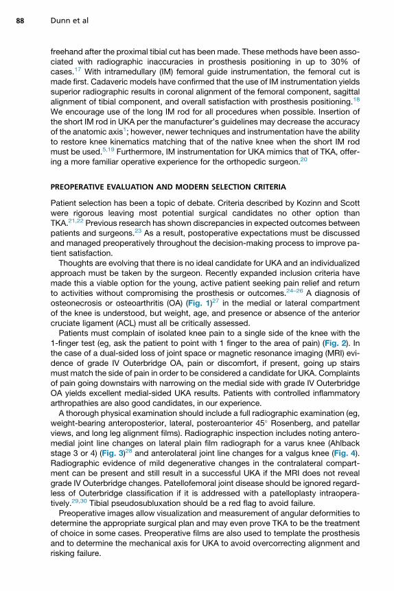
into the drill hole to decrease IM pressures before inserting the IM guide rod.43 Thelong IM rod guide with appropriate distal femoral cutting block is then inserted slowly.A short IM guide is available when a long-stem hip prosthesis is used, which is one ofthe reasons that we obtain 3 standing films preoperatively. A short guide is rarely usedunless a bowing deformity of the femur necessitates its use.The angulation of the cutting block should be adjusted according to preoperative
templating, approximately 6� for a varus knee and 3� or 4� for a valgus knee. The cut-ting block should be flush with the distal femoral condyle, which is accomplished bysoft tissue dissection. The posterior aspect of the cutting block is pinned in placewith a long-headed, 48-mm pin, and the distal femoral cut is made down to the levelof the posterior slotted pin. After the slotted pin is removed, the cut is completed(Fig. 7). The IM guide is removed in order to inspect the distal femoral cut. It is criticalthat the distal femoral cut is flat because the posterior and chamfer femoral cuts aredependent on this first cut. A large bone file from a TKA set is used. A self-retainingIM patellar retractor is placed into the IM pilot hole with the knee flexed to 90�. Align-ment is exclusively obtained with this jig, and therefore, the importance of this stepcannot be overemphasized. The posterior surface of the femoral jig must be perpen-dicular to the femur, ignoring the tibial surface. It is crucial to release the tibial surfacefor a medial UKA, to avoid a varus knee and a failed UKA. For a valgus knee, we advisea release of the IT band in all patients because the valgus knee heals back to bonewithin several weeks.
Proximal Tibial Cut with Extramedullary Guide
The extramedullary tibial guide is positioned in line with the long axis of the tibia usingankle clamps, similar to the same step in a TKA. Minimizing padding at the ankle al-lows for easier palpation of the distal tibia. The guide should be either medial or lateralto the tibial crest to ensure correct alignment depending on a varus or valgus knee. Alateral patellar incision is used for our valgus knees. The posterior slope should mirrorthe patient’s native anatomy, as determined during preoperative planning. A posteriorslope of approximately 5� to 7� is recommended, with a slope at a lesser degree for anACL-deficient knee. In the presence of an extension lag, the cut should be more
Fig. 7. Completion of distal femoral cut. It is important to protect the soft tissues whencompleting the distal femoral cut.

UKA: Intramedullary Technique 95
anterior than posterior, despite fears of creating hyperextension. This strategy allowsthe contracted hamstrings a chance to be stretched out during the early postoperativeperiod. When proceeding in this fashion, loss of extension, common in patients70 years or older, is not a contraindication for a UKA, in our experience (Fig. 8).The cutting block is centered over the medial or lateral tibial surface without over-
lapping the patellar tendon to avoid cutting it with the saw. A 4-mm slotted stylus isused to measure the appropriate amount of resection on the deepest portion of thetibial plateau, which is easily visible after the resection of the distal femur has beencompleted. The proximal tibial cutting block should be secured with 2 headless,threaded, long pins and the extramedullary guide left in place for added securitywith a long-headed, threaded pin. Three pins are used to secure the tibial cuttingblock; there have been no postoperative stress fractures.44
The proximal tibial cut is made with the tibial cutting block and extramedullary guidewith the knee positioned in 90�. Hyperflexion should always be avoided whencompleting this cut. Care is taken to protect the MCL with a Z retractor placed metic-ulously for a medial UKA and on the lateral side to protect vital structures for a lateralUKA. A sagittal sawwith a triangular tip is used to avoid a small bone bridge on the ante-rolateral or medial aspect of the tibial plateau where the sagittal and proximal tibial cutsmeet. The sawblade should be placed lateral ormedial to the guide, hugging themedialor lateral aspect of the intercondylar notch. Extreme caution must be used when usingthis saw to avoid penetrating the posterior capsule. In addition, care must be taken toavoid undercutting the tibial spine when vertically pushing to avoid a fracture.When the cut of bone is removed as 1 piece, it can be used as a guide to determine
the proper tibial tray size. To successfully do this, a cobb is placed on top with a1.27-cm (0.5-inch) flat osteotome underneath. It is essential not to wedge the osteo-tome. The leg should be placed in external rotation and 90� flexion and the cobb usedon the medial side to remove the bone fragment. For a lateral UKA, a figure-of-four po-sition can be used to successfully complete this step. When the fragment is difficult toremove, a knife can be used to remove the capsular meniscal attachments, whichshould have been removed at the beginning of the procedure.It is important to verify the gaps at this point in the procedure in order to decrease
the odds of a gap imbalance when trialing components. An 8-mm or 10-mm spacerguide is placed to ensure that full extension has been maintained as well as no flexiontightness.
Fig. 8. Extramedullary tibial guided positioned for a medial UKA. The angel wing verifiesthe level of cut with soft tissue protection.
Dunn et al96
Posterior Femoral and Chamfer Cuts
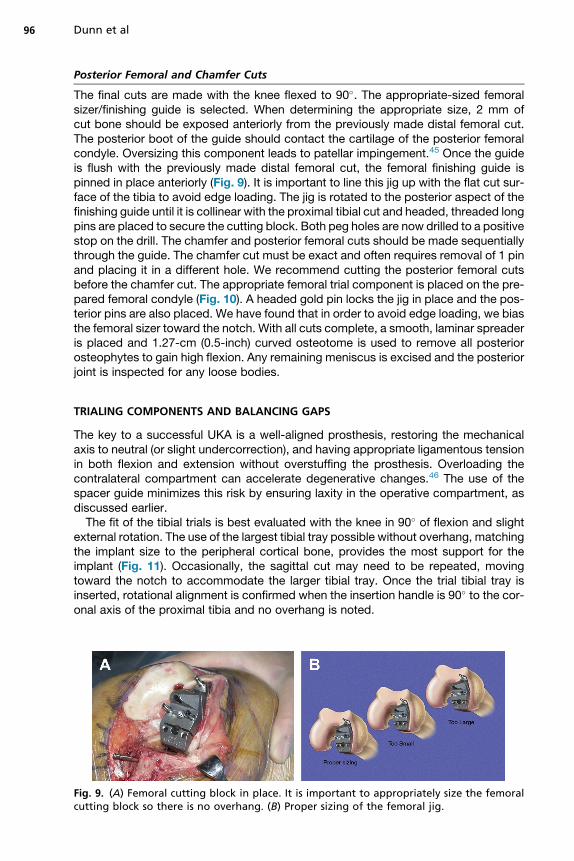
The final cuts are made with the knee flexed to 90�. The appropriate-sized femoralsizer/finishing guide is selected. When determining the appropriate size, 2 mm ofcut bone should be exposed anteriorly from the previously made distal femoral cut.The posterior boot of the guide should contact the cartilage of the posterior femoralcondyle. Oversizing this component leads to patellar impingement.45 Once the guideis flush with the previously made distal femoral cut, the femoral finishing guide ispinned in place anteriorly (Fig. 9). It is important to line this jig up with the flat cut sur-face of the tibia to avoid edge loading. The jig is rotated to the posterior aspect of thefinishing guide until it is collinear with the proximal tibial cut and headed, threaded longpins are placed to secure the cutting block. Both peg holes are now drilled to a positivestop on the drill. The chamfer and posterior femoral cuts should be made sequentiallythrough the guide. The chamfer cut must be exact and often requires removal of 1 pinand placing it in a different hole. We recommend cutting the posterior femoral cutsbefore the chamfer cut. The appropriate femoral trial component is placed on the pre-pared femoral condyle (Fig. 10). A headed gold pin locks the jig in place and the pos-terior pins are also placed. We have found that in order to avoid edge loading, we biasthe femoral sizer toward the notch. With all cuts complete, a smooth, laminar spreaderis placed and 1.27-cm (0.5-inch) curved osteotome is used to remove all posteriorosteophytes to gain high flexion. Any remaining meniscus is excised and the posteriorjoint is inspected for any loose bodies.
TRIALING COMPONENTS AND BALANCING GAPS
The key to a successful UKA is a well-aligned prosthesis, restoring the mechanicalaxis to neutral (or slight undercorrection), and having appropriate ligamentous tensionin both flexion and extension without overstuffing the prosthesis. Overloading thecontralateral compartment can accelerate degenerative changes.46 The use of thespacer guide minimizes this risk by ensuring laxity in the operative compartment, asdiscussed earlier.The fit of the tibial trials is best evaluated with the knee in 90� of flexion and slight
external rotation. The use of the largest tibial tray possible without overhang, matchingthe implant size to the peripheral cortical bone, provides the most support for theimplant (Fig. 11). Occasionally, the sagittal cut may need to be repeated, movingtoward the notch to accommodate the larger tibial tray. Once the trial tibial tray isinserted, rotational alignment is confirmed when the insertion handle is 90� to the cor-onal axis of the proximal tibia and no overhang is noted.
Fig. 9. (A) Femoral cutting block in place. It is important to appropriately size the femoralcutting block so there is no overhang. (B) Proper sizing of the femoral jig.

Fig. 10. Femoral trial sizing is completed before the trial component is placed on the pre-pared femoral surface.
UKA: Intramedullary Technique 97
A curved device is included in the set to feel around the back of the knee with thesizing guide to ensure that the correct tibial prosthesis is selected. The tibial keel isprepared with the provided keel punch and then inserted (Fig. 12A). It is importantwhen creating this trough to first set it flush with the tibial cut surface, then send it pos-teriorly with good coverage, before drilling the 2, 20�-angled posterior tibial holes (seeFig. 12B). The trial femoral component and the trial polyethylene component are theninserted and the patellar retractor is removed.After the trial components are inserted, the flexion extension gaps and tracking are
evaluated. The knee is moved through its full ROM without the tension gauge toassess tracking between the femoral and tibial components. The tension gauge isthen inserted, with the knee flexed to 90�. The tension gauge should slip in and outof the joint with 2 fingers and slight resistance without movement of the tibial trialon the 2-mm side. Movement of the tibial trial is an indication that the flexion gapmay be too tight. This step should also be repeated with the knee in full extension.If the tibial trial moves when in extension, the extension gap may be too tight. Atthis point, necessary adjustments should be made to balance the gaps and properlyalign the components.
Fig. 11. Templates to find the largest tibial tray are completed.

Fig. 12. (A) The tibial keel is placed with an appropriate tibial tray to finalize preparation ofthe tibial surface. (B) The cut tibial surface is completed, and the posterior tibial holes areprepared.
Dunn et al98
FINAL PREPARATION AND CEMENTING OF FINAL IMPLANTS
In final preparation for implant placement, the prepared bony surfaces are copiouslyirrigated with antibiotic solutions and the joint is reinspected for debris. The bonysurfaces are dried and a 10.2-cm � 20.3-cm (4-inch � 8-inch) sponge held by aZ retractor is placed in the posterior aspect of the joint in addition to a thrombin spray.The cement should be compressed into the tibia with a finger or a ganglion knife, mak-ing sure not to place excessive cement posteriorly. Excess cement is difficult toremove after the implants are in place. We advise placing a small amount of cementonly on the posterior aspect of the prosthesis and the anterior section of the tibia.The final tibial component is then inserted and impacted into place. A small, curvedcurette and a knife are used to remove excess cement from the posterior aspect ofthe joint. The process is then repeated with the femoral component, and the trial poly-ethylene component is placed (Fig. 13).
Fig. 13. The tibia is cemented in place, and the femoral surface is prepared with cement toaccept the real prosthesis.

UKA: Intramedullary Technique 99
The leg is moved into extension to compress the components while the cementcures. The cement is monitored, and when appropriate, the previously packed spongein the posterior aspect of joint is removed. Excess posterior cement should be broughtinto view for removal (Fig. 14). If the Z retractor is placed back in the joint, it should al-ways be checked first to make sure that it does not have any excess cement. The trialpolyethylene component is removed when the cement hardens, and the real polyeth-ylene is inserted after checking the gaps (Fig. 15). We advise avoiding making the kneetoo tight. TheMCL is checked in extension and flexion to ensure appropriate laxity. Thewound should be copiously irrigated before closure of the incision in the surgeon’spreferred standard fashion; our preference is a subcuticular monocryl (Fig. 16).
COMPLICATIONS AND OUTCOMES
Please see “Outcomes and Complications of Unicondylar Arthroplasty” by Della Valleet al, elsewhere in this issue for detailed outcomes and complications.Long-term outcomes of UKA performed with IM instrumentation are good. Berger
and colleagues reported 98% survivorship at 10 years and 95.7% at 13 years whenconversion to TKA was used as the end point in 62 consecutive patients. Whenaspetic loosening was used as an end point, survivorship increased to 100% at13 years; 80% of patients reported excellent satisfaction, 12% reported good satis-faction, and 8% reported fair satisfaction. The 2 failures were because of progressionof symptomatic arthritis in other compartments, albeit at a slow progression rate of7 and 11 years.Patients with UKA outperform their peers with TKA on reported symptoms, activities
of daily living, and sport and recreation.47,48 Patients with UKA show greater kneeROM and report less difficulty with activities that involve bending their knee.48,49
Body mass index (BMI, calculated as weight in kilograms divided by the square ofheight in meters) does not adversely affect outcomes50 and persons with BMI greaterthan 35 continue to show functional improvement for at least 2 years after surgery.51
In a recent review of our series, 52 of 56 patients (93%) who participated in sportspreoperatively and underwent UKA returned to sports within 6 months after UKA. The3 patients who did not resume sports postoperatively did not resume sports for rea-sons unrelated to their knee. All patients who participated in tennis (N5 14) and skiing(N 5 23) preoperatively resumed after UKA without complication.
Fig. 14. The femoral and tibial components are cemented in place and any excessive cementoutside the components or under the engaging locking mechanism is safely removed.

Fig. 15. The real polyethylene insert is snapped into place with an audible sound.
Dunn et al100
POSTOPERATIVE PROTOCOL AND REHABILITATION
ROM exercises are initiated immediately postoperatively in the postanesthesia careunit, and a multimodal pain control regimen is routinely used, including nonsteroidalantiinflammatory drugs and 1-time intravenous steroid intraoperatively to controlswelling.52 A Hemovac drain is also used and sewn in the knee. We have found thatthe number 1 complication in our patients is too much activity too fast too soonwith hematoma collection. For this reason, a drain is left in for several days, and wehave had no infections. A CPM device and extensive icing are used to return our pa-tients to activities in an accelerated fashion. The drain is taken out in the office andambulation is partial weight bearing with crutches or a walker for the octogenarians.Deep vein thrombosis prophylaxis is based on risk stratification in accordance withthe recommendations from the American Academy of Orthopaedic Surgeons.53 Weprefer Coumadin for 6 weeks with thigh-high compression stockings.A formal outpatient rehabilitation program is prescribed including wall slides and
stationary bike riding without tension and the seat placed high to avoid excessiveflexion starting on day 1. Emphasis is placed on ROM, restoration of normalmechanics, and strengthening of the core, hip, and quadriceps, which has beenshown to decrease rates of anterior knee pain and improve outcomes.54–56 Strength
Fig. 16. Final UKA with real polyethylene liner. (A) Knee in full flexion. (B) Knee in fullextension.

UKA: Intramedullary Technique 101
training is not initiated until 6 weeks postoperatively. Patients are permitted to return totheir desired activities without restrictions after the completion of their physical ther-apy program. We allow patients to return to golf (riding in a cart) at 8 to 12 weeks,depending on swelling. Singles tennis and downhill skiing are permitted at 6 months.
SUMMARY
UKA is a reliable method of alleviating pain and restoring function in patients with kneearthritis with the appropriate indications and meticulous surgical technique. Use of IMinstrumentation on the femur provides a similar operative experience to TKA, allowingmost surgeons the comfort of a procedure known well to them. Preoperative goalsetting and postoperative rehabilitation involving core, hip, and quadriceps strength-ening are nonsurgical adjuncts, which enhance outcomes and patient satisfaction inthis already successful procedure.
REFERENCES
1. McKeever DC. Tibial plateau prosthesis. Clin Orthop Relat Res 1960;18:86–95.2. MacIntosh DL, Hunter GA. The use of the hemiarthroplasty prosthesis for
advanced osteoarthritis and rheumatoid arthritis of the knee. J Bone JointSurg Br 1972;54(2):244–55.
3. Laurencin CT, Zelicof SB, Scott RD, et al. Unicompartmental versus total kneearthroplasty in the same patient. A comparative study. Clin Orthop Relat Res1991;(273):151–6.
4. Knutson K, Lindstrand A, Lidgren L. Survival of knee arthroplasties. A nation-wide multicentre investigation of 8000 cases. J Bone Joint Surg Br 1986;68(5):795–803.
5. Patil S, Colwell CW Jr, Ezzet KA, et al. Can normal knee kinematics be restoredwith unicompartmental knee replacement? J Bone Joint Surg Am 2005;87(2):332–8.
6. Noticewala MS, Geller JA, Lee JH, et al. Unicompartmental knee arthroplasty re-lieves pain and improves function more than total knee arthroplasty.J Arthroplasty 2012;27(Suppl 8):99–105.
7. Rougraff BT, Heck DA, Gibson AE. A comparison of tricompartmental and uni-compartmental arthroplasty for the treatment of gonarthrosis. Clin Orthop RelatRes 1991;(273):157–64.
8. Oral E, Neils AL, Rowell SL, et al. Increasing irradiation temperature maximizesvitamin E grafting and wear resistance of ultrahigh molecular weight polyeth-ylene. J Biomed Mater Res B Appl Biomater 2013;101(3):436–40.
9. Price AJ, Waite JC, Svard U. Long-term clinical results of the medial Oxfordunicompartmental knee arthroplasty. Clin Orthop Relat Res 2005;(435):171–80.
10. Naudie D, Guerin J, Parker DA, et al. Medial unicompartmental knee arthro-plasty with the Miller-Galante prosthesis. J Bone Joint Surg Am 2004;86-A(9):1931–5.
11. Berger RA, Meneghini RM, Jacobs JJ, et al. Results of unicompartmental kneearthroplasty at a minimum of ten years of follow-up. J Bone Joint Surg Am 2005;87(5):999–1006.
12. Argenson JN, Chevrol-Benkeddache Y, Aubaniac JM. Modern unicompartmen-tal knee arthroplasty with cement: A three to ten-year follow-up study. J BoneJoint Surg Am 2002;84-A(12):2235–9.

Dunn et al102
13. Keblish PA, Briard JL. Mobile-bearing unicompartmental knee arthroplasty: A2-center study with an 11-year (mean) follow-up. J Arthroplasty 2004;19(7Suppl 2):87–94.
14. Cartier P, Sanouiller JL, Grelsamer RP. Unicompartmental knee arthroplasty sur-gery. 10-year minimum follow-up period. J Arthroplasty 1996;11(7):782–8.
15. Hernigou P, Deschamps G. Protheses unicompartimentales du genou. Rev ChirOrthop Reparatrice Appar Mot 1996;82(Suppl):23–60 [in French].
16. Lootvoet L, Burton P, Himmer O, et al. A unicompartment knee prosthesis: Theeffect of the positioning of the tibial plate on the functional results. Acta OrthopBelg 1997;63(2):94–101.
17. Tabor OB Jr, Tabor OB. Unicompartmental arthroplasty: A long-term follow-upstudy. J Arthroplasty 1998;13(4):373–9.
18. Jenny JY, Boeri C. Accuracy of implantation of a unicompartmental total kneearthroplasty with 2 different instrumentations: A case-controlled comparativestudy. J Arthroplasty 2002;17(8):1016–20.
19. Whiteside LA, McCarthy DS. Laboratory evaluation of alignment and kinematicsin a unicompartmental knee arthroplasty inserted with intramedullary instrumen-tation. Clin Orthop Relat Res 1992;(274):238–47.
20. Della Valle CJ, Berger RA, Rosenberg AG. Minimally invasive unicompartmentalknee arthroplasty using intramedullary femoral alignment. Oper Tech Orthop2006;16(3):186–94.
21. Stern SH, Becker MW, Insall JN. Unicondylar knee arthroplasty. An evaluation ofselection criteria. Clin Orthop Relat Res 1993;(286):143–8.
22. Kozinn SC, Scott R. Unicondylar knee arthroplasty. J Bone Joint Surg Am 1989;71(1):145–50.
23. Greene KA, Harwin SF. Maximizing patient satisfaction and functional results af-ter total knee arthroplasty. J Knee Surg 2011;24(1):19–24.
24. Berend KR, Lombardi AV Jr. Liberal indications for minimally invasive Oxfordunicondylar arthroplasty provide rapid functional recovery and pain relief.Surg Technol Int 2007;16:193–7.
25. Pennington DW, Swienckowski JJ, Lutes WB, et al. Unicompartmental knee ar-throplasty in patients sixty years of age or younger. J Bone Joint Surg Am 2003;85-A(10):1968–73.
26. Borus T, Thornhill T. Unicompartmental knee arthroplasty. J Am Acad OrthopSurg 2008;16(1):9–18.
27. Parratte S, Argenson JN, Dumas J, et al. Unicompartmental knee arthroplastyfor avascular osteonecrosis. Clin Orthop Relat Res 2007;464:37–42.
28. Ahlback S. Osteoarthrosis of the knee. A radiographic investigation. Acta RadiolDiagn (Stockh) 1968;(Suppl 277):7–72.
29. Beard DJ, Pandit H, Ostlere S, et al. Pre-operative clinical and radiologicalassessment of the patellofemoral joint in unicompartmental knee replace-ment and its influence on outcome. J Bone Joint Surg Br 2007;89(12):1602–7.
30. Outerbridge RE. The etiology of chondromalacia patellae. J Bone Joint Surg Br1961;43-B:752–7.
31. AOA. The Australian Orthopaedic Association national joint replacement regis-try. Annual report. 2012.
32. Younger AS, Duncan CP, Masri BA. Surgical exposures in revision total kneearthroplasty. J Am Acad Orthop Surg 1998;6(1):55–64.
33. Aglietti P, Baldini A, Sensi L. Quadriceps-sparing versus mini-subvastusapproach in total knee arthroplasty. Clin Orthop Relat Res 2006;452:106–11.

UKA: Intramedullary Technique 103
34. Bonutti PM, Zywiel MG, Ulrich SD, et al. A comparison of subvastus and midvas-tus approaches in minimally invasive total knee arthroplasty. J Bone Joint SurgAm 2010;92(3):575–82.
35. Dalury DF, Snow RG, Adams MJ. Electromyographic evaluation of the midvastusapproach. J Arthroplasty 2008;23(1):136–40.
36. Biedert RM, Sanchis-Alfonso V. Sources of anterior knee pain. Clin Sports Med2002;21(3):335–47, vii.
37. Kumar D, Alvand A, Beacon JP. Impingement of infrapatellar fat pad (Hoffa’sdisease): results of high-portal arthroscopic resection. Arthroscopy 2007;23(11):1180–6.e1.
38. Liu ZT, Fu PL, Wu HS, et al. Patellar reshaping versus resurfacing in total kneearthroplasty–results of a randomized prospective trial at a minimum of 7 years’follow-up. Knee 2012;19(3):198–202.
39. Hoteya K, Kato Y, Motojima S, et al. Association between intercondylar notchnarrowing and bilateral anterior cruciate ligament injuries in athletes. ArchOrthop Trauma Surg 2011;131(3):371–6.
40. Dennis DA, Komistek RD, Kim RH, et al. Gap balancing versus measured resec-tion technique for total knee arthroplasty. Clin Orthop Relat Res 2010;468(1):102–7.
41. Shakespeare D, Waite J. The Oxford medial partial knee replacement. The ratio-nale for a femur first technique. Knee 2012;19(6):927–32.
42. Wangroongsub Y, Cherdtaweesup S. Proper entry point for femoral intrame-dullary guide in total knee arthroplasty. J Med Assoc Thai 2009;92(Suppl 6):S1–5.
43. Amro RR, Nazarian DG, Norris RB, et al. Suction instrumentation decreases in-tramedullary pressure and pulmonary embolism during total knee arthroplasty.University of Pennsylvania Orthopaedic Journal 2001;14:55–9.
44. Brumby SA, Carrington R, Zayontz S, et al. Tibial plateau stress fracture: Acomplication of unicompartmental knee arthroplasty using 4 guide pinholes.J Arthroplasty 2003;18(6):809–12.
45. Hernigou P, Deschamps G. Patellar impingement following unicompartmentalarthroplasty. J Bone Joint Surg Am 2002;84-A(7):1132–7.
46. Koeck FX, Beckmann J, Luring C, et al. Evaluation of implant position and kneealignment after patient-specific unicompartmental knee arthroplasty. Knee2011;18(5):294–9.
47. Walton NP, Jahromi I, Lewis PL, et al. Patient-perceived outcomes and return tosport and work: TKA versus mini-incision unicompartmental knee arthroplasty.J Knee Surg 2006;19(2):112–6.
48. Lygre SH, Espehaug B, Havelin LI, et al. Pain and function in patients after pri-mary unicompartmental and total knee arthroplasty. J Bone Joint Surg Am 2010;92(18):2890–7.
49. Amin AK, Patton JT, Cook RE, et al. Unicompartmental or total knee arthro-plasty?: Results from a matched study. Clin Orthop Relat Res 2006;451:101–6.
50. Naal FD, Neuerburg C, Salzmann GM, et al. Association of body mass indexand clinical outcome 2 years after unicompartmental knee arthroplasty. ArchOrthop Trauma Surg 2009;129(4):463–8.
51. Thompson SA, Liabaud B, Nellans KW, et al. Factors associated with poor out-comes following unicompartmental knee arthroplasty: Redefining the “classic”indications for surgery. J Arthroplasty 2013; Mar 21. [Epub ahead of print].
52. Parvizi J, Miller AG, Gandhi K. Multimodal pain management after total jointarthroplasty. J Bone Joint Surg Am 2011;93(11):1075–84.

Dunn et al104
53. Mont MA, Jacobs JJ, Boggio LN, et al. Preventing venous thromboembolicdisease in patients undergoing elective hip and knee arthroplasty. J Am AcadOrthop Surg 2011;19(12):768–76.
54. Earl JE, Hoch AZ. A proximal strengthening program improves pain, function,and biomechanics in women with patellofemoral pain syndrome. Am J SportsMed 2011;39(1):154–63.
55. Shirey M, Hurlbutt M, Johansen N, et al. The influence of core musculatureengagement on hip and knee kinematics in women during a single leg squat.Int J Sports Phys Ther 2012;7(1):1–12.
56. Chiu JK, Wong YM, Yung PS, et al. The effects of quadriceps strengthening onpain, function, and patellofemoral joint contact area in persons with patellofe-moral pain. Am J Phys Med Rehabil 2012;91(2):98–106.