Embed Size (px)
Citation preview

Trans. Nat. Acad. Sci. & Tech. (Phils.) J 98Q.J I:J J ).J 27
CELLULOSE BIODEGRADATION STUDIES: APPLICATION OF rONA AND PROTOPLAST FUSION TECHNIQUES
Saturnina C. Halos Natural Sciences Research Institute
Univenity ol the Philippines Diliman. Quezon City. Philippines
Introduction
Cellulose biodegradation refers to the breakdown of cellulose to its component glucose units through the action of enzymes. This process has attracted scientific attention because of the complexity of the enzymes involved and the environmental, as well as economic, signitlcance of the process.
Cellulose, the major structural polysaccharide of plants, is a hydrophilic linear glucose polymer with the anhydroglucose units bonded by B-IA, glucosidic linkage (Ghose and Mishra, 1984). The number of glucose units may vary from 15 (a-cellulose) to more than 10,000 (a-cellulose) per molecule. The polymer has both crystalline and amorphous regions, the former referring to the portion more resistant to chemical/biochemical attack and the latter, to the portion of the cellulose chain that is prone to easy hydrolysis (Muhlenthaler, 1967).
Cellulose biodegradation is mediated by several enzyme systems. The more studied are the extracellular cellulase systems in fungi that have three components: endoglucanases (EC 3.2.1.4), cellobiohydroJases (EC 3.2_1.91) and B-glucosidases (EC 3.2.1.21) (Coughlan and Ljungdahl, 1988). The endoglucanases are found to be inactive against crystalline cellulose when acting alone, but hydrolyze amorphous cellulose and soluble derivatives of carboxymethyl cellulose (CM-cellulose). Endoglucanases are also referred to as CM-cellu1ases. Their attack on amorphous cellulose is characterized by random cleavage of B-glycosidic linkages. By contrast, cellobiohydrolases degrade amorphous cellulose by consecutive removal 'of cellobiose units from the non-reducing end of the substrate. Endoglucanases and cellobiohydrolases act together to degrade crystalline cellulose and the B-glucosidades complete the hydrolytic process by converting the resultant cellobiose to glucose or removing glucose from the non-red udng end of short cellooligosaccharides. Exoglucohydrolases (EC 3.2 . 1.74) from Penicillillm funiculosum and Talaromyces emersonii catalyze the removal of glucose residues from the non-reducing end of cellodextrins but do not interact synergistically wirh endoglucanases in the hydrolysis of cellulose. Oxidative enzymes such as cellobiose oxidase (EC 1.199.18) and cellobiose: quinone oxidoreductase (EC 1.3.5.1) have been found to participate in cellulose degradation. Most, if not all, of the extracellular hydrolytic enzymes of
113

114 Transactions National Academy of Science
fungi arc glycoproteins. Of the known aerobic cellulolytic bacteria, only three, Clostridum thermocellwn. Thermonospora sp., and Microbispora hispora possess culture filtrates active against crystalline cellulose. In anaerobic bacteria, an active cellulase complex referred to as cellulosome (Lamed et al, 1983) is bound to the cell surface. The bound enzymes are believed to be the more active cellulases in bacteria. However, one can see a more complex structure. The (.:ellulosomes comprise 35 polypeptides ranging from 45kDA to about 200 kDA (Lamed, e[ al, 1983 and Hon-Nam, et al, 1986).
This paper describes our research work done a t the Natural Sciences Research Institute (NSRI) at the U .P. Diliman campus and at the National InstitUT.e of Biotechnology and Applied Microbiology (BIOTECH) at the U.P. Los Banos campus. Research done at the NSRI mainly involves the use of rONA techniques in studying cellulose biodegradation whereas at the BIOTECH, our work involved the use of protoplast fusion in improving cellulose biodegradation in fungi.
I. The use ofrecombina1l1 DNA in cellulose degradation studies
Recombinant DNA techniques refer to a set of procedures that obtains a piece of DNA from a donor species and inserts this piece to a self-replicating DNA molecule referred to as a vector that has been constructed to easily enter and multiply in a host cell.
At present, a number of laboratories worldwide have employed the recombinant DNA techniques in studying the cellulases of fungi and bacteria. Tltis is based on the underlying concept of molecular biology that the structure determines the function. Primarily, people wanted to examine the structure of the cellulase genes to explain such phenomena as the proper and functional aggregation of a multicQmponent complex or the varied activity of the enzyme depending on the source of substrate. In our studies, we wanted to do three things:
One was to clone the cellulase genes from a depressed mutant of Cellulomonas fimi and compare it with the genes from the wild type strain. The results would tell us if the genes themselves contain sequences involved in glucose repression and where these sequences would be located.
Two, we wanted to place the cellulase genes in a multicopy plasmid, place it back into the Cellulomonas cell to produce a high cell ulase producer.
Three, we wanted to identify the exact gene products of the genes we isolated. We started our work in May, 1986 and with the funds obtained from the U.S.
Agency for International Development, we cloned and obtained restriction maps of two cellulase genes, did partial sequencing, identified our constraint in placing the cellulase genes back in the Cellulomonas and are still in the process of identifying the exact gene products of our two genes.
Our procedures involved construction of a Cellulomonas DNA library, molecular cloning and subcloning, restriction mapping, Southern blot hybridization, transformation experiments, and enzyme assays. Subcloning, restriction mapping. Southern blot hybridization and partial DNA sequencing were done by one of our
Halos, Cellulose Biodegradation Studies 115
project personnel, Rose Caday, at the laboratory of Professor Seymour Fogel at the University of California, Berkeley, whereas the rest of the work was done at our NSRI laboratory. Details of our procedures were presented in three papers being prepared for publication.
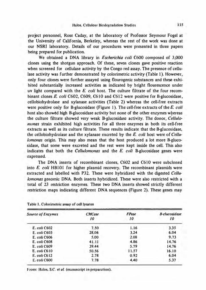
We obtained a DNA library in Escherichia coli C600 composed of 3,000 clones using the shotgun approach. Of these, seven clones gave positive reaction when screened for cellulase activity by the Congo red assay. The presence of cellulase activity was further demonstrated by colorimetric activity (Table 1). However, only four clones were further assayed using flourogenic substances and these exhibited substantially increased activities as indicated by bright flourescence under uv light compared with the E. coli host. The culture filtrate of the four recombinant clones E. coli C602, C609, C610 and C612 were positive for B-glucosidase, cellobiohydrolase and xylanase activities (Table 2) whereas the cell-free extracts were positive only for B-glucosidase (Figure 1). The cell-free extracts of the E. coli host also showed high B-glucosidase activity but none of the other enzymes w~ereas the culture filtrate showed very weak B-glucosidasc activity. The donor, Cellulomonas strain exhibited high activities for all three enzymes in both its cell-free extracts as well as its culture filtrate. These results indicate that the B-glucosIdase, the cel1obiohydrolase and the xylanase excreted by the E. coli host were of Cellulomonas origin. This may also mean that the host produced a lot more B-glucosidase, that some were excreted and the rest were kept inside the cell. This also indicates that both the Cellulomonas and the E. coli B-glucosidase genes were expressed.
The DNA inserts of recombinant clones, C602 and C610 were subcloned into E. coli HBIOI for higher plasmid recovery. The recombinant plasm ids were extracted and labelled with P32. These were hybridized with the digested Cellulomonas genomic DNA. Both inserts hybridized. These were also restricted with a total of 23 restriction enzymes. These two DNA inserts showed strictly different restriction maps indicating different DNA sequences (Figure 2). These genes may
Table I. Colorimetric assay of cell Iysates
Source of Enzymes CMCase FPase 8-clucosidase 10 10 10
E. coli C602 7.50 1.16 3.35 E. coli C603 28.06 3.24 6.04 E. coli C606 5.00 2.08 9.73 E. coli C608 41.11 4.86 14.76 E. coli C609 39.44 5.79 14.76 E. coli ('610 50.56 11.57 16.10 E. coli C612 2.78 0.92 6.04 E. coli C600 7.78 4.40 5.37
From: Halos. S.c. et al. (manuscript in preparation).

116 Transactions National Academy of Science
Table 2. Cellulase activity of culture filtrate from transformed and control cells
Source of Enzymes MUG
E. coli C602 ++ E. coli C603 nt E. coli C606 nt E. coli C608 nt E. coli C609 + E. coli C610 + E. coli C612 + C. coli C600 +/-E. coli HB 294 +/-C. nmi +++
+++ ++
Strong fluorescence Medium fluorescence Weak fluorescence +
+/-·t
Very weak fluorescencl.l/c1earing Presence of halo not rested
Substrates MUCh MU.'(y
++ ++
nt nt nt nl nt nl + + + + + +
+/- +/-+/- +/-+++ +++
nt MUG MUCb MUXy
4-methylumbelliferyl B-D glucopyranosidc (B-glucosidase) 4-methylumbelliferyl B-D ceUobioyranosidc (Cellobiohydrolase) 4-methylumbclliferyl B-D xylopyranoside (Xylanase)
From: Halos, S.c. et al. (manuscript in preparation)
CMC*
+ + + + + + + + + +
Figure I. FlulHcscenn~ of crude extracts using 4 methylumbeliferyl B-D glycosides. Column I, MUG; Column 2, MlICb; Column 3, MUXy; Column 4 Mixed suhstrah:s. no enzyme. Row l. C602; Row 2. C609; Row 3, ('610; Row 4, C612, Row:'i t UB294; Row 6, C600, Row 7, Cellulomonas flmi. (Halo~. C.S., J. Claudio, D. Sanchez, V1anuscript in preparation).

Halos, Cellulose Biodegradation Studies 117
PaK 11 M UX Pv ES K h
II , I " , " ! I pRCK 220 ___ --_ ...... _ ... ___ IIIM __ .............. _
as. B& PaXh PuJ>v
pReu 100 " I II II
Figure 2. Restriction maps of cloned DNA fragments from depressed mutant of Ce/lu/omollas /lmi expressing cellulase activity. Ps, Pstl; K, KpnI; B, 8amHI; M, MIuI; H, HindIlI; X, Xbal; ?V, Pvul; E, EcoRI; S, Smal; Sa, SaIl; 8g, 8gll; A, Aval; Bs, BslxI; Pu, PvuIJ; Xh. Xhol. Pu sites were not clearly established from restriction map. (Halos, S.c. & R.A. Caday, manuscript in preparation).
represent the two separately migrating cellobiohydrolases that we have extracted from Cel/ulomonas. However, these restriction maps differed from restriction maps obtained for cellulase genes of various microorganisms, including the wild type of Cel/ulomonas limi (Figures 3-7). It is possible that we have cloned other cellulase genes and that there could be at least four cellulase genes in Cel/ulomonas limi. There were ten different DNA fragments expressing cellulolytic activities cloned for C thermocellum and six in Microbispora bispora.
The cloned genes, including those obtained from other microorganisms and inserted in pBR322 or its derivatives, were used to transform the Cel/ulomonas timi mutant. Although we were able to obtain transformation, the cells apparently could not maintain the plasmid. We have observed, however, that Cel/ulomonas has a plasmid which we can use to construct a cloning vector in the future.
H K P K P K
I II I I I I plCl • &.11 6.2 2.1 2.a
II.'
EHP n IE E H P E HO H
PoIel U I I II 1.72.1 1.a 2.2 2.4 4 1.4 2.7 •• a
piC:: 21 1.' II H HO H
1.1 2 1.4
Figure 3. Restriction maps of cellulase genes from Cellulomonag sp. H, HindIII; E, EcoRI; K, Kpnl, P, Pvull; B, BamHI. (Lohwongwatana, unpublished)

II 8 Transactions National Academy of Science
B a :: a I - I ,') I ;i Z I
III
pH1:1
I 11.1 O.S I.' I.' 1' •• '12 ,
'-S
_IU ... I _I _aa %:jBr .... _m
I .-u I I 0.' D.! 0.1 1.1
L...-.I.i I
I _m I r-.I .... 1_1 -.0 ,
r , , " r
I 1' ••• 12 t 0.:1 1.' 0 .. 0.' 0.'
•• 1
Figure 4. Restriction maps of cloned DNA fragments from Bacillus sp. expressing cellulase activity. (Horikoshi and Fukumori, 1988)
IEIU' K. K • • R II ..... II:
l" I r I I I ~l( -"TU. I
IE K II Sao Sa> n IE
I I I I If
.,,:TUO
I: Sm P " I I
pCTlOl I
II: " H • n IE
I pCT401
., I , j
R • • H .... H. KlalllE
)))~ ~r I .n-
It II " Il
I I I I
e(."'ftlOO
It H U B Ia' • II. IE
"))1 )J( I( V J penGO
c P Be P • • )11
& H .... " j-JI:::' ==:::r=:=r=:::::1 .cT1_ L-.
R II II
.cT1- tl =======~===::::j
Figure 5. Restriction map of DNA from Clostridum thermocellum encoding CMC or MUChhydrolyzing activity. B, BamHI; E, EcoRI; H, Hindlll; P, Pstl; Sa, Sail. Sm, Smal. pCT800 and pCTl300 contain 6 and 9 Hindlll sites, respectively. (Beguin et al .• 1988)
Halos, Cellulose Biodegradation Studies 119
I'" dJ
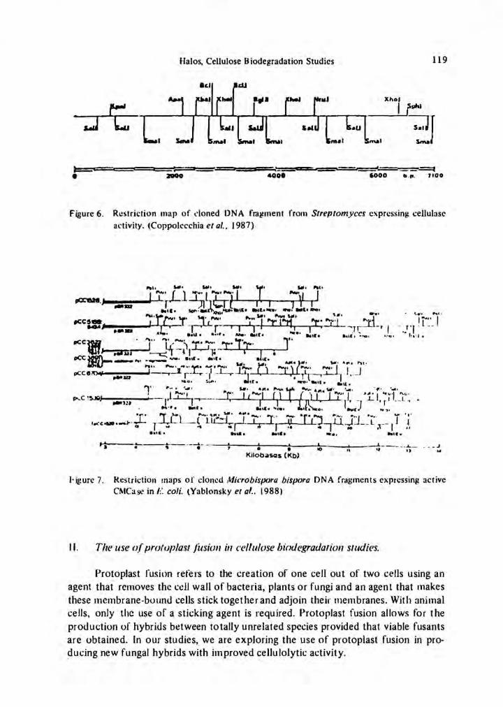
Figure 6. Rl:striction map of doned DNA fra!!ment from Streptomyces expressing cellulase activity. (Coppolecchia et 01., 1987)
hgurc 7. Restriction maps or cloned Microbispora bispora DNA fragments expressing active CMCase in I:: coli. tYablonsky et 01 .. 1988)
II. The use of protoplast fusioll in cellulose biodegradatioll sllldies.
Protoplast fusion refers to the creation of one cell out of two cells using an agent that removes the cdl wall of bacteria, plants or fungi and an agent that makes these membrane-buund cells stick together and adjoin their membranes. With animal cells, only the use of a sticking agent is required. Protoplast fusion allows for the productiun uf hybrids between totally unrelated species provided that viable fusants are obtained. In our studies, we are exploring the use of protoplast fusion in producing new fungal hybrids with improved cellulolytic activity.

120 Transactions National Academy of Science
Of the various fungaJ species producing ccllulases, the Trichoderma and the Pellicillium species are the ones currently used in the limited commercial production of cellulases. The advamage of using fungal cellulases lies in their relative stability to extraction procedures. However, the search for better sources of crllulases continues since no st rain that can be used economically to produce the enzymes has yet been developed or isolated.
(n our earliest project of screening for high cellulase producers, we identified, one isolate of Penicillium fUlliculoswn Thom No. 171 that was as good as our reference strain, Trichoderma reesei RutC30, a strain developed for high cellulolytic activity by researchers of Rutgers University in New Jcrsey. In addition, P funiculosum No. 171 produced higher levels of B-glucosidase activity than T. Ret.'sei Rut C300 (Cruz, W. T., 1986). This prompted us to explore the possibility of using protoplast fusion to develop intergeneric hybrids of Trichoderma X Penicillium that would prm.luce a hybrid cellulase complex incorporating the higher cell ulase activities of both parents.
Our study involved establishing the procedures for protoplast isolation and renegeration for P. funiculosul1l, and T. reesei RutC30 using resources available to us, identitkation of suitable selection markers, characterization of fusants and assaying r or their cellulolytic activities. Details of procedures and results are presented in two papers submitted for publication.
Of six enzyme preparations tcsted for protoplast product ion, a more efficient one was the combination of Novozyme 234 (Novo Industries, Inc.) and Zymolyaze 20T which released 3.6 X 10⁷ for T. reesei and 1.4 X 10⁷ for P. funiculosum per mg mycelia (Table 3 and Figure 8). This enzymes combination released protoplast in direct proportion to the amount of mycelia when exposed to the same concentration of enzyme (2.5 mg/ml Novozyme 234 and 5 mg/ml Zymolyase 20T) t ypicaJ of an enzymatic reaction. T. reesei was more susceptible to these enzymes in that protoplasts were first produced from T. reesei 60 minutes following treatment of mycelium compared with P. funiculosum that started releasing
Table 3. Comparison of lytic enzyme preparations for the release of Protoplasts from Tri· choderma reesei RUT C·30 and Penicillum funiculosum Thom MG 171
Enzyme System
Celluclost Cellulase (laboratory prep.) Novozyme 234 Zymolase 20T Celluclast + Zymolace 20T Novozym 234 X zymolase 20T
Protoplast yield Trichoderma reesei RUT C·30
3.7 X 10⁶
2.9 X 10⁵
0.5 X 10⁶
0.1 X 10⁶
3.6 X 10⁷
From: Pham, L. & S.c. Halos, 1988. manuscript for publication.
Penicillum funiculosum Thom MG 171
2 X 10⁶
1.7 X 10⁶
0.28 X 10⁶
0.016 X }O6 1.4 X 10 7

70
60
e 50 ... f1Q ....
0 .. ... i
40
Q, 0 ~ 30 4:
20
10
Figure 8.
Halos. CeJlu lose Biodegradation Studies 121
60 100 160 200 260 BOO
The effect of mycelial oncentration on protoplast formation of Trichoderma reesei RUT C·JO and Penicillium funiculosum Thorn MG 171. (Pham, l.. & S.c. Halos, 1988. manuscript for publication).
protoplast 90 minutes after treatment. Furthermore, the highest yield of protoplasts was obtained with Trichoderma 2 hours after enzyme exposure, whereas with P .lrmiculoswn it was 4 hours after. Also, T. reesei regenerated more at 92% than P fimiculosum which gave a 31. J 7% regeneration frequency. Regeneration occurred 3 hours after transfer to Winge medium.
. Since we had no intention of altering the genetic makeup of our parental strains, we sought for innate properties of these strains to use as selection markers. We screened their resjstance to different metal ions (Cu, Na, Co, and Hg), fungicides (Captan and Bcnlate) and antibiotic (nystatin) at different concentrations. We were able to identify complementing markers CoR and HgS for T. reesei and CoS and HgR for P. funiculosum (Table 4) as the primary selection markers (Figure 9). Fusants were then selected as CoR and HgR colonies (Figure 10).
Fusants deri .... ed were viable and exhibited different morphologies (Table 5) which combine the properties of both parents. Fusants did not exhibit uniform
characteristics which could be due to observations that mycelial fungi are multinucleated. Fusants might have represented different combinations of the 3 nuclei that are often found in one mycelial cell.

122 Transactions National Academy of Science
Table 4. The effect of Metal Ions on Protoplast Regeneration of Trichoderml1 reesei RUT C-30 and Penicillium funiculosum Thorn MG 171
Metal Ions
Cu (10 ppm) Na (10 ppm)
Cu (10 ppm) Na 00 ppm) Co 00 ppm) Hg (I ppm)
Trichoderma reesei RUT C-JO
+ +
+ + +
+ regenerated - = did not regenerate
Penicillium funicolusom Thom 1 71
+
+ +
+
From: Pham, I. & S.c. Halos, 1988. manuscript for publication)
Figure 9.
Protoplast from Penicillium funiculosum.
Regenerating protoplast from P. funiculosum.
Regeneration of Penicillium funiculosum protoplast. (Pham, L. & S.C. Halos, 1988. manuscript for publication)_

Halos, Cellulose Biodegradation Studies 123
Figure 10. Parental Strains and Fusants derived from Trichoderma reesei RutC30 and Peni· cillium {tmicu!osum Thorn MG 171. (Pham, L & S.C. Halos. 1988. manuscript for publkation). extreme left - without Co +, Hg ++
. t C + H ++ center- WIt 1 .0, g '1 'h C+H++ extreme r~ 1t - wtt out 0, g
Table 5. Cultural characteristics of fusant strains following protoplast fusion compared t n parental strains
COLONY COLONY SPORU- PIGMENT A nON FUSANTNU. CO/DR Tf.:XTURE LAT/ON
14 white cottony none red 20 graying cottony fourth day dark brown
green 24 light blue vclvetty second day sulfur
to mos:\ green yellow 34 olive green cottony fourth day red to
brick red 35 oli\'e green cottony fourth day red TO
brick clay 36 bluc to sulfur
moss green vclvetty second day yellow 37 blue to moss velvctty second day sulfur
green yelJow

124 Tran.sactions National Academy of Science
FUSANTNU.
46 47 62 66 71 74 76 77
COLONY COLOR
moss green yellow green olive green moss green moss green moss green oliv(' green moss green
PARENTAL STRAINS
T. reesei RUT C:·30
P. funiculum Thorn MG-I?1
yellow green to olive green
moss agreen
COLONY SPORU· n"XTURE LATION
velvetty velvetty velvetty irregular velvetty velvetty velvetty vclvetty
cottony fourth day
vdvetty fourthday
From: Pham, L. & S.c. Halos, 1988. manuscript for publication.
Table 6. Enzyme activities of fusants a nd parental strains
FUSANT PROTEIN CMCAS/:' FI'ASE NO. Mg/ml JUjml/mill IU/m/min
14 0.38 .157 .007 20 0.68 .055 .051 24 1.09 .565 .154 34 1.52 1.52 1.31 35 1.55 4.65 1.19 36 0.95 3.90 0.697 37 1.93 3.90 1.04 46 1.14 5.46 .36 47 .553 2.17 .36 66 1.25 5.46 .332 67 1.38 5.05 .408 71 1.59 4.83 .344 74 1. 86 5.23 .398 76 1.25 4.42 .352 77 1.45 5.62 .359 78 1.92 3.56 .334
PARENTAL STRAINS
T. reesei 2.38 7.15 2.49 RUT C·30 P. funiculosum TThom MG· 1 71 1.25 4.55 1.40
From: Pham, 1... & S.c. Halos, 1988. manuscript for publication.
PIGMENTA TJON
dark brown dark brown dark red dark rt'd brown brown dark brown dark brown
sulfur yellow
red
B·GL UCOSIDASE JU/JO min,
.101
.037
.126 7.79 4.267 1.877 1.319
.178
9.89

Halos, Cellulose Biodegradation Studies 125
The cellulolytic activity of various fusants were in general lower than their parental strains (Table 6) Pham and Halos, 1988). However, preliminary results with the second generation or the progeny of two fusants indicate that these have higher eM-cellulase and FP-cellulase activities than the parental strains (Tables 7-8)(de los Reyes, M. 1988).
Table 7. Average CMcase activity (IV/ml), glucose production (mg/ml) and specific activity (IV/mg) in rice straw of the cellulase enzyme from the fusants and parental strains
CMC(1se act. Spec. act. Glucose .vield Isolate Name/Number (IU/mI) (IU/mg) (mg/ml)
Trichoderma reesei Rut CJO 0.0 759B 0.4258 B 0.1366 P. fUlliculosum MG 171 0.0557 B 0.6724B 0.1002
BI8 0.3454A 0.8665A 0.6217 B12 0.2857A 0.7704A 0.5143 BI3 0.3639A 0.3808 A 0.6551 B14 0.360S A 0.7454A 0.6490
From: de los Reyes, e.C., 1988
Table 8. Average FPase activity (IV/ml), glucose production (mg/ml) and specific activity (IV/mg) in rice straw of the cellulase enzyme from the fusants and parental strains
FPase act. Spec. act. Glucose yield Isolate Name/Number (IU/mI) (IU/mg) (mg/ml)
Trichoderma reesei Rut C30 0.0073B 0.0431 A 0.0787 P. funiculosum MG 171 ND,B ND,A ND'
BI8 0.0793A 0.1581 A 0.8562 B12 0.0260A O.0714A 0.2809 B13 0.0733A 0.0767 A 0.7912 B14 0.0963A 0.1995 A 1.0402
1 ND, undetectable 'values within a column having similar letters indicate that they are not si~nificantJy different according to DMRT result
From: de los Reyes, ee., 1988
Conc1usion
We are just starting to explore the use of the techniques of the new genetics in cellulose hiodegradation. In the use of protoplast fusion in improving the cell u· lose biodegradation ahility of fungal strains, it appears that there is more promise

126 Transactions National Academy of Science
among the second generation fusants of T. reesei Rut (30 and P jilllicu/osom No. 171, whereby four second generation fusants exhibited higher cellulolytic activities than their parental strains. In using rONA techniques, we are contlrming results we obtained with other procedures on the presence of at least two genes for cellulascs. These genes exhibit different sequences and their protein products exhibited different mobilities upon electrophoresis. Originally, we proposed to place one of these cloned genes back into the Cel/u/otnonos cell; however, the Cel/ulomonos cannot retain the pBR322 plasmid. Hence, we are currently studying the Cellulomonas plasmid as a possible vector for the cloned cellulase gene.
Acknow iedgments
I wish to thank R.A. Caday, J .0. Claudio, D.R. Sanchez, L.J. Pham and 'I.e. Hagan for their technical assistance. This work was supported by USAJD Grant Number 492-5542-G-SS-6007-00 and BIOTECH.
literature Cited:
I. Beguin, P. et al The cell (Cellulose Degradation) Genes of Clostridium thermucellum. p. 267-282. In Biochemistry and Genetics Cellulose Degradation. eds: Aubert, J.P. Pt. 01 .. London: Harcourt Brace Jovanovich, Publishes, c 1988.
2. Coppolecchia, M.R. et aL 1987. Cloning in E coli of a Streptomyces Cellulase Gene. Biotech. Letters. 9(7): 495-500.
3. Coughlan, M.P. and L.G. Ljungdahl. Comparative Biochcmmistry of Fungal and Bacterial Cellulolytic Enzyme Systems. p. 11-30. In: Biochemistry and Genetics of Cellulose Degradation. cds. Aubert, J.P. ct al. London: Harcourt Brace Jovanovich, Publishers. c 1988.
4. Cruz, W.T. J 986. Isolation, Screening, Characterization, and Identification of Local Lellulase-Producing Fungi and OptimiZation of Enzyme Production. Master's Thesis. University of the Philippines, Los Banos.
5. de Los Reyes, C.C. 1988. Comparative Cellulase Activities of Fusants of Tridlotierma reesd Rut C30 x Penicillium funiculosum MGI7l and their Parental Strains. Undergraduate Thesis. University of the Philippines, Los Banos.
6. Ghose, T .K. and S. Mishra. 1984. Bioconversion or Lignocellulose to Ethanol p. 12 3-l4 7. In: Proc. Role of Biochemistry in Food and Energy Production Symposium. Manila Philippines.
7. lIalos, S.c., J.O. Claudio and D.R. Sanchez. Manuscript in preparation. 8. Halos, S.c. and R.A. Caday. Manuscript in preparation. 9. Hon-Nami. K. el 01., 1986. As cited by Coughlan, M.P. and L.G. Ljungdahl. Comparative
Biochemistry of l:ungal and Bacterial Cellulolytic Enzyme Systcm~. p. J 1-30. In: Bio· chemistry and Genetics of Cellulose Degradation. eds: Aubert, J.P. et. 01., London: Harcourt Brace Jovanovich, Publishers. l'. 1981:1.
10. Horikoshi, K. and F. Fukumori. Modification and E.\ rHession of Alkaline Cellui;lse Genes of Alkalophilic Bacillus strains. In: Biochemistry ,it.d Genetics of Cellulose Degradation. eds: Aubert, J.P. et 01. London: Harcourt Brace Jovanovich, Publishers. c. 1988.
11. Lamed, R. et 01. 1983. as cited by Coughlan, M.P. and L.G. Ljungdahl. Comparative Biochemistry of Fungal and Bacterial CEllulolytic Enzyme Systems. p. 11-30. In: Bio-

Halos. Cellulose Biodegradation Studies 127
chemistry and Genetics of Cellulose Degradation. eds: Aubert, J.P. et al .. London: Harcourt Brace Jovanovich, Publishers. c 1988.
12. Lohwongwatana, J.et ai., Cloning of B-glucosidase gene from Cellulomollas sp. (wlpub. Iished).
13. ~uhlenthaler, R. 1967. Ultrastructure and Formation of Plant Cell Walls. Ann. Rev. Plant 18:1-24.
14. Pham, L.J. and S.c. Halos. 1988. Manuscript (for publication). 15. Yablonsky. M. D. et al Characterization and cloning of the Cellulase Complex of
Microbispora bispora. In: Biochemistry and Genetics of Cellulose Degradation. eds: Auber, J.P. et al. London: Harcourt Brace Jovanovich, Publishers. c 1988.