Embed Size (px)
Citation preview

NE35CH02-Blackstone ARI 14 May 2012 10:46
Cellular Pathways ofHereditary Spastic Paraplegia∗
Craig BlackstoneNeurogenetics Branch, National Institute of Neurological Disorders and Stroke, NationalInstitutes of Health, Bethesda, Maryland, USA; email: [email protected]
Annu. Rev. Neurosci. 2012. 35:25–47
First published online as a Review in Advance onApril 20, 2012
The Annual Review of Neuroscience is online atneuro.annualreviews.org
This article’s doi:10.1146/annurev-neuro-062111-150400
∗This is a work of the U.S. Government and is notsubject to copyright protection in the UnitedStates.
Keywords
spasticity, lipid droplet, BMP, cytokinesis, endosome, endoplasmicreticulum
Abstract
Human voluntary movement is controlled by the pyramidal motorsystem, a long CNS pathway comprising corticospinal and lower mo-tor neurons. Hereditary spastic paraplegias (HSPs) are a large, geneti-cally diverse group of inherited neurologic disorders characterized by alength-dependent distal axonopathy of the corticospinal tracts, resultingin lower limb spasticity and weakness. A range of studies are convergingon alterations in the shaping of organelles, particularly the endoplasmicreticulum, as well as intracellular membrane trafficking and distributionas primary defects underlying the HSPs, with clear relevance for otherlong axonopathies affecting peripheral nerves and lower motor neurons.
25
Ann
u. R
ev. N
euro
sci.
2012
.35:
25-4
7. D
ownl
oade
d fr
om w
ww
.ann
ualr
evie
ws.
org
by N
atio
nal I
nstit
utes
of
Hea
lth L
ibra
ry (
NIH
) on
06/
21/1
2. F
or p
erso
nal u
se o
nly.
NE35CH02-Blackstone ARI 14 May 2012 10:46
HSP: hereditaryspastic paraplegia
Contents
INTRODUCTION . . . . . . . . . . . . . . . . . . 26COMMON CELLULAR
PATHOGENIC THEMES. . . . . . . . 28Axon Pathfinding . . . . . . . . . . . . . . . . . . 28Myelination . . . . . . . . . . . . . . . . . . . . . . . 28Endoplasmic Reticulum
Network Morphology . . . . . . . . . . . 31Lipid Synthesis and Metabolism . . . . 34Endosomal Dynamics . . . . . . . . . . . . . . 35Motor-Based Transport . . . . . . . . . . . . 37Mitochondrial Function . . . . . . . . . . . . 37
RELATED DISORDERS . . . . . . . . . . . . 37E PLURIBUS UNUM?. . . . . . . . . . . . . . . 38
Bone MorphogeneticProtein Signaling . . . . . . . . . . . . . . . 38
Interorganelle Contactsand Communication . . . . . . . . . . . . 39
CONCLUDING REMARKS . . . . . . . . . 39
INTRODUCTION
Voluntary movement in humans relies on thepyramidal motor system, a tortuous, multisy-naptic pathway in the CNS that extends fromthe cerebral motor cortex to neuromuscularjunctions innervating skeletal muscle. This sys-tem is arranged in two main stages (Figure 1).First, axons of large pyramidal neurons origi-nating in layer V of the cerebral motor cortexcourse through the medullary pyramids, wheremost fibers decussate in the caudal medulla be-fore descending as lateral corticospinal tractswithin the spinal cord. Although some corti-cospinal axons establish synapses directly withlower motor neurons in the spinal cord ante-rior horn, the vast majority synapse with spinalinterneurons, which then establish connectionswith lower motor neurons. In the next stage,lower motor neurons terminate in specializedsynapses at neuromuscular junctions through-out the body to regulate skeletal muscle con-tractility (Figure 1) (Carpenter 1991).
Distances traversed by corticospinal andlower motor neurons are among the furthest
in the body; their axons extend up to 1 m inlength and the axoplasm comprises >99% oftotal cell volume. This length has evolved topermit the very rapid relay of action potentials,enabling timely voluntary movement, but itcomes at great expense to the neuron: Complexintracellular machineries are required for sort-ing and distributing proteins, lipids, mRNAs,organelles, and other molecules over suchlong distances. These machineries utilize anelaborate neuronal cytoskeletal scaffold alongwhich motor proteins target and deliver com-ponents selectively throughout the cell; axonaltransport machineries rely on microtubulesin particular, which function as polarizedtracks with their plus ends oriented toward theaxon terminal. A variety of mechanoenzymeswithin the kinesin, dynein, and myosin proteinsuperfamilies mediate much of the anterogradeand retrograde transport specificity throughselective cargo interactions. Additional speci-ficity and regulatory control are contributedby various adaptor proteins. The interaction ofintracellular cargoes with these complexes per-mits tightly regulated, selective allocations oforganelles, proteins, lipids, and other moleculesto growth cones during axonal developmentand to specialized axon domains such as branchpoints, internodal segments, and presynapticterminals in mature neurons (Goldstein et al.2008, Arnold 2009, Hirokawa et al. 2010).
Not surprisingly, long axons are an Achilles’heel of the nervous system; length-dependentdefects in axon development and maintenancegive rise to a host of neurological disorders,both acquired and inherited. Acquired disor-ders are numerous and highly varied, with eti-ologies encompassing injuries, nutritional defi-ciencies, endocrine and metabolic disturbances,infections, and environmental toxins, to namea few; these are not discussed here. The focusof this review is on inherited Mendelian dis-orders, as exemplified by the hereditary spasticparaplegias (HSPs). Although these are amongthe most genetically diverse of all diseases, withnearly 50 distinct loci and more than 20 geneproducts identified to date, they are unified bythe defining, predominant clinical feature of
26 Blackstone
Ann
u. R
ev. N
euro
sci.
2012
.35:
25-4
7. D
ownl
oade
d fr
om w
ww
.ann
ualr
evie
ws.
org
by N
atio
nal I
nstit
utes
of
Hea
lth L
ibra
ry (
NIH
) on
06/
21/1
2. F
or p
erso
nal u
se o
nly.

NE35CH02-Blackstone ARI 14 May 2012 10:46
progressive lower limb spasticity and weakness,with sparing of the upper limbs to a large extent(Fink 2006, Depienne et al. 2007, Salinas et al.2008, Dion et al. 2009, Blackstone et al. 2011,Lang et al. 2011).
HSPs are uncommon but not rare, with aprevalence of ∼3–9/100,000 in most popula-tions, and thus likely afflict several hundredthousand individuals worldwide. Inheritancecan be X-linked recessive, autosomal recessive,or autosomal dominant, and age at onset canvary widely, from early childhood to late inlife. HSPs have historically been classified aspure or complicated on the basis of the absence(pure) or presence (complicated) of associatedclinical features such as distal amyotrophy,cognitive dysfunction, retinopathy, ataxia, thincorpus callosum, and peripheral neuropathy(Harding 1983). Even in pure forms, urinarysymptoms and mild dorsal column sensorydeficits are frequently encountered. Morerecently, a numeric labeling scheme has takenhold, and HSPs are increasingly referred tomainly by their genetic classification, SPG1-48(Depienne et al. 2007, Salinas et al. 2008, Dionet al. 2009, Blackstone et al. 2011).
Because most patients with HSP have anormal life span, a limited number of neu-ropathologic evaluations of HSPs have beenpublished, particularly for the most instructivepure forms with a genetic diagnosis. Still, thesestudies have typically shown evidence of axonaldegeneration, principally involving the longestascending sensory fibers and descending cor-ticospinal tract axons in a distal, “dying-back”manner (DeLuca et al. 2004). Because thelongest corticospinal axons control the lowermotor neurons innervating muscles of thelower limbs, such findings are concordant withthe cardinal clinical features of HSP; sensorymanifestations tend to be clinically mild. Thereis usually little neuronal death even late inthe disease course, especially in pure forms,so HSPs are a prototype for understandingdisorders that impair axons (Soderblom &Blackstone 2006). Importantly, HSPs are fun-damentally diseases of massive scale, affecting
Pons
Caudalmedulla
Lumbarspinal cord
Skeletalmuscle
Midbrain
Corticospinalaxon
Decussationof pyramids
Upper motor neuron
cell body
Figure 1Schematic diagram of the corticospinal tract emphasizing its descent throughthe CNS. Although most fibers decussate in the caudal medulla, a minority offibers descend uncrossed as the ventral corticospinal tract (not shown).
www.annualreviews.org • Hereditary Spastic Paraplegia 27
Ann
u. R
ev. N
euro
sci.
2012
.35:
25-4
7. D
ownl
oade
d fr
om w
ww
.ann
ualr
evie
ws.
org
by N
atio
nal I
nstit
utes
of
Hea
lth L
ibra
ry (
NIH
) on
06/
21/1
2. F
or p
erso
nal u
se o
nly.

NE35CH02-Blackstone ARI 14 May 2012 10:46
Spasticity: increase inmuscle tone associatedwith hyperactivetendon stretch reflexes
Corpus callosum:arched bridge of nervefibers connecting theleft and right cerebralhemispheres
predominantly the longest neurons that are or-ders of magnitude larger than most other cells.
COMMON CELLULARPATHOGENIC THEMES
The common clinical and pathological featuresof different HSPs prefigure a small number ofcommon themes at the cellular level, and thegenetic heterogeneity provides a significantadvantage in identifying these convergentthemes. Indeed, published studies have indi-cated that HSP disease proteins cluster withina small number of predicted cellular processes(Table 1 and Figure 2) (Soderblom &Blackstone 2006, Depienne et al. 2007, Salinaset al. 2008, Dion et al. 2009, Blackstone et al.2011). Although we discuss HSP proteins bytopic, one must remember that many of themappear to function in a number of differentpathways; thus pathogenic groupings mayevolve over time.
Axon Pathfinding
Among the first HSP mutations described werein the L1CAM gene, encoding a cell surface gly-coprotein of the immunoglobulin (Ig) super-family. Loss-of-function mutations in L1CAMare implicated not only in X-linked, early-onset,complicated HSP (SPG1), but also in otherX-linked syndromes including MASA (for men-tal retardation, aphasia, shuffling gait, and ad-ducted thumbs), hydrocephalus, and agenesis ofthe corpus callosum ( Jouet et al. 1994, Weller& Gartner 2001). Each of these disorders dis-plays clinical and pathological evidence of cor-ticospinal tract impairment; the disorders areconsidered together along a disease spectrumknown as L1 disease or CRASH syndrome(for corpus callosum hypoplasia, retardation,adducted thumbs, spastic paraplegia or shuf-fling gait, and hydrocephalus) (Soderblom &Blackstone 2006).
The L1CAM protein is more than 1200amino acid residues in size, with a large extra-cellular segment harboring 6 Ig-like domainsand 5 fibronectin type III domains, a singletransmembrane domain, and a short cytoplas-
mic tail. L1CAM participates in a complex setof extracellular and intracellular interactions,binding not only other L1CAM molecules butalso a host of extracellular ligands—includingother cell adhesion molecules, integrins, andproteoglycans—as well as intracellular proteinssuch as ankyrins. Disease mutations are foundthroughout the protein, and partial or completeloss of L1CAM function seems critical for theL1 disease phenotype. In L1CAM null mice, thecorticospinal tracts are abnormal, arising froma conspicuous failure to decussate within themedulla (Dahme et al. 1997, Cohen et al. 1998).
How does this pathfinding defect occur? Inthe developing CNS, L1CAM associates withneuropilin-1 (Nrp1), which itself interacts withPlexin-A proteins to form the Semaphorin3A(Sema3A) receptor complex. Upon Sema3Abinding to Nrp1, L1CAM and Nrp1 are coin-ternalized in a L1CAM-dependent manner.Sema3A is a repulsive guidance cue releasedfrom cells in the ventral spinal cord to steer cor-ticospinal neurons away from the midline spinalcord/medullary junction, and L1CAM muta-tions may affect Sema3A signaling when axonsare crossing the midline by interfering with re-ceptor internalization and signaling at growthcones (Castellani et al. 2004). In fact, the as-sociation of Nrp1 with L1CAM mediates theactivation of a focal adhesion kinase-mitogen-activated protein kinase pathway controlling acritical aspect of the repulsive behavior, the dis-assembly of adherent zones in growth conesand their subsequent collapse (Bechara et al.2008). This compelling role of L1CAM in axonpathfinding during development is consistentwith the early onset of SPG1.
Myelination
A distinguishing feature of axons in the centraland peripheral nervous systems is an insulatingmyelin sheath, a specialization important forincreasing the speed of electrical impulsepropagation. Schwann cells supply myelin forperipheral neurons, whereas oligodendrocytesmyelinate axons of CNS neurons. Spasticparaplegia as a manifestation of abnormal
28 Blackstone
Ann
u. R
ev. N
euro
sci.
2012
.35:
25-4
7. D
ownl
oade
d fr
om w
ww
.ann
ualr
evie
ws.
org
by N
atio
nal I
nstit
utes
of
Hea
lth L
ibra
ry (
NIH
) on
06/
21/1
2. F
or p
erso
nal u
se o
nly.

NE35CH02-Blackstone ARI 14 May 2012 10:46
Table 1 Identified HSP genes, grouped functionallya,b
Disease/geneb Protein name Inheritance Cellular functionsMembrane traffic and organelle shaping
SPG3A/ATL1 Atlastin-1 AD ER morphogenesisBMP signaling
SPG4/SPAST Spastin (M1 and M87 isoforms) AD Microtubule severingER morphogenesisEndosomal trafficBMP signalingCytokinesis
SPG6/NIPA1 NIPA1 AD Endosomal trafficMg2+ transportBMP signaling
SPG8/KIAA0196 Strumpellin AD Endosomal trafficCytoskeletal (actin) regulation
SPG10/KIF5A KIF5A AD Microtubule-based motor proteinSPG11 Spatacsin AR Endosomal trafficSPG12/RTN2 Reticulon 2 AD ER morphogenesisSPG15/ZFYVE26 Spastizin/
ZFYVE26/FYVE-CENT
AR Endosomal trafficCytokinesisAutophagy
SPG17/BSCL2 Seipin/BSCL2 AD Lipid droplet biogenesis at ERSPG18/ERLIN2 Erlin2 AR ER-associated degradation
Lipid raft-associatedSPG20 Spartin AR Endosomal traffic
BMP signalingCytokinesisLipid droplet turnoverMitochondrial regulation
SPG21 Maspardin AR Endosomal trafficSPG31/REEP1 REEP1 AD ER morphogenesis
ER-microtubule interactionSPG48/KIAA0415 KIAA0415 (AP-5 subunit) AR Endocytic adaptor protein complexAP-4 deficiency/AP4S1, AP4B1, AP4E1 AP-4 S1, B1, and E1 subunits AR Endocytic adaptor protein complexJPLS/ALS2 Alsin AR Endosomal traffic
Mitochondrial regulationSPG7 Paraplegin AR Mitochondrial m-AAA ATPaseSPG13/HSPD1 HSP60 AD Mitochondrial chaperonin
Myelination and lipid/sterol modificationSPG2/PLP1 Proteolipid protein X-linked Major myelin proteinSPG5/CYP7B CYP7B1 AR Cholesterol metabolismSPG35/FA2H Fatty acid 2-hydroxylase AR Myelin lipid hydroxylationSPG39/PNPLA2 Neuropathy target esterase AR Phospholipid homeostasisSPG42/SLC33A1 SLC33A1 AD Acetyl-CoA transporterSPG44/GJC2 Connexin-47 AR Intercellular gap junction channel
Axon PathfindingSPG1/L1CAM L1CAM X-linked Cell adhesion and signalingaAbbreviations: AD, autosomal dominant; AR, autosomal recessive; BMP, bone morphogenetic protein; ER, endoplasmic reticulum.bWhen different from disease name.
www.annualreviews.org • Hereditary Spastic Paraplegia 29
Ann
u. R
ev. N
euro
sci.
2012
.35:
25-4
7. D
ownl
oade
d fr
om w
ww
.ann
ualr
evie
ws.
org
by N
atio
nal I
nstit
utes
of
Hea
lth L
ibra
ry (
NIH
) on
06/
21/1
2. F
or p
erso
nal u
se o
nly.

NE35CH02-Blackstone ARI 14 May 2012 10:46
Myelin/oligodendrocytes(PLP, CX47, FA2H)
Microtubules(spastin, REEP1)
Endosomes(NIPA1, spastizin, spartin,
spatacsin, spastin, strumpellinmaspardin, AP4, AP5, alsin)
Anterograde transport(KIF1A, KIF5A)
Tubular ER(atlastin-1, erlin2,
seipin, spastin,REEP1, reticulon 2)
Golgi(SLC33A1)
Mitochondria(paraplegin,
HSP60, spartin)
Figure 2Common pathogenic themes in the HSPs. This schematic representation of a corticospinal motor neuron emphasizes where HSP geneproducts as listed in Table 1 are proposed to function. L1CAM is an integral membrane protein localized to the plasma membrane.CYP7B1 and NTE distributions are not shown, pending more detailed studies of their sites of action.
Intraperiod lines:fused outer leaflets ofcontiguous plasmamembranes in themyelin sheath
myelination in the CNS is not uncommon; forexample, this occurs in multiple sclerosis and avariety of acquired and inherited leukodystro-phies. Mutations in the PLP1 gene encodingthe tetraspan integral membrane proteolipidprotein (PLP) and its smaller DM20 isoformgive rise to two major diseases along a clinicalspectrum: a pure or complicated HSP (SPG2)and the generally much more severe Pelizaeus-Merzbacher disease (PMD) (Inoue 2005).
Although PLP and DM20 are the major pro-tein constituents of CNS myelin (∼50% of thetotal protein), PLP1 duplications paradoxicallycause more severe disease than do deletions,whereas complete absence of PLP/DM20 istypically associated with SPG2 or mild presen-tations of PMD. Plp1 null mice in particularhave been widely studied as a model for SPG2.Unexpectedly, in these mice the myelin sheathmaintains its normal thickness, though withsubtle anomalies of the intraperiod lines. Inthe underlying axons, anterograde transport isimpaired, and cargoes undergoing retrogradetransport become stuck at distal juxtaparanodal
regions (Edgar et al. 2004, Gruenenfelderet al. 2011). It seems reasonable to postulatethat oligodendrocytes modulate the activity ofmotor proteins involved in intracellular cargotransport via signaling cascades in the under-lying axon and that this modulation is sensitiveto PLP/DM20 (Gruenenfelder et al. 2011).
Mutations in a more recently identifiedHSP gene similarly define a disease spectrumcomprising HSP and PMD-like disease wherecell-cell communication is altered. The slowlyprogressive, complicated SPG44 is causedby homozygous mutations in the GJC2 geneencoding connexin 47 (CX47). Connexins (typ-ically numbered based on predicted molecularweight) are oligomeric proteins forming gapjunction channels, which establish connectionsbetween apposed cell membranes to permitthe intercellular diffusion of ions and smallmolecules (typically <1000 Da). CX47 formsconnections between astrocytes and oligo-dendrocytes in concert with CX43. BecauseCX47/CX43 heterotypic channels appearessential for the maintenance of CNS myelin,
30 Blackstone
Ann
u. R
ev. N
euro
sci.
2012
.35:
25-4
7. D
ownl
oade
d fr
om w
ww
.ann
ualr
evie
ws.
org
by N
atio
nal I
nstit
utes
of
Hea
lth L
ibra
ry (
NIH
) on
06/
21/1
2. F
or p
erso
nal u
se o
nly.

NE35CH02-Blackstone ARI 14 May 2012 10:46
alterations in CX47 that result in CX47/CX43channel dysfunction likely underlie SPG44(Orthmann-Murphy et al. 2009).
A third HSP with a compelling link todysmyelination is autosomal recessive SPG35.This disorder sits along a disease spectrumspanning neurodegeneration with brain ironaccumulation, leukodystrophy, and HSP andresults from loss-of-function mutations in thefatty acid-2 hydroxylase gene FA2H (Dick et al.2010, Schneider & Bhatia 2010). The FA2Hprotein is a nicotinamide adenine dinucleotidephosphate (NADPH)–dependent monooxyge-nase that converts free fatty acids to 2-hydroxyfatty acids. These are incorporated into myelingalactolipids containing hydroxy fatty acid asthe N-acyl chain, which maintains the myelinsheath. Fa2h null mice have been developedas a model for SPG35, and these animalsexhibit significant demyelination, axon lossor enlargement, cerebellar defects, and spatiallearning and memory deficits. Animals lackingFa2h only in oligodendrocytes and Schwanncells do not exhibit memory deficits, indicatingthat some neurological manifestations mayderive from a lack of FA2H in other cell types(Potter et al. 2011).
Taken together, this subgroup of HSPsmost exemplifies a noncell-autonomous diseasepathogenesis. In this regard, oligodendrocytesfrom Plp1 null mice were able to induce a focalaxonopathy when transplanted into the dorsalcolumns of the myelin-deficient shiverer mouse(Edgar et al. 2004). Thus, HSP-associated alter-ations in oligodendrocyte-mediated myelina-tion can directly cause changes in the underly-ing axon, impairing corticospinal tract function.
Endoplasmic ReticulumNetwork Morphology
Cellular organelles have diverse but charac-teristic morphologies that are evolutionarilyconserved, indicating that the function of anorganelle is fundamentally related to its form.An obvious corollary is that disruption of formcan give rise to disease, and in fact, this has beenshown for a number of organelles. For instance,
mitochondrial morphology is shaped by theopposing processes of fission and fusion, andmultiple large GTPases involved in regulatingthis balance are mutated in autosomal dominantneurological disorders, including optic atrophytype 1 (OPA1) and Charcot-Marie-Tooth type2A neuropathy (MFN2) (Westermann 2010).
An analogous situation occurs in the en-doplasmic reticulum (ER), and the roles ofcommon HSP gene products in shaping thetubular ER network have indicated this maybe the most common pathogenic theme (Parket al. 2010, Montenegro et al. 2012). The ER isamong the most distinctive organelles becauseof its large size, morphological heterogeneity,and extension throughout the cell. Althoughit is a continuous membrane-bound luminalsystem, it comprises the distinct morphologiesof the nuclear envelope (with thousandsof specialized pores), peripheral sheet-likestructures studded with polyribosomes, and apolygonal network of interconnected smoothtubules distributed widely throughout the cell.Concordant with this structural heterogeneity,the ER is a multifunctional organelle involvedin the synthesis, modification, quality control,and trafficking of integral membrane andsecreted proteins. It is critical as well for Ca2+
sequestration and release, signaling, sterol syn-thesis, and lipid synthesis and distribution. Inneurons, the ER plays crucial roles in the mas-sive polarized membrane expansion that occursduring axon and dendrite genesis and as an in-tracellular Ca2+ store integrated with pre- andpostsynaptic signaling pathways (Verkhratsky2005, Park & Blackstone 2010, Renvoise &Blackstone 2010, Lynes & Simmen 2011).
The three most common autosomal dom-inant HSPs—SPG3A, SPG4, and SPG31—aswell as the less common SPG12 result frommutations in proteins directly implicated in theformation of the tubular ER network, which isoverwhelmingly smooth ER (Park et al. 2010,Montenegro et al. 2012). Mutations in theSPG3A gene ATL1 are the second most com-mon cause of HSP and are the most commoncause of early-onset disease. The SPG3A pro-tein atlastin-1 is a member of a family of large
www.annualreviews.org • Hereditary Spastic Paraplegia 31
Ann
u. R
ev. N
euro
sci.
2012
.35:
25-4
7. D
ownl
oade
d fr
om w
ww
.ann
ualr
evie
ws.
org
by N
atio
nal I
nstit
utes
of
Hea
lth L
ibra
ry (
NIH
) on
06/
21/1
2. F
or p
erso
nal u
se o
nly.

NE35CH02-Blackstone ARI 14 May 2012 10:46
AAA
MIT
MTB
GTP
ER lumen
Rough ERNuclear envelope
Smooth ER
Smooth tubular ER
Golgi
Atlastin-1
M1 spastin
REEP 1-4
Atlastin-1REEP5-6
reticulon
Figure 3Spastin, atlastin, REEP, and reticulon proteins interact and shape the endoplasmic reticulum (ER) network. (Left) Schematic diagram ofa neuron showing the distribution of different ER domains. Below this schematic is an enlargement of a three-way tubular ER junction.REEP and reticulon proteins form large oligomers to shape the tubular ER. Atlastin proteins are enriched in puncta along the tubules,including at three-way junctions. (Right) Proposed membrane topologies for protein families involved in generating the tubular ERnetwork. GTP, atlastin GTPase domain; MTB, microtubule-binding domain.
Dynaminsuperfamily: large,multimeric GTPasesinvolved in membranefission or fusion
Root hairs: long,thin, tubularoutgrowths of plantroot epidermal cellsthat absorb water andminerals from soil
oligomeric GTPases related to the dynaminsuperfamily. Atlastin-1 is one of three homol-ogous proteins in mammals (atlastin-1, -2 and-3) thought to be paralogs, but it is the onlyform expressed highly in the CNS (Zhu et al.2003, Rismanchi et al. 2008). Atlastin-relatedGTPases are found in all eukaryotic cells andinclude Sey1p in Saccharomyces cerevisiae androot hair defective 3 (RHD3) in Arabidopsis (Huet al. 2009). In contrast with mammals, speciessuch as S. cerevisiae and Drosophila melanogasterhave only a single atlastin ortholog. Acrossspecies, the atlastins can diverge considerablyat the sequence level, but all share a similardomain organization: a large cytoplasmicN-terminal domain containing a tripartiteGTP-binding domain, two very closely spacedhydrophobic segments, and a small cytoplasmicC-terminal tail (Figure 3). These multimeric,integral membrane GTPases localize predom-inantly to the tubular ER but are also found in
the ER-Golgi intermediate compartment andin the cis-Golgi apparatus in some cell types(Zhu et al. 2003, 2006; Rismanchi et al. 2008).
Atlastin GTPase activity is required for theformation of the three-way junctions in ERtubules in a wide range of species by directlymediating homotypic fusion of ER tubules(Rismanchi et al. 2008, Hu et al. 2009, Orsoet al. 2009, Bian et al. 2011, Byrnes &Sondermann 2011, Chen et al. 2011, Moss et al.2011). Consistent with this role, atlastins local-ize to discrete sites along ER tubules, includingat three-way junctions. Depletion of atlastin-1by shRNA in cultured cortical neurons inhibitsaxon elongation (Zhu et al. 2006), and thereis a link between proper ER morphology andthe formation and maintenance of long cellularprocesses such as axons and plant root hairs (seesidebar, ER Shaping: Plant Roots to Axons).
Mammalian atlastins and yeast Sey1p (syn-thetic enhancement of yop1) interact directly
32 Blackstone
Ann
u. R
ev. N
euro
sci.
2012
.35:
25-4
7. D
ownl
oade
d fr
om w
ww
.ann
ualr
evie
ws.
org
by N
atio
nal I
nstit
utes
of
Hea
lth L
ibra
ry (
NIH
) on
06/
21/1
2. F
or p
erso
nal u
se o
nly.

NE35CH02-Blackstone ARI 14 May 2012 10:46
Hydrophobicwedging: partitioningthe bulk ofintramembranehydrophobic domainswithin one leaflet of aphospholipid bilayer,generating membranecurvature
with the reticulon and Yop1p/DP1/REEP fam-ilies of ER-shaping proteins in the tubular ER(Hu et al. 2009, Park et al. 2010). Members ofthese families each have two long hydropho-bic stretches that form intramembrane hair-pin domains predicted to partially span thelipid bilayer, inducing and/or stabilizing high-curvature ER tubules via hydrophobic wedging(Voeltz et al. 2006, Hu et al. 2008, Shibata et al.2009, West et al. 2011). Mutations in REEP1cause an autosomal dominant, pure HSP knownas SPG31. REEP1 belongs to a family of re-lated proteins (REEP1–6 in mammals) and wasoriginally identified on the basis of its abilityto promote trafficking of olfactory receptors tothe plasma membrane surface (Saito et al. 2004).REEP1 localizes to the tubular ER and inter-acts with atlastin-1 via its predicted hydropho-bic hairpin motifs. Some members (REEP1–4)also have an extended C-terminal domain (rel-ative to REEP5–6) that binds microtubules, es-tablishing REEP1 as a member of a subfamilyof REEPs (REEP1–4) involved not only in ERshaping but also in interactions of ER tubuleswith the microtubule cytoskeleton (Park et al.2010, Blackstone et al. 2011). Very recently,mutations in the RTN2 gene encoding the ER-shaping protein reticulon 2 have been identifiedin families with autosomal dominant SPG12(Montenegro et al. 2012).
The fundamental link between the tubularER and microtubules has been appreciated fordecades (Terasaki et al. 1986), and althoughthe microtubule cytoskeleton is not absolutelyrequired for ER network formation (Shibataet al. 2009), microtubule-based ER motility iskey for the proper organization and distribu-tion of ER tubules. This is achieved througha variety of mechanisms: membrane sliding, inwhich ER tubules slide along microtubules us-ing motor activity; microtubule movement, inwhich ER tubules latch on to moving micro-tubules; and the tip attachment complex, inwhich ER attaches to growing microtubule plusends (Waterman-Storer & Salmon 1998).
Impairment of this relationship between ERtubules and the microtubule cytoskeleton as apathogenic mechanism for HSPs is supported
ER SHAPING: PLANT ROOTS TO AXONS
In the flowering plant Arabidopsis, root hairs are long processesemanating from root epidermal cells. Like axon growth, root hairtip growth is an extreme form of polarized cell expansion reg-ulated by signaling molecules, Ca2+ flux, cytoskeletal dynamics,GTPases of the Rab, Arf, and Rho/Rac families, and reactive oxy-gen species. Loss-of-function mutations in the atlastin/SPG3Aortholog RHD3 have highlighted the importance of proper ERmorphology in Arabidopsis root hair formation (Wang et al. 1997,Chen et al. 2011). rhd3 mutant plants have abnormal tubular ERbundles within short, wavy root hairs and an unusually large num-ber of vesicles in subapical (rather than apical) hair regions. Thisdefective polarized expansion may reflect decreased or misplaceddeposition of secretory vesicles during root hair elongation. Al-though ER tubules in plants are oriented mostly along actinfibers, root hair tip growth depends on microtubules also, andER morphology changes during root hair elongation (Siebereret al. 2005). Given the speed and adaptability of Arabidopsis ge-netics and conservation of atlastin/RHD3 GTPases as well asER-shaping reticulon/REEP proteins (Sparkes et al. 2011), con-tinued studies of root hair elongation will likely provide insightsinto the functions of ER in axon growth and maintenance.
further by the fact that the SPG4 proteinspastin, a microtubule-interacting and sever-ing AAA ATPase, binds atlastin-1 and REEP1as well as the ER-shaping protein reticulon1 (Evans et al. 2006, Mannan et al. 2006,Sanderson et al. 2006, Connell et al. 2009, Parket al. 2010). Spastin occurs as two main iso-forms generated by differential use of AUGstart codons: a 60-kDa form and a 67-kDa form(Mancuso & Rugarli 2008). The larger iso-form has an additional 86 amino acid stretchat the N-terminus containing a hydrophobicsegment predicted to insert in the ER mem-brane as a partially membrane-spanning hair-pin (Park et al. 2010). Interactions of M1 spastinwith REEP1 and atlastin-1 appear to be medi-ated largely through this hairpin, though flank-ing regions may also participate (Evans et al.2006, Sanderson et al. 2006). The larger spastinisoform is particularly enriched in the spinalcord, and a dysfunctional M1 spastin polypep-tide, but not one representing the shorter M87
www.annualreviews.org • Hereditary Spastic Paraplegia 33
Ann
u. R
ev. N
euro
sci.
2012
.35:
25-4
7. D
ownl
oade
d fr
om w
ww
.ann
ualr
evie
ws.
org
by N
atio
nal I
nstit
utes
of
Hea
lth L
ibra
ry (
NIH
) on
06/
21/1
2. F
or p
erso
nal u
se o
nly.
NE35CH02-Blackstone ARI 14 May 2012 10:46
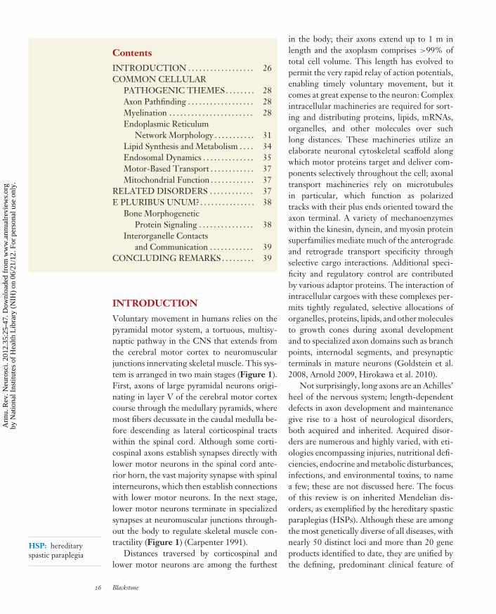
Lipid droplet (LD):dynamic cytoplasmicorganelle consistingof a phospholipidmonolayersurrounding a neutrallipid core; proteinsare found within themonolayer anddecorating its surface
form (M85 in rodents), was deleterious to axongrowth in cultured neurons (Solowska et al.2008), strengthening the evidence linking M1spastin to HSP pathogenesis. By comparison,M87 spastin appears involved in cytokinesis, se-cretion, and possibly endocytosis through itsinteractions linking microtubule dynamics tomembrane modeling in these compartments(Yang et al. 2008, Connell et al. 2009). Insum, there appear to be strong physical andfunctional links between M1 spastin, atlastin-1, reticulon 2, and REEP1 involved in shapingthe tubular ER network in concert with the mi-crotubule cytoskeleton. Even so, the ER has alarge number of functions, and it remains un-clear which are most pathogenically relevant forHSPs.
Because several other HSP proteins alsolocalize to the ER, understanding these mayclarify the role of ER in HSP pathogenesis.Mutations in the Berardinelli-Seip congenitallipodystrophy protein 2 gene BSCL2, whichencodes an integral membrane of the ERknown as BSCL2/seipin, cause two distinctdiseases. Heterozygous gain-of-function muta-tions in an N-linked glycosylation site give riseto a disease spectrum encompassing autosomaldominant SPG17 (Silver syndrome), with distalamyotrophy as a significant feature in additionto spastic paraparesis, and distal hereditarymotor neuropathy type V, characterized bymore prominent distal spinal muscular atrophy(Windpassinger et al. 2004). In contrast, auto-somal recessive loss-of-function mutations giverise to Berardinelli-Seip congenital lipodys-trophy, without spasticity or amyotrophy. Theseipin protein and its yeast ortholog, Fld1p(few lipid droplets 1), regulate the size of lipiddroplets (LDs), explaining the loss-of-functionlipodystrophy presentation (Cui et al. 2011; Feiet al. 2011a,b; Tian et al. 2011). The missensechanges underlying SPG17 have resulted inmisfolding of seipin, forming aggregates andtriggering ER stress (Ito & Suzuki 2009, Yagiet al. 2011), which may play a role in HSPpathogenesis. LDs accumulate under variouscellular stress conditions, and a number ofunfolded protein response pathways have
been implicated in LD formation (Hapalaet al. 2011). However, it will be important toinvestigate any effects of misfolded seipin onother aspects of ER structure and function.
Recently, the gene for the complicated,autosomal recessive SPG18 was identified ina consanguinous Saudi family as ERLIN2(Alazami et al. 2011). A second study inde-pendently identified loss-of-function ERLIN2mutations in a family with motor dysfunc-tion, joint contractures, and intellectual dis-ability (Yıldırım et al. 2011). The erlin2 pro-tein resides in the ER and contains a SPFHdomain—named for its presence in stom-atin, prohibitin, flotillin, and HflC/K. SPHFdomain–containing proteins share the ability toassemble into large oligomers, and they localizepreferentially to cholesterol-rich domains, in-cluding lipid rafts (Browman et al. 2006). Erlin2has been functionally linked to ER-associateddegradation (ERAD), a multistep degradativepathway encompassing ubiquitin-proteosome-mediated degradation of ER proteins, therebyregulating levels of proteins such as the inosi-tol trisphosphate receptor (Pearce et al. 2009).Also, erlin2 binds gp78, a membrane-boundubiquitin ligase similar to HRD1 that medi-ates sterol-accelerated ERAD of 3-hydroxy-3-methyl-glutaryl (HMG)-CoA reductase, a keyenzyme in the biosynthesis of cholesterol ( Joet al. 2011).
Lipid Synthesis and Metabolism
These latter two HSP proteins, erlin2 andseipin, present compelling insights into howdefects in ER shaping and distribution mightcause HSP. A key function of the ER is thesynthesis, metabolism, and distribution oflipids and sterols, employing both vesicularand nonvesicular mechanisms, and otherHSP proteins fit into this pathogenic theme.The complicated, autosomal recessive HSPTroyer syndrome (SPG20) is caused by mu-tations resulting in a loss of spartin protein(Bakowska et al. 2008). Spartin localizes toa variety of cellular structures and has beenimplicated in a number of functions, including
34 Blackstone
Ann
u. R
ev. N
euro
sci.
2012
.35:
25-4
7. D
ownl
oade
d fr
om w
ww
.ann
ualr
evie
ws.
org
by N
atio
nal I
nstit
utes
of
Hea
lth L
ibra
ry (
NIH
) on
06/
21/1
2. F
or p
erso
nal u
se o
nly.
NE35CH02-Blackstone ARI 14 May 2012 10:46
MIT domain:conserved domaincomprising threeα-helices present inmicrotubule-interacting andtrafficking proteins
ESCRT: endosomalsorting complexrequired for transport
cytokinesis and epidermal growth factor(EGF) receptor trafficking (Robay et al. 2006,Bakowska et al. 2007, Renvoise et al. 2010, Lindet al. 2011). Spartin also regulates LD biogene-sis by promoting atrophin-1 interacting protein4 (AIP4)-mediated ubiquitination of LD pro-teins (Eastman et al. 2009, Edwards et al. 2009,Hooper et al. 2010) and by recruiting PKC-ζvia the PKC-ζ -interacting proteins ZIP1(p62/sequestosome) and ZIP3 to LDs(Urbanczyk & Enz 2011). Little is knownabout any roles for LDs in axons. However,because the SPG17 protein seipin regulatesLD formation, alterations in LD biogenesisor turnover could affect lipid distribution,organelle shaping, or signaling pathwaysimportant for axonal health.
Although not directly implicated in LDbiogenesis, other HSP proteins are enzymesinvolved in related lipid and cholesterol biosyn-thetic pathways. SPG42 is caused by mutationsin the SLC33A1 gene encoding the acetyl-CoA transporter. In animals, acetyl-CoA isessential for maintaining the balance betweencarbohydrate and fat metabolism. Undernormal circumstances, acetyl-CoA from fattyacid metabolism enters the citric acid cycle,contributing to the energy supply of the cell.SLC33A1 transports acetyl-CoA into the Golgiapparatus lumen and has been directly linkedto the growth of axons because knock down ofslc33a1 in zebrafish causes defective outgrowthfrom the spinal cord (Lin et al. 2008).
Last, neuropathy target esterase (NTE) isan integral membrane protein of neuronal ERthat is mutated in autosomal recessive SPG39, acomplicated HSP with prominent amyotrophy.NTE deacylates the major membrane phos-pholipid, phosphatidylcholine. Mutation of theNTE gene PNPLA2 or chemical inhibition ofNTE with organophosphates alters membranecomposition and causes distal degeneration oflong spinal axons in mice and man (Rainieret al. 2008, Read et al. 2009). Another HSPprotein, cytochrome P450-7B1 (CYP7B1),is mutated in autosomal recessive SPG5(Tsaousidou et al. 2008) and functions incholesterol metabolism; in patients with SPG5
there is a dramatic increase in oxysterol sub-strates in plasma and cerebrospinal fluid (Schuleet al. 2010). Given the fundamental roles playedby lipids and sterols in neuronal functions, itseems very likely that more genes will be iden-tified within this category.
Endosomal Dynamics
Although changes in ER morphology encom-pass proteins altered in the majority of patientswith HSP, more HSP proteins have been im-plicated in endosomal dynamics; however, animmediate caveat is that the cell seems to co-opt some of these proteins and machineriesfor other functions (Blackstone et al. 2011). Asnoted earlier, the SPG20 protein spartin func-tions in LD turnover, but it is also required forefficient EGF receptor degradation and likelyregulates EGF signaling (Bakowska et al. 2007).Spartin may also regulate a variety of signalingpathways via ubiquitin modification throughits interactions with E3 ubiquitin ligases suchas AIP4 and AIP5. Spartin harbors an MITdomain as well and interacts selectively withIST1, a component of the ESCRT-III complex.ESCRT comprises a series of cytosolic proteincomplexes, ESCRT-0, ESCRT-I, ESCRT-II,and ESCRT-III, and the sequential activi-ties of these complexes are required to rec-ognize and sort ubiquitin-modified proteinsinto internal vesicles of multivesicular bodies(Hurley & Hanson 2010). More recently,ESCRT proteins have been implicated in othercellular functions, including viral budding andcytokinesis, and spartin has been shown to par-ticipate in cytokinesis (Renvoise et al. 2010,Lind et al. 2011).
This link to ESCRT-III is shared by theSPG4 protein spastin. Although discussed ear-lier in the context of its interactions withREEP1 and atlastin-1 to coordinate ER mem-brane modeling and microtubule interactions(Park et al. 2010), spastin harbors an MITdomain that binds the ESCRT-III subunitsCHMP1B and IST1 to couple the severing ofmicrotubules with membrane scission. TheseESCRT interactions are crucial for spastin’s
www.annualreviews.org • Hereditary Spastic Paraplegia 35
Ann
u. R
ev. N
euro
sci.
2012
.35:
25-4
7. D
ownl
oade
d fr
om w
ww
.ann
ualr
evie
ws.
org
by N
atio
nal I
nstit
utes
of
Hea
lth L
ibra
ry (
NIH
) on
06/
21/1
2. F
or p
erso
nal u
se o
nly.

NE35CH02-Blackstone ARI 14 May 2012 10:46
WASH complex:Wiscott-Aldrichsyndrome protein andSCAR (suppressor ofcAMP receptor)homolog complex
Tubular transportintermediate: small,cigar-shapedorganelles that trafficfrom one membranecompartment toanother
role in severing microtubules to complete theabscission phase of cytokinesis (Yang et al.2008, Connell et al. 2009, Guizetti et al. 2011).ESCRT proteins are also involved in centro-some stability (Morita et al. 2010), raising thepossibility that axon genesis and regulationby centrosomes could be a function of theseproteins. Other possibilities include roles forthe ESCRT-III interactions with spastin andspartin in the delivery and downregulation ofcell surface receptors to regulate signaling inaxons.
Mutations in the KIAA1096 (SPG8) genethat encodes strumpellin cause a severe, pureHSP (Valdmanis et al. 2007). Strumpellincontains few motifs or domains of identifiedfunction, aside from a region of putativespectrin repeats. The main clues to its pos-sible endosomal functions stem from itsidentification as a subunit of the WASHcomplex (Derivery & Gautreau 2010). Thiscomplex, comprising seven subunits (five corecomponents), connects tubular endosomes ofretrograde cargo sorting to the cytoskeleton,and it associates with endosomes via an inter-action with vacuolar protein sorting–associatedprotein 35 (VPS35) (Harbour et al. 2010).Along with VPS26 and VPS29, VPS35 is acomponent of retromer, an endosomal complexresponsible for sorting cargoes from endo-somes to the trans-Golgi network (Bonifacino& Hurley 2008). Depletion of members of theWASH complex cause increased tubulation atearly endosomes, impairing trafficking throughearly endosomal compartments (Derivery et al.2009, Gomez & Billadeau 2009, Jia et al. 2010).Several members of the complex—WASH1,FAM21 (family with sequence similarity 21),and actin-capping protein—regulate actin dy-namics. The WASH complex helps generate anactin network on early endosomes, for instanceby activating the Arp2/3 complex to nucleatenew actin filaments branching off extantfilaments. The increased tubulation associatedwith WASH-complex depletion may reflect alack of actin-mediated forces that are requiredfor fission of tubular transport intermediatesfrom the endosome (Campellone & Welch
2010). Thus, the WASH complex (containingstrumpellin) exemplifies another HSP proteinfunctioning in coordinating membrane model-ing and cytoskeletal organization. Strumpellininteracts with valosin-containing protein(VCP/p97), an AAA ATPase mutated infrontotemporal dementia with Paget’s diseaseof bone and inclusion body myopathy (Clemenet al. 2010), and VPS35 is mutated in a numberof families with autosomal dominant, late-onsetParkinson disease (Vilarino-Guell et al. 2011,Zimprich et al. 2011). A causative mutation inthe WASH subunit SWIP has recently beenidentified for autosomal recessive intellectualdisability (Ropers et al. 2011). Thus, roles of theWASH-retromer axis in neurological diseaseclearly extend beyond the HSPs and representa very important area for investigation.
Another emerging HSP-related complexrelated to endocytic trafficking comprises theSPG15 protein spastizin/FYVE-CENT, theSPG11 protein spatacsin, and the SPG48 pro-tein KIAA0415. Clinically, SPG11 and SPG15share a number of characteristics; both are fre-quently associated with thin corpus callosum,and both can present with juvenile parkin-sonism. Spastizin and spatacsin colocalize incytoplasmic structures and were identified asproteins that coprecipitate with the SPG48protein KIAA0415 (Slabicki et al. 2010, Murmuet al. 2011). KIAA0415 was originally proposedas a DNA helicase on the basis of sequencepredictions (Slabicki et al. 2010), but a veryrecent study provides compelling evidence thatit is a subunit of a new adaptor protein complex,AP-5, involved in endosomal dynamics (Hirstet al. 2011). Spastizin/FYVE-CENT containsa FYVE domain and binds the lipid PI(3)P,functioning along with ESCRT proteins incytokinesis (Sagona et al. 2010). Along theselines, mutations in multiple proteins of theAP-4 complex, which is involved in traffickingof amyloid precursor protein from the trans-Golgi to endosomes (Burgos et al. 2010), causeautosomal recessive syndromes with prominentclinical features ranging from intellectual dis-ability to progressive spastic paraplegia (AbouJamra et al. 2011, Moreno-De-Luca et al.
36 Blackstone
Ann
u. R
ev. N
euro
sci.
2012
.35:
25-4
7. D
ownl
oade
d fr
om w
ww
.ann
ualr
evie
ws.
org
by N
atio
nal I
nstit
utes
of
Hea
lth L
ibra
ry (
NIH
) on
06/
21/1
2. F
or p
erso
nal u
se o
nly.

NE35CH02-Blackstone ARI 14 May 2012 10:46
Amyotrophic lateralsclerosis (ALS): adegenerative disorderaffecting corticospinaland lower motorneurons
2011). Thus, these adaptor protein complexesappear highly relevant for pathogenesis ofHSPs and other neurological diseases.
Together, the expanding number of HSPgenes implicated in endosome dynamics arealready revealing new relationships amongprotein complexes, with implications thatextend beyond the primary HSPs. Indeed, anumber of patients with familial amyotrophiclateral sclerosis (ALS) resulting from anautosomal recessive mutation in the ALS2gene encoding the alsin protein, a guaninenucleotide exchange factor (GEF) for thesmall GTPases Rab5 and Rac1, have a diseasepresentation more similar to the HSPs than toALS. Examination of Als2 null mice revealedmotor impairments and a distal axonopathy ofthe corticospinal tract. Rab5-dependent endo-somal fusion is impaired in neurons from thesemice, whereas alsin overexpression in neuronsstimulates Rab5-dependent endosomal fusion,resulting in enlarged endosomes (Devon et al.2006, Deng et al. 2007, Hadano et al. 2007).
Motor-Based Transport
The identification of mutations in the KIF5Agene encoding kinesin heavy chain 5A (knownalso as kinesin-1A) in families with SPG10,a pure or complicated HSP, has provideddirect evidence for motor-based transportimpairments underlying HSPs (Reid et al.2002, Goizet et al. 2009). KIF5 proteins areATP-dependent motors that move cargoes inthe anterograde direction along axons, andmost mutations are missense changes in themotor domain. Drosophila harboring mutationsin the KIF5 ortholog Khc have posteriorparalysis, with organelle-filled axon swellingsjammed with cargoes (Hurd & Saxton 1996).In mammals, the KIF5A motor protein shuttlesneurofilament subunits along axons and possi-bly other anterograde cargoes such as vesicles.KIF5 also regulates transport of cargoes in den-drites and has roles in a number of membranetraffic pathways. The efficiency of cargo trans-port to the distal axon is thought to be affectedeither because the mutated KIF5A are slower
motors or because they have reduced micro-tubule binding affinity and act in a dominant-negative manner by competing with wild-typemotors for cargo binding (Ebbing et al. 2008).
Mitochondrial Function
Mitochondrial dysfunction has been implicatedin a host of developmental and degenerativeneurological disorders, manifesting clinically asperipheral neuropathies, movement disorders,visual disturbances, and cognitive disability (Di-Mauro & Schon 2008). Given this fundamentallink to neurological disease, it is surprisingthat so few HSP genes encode mitochondrialproteins. Two resident mitochondrial proteinsmutated in HSPs are paraplegin (autosomalrecessive SPG7) and HSP60 (autosomaldominant SPG13). Paraplegin is an m-AAAmetalloprotease of the inner mitochondrialmembrane, where it functions in ribosomalassembly and protein quality control. Muscletissue from SPG7 patients exhibits defectsin oxidative phosphorylation and Spg7 nullmice have axonal swellings with accumulatedmitochondria and neurofilaments, indicatingthat both mitochondrial function and axonaltransport are impaired (Ferreirinha et al. 2004).SPG13 is typically a late-onset, pure HSP, anda causative missense mutation (p.V98I) impairsHSP60 chaperonin activity, leading to impairedmitochondrial quality contol (Bross et al. 2008).
RELATED DISORDERS
We have discussed the convergent pathwaysof many proteins mutated in HSPs, but ithas become increasing clear that the HSPpresentation is often part of a broader diseasespectrum; thus it is important to considerrelated disorders that may share pathogenicthemes. Indeed, the importance of organellemorphology and distribution in maintainingaxons is also emphasized by other inheritedaxonopathies, particularly peripheral nervedisorders such as the Charcot-Marie-Tooth(CMT) neuropathies and hereditary sen-sory and autonomic neuropathies (HSAN).
www.annualreviews.org • Hereditary Spastic Paraplegia 37
Ann
u. R
ev. N
euro
sci.
2012
.35:
25-4
7. D
ownl
oade
d fr
om w
ww
.ann
ualr
evie
ws.
org
by N
atio
nal I
nstit
utes
of
Hea
lth L
ibra
ry (
NIH
) on
06/
21/1
2. F
or p
erso
nal u
se o
nly.

NE35CH02-Blackstone ARI 14 May 2012 10:46
Bone morphogeneticprotein (BMP):a member of thetransforming growthfactor-β superfamily
Mutations in the FAM134B gene were iden-tified in some patients with HSAN II. TheFAM134B protein is a member of the FAM134protein family, each of which contains apair of long hydrophobic segments reminis-cent of those in ER-shaping reticulon andYop1p/DP1/REEP proteins. FAM134B isenriched in the cis-Golgi apparatus, and itsdepletion causes prominent changes in Golgimorphology in neurons (Kurth et al. 2009).More recently, homozygous loss-of-functionKIF1A mutations were identified in an Afghanfamily with HSAN II (Riviere et al. 2011), andthe KIF1A protein is a motor involved in axonaltransport of synaptic vesicles. A family has alsobeen recently identified with hereditary spasticparaplegia caused by homozygous mutationin the KIF1A motor domain (Erlich et al.2011), indicating that HSP and HSANs may,in at least some cases, fall along a phenotypicspectrum. In fact, dominant missense muta-tions in the SPG3A protein atlastin-1 havebeen recently reported in hereditary sensoryneuropathy (HSN) I (Guelly et al. 2011).More generally, these disorders highlight theimplications of morphological defects in theER and the early secretory pathway as wellas distribution defects in the pathogenesis oflength-dependent axonopathies.
Pathogenic studies of CMT peripheral neu-ropathies are also instructive. CMT1 is com-posed of demyelination disorders, and CMT2 iscomposed of those that cause axonopathies. Ax-onal forms of CMT in particular can be causedby mutations in genes that encode proteinsthat function in organelle morphogenesis andtrafficking. CMT2A results from mutations inthe gene encoding mitofusin2 (MFN2), whichregulates mitochondrial morphology by medi-ating mitochondrial fusion and has also beenimplicated in mitochondrial connections withthe ER (de Brito et al. 2010). The CMT2Bprotein Rab7, a small GTPase that regulatesendosomal vesicle trafficking, interacts withthe SPG21 protein maspardin, another HSP-associated protein that localizes to endosomes(Hanna & Blackstone 2009, McCray et al. 2010,Soderblom et al. 2010). Very recently, a large
CMT2 pedigree was reported with autosomaldominant mutation in DYNC1H1, which codesfor the dynein heavy chain 1 involved in retro-grade axonal transport (Weedon et al. 2011).
Finally, ER shaping mechanisms may haveroles in related neurologic disorders suchas familial ALS, in which both corticospinaland lower motor neurons are affected. Inthe superoxide dismutase 1 (SOD1) G93Atransgenic mouse model for ALS, overexpres-sion of the ER-shaping protein reticulon-4Aselectively redistributed the ER chaperoneprotein disulfide isomerase and protectedagainst neurodegeneration. Conversely, lossof reticulon-4A increased disease severity(Yang et al. 2009). Further supporting a rolefor aberrant ER morphogenesis in neurologicdisorders, a mutant variant of vesicle-associatedmembrane protein–associated protein B (VAP-B) that underlies another familial ALS (ALS8) isassociated with the production of a novel formof organized smooth ER (Fasana et al. 2010).
E PLURIBUS UNUM?
These divisions as discussed above are, of neces-sity, somewhat arbitrary because cellular path-ways show a great deal of interdependence,and a number of HSPs can fit into severalpathogenic themes. For instance, the shapingphenomenon crosses over a number of cate-gories, from ER to endosomes. A natural ques-tion is, then, can these be unified further?
Bone MorphogeneticProtein Signaling
One compelling candidate that crosses HSPcategories and is widely implicated in neu-rodegenerative diseases is bone morphogeneticprotein (BMP) signaling (Bayat et al. 2011).HSP-associated mutations are found in at leastfour proteins—atlastin-1, NIPA1 (nonim-printed in Prader-Willi/Angelman syndrome1; SPG6), spastin, and spartin—that functionas inhibitors of BMP signaling. In Drosophilaand mammals, BMP signaling functions in reg-ulating axonal growth and synaptic function,
38 Blackstone
Ann
u. R
ev. N
euro
sci.
2012
.35:
25-4
7. D
ownl
oade
d fr
om w
ww
.ann
ualr
evie
ws.
org
by N
atio
nal I
nstit
utes
of
Hea
lth L
ibra
ry (
NIH
) on
06/
21/1
2. F
or p
erso
nal u
se o
nly.

NE35CH02-Blackstone ARI 14 May 2012 10:46
and impairment of BMP signaling in Drosophilaleads to axon transport defects (Wang et al.2007). In rodents, BMP signaling is upregulatedfollowing lesioning of the corticospinal tract,and suppression of this upregulation can pro-mote regrowth of axons (Matsuura et al. 2008).
Of the HSP proteins known to inhibit BMPsignaling, the best characterized mechanisti-cally is NIPA1, an integral membrane proteinwith 9 predicted TMDs that localizes to endo-somes and the plasma membrane and functionsin Mg2+ transport (Goytain et al. 2007).Drosophila larvae lacking spichthyin (NIPA1 or-tholog) have increased synaptic boutons at neu-romuscular junctions and increased phosphory-lated MAD (mothers against decapentaplegic),a downstream messenger of BMP signaling.These changes can be suppressed with geneticalterations that inhibit BMP signaling (Wanget al. 2007). NIPA1/spichthyin is thought toinhibit BMP signaling by promoting the inter-nalization of BMP type II receptors and theirsubsequent lysosomal degradation, and NIPA1missense changes found in SPG6 patientsinterfere with this process, upregulating signal-ing. Similarly, depletion of spartin or spastin,which both localize partially to endosomes,upregulates BMP signaling (Tsang et al. 2009).
Dysregulated BMP signaling linked to ax-onal abnormalities has also been demonstratedfor atlastin. Depletion of atlastin-1 in zebrafishresulted in abnormal spinal motor axon mor-phology, with increased branching and de-creased larval mobility. BMP signaling was up-regulated in these larvae, and pharmacologicalor genetic inhibition of BMP signaling rescuedthe atl1 null phenotype (Fassier et al. 2010). Insum, these results suggest that abnormal BMPsignaling, probably caused by abnormal BMPreceptor trafficking in many cases, could be aunifying mechanism for some classes of HSP,including the two most common, SPG4 andSPG3A, which comprise almost 50% of pa-tients. Investigating relevant HSP animal mod-els will be critical to determine whether inhi-bition of BMP signaling using small-moleculeinhibitors, several of which are available, canrescue disease phenotypes.
Interorganelle Contactsand Communication
Another possibility for linking HSP themes fur-ther is via interorganelle contacts. The ER isdistributed promiscuously throughout the cell,and to mediate its many functions, it inter-acts with other organelles at specialized con-tact sites. Such interactions occur with theplasma membrane, mitochondria, and lyso-somes/endosomes. The importance of theseconnections for functions such as interorganelleexchange of lipids/sterols, signal transduction,and mobilization of Ca2+ stores is increas-ingly appreciated (Carrasco & Meyer 2011,Toulmay & Prinz 2011). In particular, ER-mitochondrial contacts have been intensivelystudied recently, with MFN2 in mammalsand an ER-mitochondrial encounter structure(ERMES) in yeast playing crucial roles (deBrito et al. 2010, Kornmann & Walter 2010).In a model of pulmonary arterial hyperten-sion, there was decreased ER-to-mitochondriaphospholipid transfer and intramitochondrialCa2+ (Sutendra et al. 2011). The contacts be-tween mitochondria and ER were disrupted ina manner dependent on increased expressionof an ER-shaping protein of the reticulon fam-ily, Nogo-B, linking changes in an ER-shapingprotein to mitochondrial dysfunction.
CONCLUDING REMARKS
The HSPs have recently been called a“paradigmatic” example of how a disease canfoster insights into fundamental cellular pro-cesses, particularly with regard to formationof the tubular ER network (De Matteis &Luini 2011). Ongoing studies investigatinghow the ER network is shaped in neuronsusing electron microscopy reconstruction orsuper-resolution confocal microscopy willimprove our understanding of the appearance,contacts, and dynamics of ER in axons. Withthe increasing throughput and falling costof next-generation sequencing technologies,more genes for HSPs and related disorderswill assuredly be uncovered, likely many more.
www.annualreviews.org • Hereditary Spastic Paraplegia 39
Ann
u. R
ev. N
euro
sci.
2012
.35:
25-4
7. D
ownl
oade
d fr
om w
ww
.ann
ualr
evie
ws.
org
by N
atio
nal I
nstit
utes
of
Hea
lth L
ibra
ry (
NIH
) on
06/
21/1
2. F
or p
erso
nal u
se o
nly.
NE35CH02-Blackstone ARI 14 May 2012 10:46
Important insights into endocytic traffickingpathways in particular seem sure to follow.
With some compelling cellular mechanismsalready identified, pharmacologic manipulationof these pathways and evaluations in cellular andanimal models will be increasingly important.Pathways such as BMP signaling and micro-tubule stability (Orso et al. 2005, Yu et al. 2008)currently seem to be particularly attractive tar-gets because they would likely relate to a sig-nificant percentage of HSP patients and seemamenable to regulation by small molecules,which could ultimately lead to therapies.
Animal models may be a particular challengefor HSPs because successfully modeling a dis-ease of 1 m axons in small rodents is not agiven. The slow, variable rates of progression
in HSP patients will be challenges for assessingthe efficacy of therapies, but emerging noninva-sive stimulation (triple stimulation technique)and imaging modalites such as diffusion ten-sor imaging (Duning et al. 2010, Unrath et al.2010) might be useful biomarkers, particularlybecause they can detect changes in patients withknown HSP mutations at a presymptomaticphase, when disease-modifying therapies wouldbe most useful (Duning et al. 2010). The pastseveral years have yielded remarkable advance-ments in our understanding of the pathogenesisunderlying the HSPs; with increasing interestin the fascinating biology of HSP proteins andtechnological advances in genetics and imagingmoving rapidly, the future is hopeful for thoseafflicted.
DISCLOSURE STATEMENT
The author is not aware of any affiliations, memberships, funding, or financial holdings that mightbe perceived as affecting the objectivity of this review.
ACKNOWLEDGMENTS
I thank Alan Hoofring for preparing the figures. Work in the author’s laboratory is funded bythe Intramural Research Program of the National Institute of Neurological Disorders and Stroke,National Institutes of Health.
LITERATURE CITED
Abou Jamra R, Philippe O, Raas-Rothschild A, Eck SH, Graf E, et al. 2011. Adaptor protein complex 4deficiency causes severe autosomal-recessive intellectual disability, progressive spastic paraplegia, shycharacter, and short stature. Am. J. Hum. Genet. 88(6):788–95
Alazami AM, Adly N, Al Dhalaan H, Alkuraya FS. 2011. A nullimorphic ERLIN2 mutation defines a compli-cated hereditary spastic paraplegia locus (SPG18). Neurogenetics 12(4):333–36
Arnold DB. 2009. Actin and microtubule-based cytoskeletal cues direct polarized targeting of proteins inneurons. Sci. Signal. 2(83):pe49
Bakowska JC, Jupille H, Fatheddin P, Puertollano R, Blackstone C. 2007. Troyer syndrome protein spartinis mono-ubiquitinated and functions in EGF receptor trafficking. Mol. Biol. Cell 18(5):1683–92
Bakowska JC, Wang H, Xin B, Sumner CJ, Blackstone C. 2008. Lack of spartin protein in Troyer syndrome:a loss-of-function disease mechanism? Arch. Neurol. 65(4):520–24
Bayat V, Jaiswal M, Bellen HJ. 2011. The BMP signaling pathway at the Drosophila neuromuscular junctionand its links to neurodegenerative diseases. Curr. Opin. Neurobiol. 21(1):182–88
Bechara A, Nawabi H, Moret F, Yaron A, Weaver E, et al. 2008. FAK-MAPK-dependent adhesion disassemblydownstream of L1 contributes to semaphorin3A-induced collapse. EMBO J. 27(11):1549–62
Bian X, Klemm RW, Liu TY, Zhang M, Sun S, et al. 2011. Structures of the atlastin GTPase provide insightinto homotypic fusion of endoplasmic reticulum membranes. Proc. Natl. Acad. Sci. USA 108(10):3976–81
40 Blackstone
Ann
u. R
ev. N
euro
sci.
2012
.35:
25-4
7. D
ownl
oade
d fr
om w
ww
.ann
ualr
evie
ws.
org
by N
atio
nal I
nstit
utes
of
Hea
lth L
ibra
ry (
NIH
) on
06/
21/1
2. F
or p
erso
nal u
se o
nly.

NE35CH02-Blackstone ARI 14 May 2012 10:46
Blackstone C, O’Kane CJ, Reid E. 2011. Hereditary spastic paraplegias: membrane traffic and the motorpathway. Nat. Rev. Neurosci. 12(1):31–42
Bonifacino JS, Hurley JH. 2008. Retromer. Curr. Opin. Cell Biol. 20(4):427–36Bross P, Naundrup S, Hansen J, Nielsen MN, Christensen JH, et al. 2008. The Hsp60-(p.V98I) mutation
associated with hereditary spastic paraplegia SPG13 compromises chaperonin function both in vitro andin vivo. J. Biol. Chem. 283(23):15694–700
Browman DT, Resek ME, Zajchowski LD, Robbins SM. 2006. Erlin-1 and erlin-2 are novel members ofthe prohibitin family of proteins that define lipid-raft-like domains of the ER. J. Cell Sci. 119(Pt. 15):3149–60
Burgos PV, Mardones GA, Rojas AL, daSilva LLP, Prabhu Y, et al. 2010. Sorting of the Alzheimer’s diseaseamyloid precursor protein mediated by the AP-4 complex. Dev. Cell 18(3):425–36
Byrnes LJ, Sondermann H. 2011. Structural basis for the nucleotide-dependent dimerization of the large Gprotein atlastin-1/SPG3A. Proc. Natl. Acad. Sci. USA 108(6):2216–21
Campellone KG, Welch MD. 2010. A nucleator arms race: cellular control of actin assembly. Nat. Rev. Mol.Cell Biol. 11(4):237–51
Carpenter MB. 1991. Core Text of Neuroanatomy. Baltimore, MD: Wilkins & Wilkins. 4th ed.Carrasco S, Meyer T. 2011. STIM proteins and the endoplasmic reticulum-plasma membrane junctions.
Annu. Rev. Biochem. 80:973–1000Castellani V, Falk J, Rougon G. 2004. Semaphorin3A-induced receptor endocytosis during axon guidance
responses is mediated by L1 CAM. Mol. Cell. Neurosci. 26(1):89–100Chen J, Stefano G, Brandizzi F, Zheng H. 2011. Arabidopsis RHD3 mediates the generation of the tubular ER
network and is required for Golgi distribution and motility in plant cells. J. Cell Sci. 124(Pt. 13):2241–52Clemen CS, Tangavelou K, Strucksberg K-H, Just S, Gaertner L, et al. 2010. Strumpellin is a novel valosin-
containing protein binding partner linking hereditary spastic paraplegia to protein aggregation diseases.Brain 133(10):2920–41
Cohen NR, Taylor JSH, Scott LB, Guillery RW, Soriano P, Furley AJW. 1998. Errors in corticospinal axonguidance in mice lacking the neural cell adhesion molecule L1. Curr. Biol. 8(1):26–33
Connell JW, Lindon C, Luzio JP, Reid E. 2009. Spastin couples microtubule severing to membrane traffic incompletion of cytokinesis and secretion. Traffic 10(1):42–56
Cui X, Wang Y, Tang Y, Liu Y, Zhao L, et al. 2011. Seipin ablation in mice results in severe generalizedlipodystrophy. Hum. Mol. Genet. 20(15):3022–30
Dahme M, Bartsch U, Martini R, Anliker B, Schachner M, Mantei N. 1997. Disruption of the mouse L1 geneleads to malformations of the nervous system. Nat. Genet. 17(3):346–49
de Brito OM, Scorrano L. 2010. An intimate liaison: spatial organization of the endoplasmic reticulum–mitochondria relationship. EMBO J. 29(16):2715–23
De Matteis MA, Luini A. 2011. Mendelian disorders of membrane trafficking. N. Engl. J. Med. 365(10):927–38DeLuca GC, Ebers GC, Esiri MM. 2004. The extent of axonal loss in the long tracts in hereditary spastic
paraplegia. Neuropathol. Appl. Neurobiol. 30(6):576–84Deng H-X, Zhai H, Fu R, Shi Y, Gorrie GH, et al. 2007. Distal axonopathy in an alsin-deficient mouse model.
Hum. Mol. Genet. 16(23):2911–20Depienne C, Stevanin G, Brice A, Durr A. 2007. Hereditary spastic paraplegias: an update. Curr. Opin. Neurol.
20(6):674–80Derivery E, Gautreau A. 2010. Evolutionary conservation of the WASH complex, an actin polymerization
machine involved in endosomal fission. Commun. Integr. Biol. 3(3):227–30Derivery E, Sousa C, Gautier JJ, Lombard B, Loew D, Gautreau A. 2009. The Arp2/3 activator WASH
controls the fission of endosomes through a large multiprotein complex. Dev. Cell 17(5):712–23Devon RS, Orban PC, Gerrow K, Barbieri MA, Schwab C, et al. 2006. Als2-deficient mice exhibit distur-
bances in endosome trafficking associated with motor behavioral abnormalities. Proc. Natl. Acad. Sci. USA103(25):9595–600
Dick KJ, Eckhardt M, Paisan-Ruiz C, Alshehhi AA, Proukakis C, et al. 2010. Mutation of FA2H underlies acomplicated form of hereditary spastic paraplegia (SPG35). Hum. Mutat. 31(4):E1251–60
DiMauro S, Schon EA. 2008. Mitochondrial disorders in the nervous system. Annu. Rev. Neurosci. 31:91–123
www.annualreviews.org • Hereditary Spastic Paraplegia 41
Ann
u. R
ev. N
euro
sci.
2012
.35:
25-4
7. D
ownl
oade
d fr
om w
ww
.ann
ualr
evie
ws.
org
by N
atio
nal I
nstit
utes
of
Hea
lth L
ibra
ry (
NIH
) on
06/
21/1
2. F
or p
erso
nal u
se o
nly.
NE35CH02-Blackstone ARI 14 May 2012 10:46
Dion PA, Daoud H, Rouleau GA. 2009. Genetics of motor neuron disorders: new insights into pathogenicmechanisms. Nat. Rev. Genet. 10(11):769–82
Duning T, Warnecke T, Schirmacher A, Schiffbauer H, Lohmann H, et al. 2010. Specific pattern of earlywhite-matter changes in pure hereditary spastic paraplegia. Mov. Disord. 25(12):1986–92
Eastman SW, Yassaee M, Bieniasz PD. 2009. A role for ubiquitin ligases and Spartin/SPG20 in lipid dropletturnover. J. Cell Biol. 184(6):881–94
Ebbing B, Mann K, Starosta A, Jaud J, Schols L, et al. 2008. Effect of spastic paraplegia mutations in KIF5Akinesin on transport activity. Hum. Mol. Genet. 17(9):1245–52
Edgar JM, McLaughlin M, Yool D, Zhang S-C, Fowler JH, et al. 2004. Oligodendroglial modulation of fastaxonal transport in a mouse model of hereditary spastic paraplegia. J. Cell Biol. 166(1):121–31
Edwards TL, Clowes VE, Tsang HTH, Connell JW, Sanderson CM, et al. 2009. Endogenous spartin (SPG20)is recruited to endosomes and lipid droplets and interacts with the ubiquitin E3 ligases AIP4 and AIP5.Biochem. J. 423(1):31–39
Erlich Y, Edvardson S, Hodges E, Zenvirt S, Thekkat P, et al. 2011. Exome sequencing and disease-networkanalysis of a single family implicate a mutation in KIF1A in hereditary spastic paraparesis. Genome Res.21(5):658–64
Evans K, Keller C, Pavur K, Glasgow K, Conn B, Lauring B. 2006. Interaction of two hereditary spasticparaplegia gene products, spastin and atlastin, suggests a common pathway for axonal maintenance. Proc.Natl. Acad. Sci. USA 103(28):10666–71
Fasana E, Fossati M, Ruggiano A, Brambillasca S, Hoogenraad CC, et al. 2010. A VAPB mutant linked toamyotrophic lateral sclerosis generates a novel form of organized smooth endoplasmic reticulum. FASEBJ. 24(5):1419–30
Fassier C, Hutt JA, Scholpp S, Lumsden A, Giros B, et al. 2010. Zebrafish atlastin controls motility and spinalmotor axon architecture via inhibition of the BMP pathway. Nat. Neurosci. 13(11):1380–87
Fei W, Du X, Yang H. 2011a. Seipin, adipogenesis and lipid droplets. Trends Endocrinol. Metab. 22(6):204–10Fei W, Shui G, Zhang Y, Krahmer N, Ferguson C, et al. 2011b. A role for phosphatidic acid in the formation
of “supersized” lipid droplets. PLoS Genet. 7(7):e1002201Ferreirinha F, Quattrini A, Pirozzi M, Valsecchi V, Dina G, et al. 2004. Axonal degeneration in paraplegin-
deficient mice is associated with abnormal mitochondria and impairment of axonal transport. J. Clin.Invest. 113(2):231–42
Fink JK. 2006. Hereditary spastic paraplegia. Curr. Neurol. Neurosci. Rep. 6(1):65–76Goizet C, Boukhris A, Mundwiller E, Tallaksen C, Forlani S, et al. 2009. Complicated forms of autosomal
dominant hereditary spastic paraplegia are frequent in SPG10. Hum. Mutat. 30(2):E376–85Goldstein AYN, Wang X, Schwarz TL. 2008. Axonal transport and the delivery of pre-synaptic components.
Curr. Opin. Neurobiol. 18(5):495–503Gomez TS, Billadeau DD. 2009. A FAM21-containing WASH complex regulates retromer-dependent sorting.
Dev. Cell 17(5):699–711Goytain A, Hines RM, El-Husseini A, Quamme GA. 2007. NIPA1(SPG6 ), the basis for autosomal dominant
form of hereditary spastic paraplegia, encodes a functional Mg2+ transporter. J. Biol. Chem. 282(11):8060–68
Gruenenfelder FI, Thomson G, Penderis J, Edgar JM. 2011. Axon-glial interaction in the CNS: what we havelearned from mouse models of Pelizaeus-Merzbacher disease. J. Anat. 219(1):33–43
Guelly C, Zhu P-P, Leonardis L, Papic L, Zidar J, et al. 2011. Targeted high-throughput sequencing identifiesmutations in atlastin-1 as a cause of hereditary sensory neuropathy type I. Am. J. Hum. Genet. 88(1):99–105
Guizetti J, Schermelleh L, Mantler J, Maar S, Poser I, et al. 2011. Cortical constriction during abscissioninvolves helices of ESCRT-III-dependent filaments. Science 331(6024):1616–20
Hadano S, Kunita R, Otomo A, Suzuki-Utsunomiya K, Ikeda J-E. 2007. Molecular and cellular function ofALS2/alsin: implication of membrane dynamics in neuronal development and degeneration. Neurochem.Int. 51(2–4):74–84
Hanna MC, Blackstone C. 2009. Interaction of the SPG21 protein ACP33/maspardin with the aldehydedehydrogenase ALDH16A1. Neurogenetics 10(3):217–28
42 Blackstone
Ann
u. R
ev. N
euro
sci.
2012
.35:
25-4
7. D
ownl
oade
d fr
om w
ww
.ann
ualr
evie
ws.
org
by N
atio
nal I
nstit
utes
of
Hea
lth L
ibra
ry (
NIH
) on
06/
21/1
2. F
or p
erso
nal u
se o
nly.

NE35CH02-Blackstone ARI 14 May 2012 10:46
Hapala I, Marza E, Ferreira T. 2011. Is fat so bad? Modulation of endoplasmic reticulum stress by lipid dropletformation. Biol. Cell 103(6):271–85
Harbour ME, Breusegem SYA, Antrobus R, Freeman C, Reid E, Seaman MNJ. 2010. The cargo-selectiveretromer complex is a recruiting hub for protein complexes that regulate endosomal tubule dynamics.J. Cell Sci. 123(Pt. 21):3703–17
Harding AE. 1983. Classification of the hereditary ataxias and paraplegias. Lancet 1(8334):1151–55Hirokawa N, Niwa S, Tanaka Y. 2010. Molecular motors in neurons: transport mechanisms and roles in brain
function, development, and disease. Neuron 68(4):610–38Hirst J, Barlow LD, Francisco GC, Sahlender DA, Seaman MNJ, et al. 2011. The fifth adaptor protein
complex. PLoS Biol. 9(10):e1001170Hooper C, Puttamadappa SS, Loring Z, Shekhtman A, Bakowska JC. 2010. Spartin activates atrophin-1-
interacting protein 4 (AIP4) E3 ubiquitin ligase and promotes ubiquitination of adipophilin on lipiddroplets. BMC Biol. 8:72
Hu J, Shibata Y, Voss C, Shemesh T, Li Z, et al. 2008. Membrane proteins of the endoplasmic reticuluminduce high-curvature tubules. Science 319(5867):1247–50
Hu J, Shibata Y, Zhu P-P, Voss C, Rismanchi N, et al. 2009. A class of dynamin-like GTPases involved in thegeneration of the tubular ER network. Cell 138(3):549–61
Hurd DD, Saxton WM. 1996. Kinesin mutations cause motor neuron disease phenotypes by disrupting fastaxonal transport in Drosophila. Genetics 144(3):1075–85
Hurley JH, Hanson PI. 2010. Membrane budding and scission by the ESCRT machinery: It’s all in the neck.Nat. Rev. Mol. Cell Biol. 11(8):556–66
Inoue K. 2005. PLP1-related inherited dysmyelinating disorders: Pelizaeus-Merzbacher disease and spasticparaplegia type 2. Neurogenetics 6(1):1–16
Ito D, Suzuki N. 2009. Seipinopathy: a novel endoplasmic reticulum stress-associated disease. Brain132(Pt. 1):8–15
Jia D, Gomez TS, Metlagel Z, Umetani J, Otwinowski Z, et al. 2010. WASH and WAVE actin regulators ofthe Wiskott-Aldrich syndrome protein (WASP) family are controlled by analogous structurally relatedcomplexes. Proc. Natl. Acad. Sci.USA 107(23):10442–47
Jo Y, Sguigna PV, DeBose-Boyd RA. 2011. Membrane-associated ubiquitin ligase complex containing gp78mediates sterol-accelerated degradation of 3-hydroxy-3-methylglutaryl-coenzyme A reductase. J. Biol.Chem. 286(17):15022–31
Jouet M, Rosenthal A, Armstrong G, MacFarlane J, Stevenson R, et al. 1994. X-linked spastic paraplegia(SPG1), MASA syndrome and X-linked hydrocephalus result from mutations in the L1 gene. Nat. Genet.7(3):402–7
Kornmann B, Walter P. 2010. ERMES-mediated ER-mitochondria contacts: molecular hubs for the regula-tion of mitochondrial biology. J. Cell Sci. 123(Pt. 9):1389–93
Kurth I, Pamminger T, Hennings JC, Soehendra D, Huebner AK, et al. 2009. Mutations in FAM134B,encoding a newly identified Golgi protein, cause severe sensory and autonomic neuropathy. Nat. Genet.41(11):1179–81
Lang N, Optenhoefel T, Deuschl G, Klebe S. 2011. Axonal integrity of corticospinal projections to the upperlimbs in patients with pure hereditary spastic paraplegia. Clin. Neurophysiol. 122(7):1417–20
Lin P, Li J, Liu Q, Mao F, Li J, et al. 2008. A missense mutation in SLC33A1, which encodes the acetyl-CoA transporter, causes autosomal-dominant spastic paraplegia (SPG42). Am. J. Hum. Genet. 83(6):752–59
Lind GE, Raiborg C, Danielsen SA, Rognum TO, Thiis-Evensen E, et al. 2011. SPG20, a novel biomarkerfor early detection of colorectal cancer, encodes a regulator of cytokinesis. Oncogene 30(37):3967–78
Lynes EM, Simmen T. 2011. Urban planning of the endoplasmic reticulum (ER): how diverse mechanismssegregate the many functions of the ER. Biochim. Biophys. Acta 1813(10):1893–905
Mancuso G, Rugarli EI. 2008. A cryptic promoter in the first exon of the SPG4 gene directs the synthesis ofthe 60-kDa spastin isoform. BMC Biol. 6:31
Mannan AU, Boehm J, Sauter SM, Rauber A, Byrne PC, et al. 2006. Spastin, the most commonly mutatedprotein in hereditary spastic paraplegia interacts with Reticulon 1 an endoplasmic reticulum protein.Neurogenetics 7(2):93–103
www.annualreviews.org • Hereditary Spastic Paraplegia 43
Ann
u. R
ev. N
euro
sci.
2012
.35:
25-4
7. D
ownl
oade
d fr
om w
ww
.ann
ualr
evie
ws.
org
by N
atio
nal I
nstit
utes
of
Hea
lth L
ibra
ry (
NIH
) on
06/
21/1
2. F
or p
erso
nal u
se o
nly.

NE35CH02-Blackstone ARI 14 May 2012 10:46
Matsuura I, Taniguchi J, Hata K, Saeki N, Yamashita T. 2008. BMP inhibition enhances axonal growth andfunctional recovery after spinal cord injury. J. Neurochem. 105(4):1471–79
McCray BA, Skordalakes E, Taylor JP. 2010. Disease mutations in Rab7 result in unregulated nucleotideexchange and inappropriate activation. Hum. Mol. Genet. 19(6):1033–47
Montenegro G, Rebelo AP, Connell J, Allison R, Babalini C, et al. 2012. Mutations in the ER-shaping proteinreticulon 2 cause the axon-degenerative disorder hereditary spastic paraplegia type 12. J. Clin. Invest.122(2):538–44
Moreno-De-Luca A, Helmers SL, Mao H, Burns TG, Melton AMA, et al. 2011. Adaptor protein complex-4(AP-4) deficiency causes a novel autosomal recessive cerebral palsy syndrome with microcephaly andintellectual disability. J. Med. Genet. 48(2):141–44
Morita E, Colf LA, Karren MA, Sandrin V, Rodesch CK, Sundquist WI. 2010. Human ESCRT-III and VPS4proteins are required for centrosome and spindle maintenance. Proc. Natl. Acad. Sci. USA 107(29):12889–94
Moss TJ, Andreazza C, Verma A, Daga A, McNew JA. 2011. Membrane fusion by the GTPase atlastin requiresa conserved C-terminal cytoplasmic tail and dimerization through the middle domain. Proc. Natl. Acad.Sci. USA 108(27):11133–38
Murmu RP, Martin E, Rastetter A, Esteves T, Muriel M-P, et al. 2011. Cellular distribution and subcellularlocalization of spatacsin and spastizin, two proteins involved in hereditary spastic paraplegia. Mol. Cell.Neurosci. 47(3):191–202
Orso G, Martinuzzi A, Rossetto MG, Sartori E, Feany M, Daga A. 2005. Disease-related phenotypes in aDrosophila model of hereditary spastic paraplegia are ameliorated by treatment with vinblastine. J. Clin.Invest. 115(11):3026–34
Orso G, Pendin D, Liu S, Tosetto J, Moss TJ, et al. 2009. Homotypic fusion of ER membranes requires thedynamin-like GTPase atlastin. Nature 460(7258):978–83
Orthmann-Murphy JL, Salsano E, Abrams CK, Bizzi A, Uziel G, et al. 2009. Hereditary spastic paraplegia isa novel phenotype for GJA12/GJC2 mutations. Brain 132(Pt. 2):426–38
Park SH, Blackstone C. 2010. Further assembly required: construction and dynamics of the endoplasmicreticulum network. EMBO Rep. 11(7):515–21
Park SH, Zhu P-P, Parker RL, Blackstone C. 2010. Hereditary spastic paraplegia proteins REEP1, spastin, andatlastin-1 coordinate microtubule interactions with the tubular ER network. J. Clin. Invest. 120(4):1097–110
Pearce MMP, Wormer DB, Wilkens S, Wojcikiewicz RJH. 2009. An endoplasmic reticulum (ER) membranecomplex composed of SPFH1 and SPFH2 mediates the ER-associated degradation of inositol 1,4,5-trisphosphate receptors. J. Biol. Chem. 284(16):10433–45
Potter KA, Kern MJ, Fullbright G, Bielawski J, Scherer SS, et al. 2011. Central nervous system dysfunctionin a mouse model of Fa2h deficiency. Glia 59(7):1009–21
Rainier S, Bui M, Mark E, Thomas D, Tokarz D, et al. 2008. Neuropathy target esterase gene mutationscause motor neuron disease. Am. J. Hum. Genet. 82(3):780–85
Read DJ, Li Y, Chao MV, Cavanagh JB, Glynn P. 2009. Neuropathy target esterase is required for adultvertebrate axon maintenance. J. Neurosci. 29(37):11594–600
Reid E, Kloos M, Ashley-Koch A, Hughes L, Bevan S, et al. 2002. A kinesin heavy chain (KIF5A) mutation inhereditary spastic paraplegia (SPG10). Am. J. Hum. Genet. 71(5):1189–94
Renvoise B, Blackstone C. 2010. Emerging themes of ER organization in the development and maintenanceof axons. Curr. Opin. Neurobiol. 20(5):531–37
Renvoise B, Parker RL, Yang D, Bakowska JC, Hurley JH, Blackstone C. 2010. SPG20 protein spartinis recruited to midbodies by ESCRT-III protein Ist1 and participates in cytokinesis. Mol. Biol. Cell21(19):3293–303
Rismanchi N, Soderblom C, Stadler J, Zhu P-P, Blackstone C. 2008. Atlastin GTPases are required for Golgiapparatus and ER morphogenesis. Hum. Mol. Genet. 17(11):1591–604
Riviere J-B, Ramalingam S, Lavastre V, Shekarabi M, Holbert S, et al. 2011. KIF1A, an axonal transporterof synaptic vesicles, is mutated in hereditary sensory and autonomic neuropathy Type 2. Am. J. Hum.Genet. 89(2):219–30
44 Blackstone
Ann
u. R
ev. N
euro
sci.
2012
.35:
25-4
7. D
ownl
oade
d fr
om w
ww
.ann
ualr
evie
ws.
org
by N
atio
nal I
nstit
utes
of
Hea
lth L
ibra
ry (
NIH
) on
06/
21/1
2. F
or p
erso
nal u
se o
nly.

NE35CH02-Blackstone ARI 14 May 2012 10:46
Robay D, Patel H, Simpson MA, Brown NA, Crosby AH. 2006. Endogenous spartin, mutated in hereditaryspastic paraplegia, has a complex subcellular localization suggesting diverse roles in neurons. Exp. CellRes. 312(15):2764–77
Ropers F, Derivery E, Hu H, Garshasbi M, Karbasiyan M, et al. 2011. Identification of a novel candidategene for non-syndromic autosomal recessive intellectual disability: the WASH complex member SWIP.Hum. Mol. Genet. 20(13):2585–90
Sagona AP, Nezis IP, Pedersen NM, Liestøl K, Poulton J, et al. 2010. PtdIns(3)P controls cytokinesis throughKIF13A-mediated recruitment of FYVE-CENT to the midbody. Nat. Cell Biol. 12(4):362–71
Saito H, Kubota M, Roberts RW, Chi Q, Matsunami H. 2004. RTP family members induce functionalexpression of mammalian odorant receptors. Cell 119(5):679–91
Salinas S, Proukakis C, Crosby A, Warner TT. 2008. Hereditary spastic paraplegia: clinical features andpathogenetic mechanisms. Lancet Neurol. 7(12):1127–38
Sanderson CM, Connell JW, Edwards TL, Bright NA, Duley S, et al. 2006. Spastin and atlastin, two proteinsmutated in autosomal-dominant hereditary spastic paraplegia, are binding partners. Hum. Mol. Genet.15(2):307–18
Schneider SA, Bhatia KP. 2010. Three faces of the same gene: FA2H links neurodegeneration with brain ironaccumulation, leukodystrophies, and hereditary spastic paraplegias. Ann. Neurol. 68(5):575–77
Schule R, Siddique T, Deng H-X, Yang Y, Donkervoort S, et al. 2010. Marked accumulation of 27-hydroxycholesterol in SPG5 patients with hereditary spastic paresis. J. Lipid Res. 51(4):819–23
Shibata Y, Hu J, Kozlov MM, Rapoport TA. 2009. Mechanisms shaping the membranes of cellular organelles.Annu. Rev. Cell Dev. Biol. 25:329–54
Sieberer BJ, Ketelaar T, Esseling JJ, Emons AMC. 2005. Microtubules guide root hair tip growth. New Phytol.167(3):711–19
Słabicki M, Theis M, Krastev DB, Samsonov S, Mundwiller E, et al. 2010. A genome-scale DNA repairRNAi screen identifies SPG48 as a novel gene associated with hereditary spastic paraplegia. PLoS Biol.8(6):e1000408
Soderblom C, Blackstone C. 2006. Traffic accidents: molecular genetic insights into the pathogenesis of thehereditary spastic paraplegias. Pharmacol. Ther. 109(1–2):42–56
Soderblom C, Stadler J, Jupille H, Blackstone C, Shupliakov O, Hanna MC. 2010. Targeted disruption of theMast syndrome gene SPG21 in mice impairs hind limb function and alters axon branching in culturedcortical neurons. Neurogenetics 11(4):369–78
Solowska JM, Morfini G, Falnikar A, Himes BT, Brady ST, et al. 2008. Quantitative and functional anal-yses of spastin in the nervous system: implications for hereditary spastic paraplegia. J. Neurosci. 28(9):2147–57
Sparkes I, Hawes C, Frigerio L. 2011. FrontiERs: movers and shapers of the higher plant cortical endoplasmicreticulum. Curr. Opin. Plant Biol. 14(6):658–65
Sutendra G, Dromparis P, Wright P, Bonnet S, Haromy A, et al. 2011. The role of Nogo and the mitochondria-endoplasmic reticulum unit in pulmonary hypertension. Sci. Transl. Med. 3(88):88ra55
Terasaki M, Chen LB, Fujiwara K. 1986. Microtubules and the endoplasmic reticulum are highly interdepen-dent structures. J. Cell Biol. 103(4):1557–68
Tian Y, Bi J, Shui G, Liu Z, Xiang Y, et al. 2011. Tissue-autonomous function of Drosophila Seipin in preventingectopic lipid droplet formation. PLoS Genet. 7(4):e1001364
Toulmay A, Prinz WA. 2011. Lipid transfer and signaling at organelle contact sites: the tip of the iceberg.Curr. Opin. Cell Biol. 23(4):458–63
Tsang HTH, Edwards TL, Wang X, Connell JW, Davies RJ, et al. 2009. The hereditary spastic paraplegiaproteins NIPA1, spastin and spartin are inhibitors of mammalian BMP signalling. Hum. Mol. Genet.18(20):3805–21
Tsaousidou MK, Ouahchi K, Warner TT, Yang Y, Simpson MA, et al. 2008. Sequence alterations withinCYP7B1 implicate defective cholesterol homeostasis in motor-neuron degeneration. Am. J. Hum. Genet.82(2):510–15
Unrath A, Muller H-P, Riecker A, Ludolph AC, Sperfeld A-D, Kassubek J. 2010. Whole brain-based analysisof regional white matter tract alterations in rare motor neuron diseases by diffusion tensor imaging.Hum. Brain Mapp. 31(11):1727–40
www.annualreviews.org • Hereditary Spastic Paraplegia 45
Ann
u. R
ev. N
euro
sci.
2012
.35:
25-4
7. D
ownl
oade
d fr
om w
ww
.ann
ualr
evie
ws.
org
by N
atio
nal I
nstit
utes
of
Hea
lth L
ibra
ry (
NIH
) on
06/
21/1
2. F
or p
erso
nal u
se o
nly.
NE35CH02-Blackstone ARI 14 May 2012 10:46
Urbanczyk A, Enz R. 2011. Spartin recruits PKC-ζ via the PKC-ζ-interacting proteins ZIP1 and ZIP3 tolipid droplets. J. Neurochem. 118(5):737–48
Valdmanis PN, Meijer IA, Reynolds A, Lei A, MacLeod P, et al. 2007. Mutations in the KIAA0196 gene atthe SPG8 locus cause hereditary spastic paraplegia. Am. J. Hum. Genet. 80(1):152–61
Verkhratsky A. 2005. Physiology and pathophysiology of the calcium store in the endoplasmic reticulum ofneurons. Physiol. Rev. 85(1):201–79
Vilarino-Guell C, Wider C, Ross OA, Dachsel JC, Kachergus JM, et al. 2011. VPS35 mutations in Parkinsondisease. Am. J. Hum. Genet. 89(1):162–67
Voeltz GK, Prinz WA, Shibata Y, Rist JM, Rapoport TA. 2006. A class of membrane proteins shaping thetubular endoplasmic reticulum. Cell 124(3):573–86
Wang H, Lockwood SK, Hoeltzel MF, Schiefelbein JW. 1997. The ROOT HAIR DEFECTIVE3 gene en-codes an evolutionarily conserved protein with GTP-binding motifs and is required for regulated cellenlargement in Arabidopsis. Genes Dev. 11(6):799–811
Wang X, Shaw WR, Tsang HTH, Reid E, O’Kane CJ. 2007. Drosophila spichthyin inhibits BMP signalingand regulates synaptic growth and axonal microtubules. Nat. Neurosci. 10(2):177–85
Waterman-Storer CM, Salmon ED. 1998. Endoplasmic reticulum membrane tubules are distributed by mi-crotubules in living cells using three distinct mechanisms. Curr. Biol. 8(14):798–806
Weedon MN, Hastings R, Caswell R, Xie W, Paszkiewicz K, et al. 2011. Exome sequencing identifies aDYNC1H1 mutation in a large pedigree with dominant axonal Charcot-Marie-Tooth disease. Am. J.Hum. Genet. 89(2):308–12
Weller S, Gartner J. 2001. Genetic and clinical aspects of X-linked hydrocephalus (L1 disease): mutations inthe L1CAM gene. Hum. Mutat. 18(1):1–12
West M, Zurek N, Hoenger A, Voeltz GK. 2011. A 3D analysis of yeast ER structure reveals how ER domainsare organized by membrane curvature. J. Cell Biol. 193(2):333–46
Westermann B. 2010. Mitochondrial fusion and fission in cell life and death. Nat. Rev. Mol. Cell Biol.11(12):872–84
Windpassinger C, Auer-Grumbach M, Irobi J, Patel H, Petek E, et al. 2004. Heterozygous missense muta-tions in BSCL2 are associated with distal hereditary motor neuropathy and Silver syndrome. Nat. Genet.36(3):271–76
Yagi T, Ito D, Nihei Y, Ishihara T, Suzuki N. 2011. N88S seipin mutant transgenic mice develop features ofseipinopathy/BSCL2-related motor neuron disease via endoplasmic reticulum stress. Hum. Mol. Genet.20(19):3831–40
Yang D, Rismanchi N, Renvoise B, Lippincott-Schwartz J, Blackstone C, Hurley JH. 2008. Structural basisfor midbody targeting of spastin by the ESCRT-III protein CHMP1B. Nat. Struct. Mol. Biol. 15(12):1278–86
Yang YS, Harel NY, Strittmatter SM. 2009. Reticulon-4A (Nogo-A) redistributes protein disulfide isomeraseto protect mice from SOD1-dependent amyotrophic lateral sclerosis. J. Neurosci. 29(44):13850–59
Yıldırım Y, Orhan EK, Iseri SAU, Serdaroglu-Oflazer P, Kara B, et al. 2011. A frameshift mutation of ERLIN2in recessive intellectual disability, motor dysfunction and multiple joint contractures. Hum. Mol. Genet.20(10):1886–92
Yu PB, Hong CC, Sachidanandan C, Babitt JL, Deng DY, et al. 2008. Dorsomorphin inhibits BMP signalsrequired for embryogenesis and iron metabolism. Nat. Chem. Biol. 4(1):33–41
Zhu P-P, Patterson A, Lavoie B, Stadler J, Shoeb M, et al. 2003. Cellular localization, oligomerization, andmembrane association of the hereditary spastic paraplegia 3A (SPG3A) protein atlastin. J. Biol. Chem.278(49):49063–71
Zhu P-P, Soderblom C, Tao-Cheng J-H, Stadler J, Blackstone C. 2006. SPG3A protein atlastin-1 is enriched ingrowth cones and promotes axon elongation during neuronal development. Hum. Mol. Genet. 15(8):1343–53
Zimprich A, Benet-Pages A, Struhal W, Graf E, Eck SH, et al. 2011. A mutation in VPS35, encoding a subunitof the retromer complex, causes late-onset Parkinson disease. Am. J. Hum. Genet. 89(1):168–75
46 Blackstone
Ann
u. R
ev. N
euro
sci.
2012
.35:
25-4
7. D
ownl
oade
d fr
om w
ww
.ann
ualr
evie
ws.
org
by N
atio
nal I
nstit
utes
of
Hea
lth L
ibra
ry (
NIH
) on
06/
21/1
2. F
or p
erso
nal u
se o
nly.

NE35CH02-Blackstone ARI 14 May 2012 10:46
RELATED RESOURCES
Neuromuscul. Dis. Cent. Web page. Familial spinal cord syndromes. http://neuromuscular.wustl.edu/spinal/fsp.html. A comprehensive, frequently updated resource for informationon all types of HSPs and related disorders.
Reid E, Rugarli EI. 2010. Hereditary spastic paraplegias. In The Online Metabolic and MolecularBases of Inherited Diseases, ed. D Valle, AL Beaudet, B Vogelstein, KW Kinzler, SE Antonarakis,et al., Ch. 228.1. New York: McGraw Hill. http://dx.doi.org/10.1036/ommbid.266
Spastic Paraplegia Found. Web page. http://www.sp-foundation.org. Valuable information onresearch efforts focusing on the HSPs.
www.annualreviews.org • Hereditary Spastic Paraplegia 47
Ann
u. R
ev. N
euro
sci.
2012
.35:
25-4
7. D
ownl
oade
d fr
om w
ww
.ann
ualr
evie
ws.
org
by N
atio
nal I
nstit
utes
of
Hea
lth L
ibra
ry (
NIH
) on
06/
21/1
2. F
or p
erso
nal u
se o
nly.

NE35-FrontMatter ARI 21 May 2012 11:24
Annual Review ofNeuroscience
Volume 35, 2012Contents
The Neural Basis of EmpathyBoris C. Bernhardt and Tania Singer � � � � � � � � � � � � � � � � � � � � � � � � � � � � � � � � � � � � � � � � � � � � � � � � � � � � � � � � 1
Cellular Pathways of Hereditary Spastic ParaplegiaCraig Blackstone � � � � � � � � � � � � � � � � � � � � � � � � � � � � � � � � � � � � � � � � � � � � � � � � � � � � � � � � � � � � � � � � � � � � � � � � � � � � � �25
Functional Consequences of Mutations in Postsynaptic ScaffoldingProteins and Relevance to Psychiatric DisordersJonathan T. Ting, Joao Peca, and Guoping Feng � � � � � � � � � � � � � � � � � � � � � � � � � � � � � � � � � � � � � � � � � �49
The Attention System of the Human Brain: 20 Years AfterSteven E. Petersen and Michael I. Posner � � � � � � � � � � � � � � � � � � � � � � � � � � � � � � � � � � � � � � � � � � � � � � � � � � �73
Primary Visual Cortex: Awareness and BlindsightDavid A. Leopold � � � � � � � � � � � � � � � � � � � � � � � � � � � � � � � � � � � � � � � � � � � � � � � � � � � � � � � � � � � � � � � � � � � � � � � � � � � � �91
Evolution of Synapse Complexity and DiversityRichard D. Emes and Seth G.N. Grant � � � � � � � � � � � � � � � � � � � � � � � � � � � � � � � � � � � � � � � � � � � � � � � � � � � 111
Social Control of the BrainRussell D. Fernald � � � � � � � � � � � � � � � � � � � � � � � � � � � � � � � � � � � � � � � � � � � � � � � � � � � � � � � � � � � � � � � � � � � � � � � � � � 133
Under Pressure: Cellular and Molecular Responses During Glaucoma,a Common Neurodegeneration with AxonopathyRobert W. Nickells, Gareth R. Howell, Ileana Soto, and Simon W.M. John � � � � � � � � � � � 153
Early Events in Axon/Dendrite PolarizationPei-lin Cheng and Mu-ming Poo � � � � � � � � � � � � � � � � � � � � � � � � � � � � � � � � � � � � � � � � � � � � � � � � � � � � � � � � � � 181
Mechanisms of Gamma OscillationsGyorgy Buzsaki and Xiao-Jing Wang � � � � � � � � � � � � � � � � � � � � � � � � � � � � � � � � � � � � � � � � � � � � � � � � � � � � � 203
The Restless Engram: Consolidations Never EndYadin Dudai � � � � � � � � � � � � � � � � � � � � � � � � � � � � � � � � � � � � � � � � � � � � � � � � � � � � � � � � � � � � � � � � � � � � � � � � � � � � � � � � 227
The Physiology of the Axon Initial SegmentKevin J. Bender and Laurence O. Trussell � � � � � � � � � � � � � � � � � � � � � � � � � � � � � � � � � � � � � � � � � � � � � � � � 249
Attractor Dynamics of Spatially Correlated Neural Activity in theLimbic SystemJames J. Knierim and Kechen Zhang � � � � � � � � � � � � � � � � � � � � � � � � � � � � � � � � � � � � � � � � � � � � � � � � � � � � � 267
Neural Basis of Reinforcement Learning and Decision MakingDaeyeol Lee, Hyojung Seo, and Min Whan Jung � � � � � � � � � � � � � � � � � � � � � � � � � � � � � � � � � � � � � � � � � 287
vii
Ann
u. R
ev. N
euro
sci.
2012
.35:
25-4
7. D
ownl
oade
d fr
om w
ww
.ann
ualr
evie
ws.
org
by N
atio
nal I
nstit
utes
of
Hea
lth L
ibra
ry (
NIH
) on
06/
21/1
2. F
or p
erso
nal u
se o
nly.

NE35-FrontMatter ARI 21 May 2012 11:24
Critical-Period Plasticity in the Visual CortexChristiaan N. Levelt and Mark Hubener � � � � � � � � � � � � � � � � � � � � � � � � � � � � � � � � � � � � � � � � � � � � � � � � � 309
What Is the Brain-Cancer Connection?Lei Cao and Matthew J. During � � � � � � � � � � � � � � � � � � � � � � � � � � � � � � � � � � � � � � � � � � � � � � � � � � � � � � � � � � 331
The Role of Organizers in Patterning the Nervous SystemClemens Kiecker and Andrew Lumsden � � � � � � � � � � � � � � � � � � � � � � � � � � � � � � � � � � � � � � � � � � � � � � � � � � � 347
The Complement System: An Unexpected Role in Synaptic PruningDuring Development and DiseaseAlexander H. Stephan, Ben A. Barres, and Beth Stevens � � � � � � � � � � � � � � � � � � � � � � � � � � � � � � � � 369
Brain Plasticity Through the Life Span: Learning to Learn and ActionVideo GamesDaphne Bavelier, C. Shawn Green, Alexandre Pouget, and Paul Schrater � � � � � � � � � � � � � 391
The Pathophysiology of Fragile X (and What It Teaches Us aboutSynapses)Asha L. Bhakar, Gul Dolen, and Mark F. Bear � � � � � � � � � � � � � � � � � � � � � � � � � � � � � � � � � � � � � � � � � � 417
Central and Peripheral Circadian Clocks in MammalsJennifer A. Mohawk, Carla B. Green, and Joseph S. Takahashi � � � � � � � � � � � � � � � � � � � � � � � � 445
Decision-Related Activity in Sensory Neurons: Correlations AmongNeurons and with BehaviorHendrikje Nienborg, Marlene R. Cohen, and Bruce G. Cumming � � � � � � � � � � � � � � � � � � � � � � 463
Compressed Sensing, Sparsity, and Dimensionality in NeuronalInformation Processing and Data AnalysisSurya Ganguli and Haim Sompolinsky � � � � � � � � � � � � � � � � � � � � � � � � � � � � � � � � � � � � � � � � � � � � � � � � � � � 485
The Auditory Hair Cell Ribbon Synapse: From Assembly to FunctionSaaid Safieddine, Aziz El-Amraoui, and Christine Petit � � � � � � � � � � � � � � � � � � � � � � � � � � � � � � � � 509
Multiple Functions of Endocannabinoid Signaling in the BrainIstvan Katona and Tamas F. Freund � � � � � � � � � � � � � � � � � � � � � � � � � � � � � � � � � � � � � � � � � � � � � � � � � � � � � � 529
Circuits for Skilled Reaching and GraspingBror Alstermark and Tadashi Isa � � � � � � � � � � � � � � � � � � � � � � � � � � � � � � � � � � � � � � � � � � � � � � � � � � � � � � � � � � 559
Indexes
Cumulative Index of Contributing Authors, Volumes 26–35 � � � � � � � � � � � � � � � � � � � � � � � � � � � 579
Cumulative Index of Chapter Titles, Volumes 26–35 � � � � � � � � � � � � � � � � � � � � � � � � � � � � � � � � � � � 583
Errata
An online log of corrections to Annual Review of Neuroscience articles may be found athttp://neuro.annualreviews.org/
viii Contents
Ann
u. R
ev. N
euro
sci.
2012
.35:
25-4
7. D
ownl
oade
d fr
om w
ww
.ann
ualr
evie
ws.
org
by N
atio
nal I
nstit
utes
of
Hea
lth L
ibra
ry (
NIH
) on
06/
21/1
2. F
or p
erso
nal u
se o
nly.