Embed Size (px)
Citation preview
Hereditary spastic paraplegia with mental impairment and thin corpus callosum
in Tunisia: SPG11, SPG15 and further genetic heterogeneity.
Authors: Amir Boukhris, MD, Giovanni Stevanin, PhD, Imed Feki, MD, Elodie Denis, BS, Nizar
Elleuch, MD, Mohamed Imed Miladi, MD, Jérémy Truchetto, BS, Paola Denora, PhD, Samir Belal,
MD, Chokri Mhiri, MD and Alexis Brice, MD.
Author Affiliations: From the Department of Neurology, Habib Bourguiba University Hospital,
Sfax, Tunisia (A.Bo., I.F., N.E., M.I.M, C.M.), the Faculté de Médecine de Sfax, Tunisia (A.Bo.,
I.F., N.E., M.I.M, C.M.), the INSERM, U679, Paris, France (A.Bo., G.S., J.T., P.D., A.Br.), the
Pierre and Marie Curie-Paris 6 University, UMR S679, Federative Institute for Neuroscience
Research (IFR70), Pitié-Salpêtrière Hospital, Paris, France (A.Bo., G.S., J.T., P.D., A.Br.), the AP-
HP, Pitié-Salpêtrière Hospital, Department of Genetics and Cytogenetics, Paris, France (G.S., E.D.,
A.Br.), and Department of Neurology, National Institute of Neurology, Tunis, Tunisia (S.B.).
Correspondence: Giovanni Stevanin, INSERM U679, Groupe Hospitalier Pitié-Salpêtrière, 47
boulevard de l’Hôpital, 75013 Paris, France
Tel: +33.1.42.16.21.82, Fax: +33.1.44.24.36.58
Funding/Support: This work was financially supported by the French Tunisian cooperation
project (to A.Br. and C.M.) led by INSERM (France) and DGRSRT (Tunisia), the VERUM
foundation (to A.Br.), the GIS-Maladies Rares (to G.S.) and the French Agency for Neuroscience
research (to the SPATAX network and to G.S.). A.Bo. received a fellowship from the French
Association Strümpell-Lorrain (ASL, France).
1
Acknowledgments: The authors thank the family members for their participation. They also thank
Drs Merle Ruberg and Cyril Goizet for critical reading of the manuscript, Drs Sylvie Forlani and
Sylvain Hanein for their kind help and Drs Chahnez Triki and Fatma Kammoun for clinical
examinations.
Date of the revision: December 7, 2007
Total word count: 4888 words (manuscript body without references: 2474)
2
ABSTRACT
Background: Complicated forms of hereditary spastic paraplegia (HSP) with autosomal recessive
(AR) transmission frequently associate slowly progressive spastic paraparesis and mental
deterioration with thin corpus callosum (TCC). This association has been found in families linked
to six different HSP loci; SPG7, SPG11, SPG15, SPG21, SPG32 and the locus of HSP with
epilepsy. Objective: To perform a clinical and genetic study of Tunisian families with AR-HSP-
TCC. Methods: We recruited and examined 73 subjects from 33 “apparently” unrelated Tunisian
AR-HSP families. Families with AR-HSP-TCC were subsequently tested for linkage to the
corresponding loci using microsatellite markers from the candidate intervals, followed by direct
sequencing of the KIAA1840 gene in families linked to SPG11. Results: We identified eight
Tunisian families (8/33=24%), including 19 affected patients, fulfilling the clinical criteria for HSP-
TCC. In seven families, linkage to either SPG11 (62.5%) or SPG15 (25%) was suggested by
haplotype reconstruction and positive LOD score values for microsatellite markers. The
identification of two recurrent mutations (R2034X and M245VfsX) in the SPG11 gene in five
families validated the linkage results. The neurological and radiological findings in SPG11 and
SPG15 patients were relatively similar. The remaining family, characterized by an earlier age at
onset and the presence of cataracts, was excluded for linkage to the six known loci, suggesting
further genetic heterogeneity. Conclusions: AR-HSP-TCC is a frequent subtype of complicated
HSP in Tunisia, and is clinically and genetically heterogeneous. SPG11 and SPG15 are the major
loci for this entity, but at least another genetic form with unique clinical features exists.
Keywords: Hereditary spastic paraplegia, Autosomal recessive, Thin corpus callosum, Linkage,
SPG11, SPG15.
3
INTRODUCTION
Hereditary spastic paraplegia (HSP) is a clinically and genetically heterogeneous group of
neurodegenerative disorders characterized by slowly progressive spasticity of the lower extremities.
In addition to pure forms, complicated forms involving additional neurologic features such as
mental retardation, ataxia, peripheral neuropathy, retinopathy, optic atrophy, deafness and
ichthyosis have also been reported.1,2 Pathologically, HSP is characterized by axonal degeneration
in the long descending and ascending tracts of the spinal cord, especially in their terminal portions.3
The corticospinal tracts are mainly affected, but the Goll and spinocerebellar tracts may also be
involved.
Autosomal dominant (AD), autosomal recessive (AR) and X-linked forms of HSP have been
identified.4 To date, more than 33 HSP loci and 15 spastic paraplegia genes (SPG) have been
identified.4-7 The corresponding proteins are often involved in axonal trafficking or mitochondrial
metabolism.8
Hereditary spastic paraplegia with mental impairment and thin corpus callosum (HSP-TCC)
is a frequent subtype of complicated HSP, often inherited as an AR trait. A frequency of 35%
among recessive forms has been reported in Brasil.9 Mental deterioration usually starts during the
first or second decade of life, associated with various signs of complicated HSP.10
The purpose of this study was to establish, through clinical and paraclinical investigations of
a large series of AR-HSP patients, the relative frequency of complex forms associating TCC and
mental impairment (HSP-TCC) and its molecular bases in Tunisia. We report eight Tunisian AR-
HSP-TCC families and confirm the phenotypic and genetic heterogeneity of this particular form of
HSP.
4
METHODS
Patients and families
During the last 15 years, we assessed 73 affected patients from 33 “apparently” unrelated Tunisian
families of Arab origin in the department of Neurology of the University Hospital of Sfax, the
reference centre in Neurological disorders in South Tunisia. After obtaining informed consent, all
available affected and apparently unaffected family members were assessed neurologically. Age at
symptom onset was obtained from the parents or the available medical records. Disability was
assessed on a 7-point scale in which 1 indicates minimal disability (slight stiffness of the legs); 2,
mild disability (unable to run, but full autonomy); 3, moderate disability in walking (reduced
perimeter, frequent falls); 4, severe disability (unilateral assistance required to walk); 5, bilateral
assistance required to walk; 6, wheelchair bound; and 7, bedridden. Mental impairment was
assessed by the mini mental state examination (MMSE) and was considered as mild (MMSE
between 21 and 26), moderate (MMSE between 16 and 20) or severe (MMSE < 15). TCC was
assessed by brain magnetic resonance imaging (MRI, 1.5 T) by experienced neurologists by
comparison with normal MRI after a mean disease duration of 17.8 ± 9.1 (range 3-39) years.
Only families meeting the following criteria were diagnosed as having AR-HSP-TCC: (1)
inheritance consistent with an AR trait, (2) slowly progressive spastic paraparesis and mental
impairment in at least one affected patient, (3) thinning of the corpus callosum as revealed by brain
MRI in at least one member of the family, and (4) exclusion of other disorders by brain and spinal
MRI and other laboratory tests.11
Genetic studies
DNA was extracted from blood using a standard protocol. According to the clinical presentation
and transmission mode, we tested the selected families for linkage to several loci previously
5
described for AR-HSP associated with mental impairment and TCC using appropriate microsatellite
markers (list available upon request to the authors): SPG7, SPG11, SPG15, SPG21/MAST, SPG32
and HSP-TCC-epilepsy.12-17 Genotypes were determined using standard methods and linkage
analysis was performed using Allegro (DECODE Genetics, Reykjavik, Iceland) assuming a fully
penetrant recessive disease with similar male-female recombination frequencies and equal allele
frequencies. Gene frequency values of 0.0005 or 0.002 were used and did not affect the LOD score
results. Young unaffected individuals (below the mean age at onset of the affected patients in the
family) were considered to have unknown status for LOD score calculations. Haplotypes
segregating at each locus were reconstructed manually by minimizing the number of recombination
events.
The index patient from each family with putative linkage to SPG11 was screened for
mutations in all coding exons and splice junctions of the KIAA1840 gene, as described previously.6
RESULTS
Frequency and clinical features of AR-HSP-TCC in Southern Tunisia
Among the 33 families with AR-HSP that could be assessed, we identified eight that fulfilled the
criteria for HSP-TCC (24%). Consanguinity was present in seven families. Two cases were
apparently sporadic but both were consanguineous. Onset in 19 patients (10 men, 9 women) from
the 8 AR-HSP-TCC families occurred at 10.6 ± 5.1 years (range 2-16). In all patients, the
presenting symptoms were gait abnormalities due to the insidious appearance of stiffness and
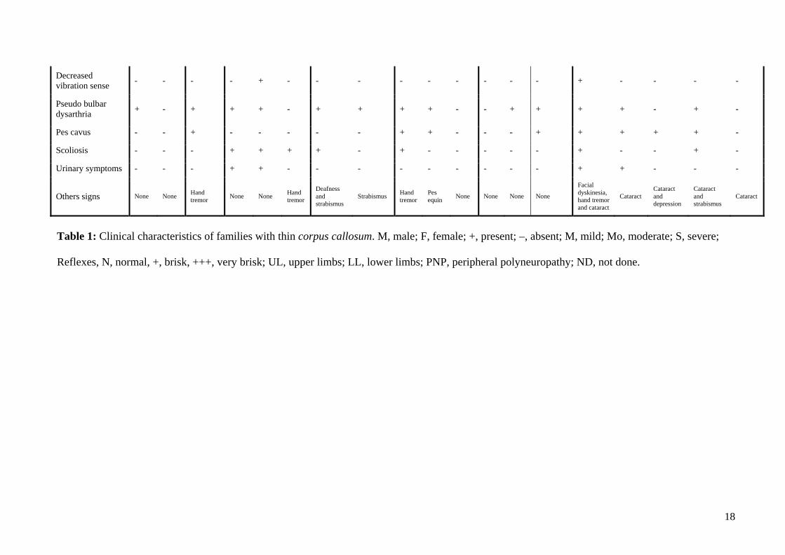
weakness in the lower extremities (Table 1).
After a mean disease duration of 18.9 ± 7.8 years (range 6-33), cognitive decline was present
in all affected individuals except one who had only 7 years of evolution, in addition to lower limb
(LL) hyperreflexia and bilateral Babinski signs. Others signs related to pyramidal tract dysfunction
were found in several patients: pseudobulbar dysarthria, upper limb (UL) spasticity and bladder
6
dysfunction. Additional signs were occasionally observed such as cerebellar ataxia (7/19), LL distal
amyotrophy (5/19) cataract (5/19) and decreased vibration sense (2/19).
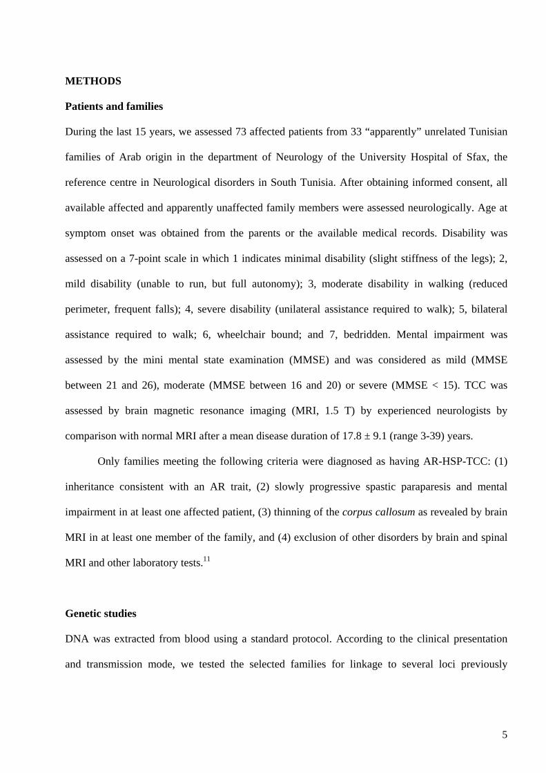
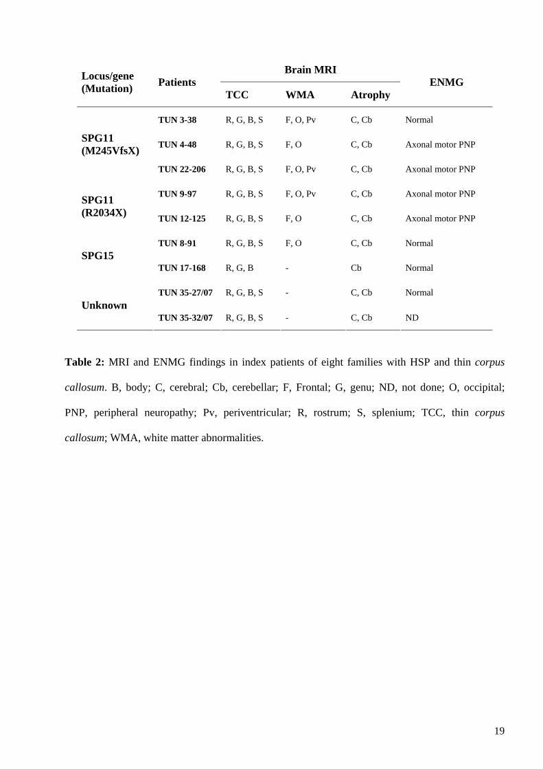
MRI was performed at the mean age of 26.5 ± 5.4 years (range 20-31) and after a mean
disease duration of 16.3 ± 6.4 years (range 6-26). Consistent with the clinical findings, all of the
nine patients studied by brain MRI had TCC, most prominently in the rostrum, genu and body,
associated with cerebral and cerebellar atrophy (Table 2). White matter changes were found
frequently (6/9 patients, 66%). In three individuals, hyperintense T2-weighted lesions were
restricted to occipital and frontal regions, but in the other cases there were diffuse abnormalities
involving periventricular regions (Figure 1). Cortical cerebral atrophy with frontal predominance
seemed to be a late feature, since it was absent in patient TUN17-168 who had the shortest disease
duration when examined (6 years). Electroneuromyograms (ENMG) showed electrophysiological
signs of predominantly axonal motor peripheral neuropathy in 4/8 patients tested (Table 2). Muscle
and nerve biopsies of these 4 patients showed signs of axonal degeneration associated with chronic
neurogenic changes without specific alterations.
Genetic findings
We first evaluated linkage to known genes (SPG7, SPG11 and SPG21) and subsequently looked for
mutations in linked families. Further explorations in non-mutated and non-linked families included
loci SPG15, SPG32 and the locus for HSP-TCC-epilepsy.
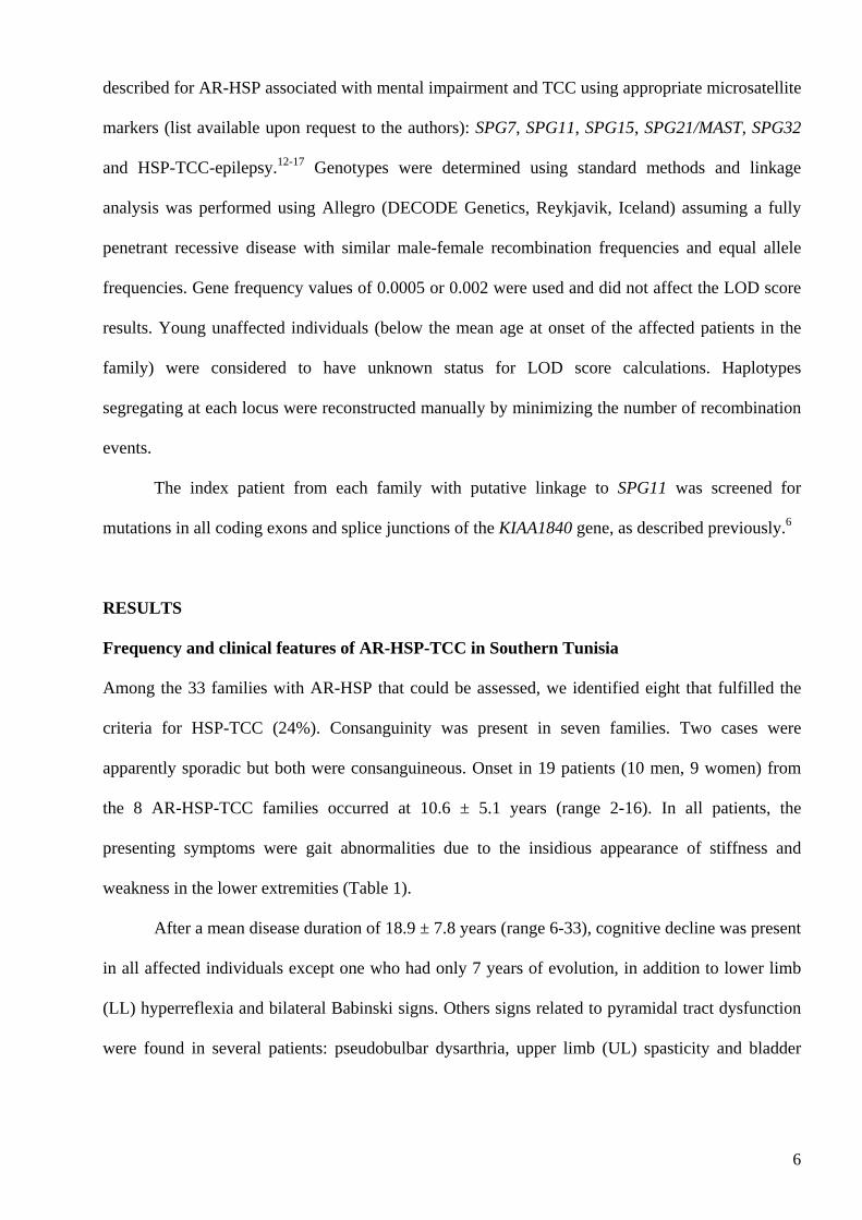
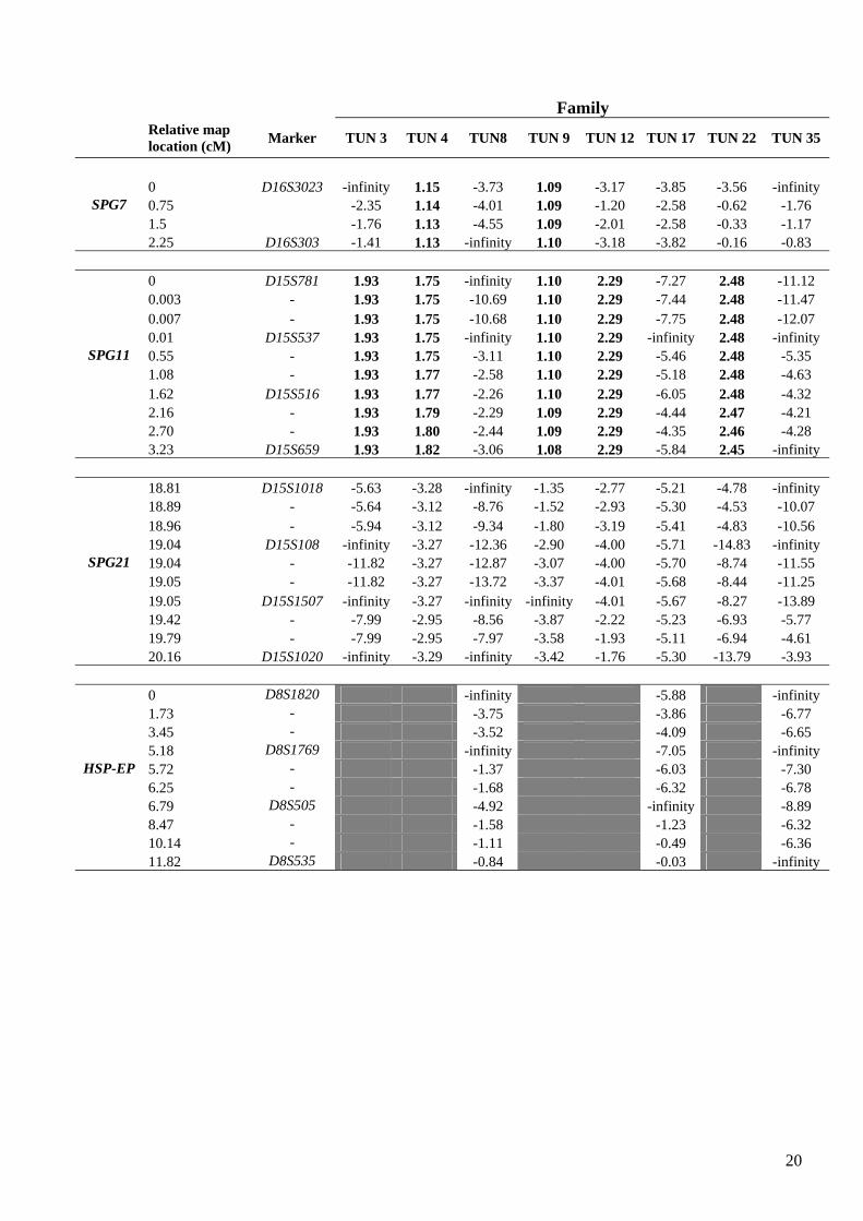
In five families (62.5%), linkage to SPG11 was suggested by identical homozygosity at
several markers and positive multipoint LOD score values from 1.1 to 2.5 reaching the maximal
expected values in the pedigrees (table 3). Patients in families TUN 3, TUN 4 and TUN 22 had
partially similar haplotypes encompassing the SPG11 gene, suggesting inheritance of a common
mutated ancestral chromosome. Similarly, families TUN 9 and TUN 12 shared the same
homozygous alleles for the whole SPG11 interval. In accordance with these results, direct
7
sequencing of all exons and splice junctions of the SPG11 gene in the five chromosome 15q-linked
families revealed two recurrent mutations: c.733_734delAT (exon 4) / p.M245VfsX in families
TUN 3, TUN 4 and TUN 22 as well as c.6100C>T (exon 32) / p.R2034X in families TUN 9 and
TUN12 (Figure 2). The mutations segregated with the disease in all families but were also present
in the homozygous state in individual TUN 3-42 who was still unaffected at 11 years of age. This
individual, only had brisk reflexes on examination and is still younger than the age at onset of his
affected relatives. Because of his young age, no further investigations could be performed.
In all three non-SPG11 families, haplotype reconstructions excluded linkage to SPG7,
SPG21, SPG32 and the locus for HSP-TCC-epilepsy because there were no haplotypes identical by
descent in the affected patients of each kindred and because the multipoint LOD scores for the
candidate intervals were negative (Table 3).
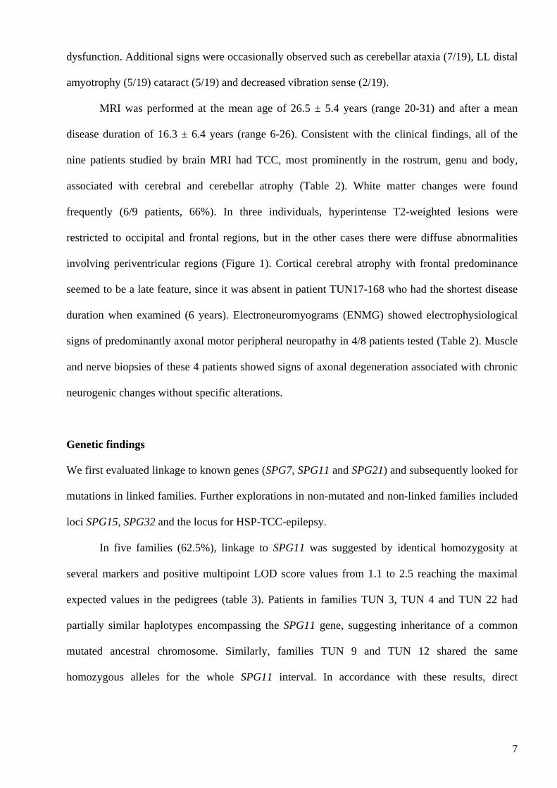
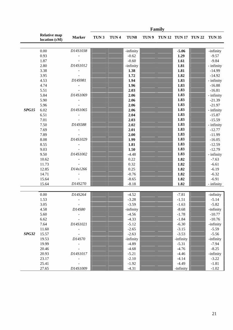
The exclusion of other known HSP-TCC loci and the co-segregation with the disease of
chromosome 14q-haplotypes between markers D14S1038 and D14S270 in two families (TUN 8 and
TUN 17, 25%) suggested their linkage to SPG15 (Figure 3A). Homozygosity was observed in the
whole interval in family TUN 17 and between markers D14S981 and D14S1002 in family TUN 8.
In addition, linkage to SPG15 was supported by positive LOD scores of 2.0 and 1.8 in families
TUN 8 and TUN 17, respectively. The haplotypes segregating with the disease were different in the
two families, suggesting the possibility of different allelic mutations.
In the remaining family (TUN 35, Figure 3B), linkage to the six known loci of HSP-TCC
was excluded (Table 3), suggesting the existence of another genetic locus for this condition.
Phenotype-genotype correlations
The mean age at onset (12.4 versus 14.3 years) and the clinical profile of patients with SPG11
(n=11) and SPG15 (n=3) were similar, but there were a few subtle differences between them.
SPG11 patients tended to have a more severe disease than SPG15 patients (mean severity score
8
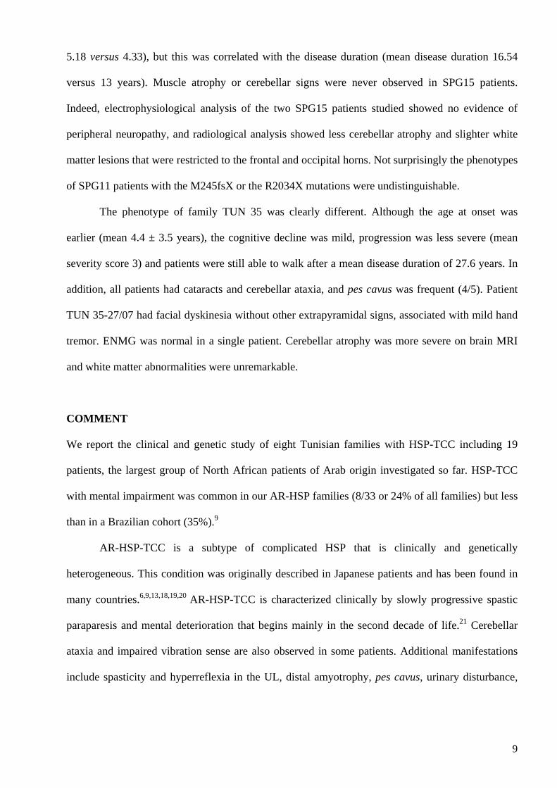
5.18 versus 4.33), but this was correlated with the disease duration (mean disease duration 16.54
versus 13 years). Muscle atrophy or cerebellar signs were never observed in SPG15 patients.
Indeed, electrophysiological analysis of the two SPG15 patients studied showed no evidence of
peripheral neuropathy, and radiological analysis showed less cerebellar atrophy and slighter white
matter lesions that were restricted to the frontal and occipital horns. Not surprisingly the phenotypes
of SPG11 patients with the M245fsX or the R2034X mutations were undistinguishable.
The phenotype of family TUN 35 was clearly different. Although the age at onset was
earlier (mean 4.4 ± 3.5 years), the cognitive decline was mild, progression was less severe (mean
severity score 3) and patients were still able to walk after a mean disease duration of 27.6 years. In
addition, all patients had cataracts and cerebellar ataxia, and pes cavus was frequent (4/5). Patient
TUN 35-27/07 had facial dyskinesia without other extrapyramidal signs, associated with mild hand
tremor. ENMG was normal in a single patient. Cerebellar atrophy was more severe on brain MRI
and white matter abnormalities were unremarkable.
COMMENT
We report the clinical and genetic study of eight Tunisian families with HSP-TCC including 19
patients, the largest group of North African patients of Arab origin investigated so far. HSP-TCC
with mental impairment was common in our AR-HSP families (8/33 or 24% of all families) but less
than in a Brazilian cohort (35%).9
AR-HSP-TCC is a subtype of complicated HSP that is clinically and genetically
heterogeneous. This condition was originally described in Japanese patients and has been found in
many countries.6,9,13,18,19,20 AR-HSP-TCC is characterized clinically by slowly progressive spastic
paraparesis and mental deterioration that begins mainly in the second decade of life.21 Cerebellar
ataxia and impaired vibration sense are also observed in some patients. Additional manifestations
include spasticity and hyperreflexia in the UL, distal amyotrophy, pes cavus, urinary disturbance,
9
dysarthria, nystagmus, congenital cataracts, seizures and extrapyramidal signs.10,11,21 On brain MRI,
progressive thinning of the corpus callosum is the neuroradiologically distinctive feature of this
syndrome.9 Periventricular white matter changes and late cortical atrophy are additional and
frequent features. Cerebral and cerebellar atrophy are slowly progressive and abnormalities of
cerebral white matter with frontal predominance have been observed in long-standing cases.22
It is clear from molecular genetic analyses that there are several underlying causes of this
syndrome,23 with five genetic loci identified (SPG11, SPG15, SPG21, SPG32 and HSP-TCC-
epilepsy). SPG7 has also been associated with a similar phenotype. The major locus is, however,
SPG11 (62% in Tunisia), a frequency similar to Japanese (77%, 11/13), Mediterranean (60%, 6/10)
and Italian (41%, 5/12) families.11,13,24,25 Results of linkage analysis were confirmed by the
subsequent identification of two recurrent mutations: R2034X and M245VfsX, in two and three
Tunisian families, respectively, suggesting founder effects.6 R2034X-segregating haplotypes were
similar in a previously published Moroccan kindred with the same mutation, suggesting inheritance
of a common ancestral mutation.6 On the contrary, the segregating haplotypes in French and Italian
kindreds carrying the M245VfsX mutation were different indicating independent ancestral events or
a very ancient mutation.6,26 SPG15, responsible for 25% of AR-HSP-TCC in our series, was the
second most important locus, in agreement with a frequency of 15% reported in another study of
families from the Mediterranean basin.27 The similarity between the neurological and radiological
findings in SPG11 and SPG15 patients suggests that the responsible genes may be functionally
related. Although a few subtle differences were observed, such as the absence of LL/UL
amyotrophy, peripheral neuropathy, or cerebellar signs in some SPG15 patients, who also had
severe lesions of the cerebellum, these features may be present in other SPG15 patients,27 and
cannot be used to distinguish SPG11 from SPG15-linked families on a clinical basis.
One family in our series was not linked to any of the loci tested, suggesting that an as yet
unmapped locus is responsible for their disease. Interestingly, this family, which represents a new
10
genetic entity, also had a unique phenotype, that differed from that of SPG11 and SPG15 by an
earlier onset and a slower progression of spastic gait, as well as the presence of cataracts and
cerebellar ataxia.
In conclusion, we describe a group of Arab families from North Africa with AR-HSP-TCC.
We demonstrate that this clinical-radiological syndrome is a frequent form of HSP and that SPG11
and SPG15 are the major loci for this clinical entity. In addition, we show the existence of a novel
genetic form of AR-HSP-TCC with unique and different clinical features.
11
REFERENCES
1. McDermott C, White K, Bushby K, Shaw P. Hereditary spastic paraparesis: a review of new
developments. J Neurol Neurosurg Psychiatry. 2000;69:150-160.
2. Harding AE. Classification of the hereditary ataxias and paraplegias. Lancet. 1983;1:1151-1155.
3. Behan WM, Maia M. Strumpell's familial spastic paraplegia: genetics and neuropathology. J
Neurol Neurosurg Psychiatry. 1974;37:8-20.
4. Fink JK. Hereditary spastic paraplegia. Curr Neurol Neurosci Rep. 2006;6:65-76.
5. Valdmanis PN, Meijer IA, Reynolds A, et al. Mutations in the KIAA0196 gene at the SPG8
locus cause hereditary spastic paraplegia. Am J Hum Genet. 2007;80:152-161.
6. Stevanin G, Santorelli FM, Azzedine H, et al. Mutations in SPG11, encoding spatacsin, are a
major cause of spastic paraplegia with thin corpus callosum. Nat Genet. 2007;39:366-372.
7. Mannan AU, Krawen P, Sauter SM, et al. ZFYVE27 (SPG33), a novel spastin-binding protein,
is mutated in hereditary spastic paraplegia. Am J Hum Genet. 2006;79:351-7.
8. Reid E. Science in motion: common molecular pathological themes emerge in the hereditary
spastic paraplegias. J Med Genet. 2003;40:81-86.
9. Franca MC Jr, D'Abreu A, Maurer-Morelli CV, et al. Prospective neuroimaging study in
hereditary spastic paraplegia with thin corpus callosum. Mov Disord. 2007;22:1556-62.
10. Ueda M, Katayama Y, Kamiya T, et al. Hereditary spastic paraplegia with a thin corpus
callosum and thalamic involvement in Japan. Neurology. 1998;51:1751-1754.
11. Shibasaki Y, Tanaka H, Iwabuchi K, et al. Linkage of autosomal recessive hereditary spastic
paraplegia with mental impairment and thin corpus callosum to chromosome 15q13-15. Ann
Neurol. 2000;48:108-112.
12. De Michele G, De Fusco M, Cavalcanti F, et al. A new locus for autosomal recessive hereditary
spastic paraplegia maps to chromosome 16q24.3. Am J Hum Genet. 1998;63:135-139.
12
13. Casali C, Valente EM, Bertini E, et al. Clinical and genetic studies in hereditary spastic
paraplegia with thin corpus callosum. Neurology. 2004;62:262-268.
14. Hughes CA, Byrne PC, Webb S, et al. SPG15, a new locus for autosomal recessive complicated
HSP on chromosome 14q. Neurology. 2000;56:1230-1233.
15. Simpson MA, Cross H, Proukakis C, et al. Maspardin is mutated in mast syndrome, a
complicated form of hereditary spastic paraplegia associated with dementia. Am J Hum Genet.
2003;73:1147-1156.
16. Stevanin G, Paternotte C, Coutinho P, et al. A new locus for autosomal recessive spastic
paraplegia (SPG32) on chromosome 14q12-q21. Neurology. 2007;68:1837-1840.
17. Al-Yahyaee S, Al-Gazali LI, De Jonghe P, et al. A novel locus for hereditary spastic paraplegia
with thin corpus callosum and epilepsy. Neurology. 2006;66:1230-1234.
18. Winner B, Uyanik G, Gross C, et al. Clinical progression and genetic analysis in hereditary
spastic paraplegia with thin corpus callosum in spastic gait gene 11 (SPG11). Arch Neurol.
2004;61:117-121.
19. Tang BS, Chen X, Zhao GH, et al. Clinical features of hereditary spastic paraplegia with thin
corpus callosum: report of 5 Chinese cases. Chin Med J. 2004; 117:1002-1005.
20. Lossos A, Stevanin G, Meiner V, et al. Hereditary spastic paraplegia with thin corpus callosum:
reduction of the SPG11 interval and evidence for further genetic heterogeneity. Arch Neurol.
2006;63:756-760.
21. Nakamura A, Izumi K, Umehara F, et al. Familial spastic paraplegia with mental impairment
and thin corpus callosum. J Neurol Sci. 1995;131:35-42.
22. Ohnishi J, T omoda Y, Yokoyama K. Neuroradiological findings in hereditary spastic
paraplegia with a thin corpus callosum. Acta Neurol Scand. 2001;104:191-192.
13
23. Brockmann K, Simpson MA, Faber A, Bonnemann C, Crosby AH, Gartner J. Complicated
hereditary spastic paraplegia with thin corpus callosum (HSP-TCC) and childhood onset.
Neuropediatrics. 2005;36:274-278.
24. Stevanin G, Montagna G, Azzedine H, et al. Spastic paraplegia with thin corpus callosum:
description of 20 new families, refinement of the SPG11 locus, candidate gene analysis and
evidence of genetic heterogeneity. Neurogenet. 2006;7:149-156.
25. Stevanin G, Azzedine H, Denora P, et al. Mutations in SPG11 are frequent in autosomal
recessive spastic paraplegia with thin corpus callosum, cognitive decline and lower motor
neuron degeneration. Brain (in press).
26. Del Bo R, Di Fonzo A, Ghezzi S, et al. SPG11: a consistent clinical phenotype in a family with
homozygous Spatacsin truncating mutation. Neurogenet. 2007;8:301-305.
27. Elleuch N, Bouslam N, Hanein S, et al. Refinement of the SPG15 candidate interval and
phenotypic heterogeneity in three large Arab families. Neurogenet. 2007;8:307-315.
14
FIGURES LEGENDS
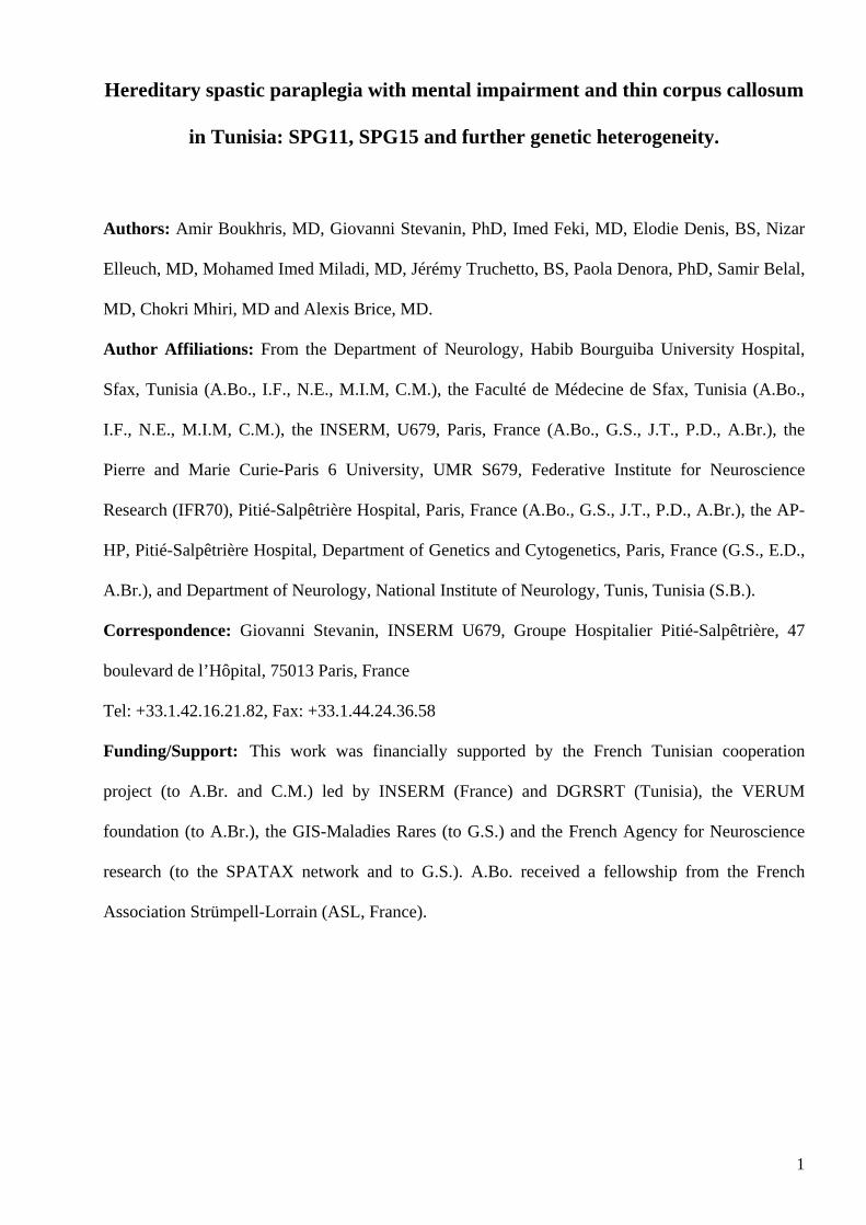
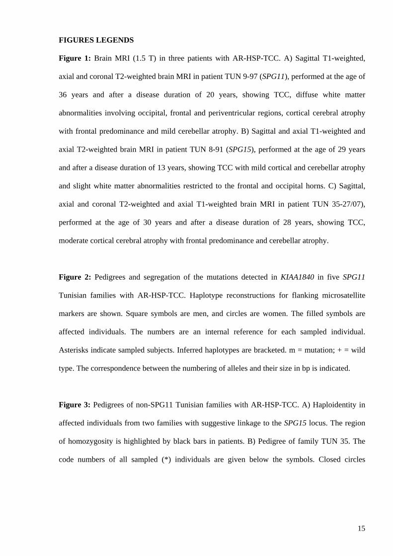
Figure 1: Brain MRI (1.5 T) in three patients with AR-HSP-TCC. A) Sagittal T1-weighted,
axial and coronal T2-weighted brain MRI in patient TUN 9-97 (SPG11), performed at the age of
36 years and after a disease duration of 20 years, showing TCC, diffuse white matter
abnormalities involving occipital, frontal and periventricular regions, cortical cerebral atrophy
with frontal predominance and mild cerebellar atrophy. B) Sagittal and axial T1-weighted and
axial T2-weighted brain MRI in patient TUN 8-91 (SPG15), performed at the age of 29 years
and after a disease duration of 13 years, showing TCC with mild cortical and cerebellar atrophy
and slight white matter abnormalities restricted to the frontal and occipital horns. C) Sagittal,
axial and coronal T2-weighted and axial T1-weighted brain MRI in patient TUN 35-27/07),
performed at the age of 30 years and after a disease duration of 28 years, showing TCC,
moderate cortical cerebral atrophy with frontal predominance and cerebellar atrophy.
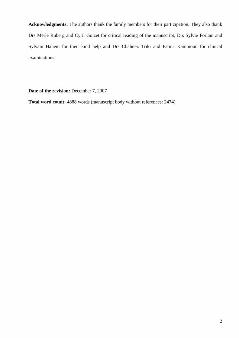
Figure 2: Pedigrees and segregation of the mutations detected in KIAA1840 in five SPG11
Tunisian families with AR-HSP-TCC. Haplotype reconstructions for flanking microsatellite
markers are shown. Square symbols are men, and circles are women. The filled symbols are
affected individuals. The numbers are an internal reference for each sampled individual.
Asterisks indicate sampled subjects. Inferred haplotypes are bracketed. m = mutation; + = wild
type. The correspondence between the numbering of alleles and their size in bp is indicated.
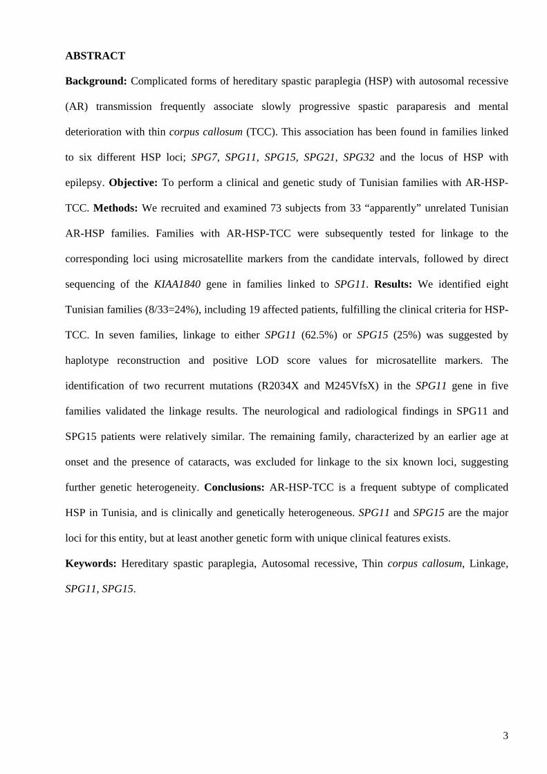
Figure 3: Pedigrees of non-SPG11 Tunisian families with AR-HSP-TCC. A) Haploidentity in
affected individuals from two families with suggestive linkage to the SPG15 locus. The region
of homozygosity is highlighted by black bars in patients. B) Pedigree of family TUN 35. The
code numbers of all sampled (*) individuals are given below the symbols. Closed circles
15

16
(women) and squares (men) indicate affected members. Arrowheads indicate the positions of
recombination events.
17
Locus/gene (Mutation)
SPG11 (M245VfsX)
SPG11 (R2034X) SPG15 Unknown
Family TUN 3 TUN 4 TUN 22 TUN 9 TUN 12 TUN 8 TUN 17 TUN 35
Consanguinity (degree)
Yes (1)
Yes (1)
Yes (1)
Yes (1)
Yes (3)
Yes (1) No No Yes
(2) Yes (2)
Yes (2)
Yes (1)
Yes (1)
Yes (1)
Yes (1)
Yes (1)
Yes (1)
Yes (1)
Yes (1)
Individual N° (sex)
38 (M)
40 (M)
48 (F)
201 (M)
206 (F)
212 (M)
97 (M)
98 (M)
125 (M)
129 (F)
130 (F)
91 (M)
92 (M)
168 (F)
27/07 (M)
26/07 (F)
32/07 (F)
33 (F)
36/07 (F)
Age at examination, years 25 21 24 36 21 37 36 34 31 34 21 29 33 20 30 32 35 31 32
Age at onset, years 15 14 8 13 4 10 16 15 15 14 13 16 13 14 2 2 2 6 10
Disease duration, years 10 7 16 23 16 27 20 19 16 20 8 13 20 6 28 30 33 25 22
Disability scale 4/7 3/7 4/7 6/7 6/7 5/7 6/7 5/7 6/7 6/7 6/7 4/7 6/7 3/7 2/7 3/7 4/7 3/7 3/7
LL spasticity S S S S S M S S S S S Mo S Mo S Mo S S M
LL reflexes +++ +++ +++ + + + +++ +++ +++ +++ +++ +++ + + +++ +++ +++ +++ +++
LL weakness Mo Mo - Mo Mo M S S S S S Mo S Mo Mo Mo Mo M Mo
LL distal amyotrophy - - M M Mo - - - Mo M - - - - - - - - -
Extensor plantar reflexes + + + + + + + + + + + + + + + + + + +
UL spasticity - - M - - - Mo M - - M - - M M M M Mo M
UL reflexes N N + N N N + + + + + + N N + +++ + + +
UL weakness - - - - - - M - - M - - - - M - - - -
UL distal amyotrophy - - - M - - - - M M - - - - - - - - -
Cognitive decline Mo - Mo Mo Mo Mo Mo Mo Mo S Mo Mo Mo M M M M M M
Cerebellar signs + - - - - + - - - - - - - - + + + + +
18
Decreased vibration sense - - - - + - - - - - - - - - + - - - -
Pseudo bulbar dysarthria + - + + + - + + + + - - + + + + - + -
Pes cavus - - + - - - - - + + - - - + + + + + -
Scoliosis - - - + + + + - + - - - - - + - - + -
Urinary symptoms - - - + + - - - - - - - - - + + - - -
Others signs None None Hand tremor None None Hand
tremor
Deafness and strabismus
Strabismus Hand tremor
Pes equin None None None None
Facial dyskinesia, hand tremor and cataract
Cataract Cataract and depression
Cataract and strabismus
Cataract
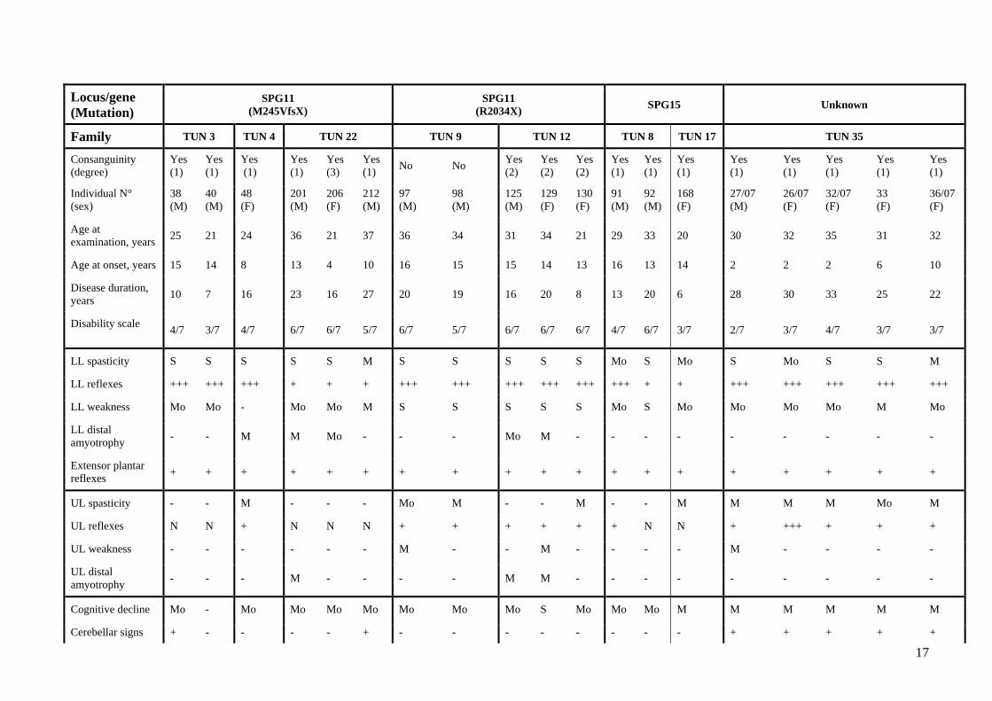
Table 1: Clinical characteristics of families with thin corpus callosum. M, male; F, female; +, present; –, absent; M, mild; Mo, moderate; S, severe;
Reflexes, N, normal, +, brisk, +++, very brisk; UL, upper limbs; LL, lower limbs; PNP, peripheral polyneuropathy; ND, not done.
Brain MRI Locus/gene (Mutation) Patients
TCC WMA Atrophy ENMG
TUN 3-38 R, G, B, S F, O, Pv C, Cb Normal
TUN 4-48 R, G, B, S F, O C, Cb Axonal motor PNP SPG11 (M245VfsX)
TUN 22-206 R, G, B, S F, O, Pv C, Cb Axonal motor PNP
TUN 9-97 R, G, B, S F, O, Pv C, Cb Axonal motor PNP SPG11 (R2034X) TUN 12-125 R, G, B, S F, O C, Cb Axonal motor PNP
TUN 8-91 R, G, B, S F, O C, Cb Normal SPG15
TUN 17-168 R, G, B - Cb Normal
TUN 35-27/07 R, G, B, S - C, Cb Normal Unknown
TUN 35-32/07 R, G, B, S - C, Cb ND
Table 2: MRI and ENMG findings in index patients of eight families with HSP and thin corpus
callosum. B, body; C, cerebral; Cb, cerebellar; F, Frontal; G, genu; ND, not done; O, occipital;
PNP, peripheral neuropathy; Pv, periventricular; R, rostrum; S, splenium; TCC, thin corpus
callosum; WMA, white matter abnormalities.
19
Family
Relative map location (cM) Marker TUN 3 TUN 4 TUN8 TUN 9 TUN 12 TUN 17 TUN 22 TUN 35
0 D16S3023 -infinity 1.15 -3.73 1.09 -3.17 -3.85 -3.56 -infinity
SPG7 0.75 -2.35 1.14 -4.01 1.09 -1.20 -2.58 -0.62 -1.76 1.5 -1.76 1.13 -4.55 1.09 -2.01 -2.58 -0.33 -1.17 2.25 D16S303 -1.41 1.13 -infinity 1.10 -3.18 -3.82 -0.16 -0.83 0 D15S781 1.93 1.75 -infinity 1.10 2.29 -7.27 2.48 -11.12 0.003 - 1.93 1.75 -10.69 1.10 2.29 -7.44 2.48 -11.47 0.007 - 1.93 1.75 -10.68 1.10 2.29 -7.75 2.48 -12.07 0.01 D15S537 1.93 1.75 -infinity 1.10 2.29 -infinity 2.48 -infinity
SPG11 0.55 - 1.93 1.75 -3.11 1.10 2.29 -5.46 2.48 -5.35 1.08 - 1.93 1.77 -2.58 1.10 2.29 -5.18 2.48 -4.63 1.62 D15S516 1.93 1.77 -2.26 1.10 2.29 -6.05 2.48 -4.32 2.16 - 1.93 1.79 -2.29 1.09 2.29 -4.44 2.47 -4.21 2.70 - 1.93 1.80 -2.44 1.09 2.29 -4.35 2.46 -4.28 3.23 D15S659 1.93 1.82 -3.06 1.08 2.29 -5.84 2.45 -infinity 18.81 D15S1018 -5.63 -3.28 -infinity -1.35 -2.77 -5.21 -4.78 -infinity 18.89 - -5.64 -3.12 -8.76 -1.52 -2.93 -5.30 -4.53 -10.07 18.96 - -5.94 -3.12 -9.34 -1.80 -3.19 -5.41 -4.83 -10.56 19.04 D15S108 -infinity -3.27 -12.36 -2.90 -4.00 -5.71 -14.83 -infinity
SPG21 19.04 - -11.82 -3.27 -12.87 -3.07 -4.00 -5.70 -8.74 -11.55 19.05 - -11.82 -3.27 -13.72 -3.37 -4.01 -5.68 -8.44 -11.25 19.05 D15S1507 -infinity -3.27 -infinity -infinity -4.01 -5.67 -8.27 -13.89 19.42 - -7.99 -2.95 -8.56 -3.87 -2.22 -5.23 -6.93 -5.77 19.79 - -7.99 -2.95 -7.97 -3.58 -1.93 -5.11 -6.94 -4.61 20.16 D15S1020 -infinity -3.29 -infinity -3.42 -1.76 -5.30 -13.79 -3.93
0 D8S1820 -infinity -5.88 -infinity
1.73 - -3.75 -3.86 -6.77 3.45 - -3.52 -4.09 -6.65 5.18 D8S1769 -infinity -7.05 -infinity
HSP-EP 5.72 - -1.37 -6.03 -7.30 6.25 - -1.68 -6.32 -6.78 6.79 D8S505 -4.92 -infinity -8.89 8.47 - -1.58 -1.23 -6.32 10.14 - -1.11 -0.49 -6.36 11.82 D8S535 -0.84 -0.03 -infinity
20
Family
Relative map location (cM) Marker TUN 3 TUN 4 TUN8 TUN 9 TUN 12 TUN 17 TUN 22 TUN 35
0.00 D14S1038 -infinity -5.06 -infinity 0.93 - -0.62 1.29 -9.57 1.87 - -0.60 1.61 -9.84 2.80 D14S1012 -infinity 1.81 - infinity 3.38 - 1.38 1.81 -14.99 3.95 - 1.72 1.82 -14.92 4.53 D14S981 1.94 1.83 - infinity 4.74 - 1.96 1.83 -16.88 5.51 - 2.03 1.83 -16.81 5.84 D14S1069 2.06 1.83 - infinity 5.90 - 2.06 1.83 -21.39 5.96 - 2.06 1.83 -21.97
SPG15 6.02 D14S1065 2.06 1.83 - infinity 6.51 - 2.04 1.83 -15.87 7.01 - 2.03 1.83 -15.59 7.50 D14S588 2.02 1.83 - infinity 7.69 - 2.01 1.83 -12.77 7.89 - 2.00 1.83 -11.99 8.08 D14S1029 1.99 1.83 -16.05 8.55 - 1.81 1.83 -12.59 9.03 - 1.50 1.83 -12.79 9.50 D14S1002 -4.48 1.83 - infinity 10.62 - 0.22 1.82 -7.63 11.73 - 0.32 1.82 -6.61 12.85 D14s1266 0.25 1.82 -6.19 14.71 - -0.76 1.82 -6.32 15.64 - -8.65 1.82 -6.91 15.64 D14S270 -8.18 1.82 - infinity 0.00 D14S264 -4.52 -7.81 -infinity 1.53 - -3.28 -1.51 -5.14 3.05 - -3.59 -1.63 -5.82 4.58 D14S80 -infinity -8.68 -infinity 5.60 - -4.56 -1.78 -10.77 6.62 - -4.33 -1.84 -10.76 7.64 D14S1021 -5.12 -6.30 -infinity 11.60 - -2.65 -3.15 -5.59
SPG32 15.57 - -2.63 -3.53 -5.56 19.53 D14S70 -infinity -infinity -infinity 19.99 - -4.89 -5.31 -7.94 20.46 - -4.68 -4.76 -8.25 20.93 D14S1017 -5.21 -4.46 -infinity 23.17 - -2.10 -4.14 -3.22 25.41 - -1.92 -4.49 -1.81 27.65 D14S1009 -4.31 -infinity -1.02
21
22
Table 3: Results of multipoint linkage analysis with markers covering the SPG7, SPG11, SPG15,
SPG21, SPG32 and HSP-TCC-Epilepsy (HSP-EP) candidate intervals. The multipoint LOD scores
calculated with ALLEGRO are indicated for each family relative to the position of the genetic
markers. Positive LOD score values are indicated in bold.
Nota Bene: Families TUN 3, TUN 4, TUN 9, TUN 12 and TUN 22, in which causative mutations in
SPG11 segregated with the disease, were not tested for linkage to SPG15, SPG32 and the locus for
HSP-TCC-epilepsy.