Embed Size (px)
Citation preview

CELL DISCOVERY AND THEORY7.1
History of the Cell Theory
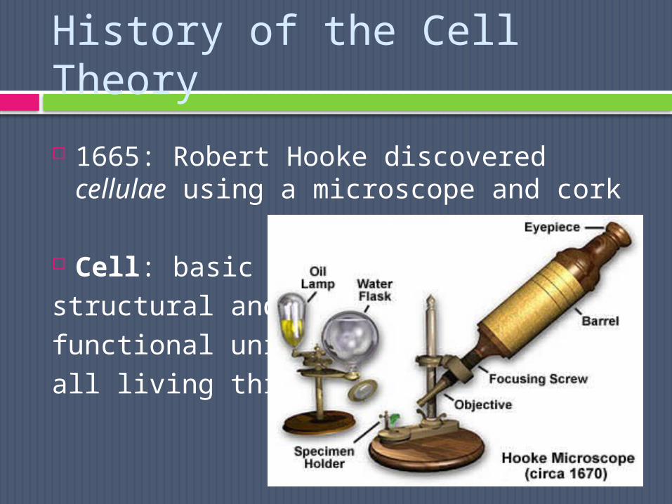
1665: Robert Hooke discovered cellulae using a microscope and cork
Cell: basic structural and functional unit of all living things

The Cell Theory
fundamental idea of modern biology
Consists of 3 principles: All living organisms composed of 1+
cells Cells are basic unit of
structure/organization of all living organisms
Cells arise only from previously existing cells, with cells passing copies of their genetic material on to their daughter cells

As magnification and resolution increases, so do the details we can
see!
Microscope Technology
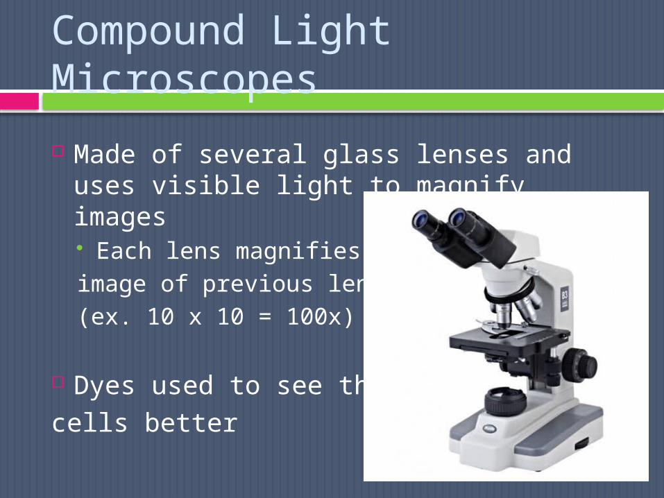
Compound Light Microscopes
Made of several glass lenses and uses visible light to magnify images Each lens magnifies image of previous lens (ex. 10 x 10 = 100x)
Dyes used to see the cells better

Limitations: objects cause light to scatter, blurring images Max magnification without blurring is
~1000x


Electron Microscopes
Developed during 1940s
Uses magnets to aim a beam of electrons at thin slices of cells
SEM, TEM, STM

Transmission Electron Microscope (TEM)
electrons passed through a specimen to a fluorescent screen
thick parts of specimen absorb more electrons, creating shaded image
*Shows internal details (more like an xray)

can magnify up to 500,000x but specimen must be dead, sliced very thin, and stained with heavy metals

Scanning Electron Microscope (SEM)
Sends electrons over specimen’s surface,creating a 3D image
Disadvantages: TEM and SEM only used for non-living cells/tissues

Scanning Tunneling Electron Microscope (STM)
Charged tip of probe brought to specimen so that electron “tunnel” through small gap between them
Allows scientists to create 3D models of
objects as small as atoms
Advantage: Can be used with live specimens


Atomic Force Microscope (AFM)
Measures forces between tip of probe and cell surface
Image is a scan of a crystal of satellite tobacco mosaic virus particles

Basic Cell Types
Cells have different shapes/sizes and differ based on function
All cells have a plasma membrane! special boundary that helps control what
enters/exits the cell
All cells have some functions in common Most have genetic material that provides
instructions for making substances that the cell needs

2 main groups: eukaryotic and prokaryotic Eukaryotic cells are ~100x larger than prokaryotic
Organelles: specialized structures that carry out specific functions Bound by membranes Allow functions to occur in different places at
the same time
Nucleus: a central organelle that contains cells genetic material (DNA)

Eukaryotic cells believed to have evolved from prokaryotic cells
PROKARYOTIC CELLS EUKARYOTIC CELLSPlasma membrane Plasma membrane
No organelles Has organellesOrganisms prokaryotes Organisms eukaryotesMake up most unicellular
organisms (bacteria)Make up most organisms and some unicellular organisms
(algae and yeast)

Cell Size
Cells are TINY! Some are as small as 1/1000th of a millimeter.
Small cells have more surface area per volume More surface area makes it easier to move material
in/out of the cell
Large cells have less surface area per volume Requires more nutrients, produces more wastes
CONCLUSION: Cells need a certain surface area to properly function (exchange materials). Surface area to volume considerations are the reason why cells stay small!