Embed Size (px)
Citation preview

Cell communication
Chapter 9Genes and Development

Fig. 9.1
Copyright © The McGraw-Hill Companies, Inc. Permission required for reproduction or display.
CytoplasmExternal environment
Membrane receptor
Intracellular receptor
Plasma membrane
Signaltransduction
pathway
Signaltransduction
pathway
Cellularresponse
Cellularresponse
Hydrophobicligand
Hydrophilicligand

Fig. 9.2a
Copyright © The McGraw-Hill Companies, Inc. Permission required for reproduction or display.
a.
Direct Contact
Adjacentplasmamembrane
Plasmamembrane
Adjacentplasmamembrane
Plasmamembrane

Fig. 9.2b
Copyright © The McGraw-Hill Companies, Inc. Permission required for reproduction or display.
b.
Paracrine Signaling
Secretory cell
Adjacenttarget cells
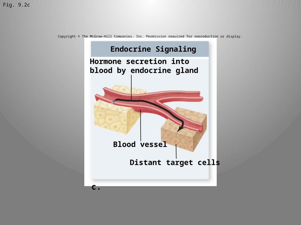
Fig. 9.2c
Copyright © The McGraw-Hill Companies, Inc. Permission required for reproduction or display.
c.
Endocrine Signaling
Blood vessel
Distant target cells
Hormone secretion intoblood by endocrine gland

Fig. 9.2d
Copyright © The McGraw-Hill Companies, Inc. Permission required for reproduction or display.
d.
Synaptic Signaling
Nerve cellNeurotransmitter
Synaptic gap
Target cell

Fig. 9.2
Copyright © The McGraw-Hill Companies, Inc. Permission required for reproduction or display.
a. b. c. d.
Synaptic SignalingEndocrine SignalingParacrine Signaling
Nerve cellNeurotransmitter
Synaptic gap
Target cell
Blood vessel
Distant target cells
Secretory cell
Direct Contact
Adjacentplasmamembrane
Plasmamembrane
Adjacenttarget cells
Hormone secretion intoblood by endocrine gland
Adjacentplasmamembrane
Plasmamembrane

Fig. 9.3Copyright © The McGraw-Hill Companies, Inc. Permission required for reproduction or display.
Kinase
Phosphatase
Kinase
Phosphatase
O
P
O–
O O–
O
P
O–
O O–
OH
OHTyr
ADP
Pi
ADP
ATP
ATP
Seror
Thr
Seror
Thr
Tyr
Pi

Fig. 9.4a
Copyright © The McGraw-Hill Companies, Inc. Permission required for reproduction or display.
a.
Ions
Ions
Ligand(signal)
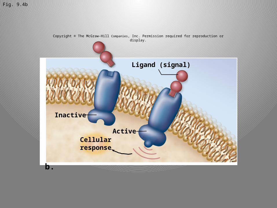
Fig. 9.4b
Copyright © The McGraw-Hill Companies, Inc. Permission required for reproduction or display.
b.
Ligand (signal)
Inactive
ActiveCellular
response

Fig. 9.4c
Copyright © The McGraw-Hill Companies, Inc. Permission required for reproduction or display.
c.
gb
g
a a
a
b
GDP GTP GTP
Ions
G protein EnzymeIon channel
GPCR
G protein activateseither enzyme or ion channel
Ligand(signal)
b g
a
bg
a
a


Fig. 9.4
Copyright © The McGraw-Hill Companies, Inc. Permission required for reproduction or display.
a.
c.
b.
Ions
Ions
Ligand (signal)
Inactive
Active
gb
g
a a
a
b
GDP GTP GTP
Ions
G protein EnzymeIon channel
GPCR
G protein activateseither enzyme or ion channel
Ligand(signal)
Cellularresponse
Ligand(signal)
b g
a
bg
a
a

Fig. 9.5
Copyright © The McGraw-Hill Companies, Inc. Permission required for reproduction or display.
Hormone
Inhibitor
Inhibitor
1. Hormones cross plasma membrane and bind to cytoplasmic receptors.
2. Hormone binding alters receptor conformation so it no longer binds inhibitor.
5. Cellular response is a change in gene expression.
3. Hormone–receptor complex translocates to nucleus.
4. Hormone–receptor complex binds to DNA. This usually turns on transcription, but can also turn it off.
Signal molecule-binding domain
DNA-bindingsite exposed
DNA-bindingsite blocked
Transcription-activating domainGenetranscription

Fig. 9.6
Copyright © The McGraw-Hill Companies, Inc. Permission required for reproduction or display.
Ligands
Phosphorylated proteinIntracellular kinase domain
Extracellular ligand-binding domain
P
P
P
P
P
P
P
P P
P
P
P
PP
P
P
P
P
Phosphategroups
Transmembrane RTK proteins
1. Ligand binds to the receptor.
2. Two receptors associate (dimerize) and phosphorylate each other (autophosphorylation).
3. Response proteins bind to phospho- tyrosine on receptor. Receptor can phosphorylate other response proteins.
Dimerization andautophosphorylation
Cellularresponse
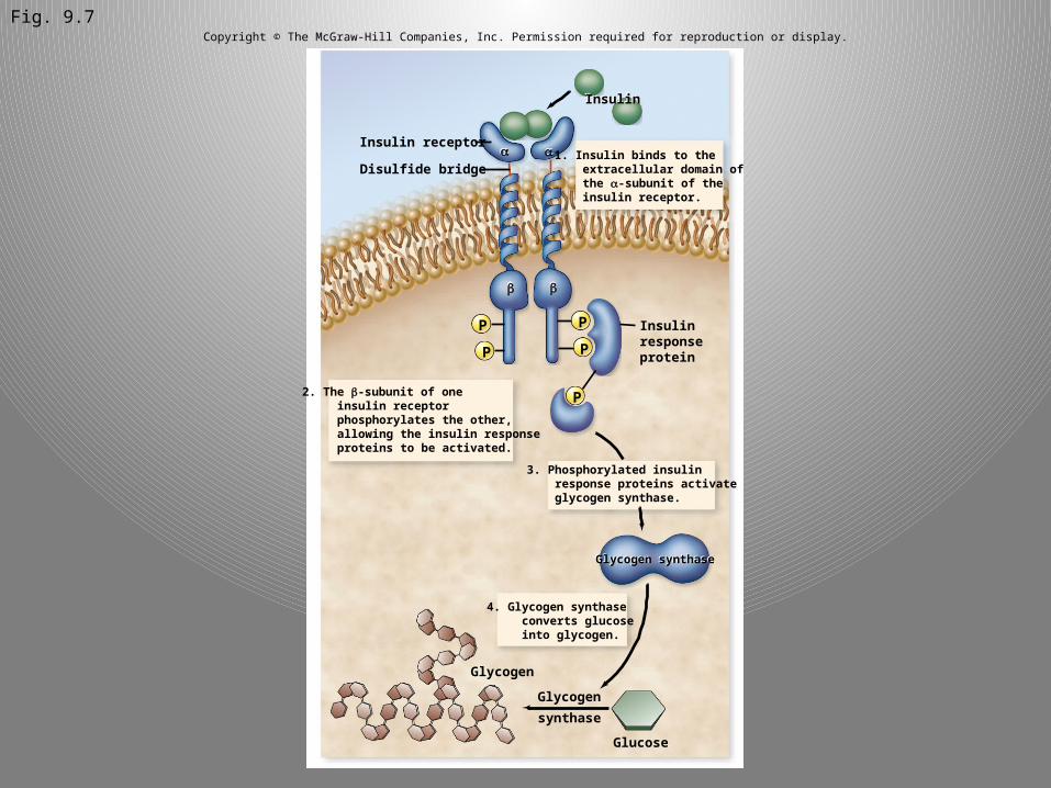
Fig. 9.7Copyright © The McGraw-Hill Companies, Inc. Permission required for reproduction or display.
Glycogen
Glycogen synthase
Glycogen
synthase
Insulin receptor
Disulfide bridge
Insulin
Glucose
aa
bb
P P
PP
P
1. Insulin binds to the extracellular domain of the a-subunit of the insulin receptor.
2. The b-subunit of one insulin receptor phosphorylates the other, allowing the insulin response proteins to be activated.
3. Phosphorylated insulin response proteins activate glycogen synthase.
4. Glycogen synthase converts glucose into glycogen.
Insulinresponseprotein
Glycogen synthase
Insulin
a a
b b

Fig. 9.8aCopyright © The McGraw-Hill Companies, Inc. Permission required for reproduction or display.
Response
Signal
Receptor
Activator
ActiveInactive
Inactive
Inactive
Active
Active
MKKMKK
MKMK
MKKKMKKK
a.
Ras
PP
P
PP
P
MAP kinase cascade
Firstkinase
Secondkinase
MAPkinase
Responseproteins
Cellularresponse
Responseproteins
Responseproteins
MKKK
MKK
MK
MKKK
MKK
MK

Fig. 9.10
Copyright © The McGraw-Hill Companies, Inc. Permission required for reproduction or display.
Ras
GT
1. Proteins bound to receptor activate Ras by exchanging GDP for GTP.
2. Ras activates the first kinase (Raf)
3. Raf activates the second kinase (MEK)
4. MEK activates MA P kinases (ERK) 5. MAP kinase (ERK) activates
proteins to produce cellular responses, including transcription factors that alter gene expression
Activatestranscriptionfactors
NuclearmembraneCellular
response
Activatestranscriptionfactors
GDP
Ras
Raf
Raf
MEK
P
PP
P
P
P
P
P
P
P
PP P
P
PResponse
proteinResponse
protein
Responseprotein
Responseprotein
Responseprotein
Responseprotein
P
MEK
ERK
ERK

Fig. 9.11
Copyright © The McGraw-Hill Companies, Inc. Permission required for reproduction or display.
GPCR
Ligand
GPCR
ab g
a
b g b g
a
GDPInactive G
protein GTP
GDP
Cellularresponse
Effector protein
GTPGTP
gb
a ab g b g
a
Active Gprotein
Pi

Fig. 9.12
Copyright © The McGraw-Hill Companies, Inc. Permission required for reproduction or display.
P
–O
–O O
O
P
–O –O
O
O
P O CH2
O
O
a.
b.
Extracellular space
Cytoplasm
Phospholipase C
PIP2
O
C
O C
O
C C
OC
O
P
O
O–O
P
O
O O–
O–
P
O–
O O–
OOH OHOH
DAG + IP3
O
C
O C
O
C C
OC
OH
P
O
O
OO–
P
O
O O–
O–
P
O–
O O–
OOH OHOH
P
–O
O O
OO
CH2
DAG
IP3
P
O–
–O O
O
P
O–
O–
OPP i
cAMP
Adenylyl cyclase
NH2
N
NH2
N
ATP cAMP + PPi
Plasmamembrane
Cleaved byphospholipase C

Fig. 9.13Copyright © The McGraw-Hill Companies, Inc. Permission required for reproduction or display.
Ligand
GPCR
GDP GTP
ATP
Activates PKA
Cellularresponse
Responseprotein
Responseprotein
Adenylyl cyclase
Cytoplasm
Nucleus Nucleus
cAMP
b g
a
b gb g
a
b g

Fig. 9.15 Copyright © The McGraw-Hill Companies, Inc. Permission required for reproduction or display.
Ca2+
ER
Cytoplasm
Phospholipase CGPCR
DAG
PIP
IP3
b g b ga
Cellularresponse
Ca2+
bindingprotein
gb ga
b
Ligand
GDP GTP

Fig. 9.16
Copyright © The McGraw-Hill Companies, Inc. Permission required for reproduction or display.
Ca2+
Calmodulin
Calmodulin
a. b.
Activeprotein
Inactiveprotein
Ca2+ Ca2+
Ca2+
Ca2+

Fig. 9.17 Copyright © The McGraw-Hill Companies, Inc. Permission required for reproduction or display.
P
Cytoplasm
Epinephrine
Glucagon
P P P
P P P P
Glycogen
CH2
GDP
GDP
GPCR
PKA
cAMP
Glucose-6-phosphate
Adenylylcyclase
Adenylylcyclase
Phosphorylasekinase
Glycogenphosphorylase
GTPGTP
P
ATPATP