Embed Size (px)
Citation preview
~~~~~C:~d Copyriaht 1910. Spectrum Publications. 1114:. ) ~ Mapaium in Health and Discalc
INTRODUCTION
52
Speculations on Renal Hormonal, and Metabolic Aberrations in a Patient
with Marginal Magnesium Deficiency
M.S. Seelig A.R. Berger
L.A. Avioli
A postmenopausal woman, with normocalcemic latent tetany of marginal magnesium (Hg) deficiency (Seelig~·· 1975), had had previously disregarded electrolyte aberrations, including slight hypochloremia, hypokalemia, and hypercapnea. Her serum sodium (Na) levels were within normal limits or were slightly elevated, despite salt restriction to control her peripheral edema. Specific Hg malabsorption was ruled out as the cause of her failure to maintain clinical improvement on oral Hg supplements, that had been achieved by intramuscular (l.m. ) administration of HgS04. Her subsequent responses to i.m . Hg were transitory. Re-evaluation disclosed renal wasting of Mg, and increased aldosterone and renin secretion when her Hg-deficit was intensified by severe dietary Mg restriction or hormonal challenge. Since her blood pressure was normal or slightly low, we propose that she may have a defect in the ascending limb of the loop of Henle (ALLH), that is responsible for both chloride (Cl) and Hg loss, and that her Mg deficiency may have contributed to and intensified the clinical syndrome. It is possible that her retention of Na and peripheral edema may reflect failure of the Na/potassium (K) exchange mechanism at the cellular level resulting from Hg deficiency . Subsequent contraction of the circulating blood volume may be contributory to her normotensive aldosteronism. Her additional abnormalitie~, which include generalized pruritus, depressive anxiety, advanced osteoporosis, and recurrent thrombophlebitis, are all manifestations that have been associated with Mg deficiency.
CASE REPORT
A 57 year old woman has suffered for 9 years (since total hysterectomy) from weakness, depression, insomnia, anxiety, peripheral edema, and generalized pruritus; the latter two findings are intensified by salt, and are only partially manageable by high dosage antihistaminic and mild tranquilizer (Valium) therapy. Hypokalemia and hypochloremia became noteworthy during diuretic treatment of her edema of unknown origin, despite concomitant KCl administration. Hypercapnea was also noted at the time her marginal hypomagnesemia (1.67 meq/liter; normal
\.. , "-..:- .... ..........
460 MAGNESIUM IN HEALTH AND DISEASE
range at the hospital • 1.9-2 . 5*) waa detected. Her serum Na was normal. despite prolonged salt restriction. Her dietary history suggested long-term low intakes of Mg. calcium (Ca). and vitamin D. Magnesium malabsorption was ruled out by metabolic balance studies.
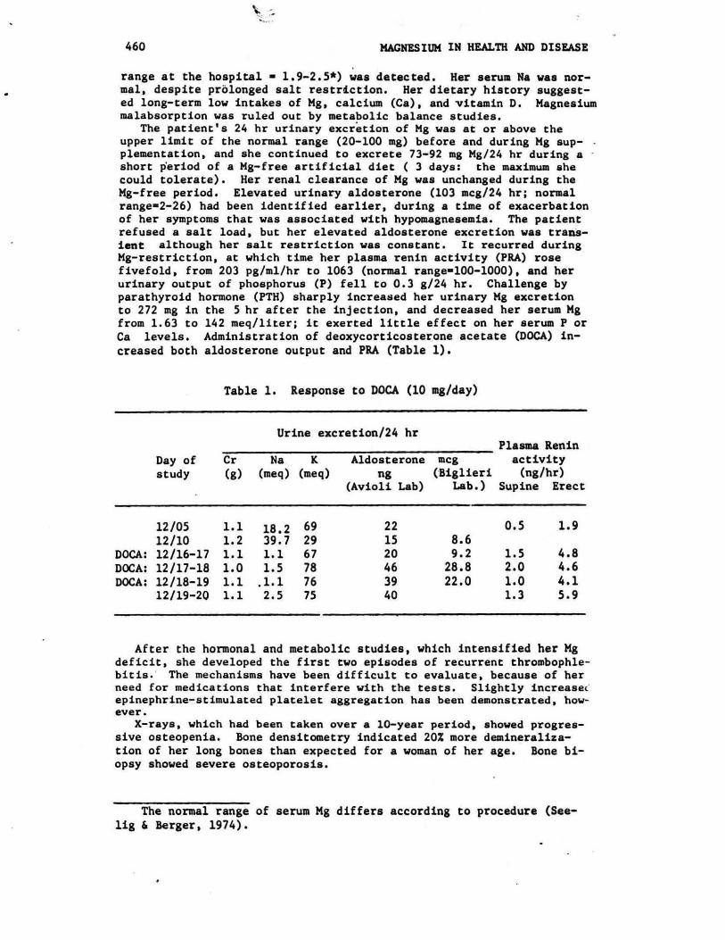
The patient ' s 24 hr urinary excretion of Mg was at or above the upper limit of the normal range (20-100 mg) before and during Hg supplementation. and she continued to excrete 73-92 mg Hg/24 hr during a short period of a Hg-free artificial diet ( 3 days: the maximum she could tolerate) . Her renal clearance of Hg was unchanged during the Mg-free period. Elevated urinary aldosterone (103 mcg/24 hr; normal range•2-26) had been identified earlier. during a time of exacerbation of her symptoms that was associated with hypomagnesemia. The patient refused a salt load. but her elevated aldosterone excretion was transient although her salt restriction was constant. It recurred during Hg-restriction. at which time her plasma renin activity (PRA) rose fivefold. from 203 pg/ml/hr to 1063 (normal range•l00-1000). and her urinary output of phosphorus (P) fell to 0.3 g/24 hr. Challenge by parathyroid hormone (PTH) sharply increased her urinary Hg excretion to 272 mg in the 5 hr after the injection. and decreased her serum Mg from 1.63 to 142 meq/liter; it exerted little effect on her serum P or Ca levels. Administration of deoxycorticosterone acetate (DOCA) increased both aldosterone output and PRA (Table 1).
Table 1. Response to DOCA (10 mg/day)
Urine excretion/24 hr Plasma Renin
Day of Cr Na K Aldosterone meg activity study (g) (meq) (meq) ng (Biglieri (ng/hr)
(Avioli Lab) Lab.) Supine Erect
12/05 1.1 18 . 2 69 22 0.5 1.9 12/10 1.2 39.7 29 15 8.6
DOCA: 12/16-17 1.1 1.1 67 20 9.2 1.5 4.8 DOCA: 12/17-18 1.0 1.5 78 46 28.8 2.0 4.6 DOCA: 12/18-19 1.1 ,1.1 76 39 22.0 1.0 4 . 1
12/19-20 1.1 2.5 75 40 1.3 5.9
After the hormonal and metabolic studies. which intensified her Hg deficit. she developed the first two episodes of recurrent thrombophlebitis . The mechanisms have been difficult to evaluate. because of her need for medica tions that interfere with the tests. Slightly increase,· epinephrine-stimulated platelet aggregation has been demonstrated. however.
X-rays. which had been taken over a lQ-year period. showed progressive osteopenia. Bone densitometry indicated 20% more demineralization of her long bones than expected for a woman of her age . Bone biopsy showed severe osteoporosis.
The normal range of serum Mg differs according to procedure (Seelig & Berger. 1974).
\.._:..
SEELIG, BERGER, AND. AVIOLI 461
DISCUSSION
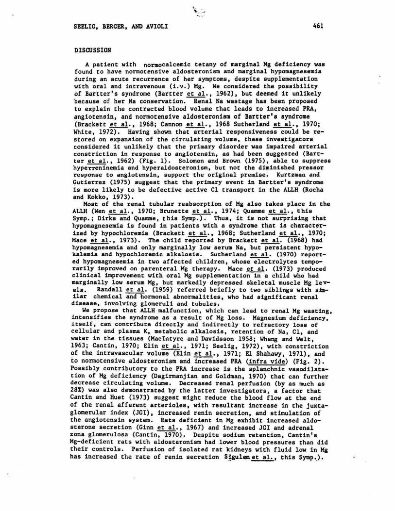
A patient with normocalcemic tetany of marginal Mg deficiency was found to have normotensive aldosteronism and marginal hypomagnesemia during an acute recurrence of her symptoms, despite supplementation with oral and intravenous (i.v.) Mg. We considered the possibility of Bartter's syndrome (Bartter et al., 1962), but deemed it unlikely because of her Na conservation.--aenal Na wastage has been proposed to explain the contracted blood volume that leads to increased PRA, angiotensin, and normotensive aldosteronism of Bartter's syndrome (Brackett et al., 1968; Cannon et al., 1968 Sutherland~., 1970; White, 1972). Having shown that arterial responsiveness could be restored on expansion of the circulating volume, these investigators considered it unlikely that the primary disorder was impaired arterial constriction in response to angiotensin, as had been suggested (Bartter et al . , 1962) (Fig. 1). Solomon and Brown (1975), able to suppress hyperreninemia and hyperaldosteronism, but not the diminished pressor response to angiotensin~ support the original premise. Kurtzman and ~utierrez (1975) suggest that the primary event in Bartter's syndrome is more likely to be defective active Cl transport in the ALLH (Rocha and Kokko, 1973).
Most of the renal tubular reabsorption of Mg also takes place in the ALLH (Wen et al . , 1970; Brunette et al., 1974 ; Quamme et al., this Symp.; Dirks and Quamme, this Sym~ Thus, it is not surprising that hypomagnesemia is found in patients with a syndrome that is characterized by hypochloremia (Brackett et al., 1968; Sutherland!!_!!., 1970; Mace et al., 1973). The child reported by Brackett et al . (1968) had hypom~emia and only marginally low serum Na, but persistent hypokalemia and hypochloremic alkalosis. Sutherland !!_!!. (1970) reported hypomagnesemia in two affected children, whose electrolytes temporarily improved on parenteral Mg therapy. Mace et al. (1973) produced clinical improvement with oral Mg supplementation in a child who had marginally low serum Mg, but markedly depressed skeletal muscle Mg levels. Randall et al. (1959) referred briefly to two siblings with similar chemical and hormonal abnormalities, who had significant renal disease, involving glomeruli and tubules.
We propose that ALLH malfunction, which can lead to renal Mg wasting, intensifies the syndrome as a result of Mg loss . Magnesium deficiency, itself, can contribute directly and indirectly to refracrory loss of cellular and plasma K, metabolic alkalosis , retention of Na, Cl, and water in the tissues (Macintyre and Davidsson 1958; Whang and Welt, 1963; Cantin, 1970; Elin et al., 1971; Seelig, 1972), with constriction of the intravascular volume (Elin et al., 1971; El Shahawy, 1971), and to normotensive aldosteronism and ~ased PRA (infra vide) (Fig. 2). Possibly contributory to the PRA increase is the splanchnic vasodilatation of Kg deficiency (Dagirmanjian and Goldman, 1970) that can further decrease circulating volume. Decreased renal perfusion (by as much as 28%) was also demonstrated by the latter investigators, a factor that Cantin and Huet (1973) suggest might reduce the blood flow at the end of the renal afferent arterioles, with resultant increase in the juxtaglomerular index (JGI), increased renin secretion, and stimulation of the angiotensin system. Rats deficient in Mg exhibit increased aldosterone secretion (Ginn et al., 1967) and increased JGI and adrenal zona glomerulosa (Cantin, 1970). Despite sodium retention, Cantin's Kg-deficient rats with aldosteronism had lower blood pressures than did their controls. Perfusion of isolated rat kidneys with fluid low in Mg has increased the rate of renin secretion S~le.m et al., this Symp .,).

HYPOKALEMIC METABOLIC ALKALOSIS HYPO'DIESIS:
NORMOTENSIVE HYPERALDOSTERONISM PRIMARY ARTERIAL UNRESPONSIVENESS TO ANGIOTENSIN*
HYPERPLASIA: HYPERTROPHY OF JUXTAGLOMERULAR APPARATUS {With "alack" Circulation, Sensed by Kidne: .. as Contraction of Effeetive Arterial Volume)
RESISTANCE TO INFUSED ANGIOTENSIN SECONDARY INCREASED JGI + t RENIN SECRETION t ANGIOTENSIN
RESULTANT t ALDOSTERONE + 1C Wastage; Alkalosis *Theory Questioned when Expansion of Extracellular Volume {by(Saline Infusion) Produced Normal Pressor Response to Angiotensin. {Albumin 11
1. IMPAIRED SODIUM RENAL TUBULAR TRANSPORT
ALTERNATIVE THEORIES
+ lNATRIURESIS HYPONATREMIA * CIRCULATING VOLUME
*But neither aldosterone-inhibitor, nor K+ supplementation corrects hypokalemia.
+ iRENIN
AL~STERONE* + K WASTAGE
2. DEFECTIVE CHLORIDE TRANSPORT - ABNORMALITY IN ALLH {Ascending Limb of Loop of Henle)
NaCl and KCl lost in urine Contracted Volume t RENIN SECRETION + t ALDOSTERONE SECRETION
* HC03 EXCRETION {proximal tubule 20 to MCS)
· Fi~ure 1. Theories to explain Bartter's Syndrome
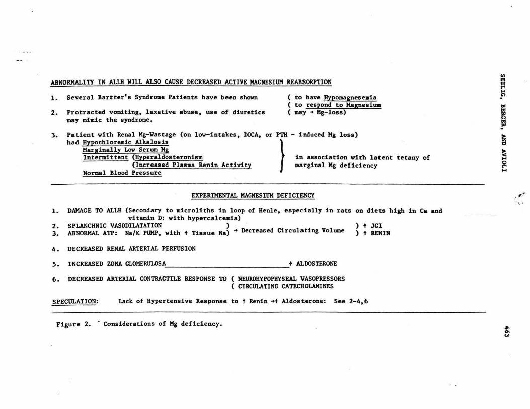
ABNORMALITY IN ALLH WILL ALSO CAUSE DECREASED ACTIVE MAGNESIUM REABSORPTION
1. Several Bartter's Syndrome Patients have been shown
2. Protracted vomiting, laxative abuse, use of diuretics may mimic the syndrome.
3. Patient with Renal Mg-Wastage (on low-intakes, DOCA, or had Hypochloremic Alkalosis
Marginally Lov Serum Mg Intermittent (Hyperaldosteronism
(Increased Plasma Renin Activity Normal Blood Pressu~e
( to have Hypomagnesemia ( to respond to Magnesium ( may + Mg-loss)
PTH- induced Mg loss)
} in association with latent tetany of marginal Mg deficiency
EXPERIMENTAL MAGNESIUM DEFICIENCY
1. DAMAGE TO ALLH (Secondary to microliths in loop of Henle, especially in rats on diets high in Ca and vitamin D: with hypercalcemia)
2. SPLANCHNIC VASODILATATION ) ) t .JGI l. ABNORMAL ATP: Na/K PUMP • with t Tissue Na) + Decreased Circulating Volume ) t RENIN
4. DECREASED RENAL ARTERIAL PERFUSION
5. INCREASED ZONA GLOMERULOSA ______________ t ALDOSTERONE
6. DECREASED ARTERIAL CONTRACTILE RESPONSE TO ( NEUROHYPOPHYSEAL VASOPRESSORS ( CIRCULATING CATECHOLAMINES
SPECULATION: Lack of Hypertensive Response to t Renin +t Aldosterone: See 2-4,6
Figure 2. · Considerations of Mg deficiency.
, ,~
' I I
464 MAGNESIUM IN HEAL'nl AND DISEAS
However, Kg-deficient dogs that were hypocalcemic showed no significant changes in JGI in one study (Rojo-Ortega et al., ~his Symp·.), whereas other Kg-deficient dogs had increased PRA and aldosterone secretion (El Shahawy, 1971).
Rats that are Kg-deficient, but have a high Ca/Kg dietary ratio, . develop Ca microlith& in Henle's loop, with resultant damage to the proximal tubules and to the ALLH (Oliver et al., 1966). Whang et al . (1973) have suggested that such damage, p~he lessened renal perfusion of Kg deficiency (supra vide), can be responsible for thereversible renal failure and aldosteronism of Mg 4eficiency in man. Re nal and hormonal evaluation of patients with primary or secondary Mg malabsorption, with and without diets rich in vitamin D or Ca, is indicated. That the rat studies are relevant to man is indicated by th observation of luminal Ca deposits in the proximal renal tubules and in the ALLH of an infant with hypocalcemia and neuromuscular irritability, who had been treated with Ca infusions and high dosage vitami D, before hypomagnesemia was recognized (Vainsel et al., 1970). It has been noted that magnesium-wasting develops in patients with renal tubular disease (Randall et al., 1959).
Whether renal Kg wastas;:-caused by an ALLH defect, is sufficient to impair response to angiotensin remains to be investigated . Invitro studies have shown that hypophyseal hormones are Hg-dependenc-~ that, in Kg-free or Kg-low media, arterial smooth muscle exhibits impaired contractile response (Somlyo et al., 1966, 1967; Altura. 1974, 1975; Altura and Altura. this Symp.)-.--
Our patient's preference for foods low in Mg, Ca. and vitamin D suggests an initiating nutritional inadequacy. The fact that her multiple complaints began only after total hysterectomy, which undoubtedly enhanced mobilization of Ca from her bones, may have allowed for a high Ca/Kg ratio in the filtered urine. Perhaps this resulted in sucJ· microlith& in the loop of Henle as have damaged the ALLH in rats (su= pra vide). Her generalized pruritus and need for high dosage antihistaminic therapy are in accord with Mg deficiency, mast cell degranulation, and histamine release having been reported in Kg-deficient rats (Belanger et al., 1957; Bois et al.~ 1963; Hungerford, 1964). Her Na retention and peripheral edema further support the likelihood that Hg deficiency participates in the etiology of her syndrome. The extent of her osteoporosis may reflect intensification of menopausal bone lesions by those caused by Mg deficiency. Magnesium-depleted animals, not excessively loaded with Ca, develop osteoporosis (Morris and O'Del 1961; Trowbridge and Seltzer. 1967; Smith and Nisbet, 1968). In addition, a child with renal Hg wastage and carpopedal spasm has recently been reported to have osteoporosis (Booth and Johanson, 1974), and earlier another child, also with carpopedal spasm and presumptive renal tubular defect in Kg absorption, was reported with osteochondritis (Klingberg, 1970).
The depression of our patient's urinary output of P, during a Hgfree period, suggests that release of PTH may have been suppressed at that time (Anast, 1977). That her target organs could respond to PTH is suggested by her low-normal serum P, and possibly by her osteopenia if one accepts the premise that estrogen deficiency allows for relativ. PTH excess (Seelig and Lehr, 1971). Her magnesiuresis, in response to PTH loading, might be relevant to the development of symptoms of hypomagnesemia (despite essentially normal blood levels) in kidney disease patients with magnesium wasting and secondary hyperparathyroidism (Randall et al., 1959).
A more recent complication, that points towards Kg deficiency as

SEELIG. BERGER. AND AVIOLI
... -'~ .. _
465
critical in our patient. is recurrent thrombophlebitis. The first two episodes occurred shortly after hormonal (desoxycorticosterone acetate) and nutritional challenges (metabolic balance studies with low Mg intakes). that intensified her Mg deficit . Such episodes have been reported in other patients with latent tetany of Mg deficiency, and have responded to Mg therapy (Durlach, 1967 a,b; DuPont et al., 1969). Magnesium administration has also been shown to lengthen thrombin generation time in man (Huntsman et al •• 1960) and has counteracted the shortened coagulation and prothrombin times caused by atherogenic diets in rats and dogs (Szelenyi et al., 1967). Magnesium also participates in platelet function (Elin. this Symp.).
Eliciting normotensive aldosteronism by measures that intensified Mg loss, recalls Conn's introduction to the metabolic findings of primary (hypertensive) aldosteronism (Conn, 1955). He commented on the hypomagnesemic form of the disease, described by Mader and Iseri (1955), and suggested that it might be an early form of the disorder,associated with hypokalemia, hypercapnea, Na retention with normal plasma lev-els. and moderate hypertension and tetany. Whether our patient (and
those with more severe hypomagnesemia caused by renal tubular wastage) (Freeman and Pearson. 1966; Gitelman et al., 1966; Klingberg, 1970; Booth and Johanson, 1974; and Paunier and Sizonenko . 1976) represents another form of the disease, in which Mg deficiency prevents the hypertensive response and causes additional complications. requires elucidation.
SUMMARY
A post-menopausal woman with multiple complaints and latent therapy of marginal Mg deficiency was found to have renal Mg wasting, even on marked restriction of Mg intake. Intermittent aldosteronism and 4 to 5-fold increased PRA was manifest on Mg restriction or loss, but not on Mg supplementation, despite which she was either normotensive or slightly hypotensive. These findings, plus her high requirement for antihistamine therapy, resemble those of experimental Mg deficiency·. She also had rapidly progressive osteoporosis. We speculate that the patient had long-term marginal Mg deficiency, that may have contributed to a renal defect, perhaps of the ascending limb of the loop of Henle, with resultant decreased tubular reabsorption of Cl and Mg. Sodium retention may have resulted in normotensive aldosteronism via increased intracellular and interstitial fluid volume, rather than by hypervolemia. It is also possible that chronic Mg deficiency contributed to impaired vasopressor response to Kg-dependent neurohypophyseal vasopressors. and to the progressive osteopenia.
ACKNOWLEDGMENTS
Appreciation is expressed to Drs. Louis Zeitz and Charles Colbert for their bone densitometry studies; to Drs. Jerome Lowenstein and E.G. Biglieri for their PRA and aldosterone determinations, respectively; and to Drs. Babette Weksler and Henrietta Lackner for their platelet studies.
466 MAGNESIUM IN HEALTH AND DISEASI
REFERENCES
Altura, B.M. Magnesium-neurohypophyseal hormone interactions in .· contraction of vascular smooth muscle. Am. J. Physiol. 223, 1615-162l (1974) .
Altura, B.M. Neurohypophyseal hormones and analogues: Magnesium dependence and contraction of arterial smooth muscle. Proc. Soc. Exp Biol. Med. 148, 1031-1037 (1975).
Altura, B.T. and Altura, B. M. Magnesium in Health and Disease (Prl 2nd Internat. Symp . on Magnesium, Montreal), this Symp.
Anast, C.S. Interrelationship of magnesium and calcium-regulating hormones, in Nutritional Imbalances in Infant and Adult Disease. Mineral , Vitamin D, and "Cholesterol. M.S. Seelig, ed. Spectrum Publ. New York, (1977), pp. 103-126.
Bartter, ·r.c., Pronove, P., Gill, J., and MacC&rdle, a.c. Hyperplasia of the juxta-glomerular complex with hyperaldosteronism and hypokalemic alkalosis. Am. J. Med. 38, 811-828 (1962).
Belanger, L.F., Van Erkel, C.A., and Jakerow, A. Behavior of the dermal mast cells in magnesium-deficient rata. Science 126, 29-30 {1957).
Bois, P., Caxon, A., and Beaulnes, A. Hiatamine-liberatin& effect of magnesium deficiency in rats. Nature 197, 501-502 {1963).
Booth, B.E. and Johanson, A. Hypomagnesemia due to renal tubular defect in reabsorption of magnesium. J, Pediatr. 84, 350-354 (1974).
Brackett, N.C., Jr., Koppe, H., Randall, R.E., Jr., and Nixon, W.P. Hyperplasia of the juxta-glomerular complex with secondary aldosteroniaawithout hypertension (Bartter's syndrome). Am. J. Med. 44, 803-819 (1968).
Brunette, M.G., Vigneault, N., and Carriere, S. Micropuncture stud of magnesium transport along the nephron in the young rat. Am. J. Phy siol. 227, 891-896 (1974).
Cannon, P.J., Leeming, J.M. , Sommers, S.C., Winters, R.M., and Laragh, J.H. Juxta-glomerular cell hyperplasia and secondary hyperaldosteronism (Bartter's syndrome): A re-evaluation of the pathphysi-ology, Medicine 47, 107-131 (1968). 1
Cantin, M. Relationship of juxtaglomerular apparatus and adrenal cortex to biochemical and extracellular fluid volume changes in magnesium deficiency. Lab. Invest • . 22, 558-568 (1970).
Cantin, M. and Huet, M. Histochemistry and ultrastructure· of the juxtaglomerular apparatus in magnesium deficient rata. Can. J. Physio Pharmacal. 51, 835-844 (1973).
Conn, J.W. Primary aldosteronism. A new clinical syndrome. ~ Clin. Hed. 45, 3-17 (1955).
Dagirmanjian, R. and Goldman, H. Magnesium deficiency and distribu tion of blood in the rat. Am. J. Physiol. 218, 1464-1467 (1970).
Dirks, J.H. and Quamme, G.A. this Symp . , p. 363-371. DuPont, B., Pony, J.C., LeBihan, G. and Leborgn~P. Maladie phlebo
thrombosante et magnesium. Sem. Hop. Paris 48, 3048-3054 (1969). Durlach, J. Le role antithrombosique physiologique du magnesium.
A propos d'une maladie par defici~ magnesium. Coeur Hed. Interne 6, 213-232 (1967).
Durlach, J, Magnesium deficiency thrombosis. Lancet 1, 1382 (1967 Elin, R.J. this Symp., p. 113-124. Elin, R.J., Armstrong, W.D. ,and Singer ,L. Body fluid electrolyte con
position of chronically magnesium deficient and control rats. Am. J. Physiol. 220, 543-548 (1971).
El Shahawy, M. The role of magnesium in thyrocardiac disorders.
SEELIG, BERGER, AND AVIOLI 467
M.S. Thesis. Univ. of Minnesota (1971). Freeman, R. H. , and Pearson, E. Hypomagnesemia of unknown etiology.
Am. J . Hed. 41, 645- 656 (1966). Ginn, H. E., Cade, R .• MacCallum, T., and Fregley, H. Aldosterone
secretions in magnesium deficient rats. Endocrinology 80, 969-971 (1967) .
Gitelman, H.J., Graham, J.B., and Welt, L. G. A new familial disorder characterized by -hypokalemia and hypomagnesemia. Trans. Assoc. Am. Physicians 79, 221- 235 (1966).
Hungerford, G.F. Role of histamine in producing the eosinophilia of magnesium deficiency. Proc. Soc . Exp. Biol. Hed. 115, 182-185 (1964).
Huntsman , R.G., Hurn, A.L. , and Lehman, H. Observations on the effects of magnesium on blood coagulation. J . Clin. Pathol. 13, 99-101 (1960).
Klingberg, W.G. Idiopathic hypomagnesemia and osteochondritis. Pediatr . Res . 4, 452 (1970).
Kurtzman, N.A. and Gutierrez, L. F. Hypothesis. The pathophysiology of Bartter's syndrome. J. Am. Med. Assoc. 234, 758-759 (1975).
Mace, J.W., ·Hambidge, K.H •• Cotlin, R. W. , DuBois, R.S., Solomons, G.S . , and Katz, F.H. Magnesium supplementation in Bartter's syndrome . Arch. Dis. Child. 48, 485-487 (1973) .
Hcinfyre-~ ·I. ·and Davl.asson ,-"'0. ··-Tlieproduction of secondary potassium depletion, sodium retention, nephrocalcinosis and hypercalcemia by magnesium deficiency. Biochem. J. 70, 456-462 (1958).
Hader, I .W. and Iser~~.T. Spontaneous hyperpotassemia, hypomagnesemia, alkalosis and tetany due to hypersecretion of cortisone-like mineralocorticoid . Am. J. Med. 19, 976-988 (1955).
Morris, E.R. and O'Dell, B.L. Magnesium deficiency in guinea pig. Mineral composition of tissues and distribution of acid-soluble phosphorus. J . Nutr. 75, 77-85 (1961).
Oliver, J., MacDowell, H., Whang , R. , and Welt, L.G. The renal lesions of electrolyte imbalance . IV. The intranephric calculosis of experimental magnesium depletion. J. Exp . Hed. 124, 263-277 (1966) .
Paunier, L. and Sizonenko, P. C. Asymptomatic chronic hypomagnesemia and hypokalemia in a child : Cell membrane disease? J. Pediatr. 88, 51-55 (1976).
Quamme, G.A., Roinel, N., Wong, N.L. H. , De Rouffignac, c .• Morel, F •• and Dirks, J.H. this Symp., p. 373-378.
Randall, R.E . , Jr., Rossmeisl , E.C . , and Bleifer, K.H. Magnesium depletion in man. Ann. Intern. Hed. 50, 257-287 (1959) .
Rocha, A. S. and Kokko, J.P. Sodium chloride and water transport in the medullary thick ascending limb ofHenle. J.Clin. Invest . 52 612~623
(1973). . • Rojo-Ortega, J .H., Brecht, H. H., Constantopoulos, G., Ganten, D. and
Genest , J •• this symp . p . 415-420. Seelig, M.S. Myocardial loss of functional magnesium. I. Effects
on mitochondrial integrity and potassium retention in myocardiology, in Recent Advances in Studies on Cardiac Structure and Metabolism. E. Bajusz and G. Rona, eds. University Park Press, Baltimore, Hd., (1972) pp . 615-625.
Seelig, ·H. S. and Berger, A. R. Range of normal serum magnesium values. N. Eng. J . Hed. 290 , 975-975 (1974).
Seelig, M.S., Berger, A.R. and Spielholz, N. Latent tetany and anxiety, marginal magnesium -deficit and normocalcemia. Dis . Nerv. ~stem 36,461-465 (1975).
468
....... -.....
MAGNESIUM IN HEAL'lll AND DISEASE
Seelig, M.s·. a·nd Lehr, D. Effects of estrogen on tissue magnesium content; possible influence on cardtovascular disease, in 1st Internat. Symp. on Magnesium. Vittel, France (1971) pp. 249-255.
Sigulem, D., Saito, H., Ajzen, H., Ramos, O.L. This Symp. ,421-426. Smith, B. S.W., and Nisbet, D.I. Magnesium deficiency in rat. d·
Comp. Pathol. 78, 149-159 (1968). Solomon, R.J. and Brown, R.S. Bartter's syndrome. New insights
into pathogenesis and treatment. Am. J. Med. 59, 575-583 (1975). Somlyo, A.V., Woo, C.Y., and Somlyo, A.P. Effect of magnesium on
posterior pituitary hormone action on vascular smooth muscle. ~· Physiol. 210, 704-714 (1966).
Somlyo, A.P., Somlyo, A.V., and Woo, C.Y. Neurohypophyseal peptide interaction with magnesium in avian vascular smooth muscle. J. Physiol. 192, 657-668 (1967).
Sutherland, L.E., Hartroft, P., Balis, J.U., Bailey, J.D., and Lynch, M.J . Bartter's syndrome. A report of four cases, including three in one sibship, with comparative evaluation of the juxtaglomerular apparatuses and glomeruli. Acta Paediat. Scand. Suppl. 201, 1-24 (1970).
Szelenyi, I., Rigo, J., Ahmed, B.O., and Sos, J. The role of magnesium in blood coagulation. Thromb. Diath. Haemorrh. 18, 626-633 (1967).
Trowbridge, H.O. and Seltzer, J.L. Formation of dentin and bone matrix in magnesium-deficient rats. J. Periodont. Res. 2, 147-153 (1967).
Vainsel, M., Vandevelde, G., Smulders, J., Vosters, M., Hubain, P., and Loeb, H. Tetany due to hypomagnesemia with secondary hypocalcemia. Arch. Dis. Child. 45, 254-258 (1970).
Wen, S.F., Evanson, N.L., and ~irks, J.H. Micropuncture study of renal tubular magnesium transport in proximal and distal tubule of the dog. Am. J . Physiol. 219, 570-576 (1970).
Whang, R. and Welt, L.G. Observations in experimental magnesium depletion. J. Clin. Invest. 42, 305-313 (1963).
Whang, R., Morosi, H.J., Rodgers, D., and Reyes, R. The influence of sustained magnesium deficiency on muscle potassium repletion. J. Lab. Clin. Med. 70, 895-902 (1967).
Whang, R., Ryan, M.P., and Aikawa, J.K. Magnesium deficiency. A cause of reversible renal failure. Lancet 1, 135-136 (1973).
White, M.G. Bartter's sundrome. A manifestation of renal tubular defects. Arch. Intern. Med. 129, 41-47 (1972).