Embed Size (px)
Citation preview


Causes of valve disease
Valve regurgitation
* Congenital *Acute rheumatic carditis *Chronic rhe. Carditis
* I E *Syphlitic aortitis *Dilated Valve ring
* Senile degeneration e.g. dilated CMP
* Traumatic valve rupture
* Damage to chordae and
papillary muscles e.g. M I
Valve stenosis
* Congenital * Senile degeneration
* Rheumatic carditis


Aetiology and pathophysiologyAetiology and pathophysiology* * Almost rheumatic in origin. 2/3 are FemalesAlmost rheumatic in origin. 2/3 are Females
Pure or predominant MS 40% of all RVDPure or predominant MS 40% of all RVD
**Heavy calcification in elderly may produceHeavy calcification in elderly may produce a form of mitral stenosisa form of mitral stenosis..
**Congenital MS { rare }Congenital MS { rare }..
_ _ Mitral vave orifice progressively diminish byMitral vave orifice progressively diminish by fibrosis, calcification of leaflets and fusion 0f cusps fibrosis, calcification of leaflets and fusion 0f cusps
and subvalvular apparatusand subvalvular apparatus . ._ _ LA dilate…LA pressure rises …pulmonary congestion LA dilate…LA pressure rises …pulmonary congestion
occurs … later pulmonary hypertension ….RVFoccurs … later pulmonary hypertension ….RVF_ _ Atrial fibrillation occurs in most patients with risk ofAtrial fibrillation occurs in most patients with risk of
thromboembolismthromboembolism.. >_ >_ 20%20% remain in sinus rhythm with small fibrotic LA remain in sinus rhythm with small fibrotic LA
andand severe PHsevere PH

* * Normal valve areaNormal valve area: 4-6 cm: 4-6 cm22
* * Mild. mitral stenosisMild. mitral stenosis _ _ MVA 1.5-2.5 cmMVA 1.5-2.5 cm22
_ _ Minimal symptomsMinimal symptoms
* * Mod. mitral stenosisMod. mitral stenosis _ _ MVA 1.0-1.5 cmMVA 1.0-1.5 cm2 2 usually does not usually does not
produceproduce _ Symptoms at rest _ Symptoms at rest
* * Severe mitral stenosisSevere mitral stenosis _ _ MVA MVA < < 1.0 cm21.0 cm2
Pathophysiology of MV:

JVP

**Dyspnia {pulmonary congestion }Dyspnia {pulmonary congestion }
** Fatigue { low COP }Fatigue { low COP }
**Oedema, ascitis {RVF }Oedema, ascitis {RVF }
**Palpitation { AF }Palpitation { AF }
**Haemoptysis {pulmonary congestion, PE }Haemoptysis {pulmonary congestion, PE }
**Cough {pulmonary congestion }Cough {pulmonary congestion }
**Chest pain {PH }Chest pain {PH }
**Symptoms of thromboembolic complication { e.g. stroke, Symptoms of thromboembolic complication { e.g. stroke, ischaemic limb} ischaemic limb}
Clinical features
Symptoms

Signs:
* Atrial fibrillation
* Mitral facies , tapping apex {Palpable 1st HS}
* Auscultation : Loud first HS, Opening snap
Mid-diastolic murmer, presyst. Accentuation
* Signs of raised pulmonary capillary pressure:
_ Crepitations, _Pulmonary oedema, Effusion
* Signs of pulmonary hypertension:
RV heave, Loud P2
Clinical features cont.

Physical examinationPhysical examination
Auscultation

Common Murmurs and Common Murmurs and Timing Timing (click on murmur to play)(click on murmur to play)
Systolic MurmursSystolic MurmursAortic stenosisAortic stenosis
Mitral insufficiencyMitral insufficiencyMitral valve prolapseMitral valve prolapse
Tricuspid insufficiencyTricuspid insufficiency
Diastolic MurmursDiastolic MurmursAortic insufficiencyAortic insufficiency
Mitral stenosisMitral stenosis
S1 S2 S1

IInvestigationsnvestigations
* *ECGECG: : LA hypertrophy , RVH , AFLA hypertrophy , RVH , AF
* * Chest X-rayChest X-ray: : Enlarged LAEnlarged LA
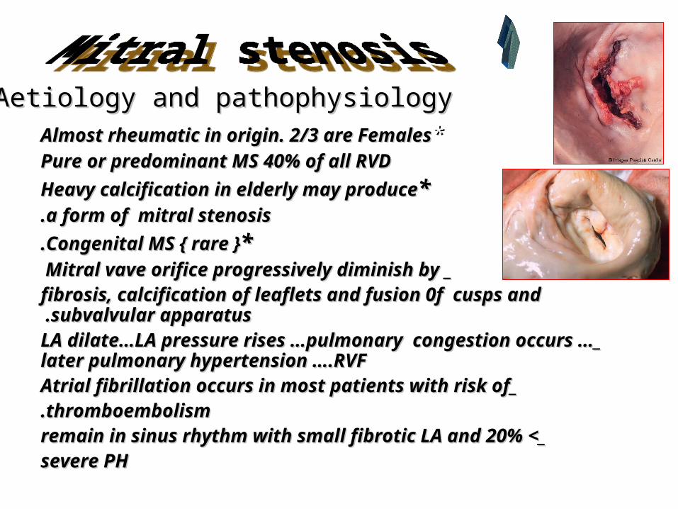
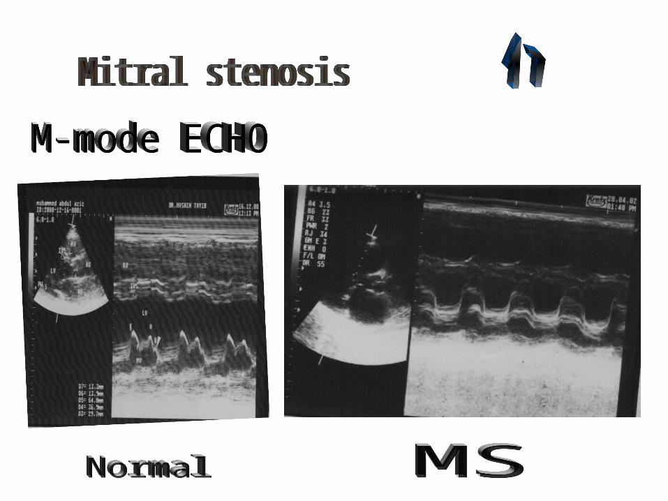
Pulmonary venous congestionPulmonary venous congestion * * ECHOECHO : :
Thick immobile cuspsThick immobile cusps Reduced valve areaReduced valve area
Reduced rate of LV diastolic fillingReduced rate of LV diastolic filling * * DopplerDoppler: :
_ _ PG across MV _ Pulmonary artery pressurePG across MV _ Pulmonary artery pressure * * Cardiac catheterizationCardiac catheterization: :
_ _ Pulmonary wedge pressurePulmonary wedge pressure _ _ PG between LA and LVPG between LA and LV

ECG MS+ AF

Chest X-ray PA view

Chest X-ray lat. View +Barium swallow
.
AO
LA
MV

2-D mode Active ECHO.

Pw Dopler

Color dopler

* * Progressive, lifelong diseaseProgressive, lifelong disease , ,
* * usually slow & stable in the early yearsusually slow & stable in the early years . .
* * Progressive acceleration in the later yearsProgressive acceleration in the later years
* * 20-4020-40 year latency from rheumatic fever to year latency from rheumatic fever to symptom onset symptom onset..
* * Additional 10 years before disabling Additional 10 years before disabling symptoms symptoms
Natural history

ManagementManagement: :
** Medical managementMedical management: : _ _ Digoxin for AF +beta blocker or calcium antagonistDigoxin for AF +beta blocker or calcium antagonist
_ _ Diuretics for pulmonary congestionDiuretics for pulmonary congestion_ _ Anticoagulant to reduce risk of systemic emboliAnticoagulant to reduce risk of systemic emboli
_ _ Antibiotic prophylaxis against IEAntibiotic prophylaxis against IE **Mitral balloon valvoplastyMitral balloon valvoplasty: :
_ _ Significant symptomsSignificant symptoms_ _ Isolated MSIsolated MS
_ _ No or trivial MRNo or trivial MR_ _ Mobile noncalcific valve /subvalvular apparatus on ECHOMobile noncalcific valve /subvalvular apparatus on ECHO
_ _ LA free of thrombusLA free of thrombus
** Mitral valve surgeryMitral valve surgery: : _ _ Closed mitral valvotomyClosed mitral valvotomy
_ _ Open mitral valvotomyOpen mitral valvotomy_ _ Mitral valve replacementMitral valve replacement
* * MechanicalMechanical * * BioprosthesisBioprosthesis

Mechanical valves