Embed Size (px)
Citation preview

CASEBOOK 2016
The Royal Australianand New ZealandCollege of Obstetriciansand Gynaecologists


CASEBOOK 2016
The Royal Australianand New ZealandCollege of Obstetriciansand Gynaecologists

DISCLAIMER
All identifying information has been omitted to preserve anonymity.
© CLINICAL EXCELLENCE COMMISSION 2017
All rights are reserved. In keeping with the NSW Government’s commitment to encouraging the availability, dissemination and exchange of information (and subject to the operation of the Copyright Act 1968), you are welcome to reproduce the information which appears in this publication, as long as the user of the information agrees to:
~ use the document for information only
~ save or print a single copy for personal use only and not to reproduce any major extract or the entire document except as permitted under Copyright Act 1968 (as amended) without the prior written permission of the State of New South Wales
~ acknowledge the source of any selected passage, table diagram or other extract reproduced
~ not make any charge for providing the information to another person or organisation without the prior written consent of the State of New South Wales and payment of an agreed copyright fee
~ not modify the Information without the express prior written permission of the State of New South Wales
~ include this copyright notice in any copy made: © Copyright – Clinical Excellence Commission for and on behalf of the Crown in right of the State of New South Wales.
NATIONAL LIBRARY OF AUSTRALIA CATALOGUING-IN PUBLICATION ENTRY
Title: Collaborating Hospitals’ Audit of Surgical Mortality (CHASM) Casebook 2016
ISBN: 2201-2990 SHPN: (CEC) 170541
Suggested citationClinical Excellence Commission (CEC) 2017. Collaborating Hospitals’ Audit of Surgical Mortality (CHASM) Casebook 2016. Sydney: CEC
CLINICAL EXCELLENCE COMMISSION
Board Chair: A/Prof Brian McCaughan AM Chief Executive: Ms Carrie Marr CHASM Chair: Prof Peter Zelas OAM
Any enquiries about, or comments on, this publication should be directed to: Manager, Special Committees Clinical Excellence Commission Locked Bag 8 HAYMARKET NSW 1240
Phone: (02) 9269 5530
Email: [email protected]
CHASM CASEBOOK 2016 | PAGE 1 CLINICAL EXCELLENCE COMMISSION
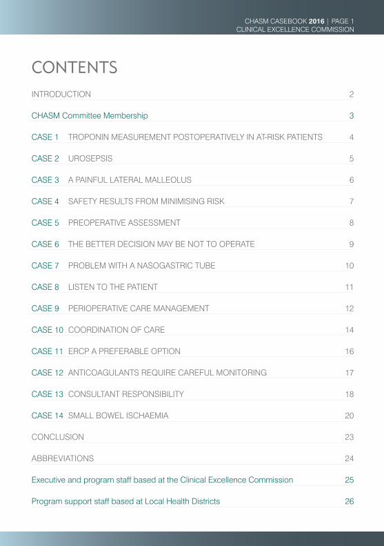
CONTENTSINTRODUCTION 2
CHASM Committee Membership 3
CASE 1 TROPONIN MEASUREMENT POSTOPERATIVELY IN AT-RISK PATIENTS 4
CASE 2 UROSEPSIS 5
CASE 3 A PAINFUL LATERAL MALLEOLUS 6
CASE 4 SAFETY RESULTS FROM MINIMISING RISK 7
CASE 5 PREOPERATIVE ASSESSMENT 8
CASE 6 THE BETTER DECISION MAY BE NOT TO OPERATE 9
CASE 7 PROBLEM WITH A NASOGASTRIC TUBE 10
CASE 8 LISTEN TO THE PATIENT 11
CASE 9 PERIOPERATIVE CARE MANAGEMENT 12
CASE 10 COORDINATION OF CARE 14
CASE 11 ERCP A PREFERABLE OPTION 16
CASE 12 ANTICOAGULANTS REQUIRE CAREFUL MONITORING 17
CASE 13 CONSULTANT RESPONSIBILITY 18
CASE 14 SMALL BOWEL ISCHAEMIA 20
CONCLUSION 23
ABBREVIATIONS 24
Executive and program staff based at the Clinical Excellence Commission 25
Program support staff based at Local Health Districts 26
PAGE 2
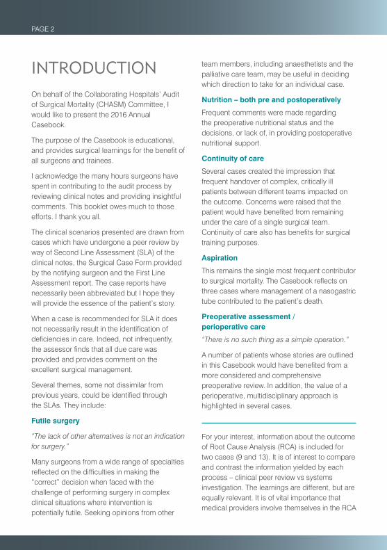
INTRODUCTIONOn behalf of the Collaborating Hospitals’ Audit of Surgical Mortality (CHASM) Committee, I would like to present the 2016 Annual Casebook.
The purpose of the Casebook is educational, and provides surgical learnings for the benefit of all surgeons and trainees.
I acknowledge the many hours surgeons have spent in contributing to the audit process by reviewing clinical notes and providing insightful comments. This booklet owes much to those efforts. I thank you all.
The clinical scenarios presented are drawn from cases which have undergone a peer review by way of Second Line Assessment (SLA) of the clinical notes, the Surgical Case Form provided by the notifying surgeon and the First Line Assessment report. The case reports have necessarily been abbreviated but I hope they will provide the essence of the patient’s story.
When a case is recommended for SLA it does not necessarily result in the identification of deficiencies in care. Indeed, not infrequently, the assessor finds that all due care was provided and provides comment on the excellent surgical management.
Several themes, some not dissimilar from previous years, could be identified through the SLAs. They include:
Futile surgery
“The lack of other alternatives is not an indication for surgery.”
Many surgeons from a wide range of specialties reflected on the difficulties in making the “correct” decision when faced with the challenge of performing surgery in complex clinical situations where intervention is potentially futile. Seeking opinions from other
team members, including anaesthetists and the palliative care team, may be useful in deciding which direction to take for an individual case.
Nutrition – both pre and postoperatively
Frequent comments were made regarding the preoperative nutritional status and the decisions, or lack of, in providing postoperative nutritional support.
Continuity of care
Several cases created the impression that frequent handover of complex, critically ill patients between different teams impacted on the outcome. Concerns were raised that the patient would have benefited from remaining under the care of a single surgical team. Continuity of care also has benefits for surgical training purposes.
Aspiration
This remains the single most frequent contributor to surgical mortality. The Casebook reflects on three cases where management of a nasogastric tube contributed to the patient’s death.
Preoperative assessment / perioperative care
“There is no such thing as a simple operation.”
A number of patients whose stories are outlined in this Casebook would have benefited from a more considered and comprehensive preoperative review. In addition, the value of a perioperative, multidisciplinary approach is highlighted in several cases.
For your interest, information about the outcome of Root Cause Analysis (RCA) is included for two cases (9 and 13). It is of interest to compare and contrast the information yielded by each process – clinical peer review vs systems investigation. The learnings are different, but are equally relevant. It is of vital importance that medical providers involve themselves in the RCA
CHASM CASEBOOK 2016 | PAGE 3 CLINICAL EXCELLENCE COMMISSION
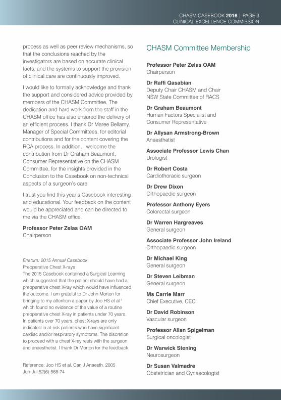
process as well as peer review mechanisms, so that the conclusions reached by the investigators are based on accurate clinical facts, and the systems to support the provision of clinical care are continuously improved.
I would like to formally acknowledge and thank the support and considered advice provided by members of the CHASM Committee. The dedication and hard work from the staff in the CHASM office has also ensured the delivery of an efficient process. I thank Dr Maree Bellamy, Manager of Special Committees, for editorial contributions and for the content covering the RCA process. In addition, I welcome the contribution from Dr Graham Beaumont, Consumer Representative on the CHASM Committee, for the insights provided in the Conclusion to the Casebook on non-technical aspects of a surgeon’s care.
I trust you find this year’s Casebook interesting and educational. Your feedback on the content would be appreciated and can be directed to me via the CHASM office.
Professor Peter Zelas OAM Chairperson
Erratum: 2015 Annual Casebook Preoperative Chest X-rays The 2015 Casebook contained a Surgical Learning which suggested that the patient should have had a preoperative chest X-ray which would have influenced the outcome. I am grateful to Dr John Morton for bringing to my attention a paper by Joo HS et al 1 which found no evidence of the value of a routine preoperative chest X-ray in patients under 70 years. In patients over 70 years, chest X-rays are only indicated in at-risk patients who have significant cardiac and/or respiratory symptoms. The discretion to proceed with a chest X-ray rests with the surgeon and anaesthetist. I thank Dr Morton for the feedback.
Reference: Joo HS et al, Can J Anaesth. 2005 Jun-Jul;52(6):568-74
CHASM Committee Membership
Professor Peter Zelas OAM Chairperson
Dr Raffi Qasabian Deputy Chair CHASM and Chair NSW State Committee of RACS
Dr Graham Beaumont Human Factors Specialist and Consumer Representative
Dr Allysan Armstrong-Brown Anaesthetist
Associate Professor Lewis Chan Urologist
Dr Robert Costa Cardiothoracic surgeon
Dr Drew Dixon Orthopaedic surgeon
Professor Anthony Eyers Colorectal surgeon
Dr Warren Hargreaves General surgeon
Associate Professor John Ireland Orthopaedic surgeon
Dr Michael King General surgeon
Dr Steven Leibman General surgeon
Ms Carrie Marr Chief Executive, CEC
Dr David Robinson Vascular surgeon
Professor Allan Spigelman Surgical oncologist
Dr Warwick Stening Neurosurgeon
Dr Susan Valmadre Obstetrician and Gynaecologist
PAGE 4
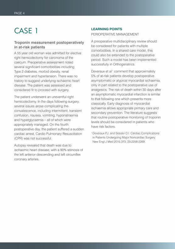
CASE 1Troponin measurement postoperatively in at-risk patients
A 55 year old woman was admitted for elective right hemicolectomy for carcinoma of the caecum. Preoperative assessment noted several significant comorbidities including Type 2 diabetes, morbid obesity, renal impairment and hypertension. There was no history to suggest underlying ischaemic heart disease. The patient was assessed and considered fit to proceed with surgery.
The patient underwent an uneventful right hemicolectomy. In the days following surgery, several issues arose complicating the convalescence, including intermittent, transient confusion, nausea, vomiting, hyponatraemia and hyperglycaemia – all of which were appropriately managed. On the fourth postoperative day, the patient suffered a sudden cardiac arrest. Cardio Pulmonary Resuscitation (CPR) was not successful.
Autopsy revealed that death was due to ischaemic heart disease, with a 90% stenosis of the left anterior descending and left circumflex coronary arteries.
LEARNING POINTS
PERIOPERATIVE MANAGEMENT
A preoperative multidisciplinary review should be considered for patients with multiple comorbidities. In a shared care model, this could also be extended to the postoperative period. Such a model has been implemented successfully in Orthogeriatrics.
Deveraux et al 1 comment that approximately 5% of at-risk patients develop postoperative asymptomatic or atypical myocardial ischaemia, only in part related to the postoperative use of analgesics. The risk of death within 30 days after an asymptomatic myocardial infarction is similar to that following one which presents more classically. Early diagnosis of myocardial ischaemia allows appropriate primary care and secondary prevention. The literature suggests that routine postoperative monitoring of troponin levels should be considered in patients who have risk factors.
1 Deveraux P.J. and Sessler D.I. Cardiac Complications in Patients Undergoing Major Noncardiac Surgery. New Engl J Med 2015;373; 23:2258-2269.

CHASM CASEBOOK 2016 | PAGE 5 CLINICAL EXCELLENCE COMMISSION
CASE 2Urosepsis
A 90 year old man whose comorbidities included diabetes, hypertension, atrial fibrillation and previous resection of colonic carcinoma, attended a urologist for investigation of macro-haematuria. The patient was admitted for a flexible cystoscopy under light sedation ten days later. He did not have a preoperative micro-urine analysis.
A long urethral stricture was found and dilated. At the time, it was noted that the patient had offensive urine and 80mg of gentamicin was administered.
In the recovery room, the patient became rapidly unwell with pyrexia, hypotension and reduced Glasgow Coma Scale (GCS). Ceftriaxone was commenced and a further dose of gentamicin administered. He was admitted to the Intensive Care Unit (ICU) where his condition deteriorated, despite treatment with fluid resuscitation, antibiotics and inotropes. He died two days following the cystoscopy.
LEARNING POINTS
The age of the patient and his comorbidities probably contributed significantly to the rapid deterioration in response to the urosepsis.
Patients undergoing a cystoscopy should have a preoperative urine culture, and this result should be followed up to confirm sterility. If the urine is infected, it should be treated appropriately and the procedure deferred until urinary sterility has been assured.
In this case, the Second Line Assessor suggested that, as a minimum on admission, nursing staff should perform a urine dipstick analysis for each patient having a cystoscopy. The procedure should be delayed if nitrites are positive, and a formal micro-urine performed and followed up. If nitrites are negative, the procedure could proceed.
Urosepsis may occur following dilatation of a urethral stricture in a patient whose preoperative urine has been sterile. Because of the potential seriousness of its sequelae, appropriate antibiotics should be administered to prevent the development of urosepsis.
PAGE 6
CASE 3A painful lateral malleolus
A man in his fifties presented to the Emergency Department (ED) with a three day history of increasing pain in the lower leg, difficulty weight bearing, rigors and vomiting. The pain had been present for seven weeks and was gradually increasing in severity. Comorbidities included insulin dependent diabetes, hepatitis C and pulmonary hypertension.
The patient had been attending a fracture clinic where the potential diagnoses of stress fracture or osteomyelitis of left fibula had been considered. Initial X-ray had demonstrated cortical thickening over the area of concern. Previous Computed Tomography (CT) and Gallium scans were inconclusive. The patient had been reviewed in the fracture clinic six days prior to his attendance at ED, where he was complaining of pain and sweating with tenderness and local swelling over the lateral malleolus. The patient was afebrile but tachycardic with a heart rate of 110 beats per minute. The white cell count (WCC) was 18.9x10⁹/L and the C-Reactive Protein (CRP) 39mg/L. X-ray showed cortical thinning and periosteal reaction over the distal third of the fibula, which confirmed deterioration when compared with the previous X-ray.
The radiologist recorded “Need to rule out infection / tumour.” The proposed management plan was for increasing analgesia and a follow up Magnetic Resonance Imaging (MRI) scan. A Cam Walker fracture boot was advised.
Following the MRI, the patient attended the ED six days later with the report stating “suspicious of osteomyelitis with intra and extra-osseous abscess”. He was admitted to the surgical ward as an ICU bed was not available. The following day he underwent surgery with the evacuation of copious amounts of pus.
Erosion of the antero-medial surface of the distal fibula was noted. The patient developed postoperative septic shock with acute kidney injury and hypotension requiring inotropes. Cultures isolated Staphylococcus aureus. The patient had clearly indicated that he did not want to be intubated or dialysed, with which the family concurred. The patient’s condition deteriorated and he died two days postoperatively.
LEARNING POINTS
There was significant delay in confirming the diagnosis. The presence of persisting and increasing pain and fever, with point tenderness over the lateral malleolus in a patient with diabetes, should alert clinicians to the possibility of underlying sepsis.
The Second Line Assessor commented that immediate admission to ICU would have been appropriate and preferable. ICU is often “full” and all surgeons have experienced the frustration of not being able to gain admission for a very ill patient. These cases should be brought to the attention of hospital management. At a minimum, obtaining the clinical advice from the ICU team whilst the patient remains in the ward is a helpful, though not ideal option.
Surgical treatment of the osteomyelitis on the day of admission should have been considered. The use of a tourniquet and avoidance of wound lavage have also been suggested as part of the strategy to decrease the chances of further dissemination of the infection.
CHASM CASEBOOK 2016 | PAGE 7 CLINICAL EXCELLENCE COMMISSION
CASE 4Safety results from minimising risk
A 90 year old man was admitted to hospital under the medical team, having sustained several superficial lacerations after a fall at home. The patient was noted to have a Haemoglobin of 68g/L. The admission plan was for a blood transfusion and referral for outpatient endoscopy. The surgical team was consulted because of persistent rectal bleeding. A diagnosis of thrombosed external haemorrhoids was made, for which an outpatient colonoscopy and banding was advised. The bleeding continued over the ensuing days and the weekend surgical registrar, who was unfamiliar with the patient, was contacted by the medical registrar. The surgical registrar suggested that he was going to examine the patient and apply rubber bands to the haemorrhoids in the ward, rather than wait for the outpatient visit. The patient had difficult intravenous access and, at the time of the examination, an intravenous line was not in place. It is unclear from the clinical notes whether the on-call consultant was notified and, if so, what the conversation entailed.
When the registrar examined the patient in his bed, he identified a large, prolapsed, pedunculated polyp with two centimeters of stalk protruding from the anus. He decided to resect the polyp in the ward. An O PDS Endoloop was applied to the stalk, but it slipped off at the time of transection. The stalk retracted and commenced bleeding. Attempts to control the bleeding were unsuccessful. The consultant attended the patient, but was also unable to control the bleeding. The patient was transferred to the operating theatre. In the anaesthetic bay the anaesthetist was unable to achieve intravenous access. The patient became unresponsive, sustaining a cardiac arrest from which he could not be resuscitated.
LEARNING POINTS
At the initial consultation, the surgeon probably mistook the prolapsed polyp for a thrombosed haemorrhoid.
Surgical interventions should be undertaken after intravenous access has been secured and in an endoscopy suite or operating theatre with availability of the appropriate instruments and resources to deal with any eventuality.
Unless there is a surgical emergency, all procedures are preferably undertaken during normal working hours. Communication between clinical staff is important in ensuring the appropriateness of care, but the ultimate responsibility rests with the treating surgeon.
The Second Line Assessor commented “…we surgeons all need to minimise risk…”
PAGE 8
CASE 5Preoperative assessment
An elderly woman was admitted for an elective incisional hernia repair, a consequence of surgery for an abdominal aortic aneurysm nine years earlier. The treating surgeon initially advised the patient that an operation was not indicated. However, the patient returned on multiple occasions requesting an operation be done. The patient desperately wanted the hernia repaired and was aware of the risks. There is no other information available as to the indications for the operation.
The patient was assessed in the preanaesthetic clinic. Comorbidities included atrial fibrillation and mild aortic stenosis. Medications included clopidogrel, metoprolol and perindoprill. The patient was assessed as American Society of Anaesthesiologists (ASA) Grade 3. The anaesthetic plan was to stop the clopidogrel preoperatively. No other comments were made.
The patient was admitted as a day only patient. The hernia was repaired with flexible composite mesh via a laparotomy, which extended from the xiphisternum to the umbilicus. During the surgery, the patient required a vasopressor for hypotension and increased oxygen was required in the latter stages of surgery to maintain an oxygen saturation above 90%.
In the recovery ward, the patient was in pain, confused, tachycardic with episodes of desaturation. An electrocardiogram (ECG) showed no acute changes. The anaesthetist discussed the patient’s condition on two occasions with the intensive care registrar. It was deemed that admission to the ICU was not necessary, but it was noted that the registrar did not see the patient. After nine hours, the patient was stable with an oxygen saturation of 95% and in the late evening, was transferred to the ward.
Nursing staff mobilised the patient to the toilet. On returning to the bed, the patient collapsed and became unresponsive. A Medical Emergency Team (MET) call was made but the patient could not be resuscitated.
LEARNING POINTS
Elderly patients often have less physiological reserve and need to be carefully evaluated before surgery. This is especially the case for elective surgery. In this case the patient was assessed by the anaesthetist as ASA 3. In light of the patient’s cardiac history, consideration could have been given to obtaining a cardiology review. This would have included implications for the risk of surgery and anticoagulant management.
If surgery is not indicated, patients should be counseled against proceeding. Patient request alone is not a sufficient justification.
In the recovery unit, where the patient had prolonged clinical complications (hypoxaemia and haemodynamic instability), admission to the ICU was indicated, but despite two anaesthetic reviews, admission was refused. Persistence, and perhaps insistence by the medical team, may have been appropriate.
The patient was booked as a day only admission. Considering the patient’s history and age and the nature of the operative procedure, consideration could have been given to arrange for inpatient care postoperatively.

CHASM CASEBOOK 2016 | PAGE 9 CLINICAL EXCELLENCE COMMISSION
CASE 6The better decision may be not to operate
A man in his nineties, able to walk 50-100 metres with a stick, with muscle invasive bladder cancer, an obstructed left kidney (nephrostomy in place) and incontinent of urine underwent a radical cystoprostatectomy and ileal conduit. The patient had been reviewed in the preanaesthetic clinic where the risks of surgery were discussed. He was documented as being ASA 4 and a “very high risk”.
The patient was hypotensive postoperatively in the ICU. Ongoing bleeding necessitated a return to the operating theatre where widespread oozing was noted and the abdomen was packed. Twenty two units of blood were transfused. The patient remained intubated. Improvement in the patient’s condition allowed a second look laparotomy on day three, with closure of the wound. On day nine, the patient became febrile and developed an entero- cutaneous fistula. This was managed conservatively and the patient was able to be extubated. However, after subsequent deterioration with sepsis, renal failure and pneumonia, the decision was made for palliation. The patient died on the nineteenth postoperative day.
LEARNING POINTS
The patient was provided with information to make an informed consent for surgery. Nevertheless, the decision whether or not to offer surgery as an option, where palliation per se is the goal, poses many considerations and management dilemmas. Communication between the surgeon and the patient, family and/or carers, is the key.
Surgeons should consider the following questions:
~ Is the surgery “futile”and in what respect? How is this to be judged?
~ Will the surgery bring some relief to the patient, even if it has no effect on the disease? What is the possibility of this outcome?
~ What are the risks of the operation?
~ Will there be a return to an acceptable quality of life?
~ What is the expectation of the natural history of the illness?
~ What are the views of the patient?
~ What is the surgeon’s opinion?
~ What are the ethics of saying “no” to an operation?
~ What are the legal implications of refusing treatment?
~ What are the alternatives to provide palliation?
~ Does the patient have an Advanced Care Directive?
~ How are futility disputes to be resolved?
The surgeon should also consider the “burden” of an operation in the context of the patient’s clinical status and wishes. Multidisciplinary consultation and “shared care” will assist in arriving at a decision.
Ultimately, it is a decision to be agreed between the patient and the surgeon.
PAGE 10
CASE 7Problem with a nasogastric tube
A man in his eighties, having had an appropriate preoperative assessment, underwent a Whipple’s procedure for a malignancy in the head of the pancreas. The patient was managed postoperatively in the ICU. A troponin leak was identified on the first postoperative day together with ECG changes consistent with myocardial ischaemia. Coronary angiography and coronary artery stenting were undertaken with appropriate anticoagulation. A pancreatic leak became evident on the following day.
On the next day, the nasogastric tube was removed on the orders of the consultant and the patient was permitted to take oral fluids. The patient became confused and tachypnoeic over the days that followed.
On day six, the patient was noted to have abdominal distension, increased work of breathing and desaturation. A nasogastric tube was inserted by a senior ICU staff member (after several unsuccessful attempts by junior staff), but there was no drainage from the stomach. The decision was taken to intubate the patient, and during this procedure, he had a massive vomit and aspirated. The nasogastric tube was reinserted and three litres of gastric contents were drained. The patient developed profound respiratory distress and died a few hours later.
LEARNING POINTS
The Second Line Assessor reflected on the wisdom of removing the nasogastric tube and leaving the patient on oral fluids in the presence of a confirmed pancreatic leak.
It is not unusual for junior staff to experience difficulty when attempting to insert a nasogastric tube, especially if the patient is very unwell. Techniques and “tricks” on how to insert a nasogastric tube successfully are always welcomed by junior staff. It would have been appropriate for ICU to communicate with the surgical team regarding the difficulties experienced with inserting the nasogastric tube.
It is highly unusual for there to be no drainage following the insertion of a nasogastric tube. It suggests that the nasogastric tube is not in the correct position and that the tube should either be advanced until there is some drainage, or be removed and reinserted.
Under the clinical circumstances outlined in this case (acute gastric dilatation with ileus), consideration must be given to ensure protection of the airway during intubation. Junior staff must be appropriately supervised.

CHASM CASEBOOK 2016 | PAGE 11 CLINICAL EXCELLENCE COMMISSION
CASE 8Listen to the patient
a. A woman in her eighties, with dementia, obesity and from a Culturally and Linguistically Diverse (CALD) background had an unwitnessed fall at home and sustained compound, displaced fractures of the third, fourth and fifth proximal phalanges with a deep laceration on the plantar surface of the foot. The wound was sutured under general anaesthesia. Postoperatively, the patient indicated she could not walk because “her legs were broken”, and began complaining of back pain. The family remained in close attendance and, together with hospital interpreters, the patient was made aware of the importance of mobilising. Even with physical encouragement, she was unable to stand.
The patient had a CT scan of the spine eighteen days later because of persisting pain and weakness. This showed a mass arising from the thoracic vertebrae at T5 to T6 which was invading the spinal canal and also extending into the adjacent ribcage. Neurological examination demonstrated a flaccid paraplegia. The family elected for minimal intervention and the patient was referred to the palliative care team. The source of the malignancy was not determined.
LEARNING POINTS 8a.
The patient was not adequately assessed postoperatively when she indicated she could not walk because of “the broken legs”. There was no note of a neurological examination being undertaken until the CT scan had been performed.
b. An elderly woman, living independently, fell at home and sustained a fractured neck of the left femur. Patient comorbidities included congestive cardiac failure, rapid atrial fibrillation, insulin dependent diabetes, previous cerebrovascular accident (CVA), poor mobility and obesity. The fracture was managed operatively two days following admission.
In the ensuing six weeks, the patient complained of severe pain in the left leg and foot. The pain was managed with analgesics, including morphine. There are several entries in the clinical charts to indicate that the pain was improved when the leg was elevated. As the legs were swollen, thromboembolic deterrent (TED) stockings were applied which exacerbated the pain, and may have contributed to a delay in diagnosis. Pressure injuries developed over the sacrum and on both heels. The patient had several X-rays of the left foot and ankle and venous ultrasound to exclude deep vein thrombosis. Arterial insufficiency was eventually diagnosed when the foot became mottled and cold.
The patient was taken to the operating theatre where an on-table angiogram demonstrated popliteal artery occlusion. Popliteal artery embolectomy restored three vessel run off to the ankle but the distal vessels were thrombosed.
Postoperatively, the embolectomy wounds failed to heal and the toes became gangrenous. The family declined the option of toe amputation and decided to proceed to palliative care.

PAGE 12
LEARNING POINTS 8b.
Anticoagulation should have been considered for a patient with rapid atrial fibrillation and a history of previous CVA, especially in a situation where there was a high risk of embolic complications.
TED stockings had been applied. This exacerbated the pain but also played a role in the delay of diagnosis of ischaemia as the feet could not be observed. Intermittent compression pumps could be utilised for these patients, but TED stockings are not indicated.
The Second Line Assessor commented that there was no consideration of arterial insufficiency as a cause for the pain until it was noted that the foot was mottled and cold. The Assessor commented that a CT angiogram should have been performed much earlier.
Both these cases reinforce the importance of giving careful consideration to the patient’s previous history and presenting symptoms. When the patient’s symptoms persist and cannot be explained, it indicates another diagnosis should be considered. A second opinion can be valuable in such situations.
CASE 9 Perioperative care management
A CALD man aged in his late seventies, was suffering from Parkinson’s disease and early dementia. Following a short course of preoperative neo-adjuvant radiotherapy, the patient was admitted for resection of a mid-rectal carcinoma.
The patient was admitted to hospital the evening prior to surgery, arriving in the overflow ward at 21:30 hrs. Although the patient was encouraged to take bowel preparation, staff doubted his compliance. The patient was noted to be confused in the early hours of the morning, and reported to have eaten 3-4 biscuits. Surgery commenced at 09:00 hrs and proceeded uneventfully with a low anterior resection (J Pouch) and loop ileostomy. Following surgery, the patient was returned to surgical ward with a continuous Fentanyl infusion prescribed for pain.
On the first postoperative day, the patient was noted to be confused and had low urine output. The urine output continued to decrease in the afternoon. No fluid resuscitation was given. The patient became drowsy and difficult to rouse in the evening.
The treating team reviewed the patient at 07:00 hrs on the second postoperative day and poor urine output overnight was noted. Oral intake was encouraged. At 08:15 hrs a MET call was initiated. The patient was found to be obtunded, hypotensive, with pinpoint pupils, GCS 3 and evidence of aspiration. The GCS improved following the administration of Naloxone.
CHASM CASEBOOK 2016 | PAGE 13 CLINICAL EXCELLENCE COMMISSION
On transfer to the ICU, the patient was intubated and food particles were noted in the respiratory tract. The clinical picture was of severe sepsis, and noradrenaline was required to maintain the blood pressure. Chest X-ray revealed right upper and lower lobe consolidation.
Flexible sigmoidoscopy showed ischaemia of the colonic pouch. The patient was returned to the operating theatre where examination of the pouch revealed staple line necrosis. This led to a laparotomy, take down of the anastomosis, and formation of a colostomy. The patient was returned to ICU.
On the third postoperative day, the patient developed multi-organ failure which did not respond to treatment and resulted in death two days later.
LEARNING POINTS
In this instance, the complexities arose from an elderly patient with comorbidities and CALD, admitted late in the evening prior to major surgery the following morning. Our system needs to have the flexibility to accommodate these clinical situations.
Confusion, and a falling urine output, were indicators of evolving complications related to analgesic overdose, dehydration and possible intra-abdominal sepsis. It is apparent from the charts that the staff noted the relevant changes in the patient’s condition, but did not recognise their significance resulting in a delay in addressing the issues. Guidelines from the CEC’s Between the Flags Program, including the Standard Adult General Observation (SAGO) Chart, should be emphasised with medical and nursing staff on an ongoing basis.
The patient received Fentanyl in excess of requirements. The surgical team should have considered this in the differential diagnosis of a postoperative confusional state. The involvement of a pain team may have led to earlier detection of this problem.
Admission to the High Dependency Unit (HDU) following major surgery is a preferable option in a patient with significant comorbidities. This may also have prevented the development of the problem with Fentanyl dosing.
The patient’s care should also have included preoperative geriatric/medical assessment, use of interpreters, and appropriate time of admission.

PAGE 14
Root Cause Analysis (RCA)
The RCA team had access to the post mortem report which confirmed that the patient died from aspiration pneumonitis and ischaemic bowel. There was general congruence between the RCA and the Second Line Assessor’s report, apart from the opinion on the timing of occurrence of the aspiration. The post mortem report confirmed the presence of fragments of food within the upper airways.
Several system issues of importance to the provision of surgical services were detected by the RCA team. There had been a delay of eight months in obtaining surgical intervention for this patient, requiring three referrals from the General Practitioner (GP) before the case was accepted by a specialist in the colorectal service. One of the contributing factors to this delay was that each specialist managed his or her own patient list. There appeared to be no system or cross referral processes for urgent cases in place, or when the waiting list for individual practitioners became too long. In addition, those patients identified through the National Bowel Cancer Screening Programme are added to the wait list in the absence of an adequate system for prioritisation.
The hospital had problems accommodating all pre-surgery admissions on the day due to bed block and had opened an overflow area in the Day Stay Unit. The patient was not allocated a bed until 18:00 hrs. His daughter left him biscuits because she was concerned that he would get hungry. The family was not aware of the requirement for bowel preparation, and were therefore unable to discuss the need for compliance with the patient.
The ‘Adult Inpatient Assessment Form’ was not completed and, therefore, the patient’s communication needs were not highlighted.
CASE 10Coordination of care
An elderly man with significant comorbidities, who was taking Dabigatran for atrial fibrillation, was admitted with a three day history of symptoms (abdominal pain and vomiting) related to a small bowel obstruction caused by a strangulated inguinal hernia. The patient had been seen six months earlier with a symptomatic hernia and a decision was taken at that time not to proceed with surgery.
The patient was reviewed by a surgical registrar. Surgery was planned for the next morning when the patient underwent a laparotomy, small bowel resection and repair of the inguinal hernia.
Postoperatively, the patient was transferred to the HDU, where the first few days were uneventful. The patient then developed an ileus and vomiting with aspiration. On the seventh postoperative day, surgical recovery was progressing satisfactorily, but severe fluid overload was evident with congestive heart failure. Dabigatran, having been ceased preoperatively, was recommenced.
On the tenth postoperative day, the critical care and medical teams continued to jointly manage the patient.
A locum physician saw the patient on the weekend and doubled the dose of frusemide to 80mg intravenously twice a day. A single dose of gentamicin 320mg was administered for gram negative organisms identified in a sputum culture. This precipitated acute kidney injury over the ensuing days during which the patient remained on the usual dose of Dabigatran.
CHASM CASEBOOK 2016 | PAGE 15 CLINICAL EXCELLENCE COMMISSION
Five days later, the patient’s blood pressure fell and a melaena stool was passed shortly thereafter. The patient became haemodynamically unstable, prompting a gastroscopy which detected no abnormality. Severe coagulopathy developed and bleeding continued. A locum surgeon was now on call. Following discussion with the family, who said they wanted everything possible done, the locum surgeon undertook a laparotomy, resected the anastomosis and performed an ileostomy. The bleeding continued postoperatively and the patient died the following day.
LEARNING POINTS
Following appropriate resuscitation, surgery for a strangulated hernia should be undertaken promptly and not deferred until the following day.
Communication between treating teams is important to ensure coordination of care and that everyone is “on the same page”. In this case, communication between the primary treating clinicians and the locum clinicians, who were overseeing care at the weekend, was of particular importance and less than ideal.
There were frequent documentation inconsistencies and omissions in the medical record. The fluid balance charts were not comprehensive. Adequate documentation is important for providing a continuing narrative of the patient’s progress.
Management of anticoagulants should be meticulous and appropriate protocols must be followed, including daily review of the patient’s coagulation status and anticoagulant requirements. At various times, the patient was receiving both Dabigatran and subcutaneous heparin.
Case assessors reflected on the adequacy of appropriate handover to on call locums, both medical and surgical. They also commented on the decision to operate on the second occasion. While it can be appreciated that a locum surgeon is at a disadvantage by not being familiar with the patient and family/carers, it would have been worth considering second opinions, either from the treating intensive care and/or medical teams, before agreeing to the requests from the family for further intervention. This may have resulted in the avoidance of what was predictably a futile operation.
Opportunities should be taken to have early discussions regarding resuscitation and end of life considerations with the patient and family and/or carers, when this is considered appropriate.
PAGE 16
CASE 11ERCP a preferable option
An elderly man was admitted from a nursing home with a history of recurrent fever, malaise and fluctuating liver function tests. The clinical picture suggested recurring cholangitis and an ultrasound showed a dilated common bile duct (CBD) probably containing a calculus. The patient had congestive cardiac failure, hypertension, severe disability and dysphasia resulting from a CVA due to atrial fibrillation. He was taking Warfarin and the International Normalising Ratio (INR) on admission was 5.7. There was evidence of poor nutrition with an albumin of 22g/L.
The patient was managed with antibiotics and the sepsis settled. The gastroenterologists, who usually provide the Endoscopic Retrograde Cholangiopancreatography (ERCP) service, were on leave and the surgeon, following lengthy discussion with the family, proceeded to a cholecystectomy. The laparoscopic approach was converted to an open operation. Pus was noted in the gall bladder. The common bile duct was explored with retrieval of some calculi, but “possibly not all”. A T tube was placed into CBD. Enoxoparine 80mg twice a day was commenced on the second postoperative day.
On day nine, a Rapid Response was requested. The patient was unwell with signs of intra- abdominal bleeding. CT scan confirmed haematoma around the liver, gall bladder bed and right paracolic gutter. At that time, the patient’s INR was 4.7 and he was still receiving enoxoparine. There was delay in addressing the patient’s deterioration. The anticoagulation was reversed and blood transfusion administered. A second surgical opinion was sought, and it was agreed to manage the patient conservatively in the ICU.
Multi Resistant Staphylococcus Aureus (MRSA) was also cultured from a wound infection. CT cholangiogram showed calculi in the CBD and an ERCP was requested.
On day 16, the patient once again became unwell with a Non-ST Elevation Myocardial Infarction (NSTEMI), rapid atrial fibrillation and acute kidney injury (which gradually improved).
On day 23, an ERCP was performed and calculi were retrieved. However, the patient remained unwell with cardiac failure and sepsis, and died on day 37 following surgery.
LEARNING POINTS
In a patient with significant comorbidities, including poor nutrition, alternatives to cholecystectomy should be carefully canvassed. An ERCP would have been the preferable option. If this was not available, and sepsis was under control, then the patient could have waited for the gastroenterologist to return. If sepsis recurred, cholecystostomy could be considered or the patient could have been transferred to a facility where ERCP was available.
While there are risks related to atrial fibrillation, these should be balanced against commencing therapeutic enoxoparine on the second day after a complicated intra-abdominal operation. There should be a daily review of the medication and anticoagulant charts and careful monitoring of the patient’s coagulation profile.
On admission, it was evident that the patient’s nutritional status was compromised. This may have had an impact on the coagulation profile. This case highlights the importance of the patient’s nutritional status in relation to surgical outcomes, and should be factored into the decision regarding surgery.
CHASM CASEBOOK 2016 | PAGE 17 CLINICAL EXCELLENCE COMMISSION
CASE 12Anticoagulants require careful monitoring
A 75 year old man underwent a 60 minute general anaesthetic for removal of three basal cell carcinomas on the leg, foot and arm. The patient had undergone a coronary artery bypass graft 25 years prior, with graft occlusion noted on an angiogram one year previously. Recent cardiac evaluation demonstrated an ejection fraction of 33% and moderate global impairment of systolic function. The patient was taking Warfarin for atrial fibrillation which was ceased four days prior to admission. The operation proceeded uneventfully.
At 07:30 hrs the day following surgery, the patient was seen by the surgeon and was feeling well. Enoxoparine 40mg was administered subcutaneously at 08:00 hrs. At 08:20 hrs, the patient complained of left sided chest pain, which together with an abnormal ECG, was consistent with angina. He was seen by the RMO, the cardiology Basic Physician Trainee and Cardiologist, and commenced on clopidogrel 300 mg, administered at 08:45 hrs and aspirin 300 mg at 08:00 hrs. Troponin levels were ordered. A heparin infusion was commenced at 16:45 hrs with a bolus of 5000 units followed by a continuous infusion (there is no note in the medical record at this time as to the troponin level). The initial aPTT at 17:00 hrs was 180 seconds, but there was uncertainty as to its validity as the blood was taken from the arm with the heparin infusion. A repeat aPTT at 18:30 hrs was 119 seconds and the rate of infusion was reduced. It is not clear from the medical records as to whether a baseline coagulation profile or renal function tests were done prior to administration of the anticoagulants.
The first report of an elevated troponin was at around 18:30 hrs and the patient was reviewed by the RMO and recorded as being well with no further chest pain. At 20:45 hrs the patient became verbally aggressive and restless and was reviewed by an RMO. The patient settled following a phone conversation with his wife. At 22:40 hrs the patient was “much worse” according to the records with a falling GCS. The patient was transferred to the ICU where he continued to deteriorate. A brain CT showed a massive cerebral haemorrhage with significant midline shift. The patient died in the early hours the following morning.
LEARNING POINTS
There is a lack of a coherent, continuous narrative in the medical record. Documentation was inadequate, and an independent review of the medical record was not able to clearly and accurately follow the course of events. Some of the clinical notes were written in retrospect.
The dosage of heparin did not follow protocol. The patient was administered enoxoparine, aspirin and clopidogrel within one hour of each other and then heparin later in the day. While the cardiology team was making the decisions, the clinical charts reveal a number of different RMOs and trainees seeing the patient during the day in question. There is a sense of discontinuity in the care provided.
It is important to closely follow the local protocols for administering heparin. Base line haemoglobin, platelet count, coagulation profile and renal function are prerequisites prior to commencing a heparin infusion. Tailoring the dose to the weight, age and renal function of the patient will also help to reduce the risk of over anticoagulation.
Vigilance in the monitoring of the coagulation levels is required, as well as prompt attention when levels are inappropriate.
This should be accompanied by regular and careful ongoing review of the medical and medication charts.
PAGE 18
CASE 13Consultant responsibility
A surgeon diagnosed a man, in his late seventies, with a carcinoma of the colon at colonoscopy, and referred the patient to his rooms for review in three weeks. A week prior to that appointment, the patient was brought to the ED by ambulance complaining of wrist pain, nausea and poor oral intake for six weeks with weight loss.
Comorbidities included atrial fibrillation (not on anticoagulants), diabetes and chronic obstructive pulmonary disease. The medical registrar noted that the patient was passing small amounts of loose stools. The abdomen was soft and not distended. The patient was admitted under the physician of the day, and was commenced on prednisone with a provisional diagnosis of gout.
A MET call was initiated at 22:00 hrs on a Friday evening, two days after admission. The patient was assessed by the medical team and noted to have a distended abdomen. Abdominal X-ray showed air fluid levels. The night surgical registrar was contacted. He reviewed the X-ray but did not attend the patient. He advised an abdominal CT scan be done.
On the Saturday morning ward round, the medical registrar reviewed the patient and noted signs of peritonism. A surgical consult was requested. The surgical registrar reviewed the patient that evening and found a distended, soft, non-tender abdomen. The CT scan demonstrated a large bowel obstruction with an incompetent ileo-caecal valve. The caecum measured 9.5cm in diameter.
The patient was discussed with the surgeon on call (who was not the original treating surgeon), who advised continuing conservative management over the weekend. He said he would notify the treating surgeon on the Monday morning. The surgeon on call did not review the patient.
Following an episode of faeculent vomiting the next day, a junior medical officer (JRMO) review was requested for possible aspiration. The JRMO discussed the situation with the surgical registrar, who advised continuation of conservative management. The patient required morphine for the abdominal pain over the weekend.
On Monday, the original treating surgeon reviewed the patient early in the morning and immediately recognised the need for urgent laparotomy. This had to be delayed because of a category 1 Caesarean Section.
Laparotomy later that day found faecal peritonitis due to a perforated caecum, with synchronous carcinomas in the sigmoid and transverse colon. A total colectomy and ileostomy were undertaken.
The patient was returned to the ICU, where he initially made steady improvement, progressing to extubation and transfer to the HDU. A wound infection developed on day ten and the skin staples were removed. Subsequently, a hypoglycaemic episode (related to insulin infusion) occurred resulting in aspiration and subsequent re-intubation. The patient deteriorated, developing multiorgan failure resulting in the withdrawal of treatment.
CHASM CASEBOOK 2016 | PAGE 19 CLINICAL EXCELLENCE COMMISSION
LEARNING POINTS
The recognition of a deteriorating patient requires the provision of appropriate support and supervision by consultant staff.
The caecum had a diameter of 9.5cm. The risk of perforation was high. There was no record in the charts to indicate whether there was any tenderness in the right iliac fossa, the presence of which, together with the diameter of the caecum, would have made an operation imperative.
It has been clearly demonstrated that clinical outcomes are improved when patients admitted as an emergency are reviewed by a consultant within 24 hours. In the United Kingdom, the expectation is that emergency admissions will have a consultant review within 14 hours of admission.
Communication between the on call surgeon and primary treating surgeon was also inadequate.
There was a significant delay in the surgical registrar reviewing the patient. “Get a CT” is a frequent response to a surgical request for an opinion. Trainees have a responsibility to attend the patient and perform an adequate review, especially when significant clinical findings have already been demonstrated.
Aspiration remains a common contributing factor to surgical morbidity and mortality. Medical practitioners need to be alert to the potential for aspiration and take appropriate steps to mitigate its occurrence.
The incidence of wound infection following laparotomy for faecal peritonitis (or similar intra-abdominal sepsis) is high. Options are to leave the skin wound open and undertake a delayed primary closure, or loosely close the wound and pack with an antiseptic gauze. Both of these strategies significantly reduce the incidence of wound infections.
While the delay to operating theatre probably did not impact on this patient’s outcome, it nevertheless continues to be a source of frustration for surgeons where operations are time critical. These matters should be escalated to the local hospital quality councils/management.
Root Cause Analysis (RCA)
The RCA focused on different issues. The team found that the patient was transferred to the ICU postoperatively for mechanical respiratory, and inotrope support for intra-abdominal sepsis and resultant shock. An insulin infusion was commenced with the patient experiencing a few days of fluctuating blood sugar levels. Ten days later, he was found unconscious with vomitus in his mask. He was treated with intravenous glucose, to which he responded, but his respiratory function deteriorated during the evening due to aspiration pneumonia.
The RCA found that there were multiple environmental and human factors that compromised the care of the patient. Among those factors were the staffing ratios and level of demand on the ICU/HDU.
Regular rounds, patient reviews, and updates did not occur. The team found that the ICU/HDU had eight full-time staff vacancies, equating to a shortage of 28-30 shifts per fortnight, with a reliance on casual staff to fill those gaps. As a result of inadequate monitoring, the patient’s deterioration was not recognised.
Recommendations made by the RCA team included the appointment of a second ICU Registrar, a review of the support and escalation processes to trigger additional resource provision at times of surge in demand for ICU services, and the implementation of a model of care that provides adequate support and supervision of junior nursing staff.

PAGE 20
CASE 14Small bowel ischaemia
Diagnosis and decision to operate
a. A 78 year old man was admitted to hospital with a two day history of epigastric pain and bilious vomiting. The patient was in good health apart from hypertension. Examination revealed tenderness in the right upper quadrant of the abdomen with a positive Murphy’s sign, leading to a provisional diagnosis of acute cholecystitis. The white cell count was mildly elevated. An upper abdominal ultrasound showed a distended gallbladder with no thickening of the gallbladder wall or stranding. Abdominal CT scan (non-contrast because of a history of nausea from previous contrast studies) demonstrated dilated loops of small bowel and right perinephric stranding.
The patient was managed with intravenous fluids and antibiotics.
Lack of clinical improvement prompted a laparoscopy three days following admission. This demonstrated purulent free fluid, dilated small bowel and a small, slightly inflamed appendix adherent in the pelvis. Appendicectomy was undertaken and a drain placed into the pelvis.
Recovery was slow, complicated by persistent fever, pain and abdominal distension. Repeat laparoscopy three days later demonstrated inflammation in the greater omentum, small bowel and caecum, with small bowel dilatation still evident. There was no further intervention. An abdominal CT scan two days later showed dilated small bowel loops.
The patient improved briefly, but three days later developed severe left sided abdominal pain, which prompted further surgical intervention.
Faecal peritonitis with multiple small bowel perforations was found at laporotomy. Small bowel resection (40cm from DJ flexure to 5cm from the caecum) without anastomosis was undertaken and the abdomen was closed. A “second look” laparotomy revealed viable small bowel and an anastomosis was performed. However, the patient developed multiorgan failure and died a few days later.
LEARNING POINTS 14a.
This case demonstrates the difficulty in diagnosing ischaemic small bowel. A high index of suspicion is required. Laparotomy should have been considered at the second laparoscopy. The Second Line Assessor commented that, in the presence of a well- established ileus, there are limitations in examining the entirety of the small bowel and also in assessing its viability.
Consideration could have been given to obtaining the opinion of a more senior colleague.
The initial laparoscopy was undertaken in the early hours of the morning. Perhaps surgery during normal working hours would have been preferable with a patient who had a difficult diagnostic problem.
The patient was managed by multiple consultants and all the procedures were undertaken by different surgeons. The Second Line Assessor commented that there was not a sense of continuity of care, which may have contributed to delay in treatment.
CHASM CASEBOOK 2016 | PAGE 21 CLINICAL EXCELLENCE COMMISSION
b. An elderly man presented to the ED with a one day history of lower abdominal pain and distension.
There was a background history of developmental delay, open cholecystectomy complicated by a paramedian incisional hernia which had been repaired several times, small bowel obstruction managed conservatively, hypertension and gout.
At 21:00 hrs, the patient was hypothermic in the ED (temperature 34.9 degrees Celsius) with a pulse rate of 110 beats per minute, respiratory rate 20 breaths per minute and a normal blood pressure. The abdomen was distended with lower abdominal tenderness to percussion. White cell count was 11.2 x10⁹/L (neutrophils 9.9).
Abdominal CT scan demonstrated a grossly distended stomach and features consistent with a closed loop small bowel obstruction with possible early ischaemic changes.
The patient was reviewed by the surgical registrar in the early hours of the morning and the findings documented. Management included intravenous fluids, insertion of a nasogastric tube and indwelling urinary catheter and administration of antibiotics. The plan was to discuss the patient with the consultant at 07:00 hrs. The consultant reviewed the patient, noting that there was no nasogastric output. The nasogastric tube was later identified not to be in the stomach and it was repositioned. The possibility of a small bowel movement during the night was also mentioned and the clinical finding of a tense abdomen was documented. The decision was made to defer surgery.
During the day, the patient’s condition deteriorated and there were MET calls for hypotension, low urine output, elevated temperature, oxygen desaturation and tachycardia. There was no documentation that a surgical review was requested.
The decision to operate was made in the early evening and surgery was undertaken at 21:00 hrs. The delay was to allow time for resuscitation and aspiration of 2 litres of fluid/ semi digested food from the stomach. At laparotomy, 60cms of ischaemic small bowel was resected (without anastomosis) and a VAC dressing applied. The patient was returned to the operating theatre the following day and the small bowel anastomosed.
Postoperatively, the patient developed Hospital Acquired Pneumonia secondary to multiple aspirations. There was some improvement in the respiratory function, but symptoms persisted despite active treatment. A decision was made, in concert with the family, that the patient would be discharged from ICU to the ward, with a plan for palliative measures should a respiratory tract infection recur. Respiratory deterioration ensued and no active treatment was pursued.

PAGE 22
LEARNING POINTS 14b.
The delay to surgery was contributed to by a lack of appreciation of the severity of the underlying pathology. Communication of the patient’s condition to a consultant did occur. The patient could not communicate effectively, but exhibited the hallmarks of peritonitis. The deterioration of the patient during the 24 hours subsequent to his admission should have prompted a sense of urgency to proceed to surgery.
Junior staff should be encouraged and supported to escalate their concerns regarding a patient’s clinical condition. In this case, a number of vital signs fell outside the normal zone of the SAGO Chart. This, together with a number of MET calls, created a strong case for referral to more senior, experienced clinicians.
There was lack of appropriate communication between junior staff and the surgical team, and also between the members of treating surgical teams.
The size of the nasogastric tube was considered by the Assessor to be too small to drain a “grossly dilated stomach with semi digested food.”
Evening handover to another surgeon in the Acute Surgical Unit caused a further delay.

CHASM CASEBOOK 2016 | PAGE 23 CLINICAL EXCELLENCE COMMISSION
CONCLUSIONMost of the learnings from this Casebook are not news to us. It is not difficult to reach similar conclusions regarding the technical issues highlighted by each case scenario. As a cohort, surgeons are highly focused on achieving good technical outcomes and this is a priority for our patients as well.
Beyond that shared interest, patients would assume that surgical teams engage in non-technical behaviours which support the desired outcomes. From the cases and learnings in this publication, it is clear that deficient non-technical skills have contributed to some of the adverse events. Operations can, and do, go wrong because of poor communication within and between teams, sub-optimal decision making by individuals and teams, and failures in team leadership and effective collaboration between members.
Non-technical behaviours and skills can present a challenge across surgical disciplines. As individuals, we need to be aware of our proficiency, or lack thereof, in the non-technical area. It remains very difficult for us to see ourselves as others see us, including members of our own operating teams.
As a starting point, perhaps we could reflect on a few non-technical behaviours, which facilitate the provision of a high standard of patient centred care:
~ Do I use the Time Out period to make sure that all team members understand what is to be done, how it will be done, and their individual roles?
~ Do I manage my operating lists so that time pressure does not negatively impact team cohesion?
~ Do I invite input from team members so that they are more likely to contribute to decision making processes, critique decisions, tell me if they do not understand something, or think things are going wrong?
~ Do I respect input from team members, even when it is incorrect?
~ Do I facilitate debriefing on non-technical skills?
~ Do I respond respectfully to assertive language from team members?
None of us go to work with the intention of creating havoc or doing harm. However, it is difficult to know what we don’t know. Self- reflection and audit may generate the feedback we need to enable us to effectively focus the actions we take to improve.
Dr Graham BeaumontHuman Factors Specialist and Consumer Representative
CHASM Committee
PAGE 24
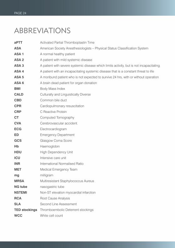
ABBREVIATIONSaPTT Activated Partial Thromboplastin Time
ASA American Society Anesthesiologists – Physical Status Classification System
ASA 1 A normal healthy patient
ASA 2 A patient with mild systemic disease
ASA 3 A patient with severe systemic disease which limits activity, but is not incapacitating
ASA 4 A patient with an incapacitating systemic disease that is a constant threat to life
ASA 5 A moribund patient who is not expected to survive 24 hrs, with or without operation
ASA 6 A brain dead patient for organ donation
BMI Body Mass Index
CALD Culturally and Linguistically Diverse
CBD Common bile duct
CPR Cardiopulmonary resuscitation
CRP C Reactive Protein
CT Computed Tomography
CVA Cerebrovascular accident.
ECG Electrocardiogram
ED Emergency Department
GCS Glasgow Coma Score
Hb Haemoglobin
HDU High Dependency Unit
ICU Intensive care unit
INR International Normalised Ratio
MET Medical Emergency Team
mg milligram
MRSA Multiresistant Staphylococcus Aureus
NG tube nasogastric tube
NSTEMI Non-ST elevation myocardial infarction
RCA Root Cause Analysis
SLA Second Line Assessment
TED stockings Thromboembolic Deterrent stockings
WCC White cell count
CHASM CASEBOOK 2016 | PAGE 25 CLINICAL EXCELLENCE COMMISSION
Executive and program staff based at the Clinical Excellence Commission
Ms Carrie Marr Chief Executive
Dr Jonny Taitz Director Patient Safety (until October 2016)
Dr Bernadette Eather Director Patient Safety (from October 2016)
Paula Cheng Manager Special Committees (until June 2016)
Dr Maree Bellamy Manager Special Committees (from June 2016)
Bruce Czerniec Data Analyst (until May 2016)
Rosemary Sullivan Data Analyst (from November 2016)
John Carrick Program Analyst (from June 2016)
Luana Oros Project Officer
Shanshan Zhao Project Officer
Katherine Asher Project Officer
Ewelina Matkowska Project Assistant
Nathaya Muadjienga Project Officer (until September 2016)
Kathy Tsoi Project Officer (until August 2016)
PAGE 26
Program support staff based at Local Health Districts
Deanne Ellis and Jillian Orr Central Coast
Briana Bartley and Pam Stuchbery Far West
Diana Whitaker and Sandra Harrison Hunter New England
Daniel Purvis Illawarra Shoalhaven
Ashley Thomas Mid North Coast
Nicole Smith Murrumbidgee
Evelyn Esquerra Nepean Blue Mountains
Craig McNally Northern NSW
Angie Pang Northern Sydney
Cyndee King and Michael Piza South Eastern Sydney
Colette Duff South Western Sydney
Jennifer Shevchuk Southern NSW
Jasmin Douglas St Vincent’s Health Network
Cassandra Chan Sydney
Vicky Scott and Shelley Hanrahan Western NSW
Kannan Srinivasan Western Sydney


Clinical Excellence CommissionCollaborating Hospitals’ Audit of Surgical Mortality
Main office locationLevel 17, McKell Building 2–24 Rawson Place SYDNEY NSW 2000
Postal CHASM Clinical Excellence Commission Locked Bag 8 HAYMARKET NSW 1240
Phone (02) 9269 5530
Email [email protected]
Web www.cec.health.nsw.gov.au