Embed Size (px)
Citation preview
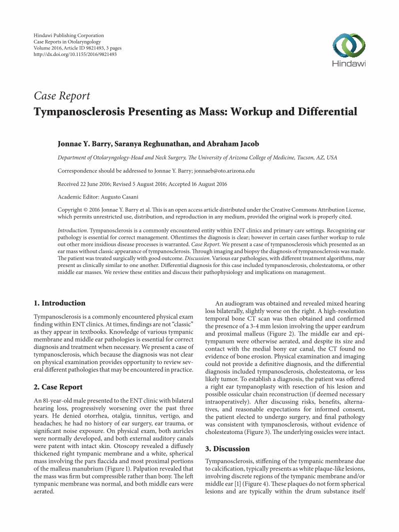
Case ReportTympanosclerosis Presenting as Mass: Workup and Differential
Jonnae Y. Barry, Saranya Reghunathan, and Abraham Jacob
Department of Otolaryngology-Head and Neck Surgery, The University of Arizona College of Medicine, Tucson, AZ, USA
Correspondence should be addressed to Jonnae Y. Barry; [email protected]
Received 22 June 2016; Revised 5 August 2016; Accepted 16 August 2016
Academic Editor: Augusto Casani
Copyright © 2016 Jonnae Y. Barry et al.This is an open access article distributed under the Creative Commons Attribution License,which permits unrestricted use, distribution, and reproduction in any medium, provided the original work is properly cited.
Introduction. Tympanosclerosis is a commonly encountered entity within ENT clinics and primary care settings. Recognizing earpathology is essential for correct management. Oftentimes the diagnosis is clear; however in certain cases further workup to ruleout other more insidious disease processes is warranted. Case Report. We present a case of tympanosclerosis which presented as anearmass without classic appearance of tympanosclerosis.Through imaging and biopsy the diagnosis of tympanosclerosis wasmade.The patient was treated surgically with good outcome.Discussion. Various ear pathologies, with different treatment algorithms, maypresent as clinically similar to one another. Differential diagnosis for this case included tympanosclerosis, cholesteatoma, or othermiddle ear masses. We review these entities and discuss their pathophysiology and implications on management.
1. Introduction
Tympanosclerosis is a commonly encountered physical examfindingwithin ENT clinics. At times, findings are not “classic”as they appear in textbooks. Knowledge of various tympanicmembrane and middle ear pathologies is essential for correctdiagnosis and treatment when necessary.We present a case oftympanosclerosis, which because the diagnosis was not clearon physical examination provides opportunity to review sev-eral different pathologies thatmay be encountered in practice.
2. Case Report
An81-year-oldmale presented to the ENT clinicwith bilateralhearing loss, progressively worsening over the past threeyears. He denied otorrhea, otalgia, tinnitus, vertigo, andheadaches; he had no history of ear surgery, ear trauma, orsignificant noise exposure. On physical exam, both auricleswere normally developed, and both external auditory canalswere patent with intact skin. Otoscopy revealed a diffuselythickened right tympanic membrane and a white, sphericalmass involving the pars flaccida and most proximal portionsof the malleus manubrium (Figure 1). Palpation revealed thatthe mass was firm but compressible rather than bony.The lefttympanic membrane was normal, and both middle ears wereaerated.
An audiogram was obtained and revealed mixed hearingloss bilaterally, slightly worse on the right. A high-resolutiontemporal bone CT scan was then obtained and confirmedthe presence of a 3-4mm lesion involving the upper eardrumand proximal malleus (Figure 2). The middle ear and epi-tympanum were otherwise aerated, and despite its size andcontact with the medial bony ear canal, the CT found noevidence of bone erosion. Physical examination and imagingcould not provide a definitive diagnosis, and the differentialdiagnosis included tympanosclerosis, cholesteatoma, or lesslikely tumor. To establish a diagnosis, the patient was offereda right ear tympanoplasty with resection of his lesion andpossible ossicular chain reconstruction (if deemed necessaryintraoperatively). After discussing risks, benefits, alterna-tives, and reasonable expectations for informed consent,the patient elected to undergo surgery, and final pathologywas consistent with tympanosclerosis, without evidence ofcholesteatoma (Figure 3).The underlying ossicles were intact.
3. Discussion
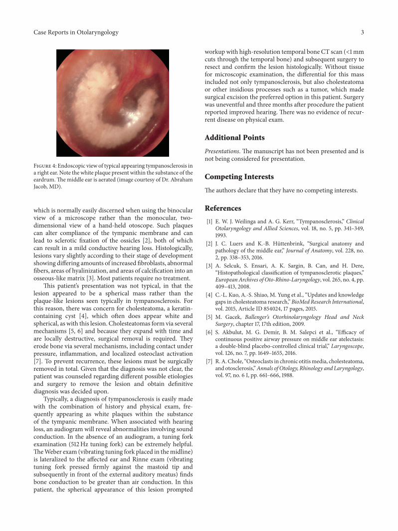
Tympanosclerosis, stiffening of the tympanic membrane dueto calcification, typically presents as white plaque-like lesions,involving discrete regions of the tympanic membrane and/ormiddle ear [1] (Figure 4).These plaques do not form sphericallesions and are typically within the drum substance itself
Hindawi Publishing CorporationCase Reports in OtolaryngologyVolume 2016, Article ID 9821493, 3 pageshttp://dx.doi.org/10.1155/2016/9821493

2 Case Reports in Otolaryngology
(a)
∗
(b)
Figure 1: (a) View of the right tympanicmembrane and lesion (arrow) utilizing the operatingmicroscope. (b) Intraoperative view of the lesion(long arrow)within the lifted tympanicmembrane (asterisk) and the underlyingmalleus (short arrow) (images courtesy ofDr. Abraham Jacob,MD).
(a) (b)
Figure 2: (a) Axial bone windowed temporal bone CT scan showing a 3-4mm hyperdense lesion (arrow); the middle ear is aerated. (b)Coronal bone windowed temporal bone CT scan showing a 3-4mm lesion lateral to the neck of the malleus (arrow).The scutum (lateral wallof epitympanum) is free of bone erosion (images courtesy of Banner University Medical Center).
(a) (b)
Figure 3: (a) Hematoxylin and eosin stained light microscopy obtained from the right earmass (10x). Fibrous infiltrate, hyaline degeneration,and calcification (arrow) without presence of squamous epithelium are seen. (b) 20x view with black arrows demonstrating giant cell reaction(images courtesy of Sarah Tang, PSF).
Case Reports in Otolaryngology 3
Figure 4: Endoscopic view of typical appearing tympanosclerosis ina right ear. Note the white plaque present within the substance of theeardrum.Themiddle ear is aerated (image courtesy of Dr. AbrahamJacob, MD).
which is normally easily discerned when using the binocularview of a microscope rather than the monocular, two-dimensional view of a hand-held otoscope. Such plaquescan alter compliance of the tympanic membrane and canlead to sclerotic fixation of the ossicles [2], both of whichcan result in a mild conductive hearing loss. Histologically,lesions vary slightly according to their stage of developmentshowing differing amounts of increased fibroblasts, abnormalfibers, areas of hyalinization, and areas of calcification into anosseous-like matrix [3]. Most patients require no treatment.
This patient’s presentation was not typical, in that thelesion appeared to be a spherical mass rather than theplaque-like lesions seen typically in tympanosclerosis. Forthis reason, there was concern for cholesteatoma, a keratin-containing cyst [4], which often does appear white andspherical, as with this lesion. Cholesteatomas form via severalmechanisms [5, 6] and because they expand with time andare locally destructive, surgical removal is required. Theyerode bone via several mechanisms, including contact underpressure, inflammation, and localized osteoclast activation[7]. To prevent recurrence, these lesions must be surgicallyremoved in total. Given that the diagnosis was not clear, thepatient was counseled regarding different possible etiologiesand surgery to remove the lesion and obtain definitivediagnosis was decided upon.
Typically, a diagnosis of tympanosclerosis is easily madewith the combination of history and physical exam, fre-quently appearing as white plaques within the substanceof the tympanic membrane. When associated with hearingloss, an audiogram will reveal abnormalities involving soundconduction. In the absence of an audiogram, a tuning forkexamination (512Hz tuning fork) can be extremely helpful.TheWeber exam (vibrating tuning fork placed in themidline)is lateralized to the affected ear and Rinne exam (vibratingtuning fork pressed firmly against the mastoid tip andsubsequently in front of the external auditory meatus) findsbone conduction to be greater than air conduction. In thispatient, the spherical appearance of this lesion prompted
workup with high-resolution temporal bone CT scan (<1mmcuts through the temporal bone) and subsequent surgery toresect and confirm the lesion histologically. Without tissuefor microscopic examination, the differential for this massincluded not only tympanosclerosis, but also cholesteatomaor other insidious processes such as a tumor, which madesurgical excision the preferred option in this patient. Surgerywas uneventful and three months after procedure the patientreported improved hearing. There was no evidence of recur-rent disease on physical exam.
Additional Points
Presentations. The manuscript has not been presented and isnot being considered for presentation.
Competing Interests
The authors declare that they have no competing interests.
References
[1] E. W. J. Weilinga and A. G. Kerr, “Tympanosclerosis,” ClinicalOtolaryngology and Allied Sciences, vol. 18, no. 5, pp. 341–349,1993.
[2] J. C. Luers and K.-B. Huttenbrink, “Surgical anatomy andpathology of the middle ear,” Journal of Anatomy, vol. 228, no.2, pp. 338–353, 2016.
[3] A. Selcuk, S. Ensari, A. K. Sargin, B. Can, and H. Dere,“Histopathological classification of tympanosclerotic plaques,”European Archives of Oto-Rhino-Laryngology, vol. 265, no. 4, pp.409–413, 2008.
[4] C.-L. Kuo, A.-S. Shiao, M. Yung et al., “Updates and knowledgegaps in cholesteatoma research,”BioMed Research International,vol. 2015, Article ID 854024, 17 pages, 2015.
[5] M. Gacek, Ballenger’s Otorhinolaryngology Head and NeckSurgery, chapter 17, 17th edition, 2009.
[6] S. Akbulut, M. G. Demir, B. M. Salepci et al., “Efficacy ofcontinuous positive airway pressure on middle ear atelectasis:a double-blind placebo-controlled clinical trial,” Laryngoscope,vol. 126, no. 7, pp. 1649–1655, 2016.
[7] R.A. Chole, “Osteoclasts in chronic otitismedia, cholesteatoma,and otosclerosis,”Annals of Otology, Rhinology and Laryngology,vol. 97, no. 6 I, pp. 661–666, 1988.

Submit your manuscripts athttp://www.hindawi.com
Stem CellsInternational
Hindawi Publishing Corporationhttp://www.hindawi.com Volume 2014
Hindawi Publishing Corporationhttp://www.hindawi.com Volume 2014
MEDIATORSINFLAMMATION
of
Hindawi Publishing Corporationhttp://www.hindawi.com Volume 2014
Behavioural Neurology
EndocrinologyInternational Journal of
Hindawi Publishing Corporationhttp://www.hindawi.com Volume 2014
Hindawi Publishing Corporationhttp://www.hindawi.com Volume 2014
Disease Markers
Hindawi Publishing Corporationhttp://www.hindawi.com Volume 2014
BioMed Research International
OncologyJournal of
Hindawi Publishing Corporationhttp://www.hindawi.com Volume 2014
Hindawi Publishing Corporationhttp://www.hindawi.com Volume 2014
Oxidative Medicine and Cellular Longevity
Hindawi Publishing Corporationhttp://www.hindawi.com Volume 2014
PPAR Research
The Scientific World JournalHindawi Publishing Corporation http://www.hindawi.com Volume 2014
Immunology ResearchHindawi Publishing Corporationhttp://www.hindawi.com Volume 2014
Journal of
ObesityJournal of
Hindawi Publishing Corporationhttp://www.hindawi.com Volume 2014
Hindawi Publishing Corporationhttp://www.hindawi.com Volume 2014
Computational and Mathematical Methods in Medicine
OphthalmologyJournal of
Hindawi Publishing Corporationhttp://www.hindawi.com Volume 2014
Diabetes ResearchJournal of
Hindawi Publishing Corporationhttp://www.hindawi.com Volume 2014
Hindawi Publishing Corporationhttp://www.hindawi.com Volume 2014
Research and TreatmentAIDS
Hindawi Publishing Corporationhttp://www.hindawi.com Volume 2014
Gastroenterology Research and Practice
Hindawi Publishing Corporationhttp://www.hindawi.com Volume 2014
Parkinson’s Disease
Evidence-Based Complementary and Alternative Medicine
Volume 2014Hindawi Publishing Corporationhttp://www.hindawi.com