Embed Size (px)
Citation preview

Int J Clin Exp Pathol 2016;9(2):2396-2401www.ijcep.com /ISSN:1936-2625/IJCEP0018900
Case ReportThyroid neuroendocrine cancer accompanied with multiple papillary thyroid carcinomas: a case report
Zengguang Liu1, Meishan Jin2, Chang Su1, Jiang Ren1, Fang Wan1, Qiang Guan3, Zhongying Miao1, Guang Chen1, Guimin Wang1
Departments of 1Thyroid Surgery, 2Pathology, The First Hospital of Jilin University, Changchun, Jilin, China; 3De-partment of General Surgery, The Central Hospital of Changchun City, Changchun, Jilin, China
Received October 31, 2015; Accepted December 26, 2015; Epub February 1, 2016; Published February 15, 2016
Abstract: Neuroendocrine tumors (NETs) are neoplasms that arise from the cells of endocrine and nervous systems. Most of them are found in the gastrointestinal tract; however, some cases of NETs have been detected in the thy-roid gland, especially the malignant NETs. In this paper, we have reported a case of NET in the thyroid gland; the tumor was accompanied with multiple papillary thyroid cancers. The patient had also developed cervical lymphatic metastasis. Before performing the operation, the lesions were detected by ultrasound examination. However, the pathological results of this case were unexpected. The patient was subjected to FNA examination. Thereafter, we performed total thyroidectomy and cervical lymph node dissection. The diagnosis was confirmed by pathological examinations. PET-CT was also performed after the operation to rule out the occurrence of secondary endocrine cancers.
Keywords: Neuroendocrine cancers, thyroid, papillary thyroid carcinomas
Introduction
Thyroid cancer is very commonly encountered in clinical practice. In general, papillary thyroid cancer is detected in most cases [1], account-ing for about 75-85% of thyroid malignancies. Although the incidence of papillary thyroid is very high, the prognosis of patients is not very poor [2]. Lymphatic and distant metastasis are usually detected in patients with multiple lesions, or in patients diagnosed with other malignant tumors; many young children also develop lymphatic and distant metastasis [3]. A high frequency ultrasound is generally used for differentiating malignant nodules from benign ones in the thyroid gland [4].
Neuroendocrine tumors (NETs) are neoplasms that arise from the endocrine and nervous sys-tems. Most NETs are benign, while some are malignant. They are most commonly encoun-tered in the intestine, where they are often known as carcinoid tumors, but they can also occur in the pancreas, lungs, and the remaining organs of the human body [5, 6]. In this paper,
we have reported a case of NET; the tumor was detected in the thyroid gland, and it was accompanied by multiple papillary thyroid carcinomas.
Case report
A 54-year-old woman was admitted to our hos-pital as she had developed a slowly growing mass in her neck. About a month ago, she had accidentally found the cervical mass but she did not experience any pain. There was no hoarseness in her voice; she also did not expe-rience any difficulties in breathing or swallow-ing. She did not show any signs of hyper- metabolism or temperature change. Physical examinations revealed a mass in the right upper neck. The mass was located under the jaw, next to the mastoid process; it was 5.0 cm × 3.0 cm × 3.0 cm in size. As this mass was oval and smooth, we thought it was a swollen lymph node. Therefore, we performed an ultrasound scan to examine the swollen mass. We did not detect any swelling in the thyroid gland through the ultrasound scan; physical examination also did not reveal any masses in the thyroid gland.
A rare case of neuroendocrine carcinoma in thyroid gland
2397 Int J Clin Exp Pathol 2016;9(2):2396-2401
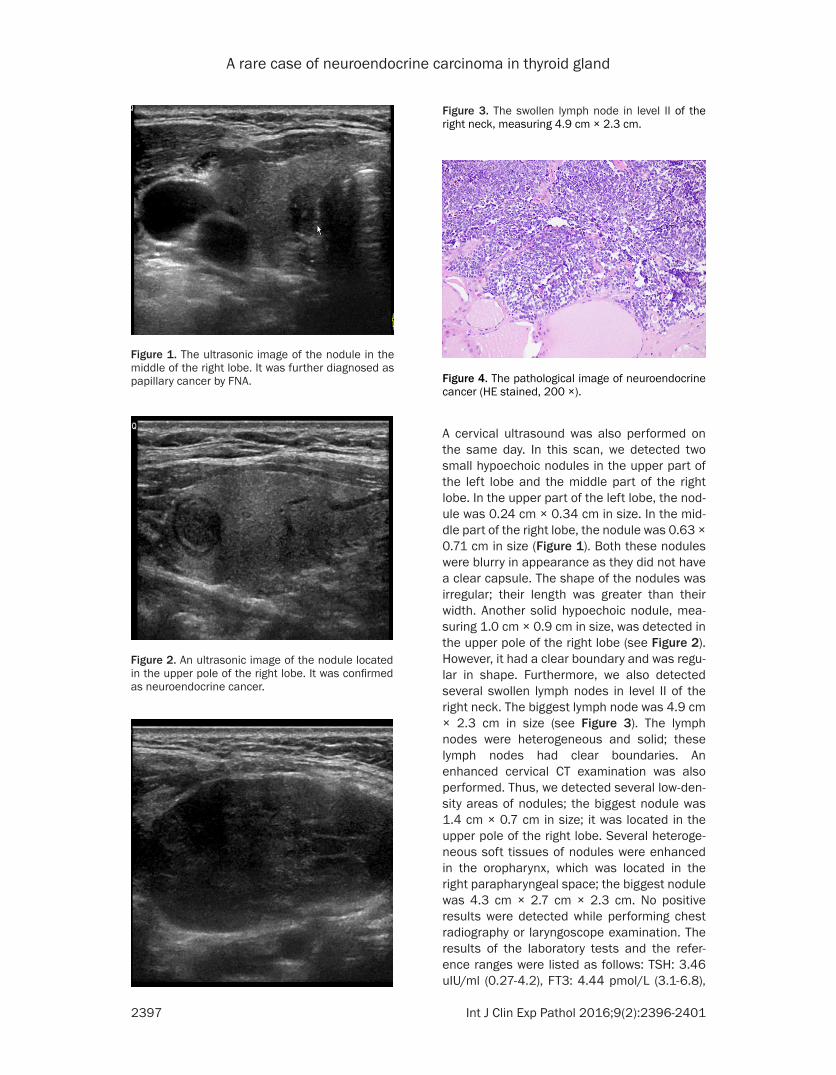
A cervical ultrasound was also performed on the same day. In this scan, we detected two small hypoechoic nodules in the upper part of the left lobe and the middle part of the right lobe. In the upper part of the left lobe, the nod-ule was 0.24 cm × 0.34 cm in size. In the mid-dle part of the right lobe, the nodule was 0.63 × 0.71 cm in size (Figure 1). Both these nodules were blurry in appearance as they did not have a clear capsule. The shape of the nodules was irregular; their length was greater than their width. Another solid hypoechoic nodule, mea-suring 1.0 cm × 0.9 cm in size, was detected in the upper pole of the right lobe (see Figure 2). However, it had a clear boundary and was regu-lar in shape. Furthermore, we also detected several swollen lymph nodes in level II of the right neck. The biggest lymph node was 4.9 cm × 2.3 cm in size (see Figure 3). The lymph nodes were heterogeneous and solid; these lymph nodes had clear boundaries. An enhanced cervical CT examination was also performed. Thus, we detected several low-den-sity areas of nodules; the biggest nodule was 1.4 cm × 0.7 cm in size; it was located in the upper pole of the right lobe. Several heteroge-neous soft tissues of nodules were enhanced in the oropharynx, which was located in the right parapharyngeal space; the biggest nodule was 4.3 cm × 2.7 cm × 2.3 cm. No positive results were detected while performing chest radiography or laryngoscope examination. The results of the laboratory tests and the refer-ence ranges were listed as follows: TSH: 3.46 uIU/ml (0.27-4.2), FT3: 4.44 pmol/L (3.1-6.8),
Figure 1. The ultrasonic image of the nodule in the middle of the right lobe. It was further diagnosed as papillary cancer by FNA.
Figure 2. An ultrasonic image of the nodule located in the upper pole of the right lobe. It was confirmed as neuroendocrine cancer.
Figure 3. The swollen lymph node in level II of the right neck, measuring 4.9 cm × 2.3 cm.
Figure 4. The pathological image of neuroendocrine cancer (HE stained, 200 ×).

A rare case of neuroendocrine carcinoma in thyroid gland
2398 Int J Clin Exp Pathol 2016;9(2):2396-2401
FT4: 16.24 pmol/L (12.0-22.0), TG-Ab: 73.36 IU/ml (< 115.0), TPO-Ab: 27.09 IU/ml (< 35.0). The levels of parathyroid hormone, carcino-embryonic antigen, and calcitonin were normal.
Fine needle aspiration (FNA) biopsy was per-formed on the nodule located in the middle of the right lobe; thyroid papillary cancer was detected in the biopsy, so the swollen lymph
nodes were considered to be metastatic in nature. Subsequently, we also performed total thyroidectomy and cervical lymph node dissec-tion. During the operation, we excised several lymph nodes surrounding the right jugular vein; the biggest lymph node was 5.0 cm × 3.0 cm in size. Frost pathological examination of the lymph nodes indicated that the carcinoma was poorly differentiated. A similar nodule was found in the upper pole of the right lobe of the
Figure 5. A-F: The immunohistochemical results of the nodule: Tg (-), CgA (+), MaxVison Syn (+), MaxVisonTTF-1 (+), MaxVisonCalcition (-), MaxVisonKi-67: 40%+.

A rare case of neuroendocrine carcinoma in thyroid gland
2399 Int J Clin Exp Pathol 2016;9(2):2396-2401
thyroid. It had many necrotic cells, and signs of fission were detected in the pathological imag-es (see Figure 4). The immunohistochemical results of the nodule were as follows: Tg (-), CgA (+), Syn (+), Calcition (-), Ki-67 (+, 40%), TTF (+), HBME-1 (+) (see Figure 5). Thus, we confirmed that the patient had developed neuroendocrine cancer in the thyroid gland. Another four micro papillary carcinomas were detected in the rest of the thyroid gland; two papillary carcinomas were found in the right lobe (measuring 0.2 cm and 0.7 cm in length and width, respectively) and the other two papillary carcinomas were found in the left lobe (both measuring 0.2 cm in length). The immunohistochemical results of the four nodules were as follows: Tg (+), CgA (-), Syn (-), Ki-67 (+, < 5%), HBME-1 (+).
In order to determine whether neuroendocrine carcinoma had developed in other parts of the patient’s body, we performed positron emis-sion tomography-computed tomography (PET-CT) scanning of the patient. The results of the scan were negative. So, the final diagnosis of this patient was primary neuroendocrine can-cer accompanied with multiple thyroid papillary cancers and cervical lymph node metastasis. The patient was prescribed an oral tablet of sinistral thyroxine to inhibit the function of the thyroid gland, and I131 radiotherapy treatment was provided to the patient. Six months after the operation, no positive findings were detect-ed during the follow-up.
Discussion
NETs arise from neuroendocrine cells, which are widely distributed in many tissues of the body. The functions of neuroendocrine cells are similar to those of neurological and endocrinal cells. So, NETs can also develop in many other organs of the human body [7]. The incidence of NETs is quite low; according to the reports of Jan Maarten, about 25 cases of NETs are detected in a population of 1000,000 [8]. Most of the NETs (about 76%) are found in the gastro-intestinal tract, lung, and bronchus [2, 9]. Although there are many kinds of NETs, they are treated as a group of tissues, because the cells of these neoplasms share common fea-tures, such as similar appearance and special secretory granules. Moreover, they often pro-duce biogenic amines and polypeptide hor-mones [10]. Malignant neuroendocrine tumors, also known as neuroendocrine carcinomas
(NEC), are divided into three types according to the extent of their differentiation. The well dif-ferentiated NECs are also called carcinoids, while the moderately differentiated NECs are called atypical carcinoids. Furthermore, the poorly differentiated NECs are called small cell carcinomas.
In very rare cases, we have detected NETs in the thyroid glands. The origin of these tumors has been associated with various kinds of cells, including the parafollicular cells, paraganglion cells, and parathyroid gland [11]. According to their origin in the thyroid gland, NETs are divid-ed into six groups: parafollicular tumors, para-ganglion tumors, complex tumors originating from both follicular and parafollicular cells, parathyroid tumors, secondary NETs, and NECs. Compared with NETs, NECs are found in extremely rare cases. After searching the data-base of our hospital, we found that only this case was finally diagnosed with NECs out of a total of 12100 patients, who had undergone surgical treatment from December, 2009 to April, 2015.
Most of the patients with NETs were older than 65 years [8]. In the early phase, the hormone produced by the tumor was very low. As a result, very few of these patients showed symptoms related to NETs. In this case, the patient was admitted at our hospital because she had developed a slowly growing mass in the neck. Using various diagnostic techniques, we con-firmed that swollen mass actually consisted of several swollen lymph nodes.
The diagnosis of thyroid NECs includes the fol-lowing two aspects: 1) The tumor arises from the thyroid gland; 2) The tumor fulfills the crite-ria of diagnosing NECs. Before performing the operation, it is very difficult to make a correct diagnosis of thyroid NCEs. Most of the cases are diagnosed on the basis of immunohisto-chemical examinations. NECs are found to be positive in NSE, CgA, and Syn. The expression of Ki-67 is associated with the differentiation of NECs. In this case, the NECs were poorly differ-entiated, because the expression of Ki-67 was higher than 40%; however, in papillary thyroid cancer tissues, the expression of Ki-67 was lower than 5%. In pathological images, poorly differentiated NECs have small cells; these cells have a deeply stained, round nucleus. For example, consider the case of “small blue cell

A rare case of neuroendocrine carcinoma in thyroid gland
2400 Int J Clin Exp Pathol 2016;9(2):2396-2401
tumors”; these tumors contain uniform cells, which have a round to oval shaped nucleus and a scanty cytoplasm consisting of pink granules. There can be anaplasia, mitotic activity, and necrosis in these tumor cells. High power exam-ination exhibits bland cytopathology. Electron microscopy can identify secretory granules, which are vital to make a correct diagnosis of the case. In this case, the patient underwent PET-CT examination after the surgery. No other tumors were detected in this scan. Although the tumor tested negative in Tg, it was positive in TTF-1, CgA, and Syn. So, it was finally con-firmed that the patient had developed NECs in the thyroid gland. Two similar cases were reported by Eusebi V in 1992 [12].
Medullary thyroid cancer (MTC) is a form of thy-roid carcinoma that originates in the parafollic-ular cells (C cells), which produce the hormone calcitonin. Medullary tumors are the most com-monly seen thyroid NETs; they are the third most common type of thyroid cancers. They make up about 3% of all thyroid cancer cases [13]. We have also come across cases in which MTCs tested negative for calcitonin [14]; how-ever, most of these cases have a family history of hyperplasia in the parafollicular cells. In this case, the tumor tested negative for calcitonin; but we also did not detect a hyperplasia of parafollicular cells.
Secondary thyroid NECs originates from the tis-sues of thyroid gland. We performed a thorough literature search to determine the incidence of secondary thyroid NECs; only 9 cases were reported until February, 2012 [15-23]. Most cases of secondary thyroid NEC originated from the larynx and long tissue [23]. Cytologically and pathologically, secondary NETs are similar to those of MTCs [16, 20, 21]. However, sec-ondary NETs are negative in calcitonin and CEA, while MTCs are positive. In this case, when the patient underwent PET-CT examina-tion after the surgery, no other NETs were found. So, we propose that the patient had developed primary NEC in the thyroid gland.
Surgery is the conventional treatment in cases diagnosed with poorly differentiated thyroid NECs. The invasiveness of such NECs is very strong, especially tumors that test negative for calcitonin [24]. In our case, the prognosis of the patient was unfavorable, as there was metasta-sis of the cervical lymph nodes. Moreover, the
patient had developed a poorly differentiated tumor. Both the metastatic lymph nodes and the poorly differentiated tumor are insensitive to chemoradiotherapy.
Disclosure of conflict of interest
None.
Address correspondence to: Dr. Guimin Wang, De- partment of Thyroid Surgery, The First Hospital of Jilin University, 71 Xinmin Avenue, Chaoyang Dis- trict, Changchun 130021, China. Tel: (+86) 431-81875286; E-mail: [email protected]
References
[1] Dinets A, Hulchiy M, Sofiadis A, Ghaderi M, Höög A, Larsson C, Zedenius J. Clinical, genet-ic, and immunohistochemical characterization of 70 Ukrainian adult cases with post-Chor-nobyl papillary thyroid carcinoma. Eur J Endo-crinol 2012; 166: 1049-60.
[2] Quaedvlieg PF, Visser O, Lamers CB, Janssen-Heijen ML, Taal BG. Epidemiology and survival in patients with carcinoid disease in The Neth-erlands. An epidemiological study with 2391 patients. Ann Oncol 2001; 12: 1295-300.
[3] Vermeer-Mens JC, Goemaere NN, Kuenen-Boumeester V, de Muinck Keizer-Schrama SM, Zwaan CM, Devos AS, de Krijger RR. Childhood papillary thyroid carcinoma with miliary pulmo-nary metastases. J Clin Oncol 2006; 24: 5788-9.
[4] Grani G, Fumarola A. Thyroglobulin in lymph node fine-needle aspiration washout: a sys-tematic review and meta-analysis of diagnostic accuracy. J Clin Endocrinol Metab 2014; 99: 1970-82.
[5] Gustafsson BI, Kidd M, Chan A, Malfertheiner MV, Modlin IM. Bronchopulmonary neuroendo-crine tumors. Cancer 2008; 113: 5-21.
[6] Oberg K, Jelic S. Neuroendocrine bronchial and thymic tumors: ESMO clinical recommen-dation for diagnosis, treatment and follow-up. Ann Oncol 2008; 19 Suppl 2: ii102-3.
[7] Jacobs C. Neuroendocrine tumors a rare find-ing: part I. Clin J Oncol Nurs 2009; 13: 21-3.
[8] van der Zwan JM, Trama A, Otter R, Larrañaga N, Tavilla A, Marcos-Gragera R, Dei Tos AP, Baudin E, Poston G, Links T; RARECARE WG. Rare neuroendocrine tumours: results of the surveillance of rare cancers in Europe project. Eur J Cancer 2013; 49: 2565-78.
[9] Biering H, Pirlich M, Bauditz J, Sandrock D, Lochs H, Gerl H. PET scan in occult ectopic ACTH syndrome: a useful tool. Clin Endocrinol (Oxf) 2003; 59: 404-5.

A rare case of neuroendocrine carcinoma in thyroid gland
2401 Int J Clin Exp Pathol 2016;9(2):2396-2401
[10] Ramage JK, Davies AH, Ardill J, Bax N, Caplin M, Grossman A, Hawkins R, McNicol AM, Reed N, Sutton R, Thakker R, Aylwin S, Breen D, Brit-ton K, Buchanan K, Corrie P, Gillams A, Lewing-ton V, McCance D, Meeran K, Watkinson A; UKNETwork for Neuroendocrine Tumours. Guidelines for the management of gastroen-teropancreatic neuroendocrine (including car-cinoid) tumours. Gut 2005; 54 Suppl 4: iv1-16.
[11] Baloch ZW, LiVolsi VA. Unusual tumors of the thyroid gland. Endocrinol Metab Clin North Am 2008; 37: 297-310, vii.
[12] Eusebi V, Damiani S, Riva C, Lloyd RV, Capella C. Calcitonin free oat-cell carcinoma of the thy-roid gland. Virchows Arch A Pathol Anat Histo-pathol 1992; 417: 267-71.
[13] DeLellis RA, Rule AH, Spiler I, Nathanson L, Tashjian AH Jr, Wolfe HJ. Calcitonin and carci-noembryonic antigen as tumor markers in medullary thyroid carcinoma. Am J Clin Pathol 1978; 70: 587-94.
[14] Modigliani E, Cohen R, Campos JM, Conte-De-volx B, Maes B, Boneu A, Schlumberger M, Big-orgne JC, Dumontier P, Leclerc L, Corcuff B, Guilhem I. Prognostic factors for survival and for biochemical cure in medullary thyroid carci-noma: results in 899 patients. The GETC Study Group. Groupe d’etude des tumeurs a calcito-nine. Clin Endocrinol (Oxf) 1998; 48: 265-73.
[15] Massani M, Caratozzolo E, Bridda A, Ruffolo C, di Pinto FC, Bonariol L, Rossi S, Bassi N. Iso-lated thyroidal metastasis as primary manifes-tation of pancreatic neuroendocrine carcino-ma. Pancreas 2010; 39: 1113-4.
[16] La Rosa S, Imperatori A, Giovanella L, Garan-cini S, Capella C. Thyroid metastases from typi-cal carcinoid of the lung differentiating be-tween medullary thyroid carcinoma and neuroendocrine tumor metastasis to the thy-roid. Thyroid 2009; 19: 521-6.
[17] Mattavelli F, Collini P, Pizzi N, Gervasoni C, Pen-nacchioli E, Mazzaferro V. Thyroid as a target of metastases. A case of foregut neuroendocrine carcinoma with multiple abdominal metasta-ses and a thyroid localization after 21 years. Tumori 2008; 94: 110-3.
[18] Bhalla R, Popp A, Nassar A. Case report: meta-static renal carcinoid to the thyroid diagnosed by fine needle aspiration biopsy. Diagn Cytopa-thol 2007; 35: 597-600.
[19] Yamada H, Hasegawa Y, Mitsudomi T, Nakashi-ma T, Yatabe Y. Neuroendocrine tumor metas-tasis to the thyroid gland. Int J Clin Oncol 2007; 12: 63-7.
[20] Maly A, Meir K, Maly B. Isolated carcinoid tu-mor metastatic to the thyroid gland: report of a case initially diagnosed by fine needle aspira-tion cytology. Acta Cytol 2006; 50: 84-7.
[21] Papi G, Corrado S, Carani C, Asa SL. Metasta-sis of a caecal neuroendocrine carcinoma to the thyroid gland. J Clin Pathol 2005; 58: 1342-3.
[22] Sivrikoz E, Ozbey NC, Kaya B, Erbil Y, Kaya S, Yilmazbayhan D, Firat P, Kapran Y. Neuroendo-crine tumors presenting with thyroid gland me-tastasis: a case series. J Med Case Rep 2012; 6: 73.
[23] Matias-Guiu X, LaGuette J, Puras-Gil AM, Rosai J. Metastatic neuroendocrine tumors to the thyroid gland mimicking medullary carcinoma: a pathologic and immunohistochemical study of six cases. Am J Surg Pathol 1997; 21: 754-62.
[24] Eusebi V, Damiani S, Riva C, Lloyd RV, Capella C. Calcitonin free oat-cell carcinoma of the thy-roid gland. Virchows Arch A Pathol Anat Histo-pathol 1990; 417: 267-71.