Embed Size (px)
Citation preview
Case ReportMycosis Fungoides of the Oral Cavity: Fungating TumorSuccessfully Treated with Electron Beam Radiation andMaintenance Bexarotene
Juri Bassuner,1 Roberto N. Miranda,1 Drew A. Emge,2 Beau A. DiCicco,3
Daniel J. Lewis,2 and Madeleine Duvic1
1The University of Texas MD Anderson Cancer Center, Houston, TX, USA2Baylor College of Medicine, Houston, TX, USA3University of Texas Medical School at Houston, Houston, TX, USA
Correspondence should be addressed to Madeleine Duvic; [email protected]
Received 13 August 2016; Revised 26 October 2016; Accepted 6 November 2016
Academic Editor: Alireza Firooz
Copyright © 2016 Juri Bassuner et al. This is an open access article distributed under the Creative Commons Attribution License,which permits unrestricted use, distribution, and reproduction in any medium, provided the original work is properly cited.
Oral involvement in mycosis fungoides is unusual and portends a poor prognosis. The clinical findings of three new cases aredescribed along with a differential diagnosis and review of the literature. For brevity, only one patient is discussed in detail belowwhereas the other two cases are solely described in table form. The patient had a four-year history of mycosis fungoides beforedeveloping an exophytic tongue tumor. He was treated with local electron beam radiation and is disease-free to date while beingon maintenance therapy with oral bexarotene. Analysis of the data collected from our review of the literature and the present casesreveal key insights.
1. Introduction
One of the most common T-cell lymphomas is mycosisfungoides (MF). It is amalignant, insidious, cutaneous, extra-nodal non-Hodgkin’s lymphoma (NHL) [1].MF encompassesabout 4% of all lymphoma cases worldwide and has anincidence of 0.36 per 100,00 [2].TheMF disease process has arelatively predictable pattern: in three phases, erythematousor eczematous patches can become infiltrated plaques andcutaneous tumors [1]. Extracutaneous manifestations of MFcan involve a wide array of sites, particularly lymph nodes[3].
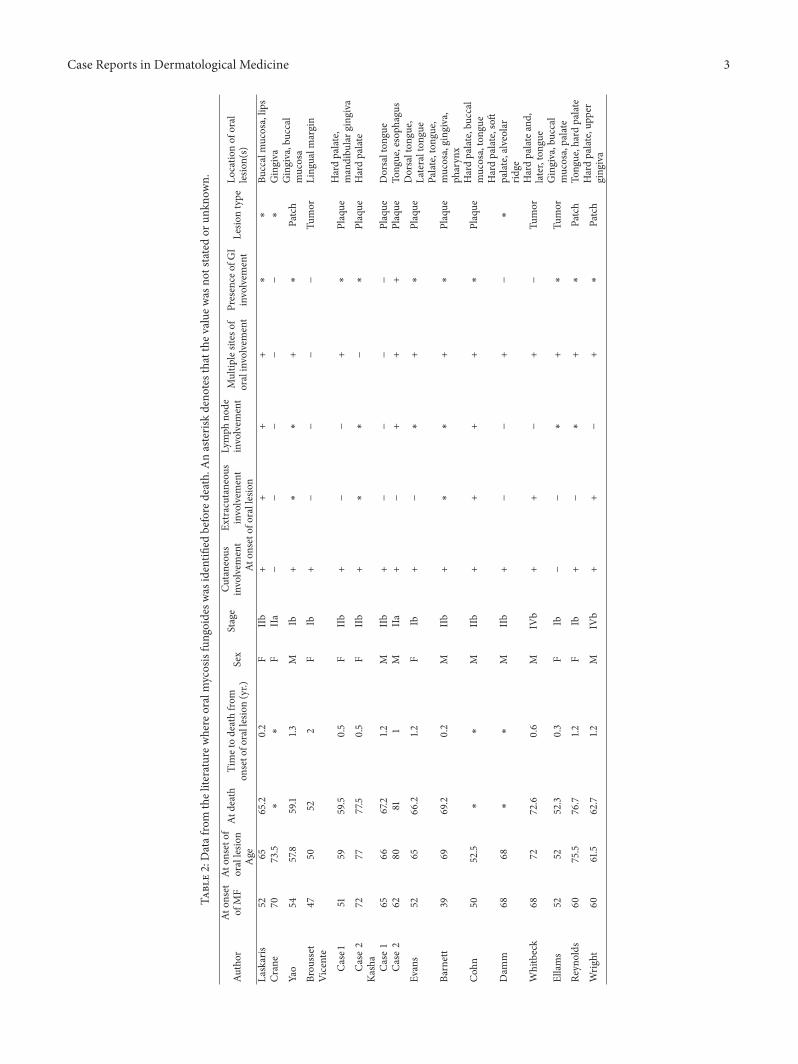
Oral cavity involvement in MF is rare, found in less than1% of patients. Interestingly, autopsy studies suggest up to 13%involvement [4]. This is thought to be a predictor of poorprognosis. Often, patients have advanced stage disease andthe majority have expired shortly after presentation [5–8].We present a case of oral MF and a review of the literature.Two additional patients with oral MF that presented toour hospital are presented in table form alongside the 45
patients with oral MF found in the literature (Table 2). Keyobservations are made from analysis of the patients.
2. Materials and Methods
We have expanded on our previous case series on oral MF(20) to include three new cases that were selected from theelectronic medical records of The University of Texas MDAnderson Cancer Center (UTMDACC). The patients weretreated at UTMDACC over periods from 2005 to present(Case 1), 2005 to 2008 (Case 2), and 2015 to present (Case 3).
3. Case Report
A 63-year-old white man (Case 1) presented in 2005 withexfoliative erythroderma. He stated that he was diagnosedwith a rash localized to his right hand three years earlier.Over the course of one and a half years, his lesions spreadwidely. On presentation, he had 90% body surface area(BSA) involvement with a 3 : 1 ratio of plaque to patch. His
Hindawi Publishing CorporationCase Reports in Dermatological MedicineVolume 2016, Article ID 5857935, 7 pageshttp://dx.doi.org/10.1155/2016/5857935
2 Case Reports in Dermatological Medicine
Table 1: Differential diagnosis of oral tumors.
Disease Oral lesion description Diagnostic cluesMalignancy/premalignancy
Squamous cell papilloma Discrete exophytic papillary lesions (verruca):occur at any intraoral site
History of human immunodeficiency virusinfection; association with cutaneous wartson fingers
Squamous cell carcinomaNonhealing ulcers, papules, or plaques: occurmost frequently at the floor of the mouth andsoft palate
History of tobacco and alcohol consumption;mechanical trauma from ill-fitting dentures
Mesenchymal neoplasms and tumor-likelesions
Fibrous and vascular overgrowths Discrete lesions of cheek or tongueHistory of chronic irritation, usually fromsome tooth-related cause or chroniccheek/tongue biting
Pyogenic granuloma Exuberant overgrowths usually at the gingivabut can occur at any intraoral site
May bleed spontaneously or followingirritation due to extreme vascularity
Odontogenic tumors and cysts
AmeloblastomaOral swellings occurring on the mandible thattypically produce multicystic appearance onradiograph
Painless and slow growing; untreated, mayreach substantial size
Odontogenic cystsOral swellings arising adjacent to teeth thatusually produce a well-demarcated cyst onradiograph
Painless and slow growing
skin exhibited indurated erythematous papular rash that wasconfluent over the upper and lower extremities with skipareas on the abdomen and relative sparing of the groin.
Flow cytometry revealed 30 × 109/L CD4 cells and 96%CD4+/CD26− cells. Biopsy of the tumor showed MF withlarge cell transformation.
The patient received numerous systemic treatmentsincluding (1) vorinostat 400mg daily that improved his pruri-tus but was accompanied by intolerable side effect of diarrheaand overall lack of response in the skin, (2) forodesinewith minor partial response, (3) combined modality withinterferon-alpha plus bexarotene and extracorporeal pho-tophoresis, (4) total body skin electron beam radiation thateffectively cleared his skin temporarily, and (5) alemtuzumabwith which he achieved durable near-complete remission.
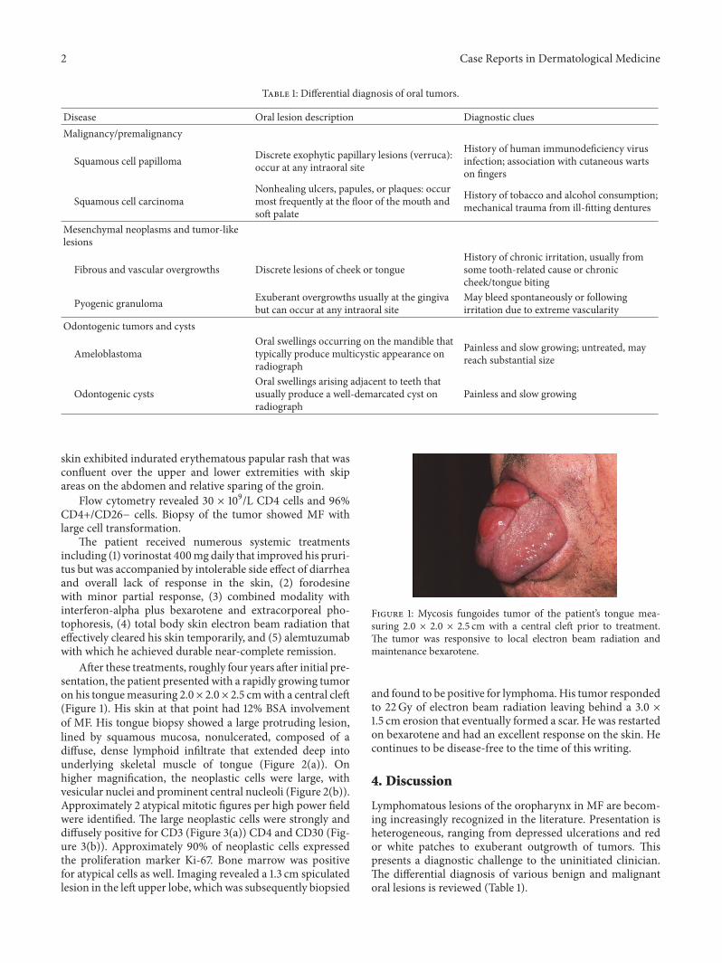
After these treatments, roughly four years after initial pre-sentation, the patient presented with a rapidly growing tumoron his tonguemeasuring 2.0 × 2.0 × 2.5 cmwith a central cleft(Figure 1). His skin at that point had 12% BSA involvementof MF. His tongue biopsy showed a large protruding lesion,lined by squamous mucosa, nonulcerated, composed of adiffuse, dense lymphoid infiltrate that extended deep intounderlying skeletal muscle of tongue (Figure 2(a)). Onhigher magnification, the neoplastic cells were large, withvesicular nuclei and prominent central nucleoli (Figure 2(b)).Approximately 2 atypical mitotic figures per high power fieldwere identified. The large neoplastic cells were strongly anddiffusely positive for CD3 (Figure 3(a)) CD4 and CD30 (Fig-ure 3(b)). Approximately 90% of neoplastic cells expressedthe proliferation marker Ki-67. Bone marrow was positivefor atypical cells as well. Imaging revealed a 1.3 cm spiculatedlesion in the left upper lobe, which was subsequently biopsied
Figure 1: Mycosis fungoides tumor of the patient’s tongue mea-suring 2.0 × 2.0 × 2.5 cm with a central cleft prior to treatment.The tumor was responsive to local electron beam radiation andmaintenance bexarotene.
and found to be positive for lymphoma.His tumor respondedto 22Gy of electron beam radiation leaving behind a 3.0 ×1.5 cm erosion that eventually formed a scar. He was restartedon bexarotene and had an excellent response on the skin. Hecontinues to be disease-free to the time of this writing.
4. Discussion
Lymphomatous lesions of the oropharynx in MF are becom-ing increasingly recognized in the literature. Presentation isheterogeneous, ranging from depressed ulcerations and redor white patches to exuberant outgrowth of tumors. Thispresents a diagnostic challenge to the uninitiated clinician.The differential diagnosis of various benign and malignantoral lesions is reviewed (Table 1).
Case Reports in Dermatological Medicine 3
Table2:Datafrom
theliterature
where
oralmycosisfungoidesw
asidentifi
edbefore
death.Anasteris
kdeno
testhatthe
valuew
asno
tstatedor
unkn
own.
Author
Aton
set
ofMF
Aton
seto
forallesio
nAt
death
Timetodeathfro
mon
seto
forallesion(yr.)
Sex
Stage
Cutaneou
sinvolvem
ent
Extracutaneous
involvem
ent
Lymph
node
involvem
ent
Multip
lesites
oforalinvolvem
ent
Presence
ofGI
involvem
ent
Lesio
ntype
Locatio
nof
oral
lesio
n(s)
Age
Aton
seto
forallesion
Laskaris
5265
65.2
0.2
FIIb
++
++
∗∗
Buccalmucosa,lip
sCr
ane
7073.5
∗∗
FIIa
−−
−−
−∗
Gingiva
Yao
5457.8
59.1
1.3M
Ib+
∗∗
+∗
Patch
Gingiva,buccal
mucosa
Brou
sset
4750
522
FIb
+−
−−
−Tu
mor
Ling
ualm
argin
Vicente
Case1
5159
59.5
0.5
FIIb
+−
−+
∗Plaque
Hardpalate,
mandibu
larg
ingiva
Case
272
7777.5
0.5
FIIb
+∗
∗−
∗Plaque
Hardpalate
Kasha
Case
165
6667.2
1.2M
IIb
+−
−−
−Plaque
Dorsalton
gue
Case
262
8081
1M
IIa+
−+
++
Plaque
Tong
ue,esoph
agus
Evans
5265
66.2
1.2F
Ib+
−∗
+∗
Plaque
Dorsalton
gue,
Lateralton
gue
Barnett
3969
69.2
0.2
MIIb
+∗
∗+
∗Plaque
Palate,ton
gue,
mucosa,ging
iva,
pharyn
x
Coh
n50
52.5
∗∗
MIIb
++
++
∗Plaque
Hardpalate,buccal
mucosa,tong
ue
Dam
m68
68∗
∗M
IIb
+−
−+
−∗
Hardpalate,soft
palate,alveolar
ridge
Whitbeck
6872
72.6
0.6
MIV
b+
+−
+−
Tumor
Hardpalateand,
later,tong
ue
Ellams
5252
52.3
0.3
FIb
−−
∗+
∗Tu
mor
Gingiva,buccal
mucosa,palate
Reyn
olds
6075.5
76.7
1.2F
Ib+
−∗
+∗
Patch
Tong
ue,hardpalate
Wrig
ht60
61.5
62.7
1.2M
IVb
++
−+
∗Patch
Hardpalate,upp
erging
iva
4 Case Reports in Dermatological Medicine
Table2:Con
tinued.
Author
Aton
set
ofMF
Aton
seto
forallesio
nAt
death
Timetodeathfro
mon
seto
forallesion(yr.)
Sex
Stage
Cutaneou
sinvolvem
ent
Extracutaneous
involvem
ent
Lymph
node
involvem
ent
Multip
lesites
oforalinvolvem
ent
Presence
ofGI
involvem
ent
Lesio
ntype
Locatio
nof
oral
lesio
n(s)
Age
Aton
seto
forallesion
Sirois Case
171
7576
1M
IVa
+∗
∗+
∗∗
Gingiva,palate,
tong
ue,lip,buccal
mucosa,tonsil
Case
244
5758
1M
III
+∗
∗−
∗∗
Tong
ueCa
se3
4649
501
MIVa
+∗
∗+
∗∗
Gingiva,ton
gue
Case
471
7475
1M
IIb
+∗
∗+
∗∗
Gingiva,palate
Case
562
6669
3F
IIb
+∗
∗+
∗∗
Gingiva,palate
Case
651
5356
3F
IVa
+∗
∗−
∗∗
Gingiva
Case
767
7381
8F
Ib−
∗∗
−∗
∗Gingiva
Case
843
5153
2M
III
+∗
∗−
∗∗
Tong
ueMcB
ride
∗63
63.1
0.1
FIIa
+∗
∗−
∗Tu
mor
Dorsalton
gue
Harman
∗57
57.6
0.6
MIIb
+−
−+
−∗
Gingiva,palate
Cawley
Case
172
7274
2M
Ib+
+−
−−
∗Hard/softpalate,
tonsils
Case
265
6565.0
0.04
MIIb
++
++
−Tu
mor
Labialcommissure,
tong
uePo
storin
oet
al.
∗60
∗∗
MIIb
+−
+−
−Plaque
Mucosa
Corbettetal.∗
∗∗
∗F
IIb
+−
−+
−Tu
mor
Softpalate,throat
Wainetal.
∗∗
∗∗
MIb
+−
−+
−Plaque
Softpalate,ton
gue,
lips
Wahieetal.
6069
∗∗
MIa
−−
−+
−∗
Suprahyoid
region
,epiglottis
Visw
anathan
6969
∗∗
MIa
+−
−+
−∗
Tong
ue,soft
palate
Luigettietal.
2738
∗∗
F∗
++
++
−Plaque
Lip,mucosa,
tong
ue,pharynx
Goldsmith
etal.
4464
∗∗
F∗
+−
−−
−Plaque
Hardpalate
Leetal.
3236
∗∗
MIIb
++
+−
−Tu
mor
Tonsil
Tillm
anetal.
60∗
∗M
∗∗
∗∗
∗∗
∗∗
Chua
etal.
8080.7
∗∗
MIb
+−
−−
−Tu
mor
Hardpalate,
ging
iva,mucosa
Case Reports in Dermatological Medicine 5
Table2:Con
tinued.
Author
Aton
set
ofMF
Aton
seto
forallesio
nAt
death
Timetodeathfro
mon
seto
forallesion(yr.)
Sex
Stage
Cutaneou
sinvolvem
ent
Extracutaneous
involvem
ent
Lymph
node
involvem
ent
Multip
lesites
oforalinvolvem
ent
Presence
ofGI
involvem
ent
Lesio
ntype
Locatio
nof
oral
lesio
n(s)
Age
Aton
seto
forallesion
Gom
ez
Case
135
4545.5
0.5
FIIb
+−
−+
−Tu
mor
Tong
ue,uvula,
orop
harynx
Case
266
70∗
∗F
Ib+
−−
+−
∗Uvula,soft
palate,
tonsils
May Ca
se1
∗40
∗∗
FIa
+−
−−
−Tu
mor
Tong
ueCa
se2
4444
∗∗
M∗
−−
−−
−∗
Tong
uePresent
repo
rtCa
se1
6074
∗∗
MIV
b+
+−
−−
Tumor
Tong
ueCa
se2
5055
55.7
0.7
MIV
b+
+−
+−
Tumor
Palate,uvula
Case
335
38∗
∗M
IVb
++
∗+
+Ulcer
Tong
ue,palate
6 Case Reports in Dermatological Medicine
(a) (b)
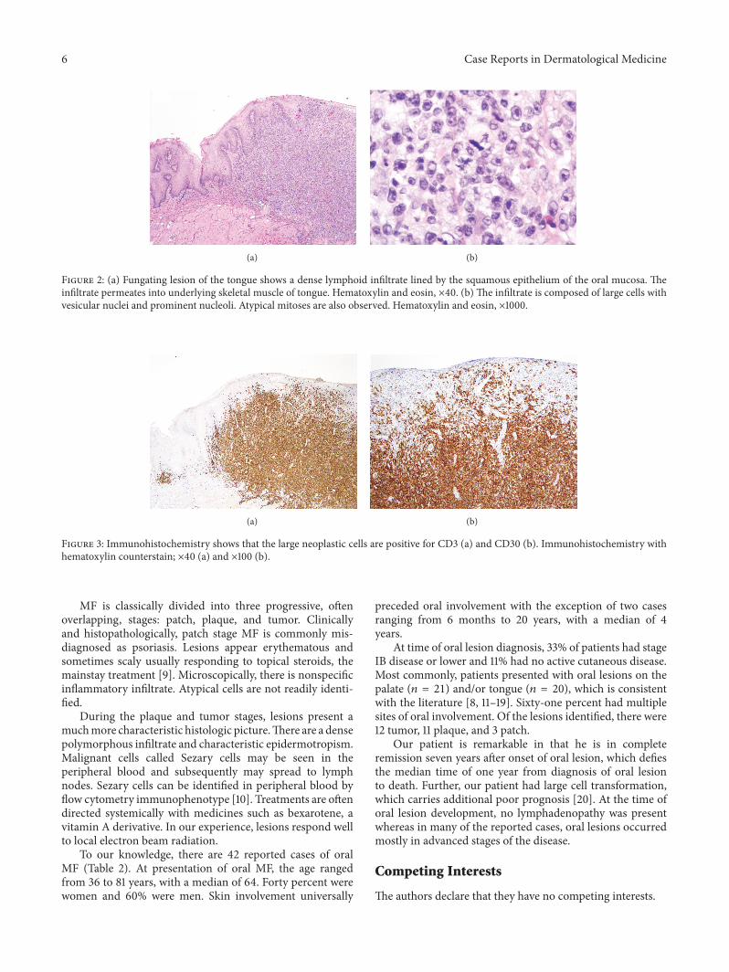
Figure 2: (a) Fungating lesion of the tongue shows a dense lymphoid infiltrate lined by the squamous epithelium of the oral mucosa. Theinfiltrate permeates into underlying skeletal muscle of tongue. Hematoxylin and eosin, ×40. (b) The infiltrate is composed of large cells withvesicular nuclei and prominent nucleoli. Atypical mitoses are also observed. Hematoxylin and eosin, ×1000.
(a) (b)
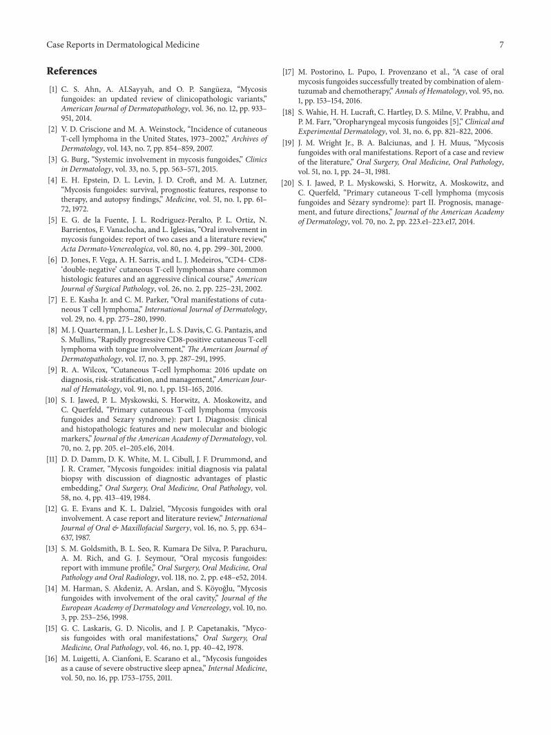
Figure 3: Immunohistochemistry shows that the large neoplastic cells are positive for CD3 (a) and CD30 (b). Immunohistochemistry withhematoxylin counterstain; ×40 (a) and ×100 (b).
MF is classically divided into three progressive, oftenoverlapping, stages: patch, plaque, and tumor. Clinicallyand histopathologically, patch stage MF is commonly mis-diagnosed as psoriasis. Lesions appear erythematous andsometimes scaly usually responding to topical steroids, themainstay treatment [9]. Microscopically, there is nonspecificinflammatory infiltrate. Atypical cells are not readily identi-fied.
During the plaque and tumor stages, lesions present amuchmore characteristic histologic picture.There are a densepolymorphous infiltrate and characteristic epidermotropism.Malignant cells called Sezary cells may be seen in theperipheral blood and subsequently may spread to lymphnodes. Sezary cells can be identified in peripheral blood byflow cytometry immunophenotype [10]. Treatments are oftendirected systemically with medicines such as bexarotene, avitamin A derivative. In our experience, lesions respond wellto local electron beam radiation.
To our knowledge, there are 42 reported cases of oralMF (Table 2). At presentation of oral MF, the age rangedfrom 36 to 81 years, with a median of 64. Forty percent werewomen and 60% were men. Skin involvement universally
preceded oral involvement with the exception of two casesranging from 6 months to 20 years, with a median of 4years.
At time of oral lesion diagnosis, 33% of patients had stageIB disease or lower and 11% had no active cutaneous disease.Most commonly, patients presented with oral lesions on thepalate (𝑛 = 21) and/or tongue (𝑛 = 20), which is consistentwith the literature [8, 11–19]. Sixty-one percent had multiplesites of oral involvement. Of the lesions identified, there were12 tumor, 11 plaque, and 3 patch.
Our patient is remarkable in that he is in completeremission seven years after onset of oral lesion, which defiesthe median time of one year from diagnosis of oral lesionto death. Further, our patient had large cell transformation,which carries additional poor prognosis [20]. At the time oforal lesion development, no lymphadenopathy was presentwhereas in many of the reported cases, oral lesions occurredmostly in advanced stages of the disease.
Competing Interests
The authors declare that they have no competing interests.
Case Reports in Dermatological Medicine 7
References
[1] C. S. Ahn, A. ALSayyah, and O. P. Sangueza, “Mycosisfungoides: an updated review of clinicopathologic variants,”American Journal of Dermatopathology, vol. 36, no. 12, pp. 933–951, 2014.
[2] V. D. Criscione and M. A. Weinstock, “Incidence of cutaneousT-cell lymphoma in the United States, 1973–2002,” Archives ofDermatology, vol. 143, no. 7, pp. 854–859, 2007.
[3] G. Burg, “Systemic involvement in mycosis fungoides,” Clinicsin Dermatology, vol. 33, no. 5, pp. 563–571, 2015.
[4] E. H. Epstein, D. L. Levin, J. D. Croft, and M. A. Lutzner,“Mycosis fungoides: survival, prognostic features, response totherapy, and autopsy findings,” Medicine, vol. 51, no. 1, pp. 61–72, 1972.
[5] E. G. de la Fuente, J. L. Rodriguez-Peralto, P. L. Ortiz, N.Barrientos, F. Vanaclocha, and L. Iglesias, “Oral involvement inmycosis fungoides: report of two cases and a literature review,”Acta Dermato-Venereologica, vol. 80, no. 4, pp. 299–301, 2000.
[6] D. Jones, F. Vega, A. H. Sarris, and L. J. Medeiros, “CD4- CD8-‘double-negative’ cutaneous T-cell lymphomas share commonhistologic features and an aggressive clinical course,” AmericanJournal of Surgical Pathology, vol. 26, no. 2, pp. 225–231, 2002.
[7] E. E. Kasha Jr. and C. M. Parker, “Oral manifestations of cuta-neous T cell lymphoma,” International Journal of Dermatology,vol. 29, no. 4, pp. 275–280, 1990.
[8] M. J. Quarterman, J. L. Lesher Jr., L. S. Davis, C. G. Pantazis, andS. Mullins, “Rapidly progressive CD8-positive cutaneous T-celllymphoma with tongue involvement,” The American Journal ofDermatopathology, vol. 17, no. 3, pp. 287–291, 1995.
[9] R. A. Wilcox, “Cutaneous T-cell lymphoma: 2016 update ondiagnosis, risk-stratification, andmanagement,”American Jour-nal of Hematology, vol. 91, no. 1, pp. 151–165, 2016.
[10] S. I. Jawed, P. L. Myskowski, S. Horwitz, A. Moskowitz, andC. Querfeld, “Primary cutaneous T-cell lymphoma (mycosisfungoides and Sezary syndrome): part I. Diagnosis: clinicaland histopathologic features and new molecular and biologicmarkers,” Journal of the American Academy of Dermatology, vol.70, no. 2, pp. 205. e1–205.e16, 2014.
[11] D. D. Damm, D. K. White, M. L. Cibull, J. F. Drummond, andJ. R. Cramer, “Mycosis fungoides: initial diagnosis via palatalbiopsy with discussion of diagnostic advantages of plasticembedding,” Oral Surgery, Oral Medicine, Oral Pathology, vol.58, no. 4, pp. 413–419, 1984.
[12] G. E. Evans and K. L. Dalziel, “Mycosis fungoides with oralinvolvement. A case report and literature review,” InternationalJournal of Oral & Maxillofacial Surgery, vol. 16, no. 5, pp. 634–637, 1987.
[13] S. M. Goldsmith, B. L. Seo, R. Kumara De Silva, P. Parachuru,A. M. Rich, and G. J. Seymour, “Oral mycosis fungoides:report with immune profile,” Oral Surgery, Oral Medicine, OralPathology and Oral Radiology, vol. 118, no. 2, pp. e48–e52, 2014.
[14] M. Harman, S. Akdeniz, A. Arslan, and S. Koyoglu, “Mycosisfungoides with involvement of the oral cavity,” Journal of theEuropean Academy of Dermatology and Venereology, vol. 10, no.3, pp. 253–256, 1998.
[15] G. C. Laskaris, G. D. Nicolis, and J. P. Capetanakis, “Myco-sis fungoides with oral manifestations,” Oral Surgery, OralMedicine, Oral Pathology, vol. 46, no. 1, pp. 40–42, 1978.
[16] M. Luigetti, A. Cianfoni, E. Scarano et al., “Mycosis fungoidesas a cause of severe obstructive sleep apnea,” Internal Medicine,vol. 50, no. 16, pp. 1753–1755, 2011.
[17] M. Postorino, L. Pupo, I. Provenzano et al., “A case of oralmycosis fungoides successfully treated by combination of alem-tuzumab and chemotherapy,”Annals of Hematology, vol. 95, no.1, pp. 153–154, 2016.
[18] S. Wahie, H. H. Lucraft, C. Hartley, D. S. Milne, V. Prabhu, andP. M. Farr, “Oropharyngeal mycosis fungoides [5],” Clinical andExperimental Dermatology, vol. 31, no. 6, pp. 821–822, 2006.
[19] J. M. Wright Jr., B. A. Balciunas, and J. H. Muus, “Mycosisfungoides with oral manifestations. Report of a case and reviewof the literature,” Oral Surgery, Oral Medicine, Oral Pathology,vol. 51, no. 1, pp. 24–31, 1981.
[20] S. I. Jawed, P. L. Myskowski, S. Horwitz, A. Moskowitz, andC. Querfeld, “Primary cutaneous T-cell lymphoma (mycosisfungoides and Sezary syndrome): part II. Prognosis, manage-ment, and future directions,” Journal of the American Academyof Dermatology, vol. 70, no. 2, pp. 223.e1–223.e17, 2014.

Submit your manuscripts athttp://www.hindawi.com
Stem CellsInternational
Hindawi Publishing Corporationhttp://www.hindawi.com Volume 2014
Hindawi Publishing Corporationhttp://www.hindawi.com Volume 2014
MEDIATORSINFLAMMATION
of
Hindawi Publishing Corporationhttp://www.hindawi.com Volume 2014
Behavioural Neurology
EndocrinologyInternational Journal of
Hindawi Publishing Corporationhttp://www.hindawi.com Volume 2014
Hindawi Publishing Corporationhttp://www.hindawi.com Volume 2014
Disease Markers
Hindawi Publishing Corporationhttp://www.hindawi.com Volume 2014
BioMed Research International
OncologyJournal of
Hindawi Publishing Corporationhttp://www.hindawi.com Volume 2014
Hindawi Publishing Corporationhttp://www.hindawi.com Volume 2014
Oxidative Medicine and Cellular Longevity
Hindawi Publishing Corporationhttp://www.hindawi.com Volume 2014
PPAR Research
The Scientific World JournalHindawi Publishing Corporation http://www.hindawi.com Volume 2014
Immunology ResearchHindawi Publishing Corporationhttp://www.hindawi.com Volume 2014
Journal of
ObesityJournal of
Hindawi Publishing Corporationhttp://www.hindawi.com Volume 2014
Hindawi Publishing Corporationhttp://www.hindawi.com Volume 2014
Computational and Mathematical Methods in Medicine
OphthalmologyJournal of
Hindawi Publishing Corporationhttp://www.hindawi.com Volume 2014
Diabetes ResearchJournal of
Hindawi Publishing Corporationhttp://www.hindawi.com Volume 2014
Hindawi Publishing Corporationhttp://www.hindawi.com Volume 2014
Research and TreatmentAIDS
Hindawi Publishing Corporationhttp://www.hindawi.com Volume 2014
Gastroenterology Research and Practice
Hindawi Publishing Corporationhttp://www.hindawi.com Volume 2014
Parkinson’s Disease
Evidence-Based Complementary and Alternative Medicine
Volume 2014Hindawi Publishing Corporationhttp://www.hindawi.com


![Significance of CD30 Expression by Epidermotropic T Cells ... · diagnosis included LyP, lymphomatoid pityriasis lichenoides and “pityriasis lichenoides-like” mycosis fungoides.[7,8]](https://img.dokumen.tips/doc/110x75/60223092b9e61714693c3a28/significance-of-cd30-expression-by-epidermotropic-t-cells-diagnosis-included.jpg)