Embed Size (px)
Citation preview

CASE REPORT
Multiple dens evaginatus: diagnosis, management,and complications: case reportElsa A. Echeverri, DDS Ming M. Wang, DDS, MS Carmen Chavaria, DDS D. Lance Taylor, DDS
AbstractDens evaginatus is a disturbance in tooth formation that appears clinically as an accessory occlusal tuberculated cusp
composed of enamel and dentin, usually enclosing pulp tissue. The tubercle often fractures or is worn away, with subsequentpulp exposure leading to pulpal inflammation, necrosis, and periapical inflammation. When this happens to an immature tooth,diagnosis may be complicated by the similarity in radiographic appearance of the periapical lesion and the dental follicle. Themanagement of such teeth may be complicated because of their immaturity. A case report of multiple abraded dens evaginatuswith pulp necrosis is presented that illustrates 1) the difficulty of diagnosing otherwise apparently healthy teeth associated withperiapical lesions and sinus tracts, and 2) their management and treatment complications including recurrent infections androot fractures. (Pediatr Dent 16:314-17, 1994)
Literature review
Dens evaginatus is an anomalous tooth develop-ment arising during morphodifferentiation. It is causedby abnormal proliferation of the inner enamel epithe-lium into the stellate reticulum of the enamel organ1
with a core of dentin surrounding a narrow extensionof the pulp tissue projecting into the tubercle.2 It is alsoreferred to as tuberculated cusp, accessory tubercle,occlusal tuberculated premolar, Leong’s premolar,evaginatus odontoma, and occlusal pearl. 3 Prevalenceranges from I to 4%.4 It appears primarily in the Mon-goloid racial group: the Paleo-Asiatics (Indians of North,Central and South America and Eskimos),s the Neo-Asiatics, and the Indonesian-Malays (Filipinos). Densevaginatus has been reported in Chinese,6 Thai,7 Eski-mos, North American Indians, s and occasionally inCaucasians.s
This anomaly, an enamel-covered tubercle on theocclusal surface between the buccal and lingual cuspsof posterior teeth, can occur unilaterally or bilaterally.It occurs primarily in premolars but also has been re-ported -- although rarely -- on molars, canines, andincisors. 9-u The occurrence is five times more frequentin the mandible than in the maxilla22
The clinical importance of this condition is that thistubercle easily fractures or is worn away, exposing thefine pulpal extension, which may lead to infection. Thetubercle may fracture or be abraded as soon as thetooth comes into occlusion. Infection and loss of toothvitality may occur before root development is com-plete13 when pulp tests often are unreliable24 Periapicallesions on the radiographs may be indistinguishablefrom or misinterpreted as developing dental follicle.Diagnosis and treatment may be delayed and severetoothache or infection may occur because there is no
obvious etiology for a pulpitis such as caries or trauma.Immature root development in a young patient makesmanaging the affected teeth a problem.
Case reportPast history
A 11-year-old Hispanic boy was referred to the Uni-versity of Texas -- Houston, Health Science Center,Dental Branch, Pediatric Dentistry Emergency Clinicby a pediatrician in November 1990. The patient’s chiefcomplaint was severe pain in the left maxillary area.The patient’s mother said the child had complained ofdiscomfort in this area about five months earlier. Theyhad consulted a dentist who diagnosed no carious le-sions and prescribed a medication that the mother couldnot recall. A week before this admission, the child com-plained of severe toothache, so he was taken to anotherdentist and, according to the mother, "some grinding"was performed on teeth in the affected area. Not re-lieved of the severe pain, the patient saw a pediatricianwho prescribed antibiotics and referred him to thisinstitution. After initial examination, an endodontistwas consulted.
Clinical examinationClinical examination revealed 27 permanent teeth,
all free of caries and restorations, with the exception ofthe mandibular right first permanent molar (#30), whichhad incipient occlusal caries. Generalized gingivitis waspresent, but periodontal probings were all within 1-3mm. The maxillary left second premolar (#13) was verytender to percussion and mobile, while the adjacentteeth were not. A parulis was present on the alveolarmucosa of both the mandibular left (#20) and right(#29) second premolars. Teeth #13, #20, and #29 didnot respond to thermal or electric pulp tests, while the
314 Pediatric Dentistry: July/August 1994 - Volume 16, Number 4

Fig 1. Periapical radiograph of tooth #13 showing immatureapex with small radiolucency similar to dental follicle.Thickening of the periodontal ligament space on the mesial sideof the tooth is also apparent. No caries or restoration observedon #13.
Fig 2. Diffuse radiolucency around the wide open apex of #20with no caries or restoration.
adjacent teeth responded normally. Periapical radio-graphs revealed no evidence of caries or restorationson #13, #20, and #29. A small, circumscribed radio-lucency similar to a dental follicle was observed aroundthe immature apex of #13. Widening of the periodontalligament space on the mesial aspect of #13 also wasobserved (Fig 1). Diffuse radiolucencies were observedaround the immature apices of #20 and #29 (Figs 2, 3).Teeth #20 and #29 were not tender to percussion. Care-ful examination revealed a small, round, flat surfaceabout 1-1.5 mm in diameter between the buccal andlingual cusps on #13, #20, and #29 with a pin-pointdark spot in the center of each flat surface (Fig 4).Abraded dens evaginatus was suspected at this time,and a tentative diagnosis of pulpal necrosis and exacer-bation of chronic apical periodontitis was made fortooth #13. The diagnosis was confirmed by performinga test cavity into the pulp chamber without anesthesiarevealing a necrotic pulp chamber. The canal wasdebrided with endodontic files, dried and packed withCa(OH)2 powder, and temporized with IRM® (DentsplyInternational, Inc., Milford, DE) The mother was in-formed that an apexification procedure would be nec-essary to induce root end closure.
A tentative diagnosis of pulpal necrosis with suppu-rative apical periodontitis was made for #20 and #29.Complete blood count, and bone and kidney panelswere ordered to rule out possible underlying systemiccomponents such as vitamin D-resistant rickets, andhyperparathyroidism. Vitamin D-resistant rickets isoften accompanied by draining periapical abscessesand pulp horns that tend to extend into the cusp tipsallowing penetration of microorganisms.15-16 Inhyperparathyroidism multiple periapical radio-lucencies (e.g., giant cell granulomas) often present asthe first clinical manifestation.17-18 The laboratory re-sults all were within normal limits.
Fig 3. Diffuse radiolucency around the wide-open apex of #29with no caries or restoration.
Fig 4. Abraded dens evaginatus on #20. No caries or restoration.A small, round, flat surface (black arrow) between the buccaland lingual cusps as a result of the abraded dens evaginatus. Aparulis on the alveolar gingiva also is present (white arrow).
Pediatric Dentistry: July/August 1994 - Volume 16, Number 4 315
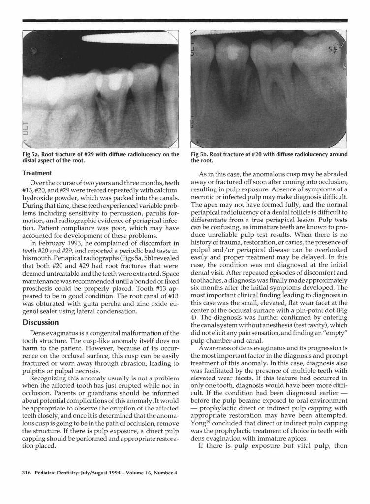
Fig 5a. Root fracture of #29 with diffuse radiolucency on thedistal aspect of the root.
Treatment
Over the course of two years and three months, teeth#13, #20, and #29 were treated repeatedly with calciumhydroxide powder, which was packed into the canals.During that time, these teeth experienced variable prob-lems including sensitivity to percussion, parulis for-mation, and radiographic evidence of periapical infec-tion. Patient compliance was poor, which may haveaccounted for development of these problems.
In February 1993, he complained of discomfort inteeth #20 and #29, and reported a periodic bad taste inhis mouth. Periapical radiographs (Figs 5a, 5b) revealedthat both #20 and #29 had root fractures that weredeemed untreatable and the teeth were extracted. Spacemaintenance was recommended until a bonded or fixedprosthesis could be properly placed. Tooth #13 ap-peared to be in good condition. The root canal of #13was obturated with gutta percha and zinc oxide eu-genol sealer using lateral condensation.
DiscussionDens evaginatus is a congenital malformation of the
tooth structure. The cusp-like anomaly itself does noharm to the patient. However, because of its occur-rence on the occlusal surface, this cusp can be easilyfractured or worn away through abrasion, leading topulpitis or pulpal necrosis.
Recognizing this anomaly usually is not a problemwhen the affected tooth has just erupted while not inocclusion. Parents or guardians should be informedabout potential complications of this anomaly. It wouldbe appropriate to observe the eruption of the affectedteeth closely, and once it is determined that the anoma-lous cusp is going to be in the path of occlusion, removethe structure. If there is pulp exposure, a direct pulpcapping should be performed and appropriate restora-tion placed.
Fig 5b. Root fracture of #20 with diffuse radiolucency aroundthe root.
As in this case, the anomalous cusp may be abradedaway or fractured off soon after coming into occlusion,resulting in pulp exposure. Absence of symptoms of anecrotic or infected pulp may make diagnosis difficult.The apex may not have formed fully, and the normalperiapical radiolucency of a dental follicle is difficult todifferentiate from a true periapical lesion. Pulp testscan be confusing, as immature teeth are known to pro-duce unreliable pulp test results. When there is nohistory of trauma, restoration, or caries, the presence ofpulpal and/or periapical disease can be overlookedeasily and proper treatment may be delayed. In thiscase, the condition was not diagnosed at the initialdental visit. After repeated episodes of discomfort andtoothaches, a diagnosis was finally made approximatelysix months after the initial symptoms developed. Themost important clinical finding leading to diagnosis inthis case was the small, elevated, flat wear facet at thecenter of the occlusal surface with a pin-point dot (Fig4). The diagnosis was further confirmed by enteringthe canal system without anesthesia (test cavity), whichdid not elicit any pain sensation, and finding an "empty"pulp chamber and canal.
Awareness of dens evaginatus and its progression isthe most important factor in the diagnosis and prompttreatment of this anomaly. In this case, diagnosis alsowas facilitated by the presence of multiple teeth withelevated wear facets. If this feature had occurred inonly one tooth, diagnosis would have been more diffi-cult. If the condition had been diagnosed earlier —before the pulp became exposed to oral environment— prophylactic direct or indirect pulp capping withappropriate restoration may have been attempted.Yong19 concluded that direct or indirect pulp cappingwas the prophylactic treatment of choice in teeth withdens evagination with immature apices.
If there is pulp exposure but vital pulp, then
316 Pediatric Dentistry: July/August 1994 - Volume 16, Number 4

apexogenesis should be the treatment goal. If at diag-
nosis the pulp tissue is necrotic, as in the reported case,an apexification procedure should be performed.
The etiology of the vertical fractures of teeth #20 and
#29 is unknown. Because of the immature develop-mental stage, the root may have been too weak to sus-
tain increasing occlusal forces as the child grew. Verti-cal fractures also may have resulted from the placement
force of the Ca(OH) 2 powder, which required somevertical condensation. If this were the case, a paste
form of Ca(OH) 2 (e.g., Ca(OH)2 powder mixed withsaline solution in a paste or Pulpdent Paste ® (Pulp-Dent Corp., Watertown, MA), which contains an aque-
ous cellulose carrier) may have been a better choice.
However, during placement of the Ca(OH) 2 powder,there was no indication of vertical root fracture (as
indicated by a popping sound or sudden sinking of thepacking instrument), and since the patient did not re-
turn for treatment until a year later when the toothbecame symptomatic, the more likely etiology for ver-
tical root fracture is occlusal forces.
Dr. Echeverri is assistant professor, division of pediatric dentistry,department of growth and development; Dr. Wang is associate pro-fessor, division of endodontics, department of stomatology; Dr.Chavaria is clinical assistant professor, division of pediatric dentistry,department of growth and development; and Dr. Taylor is resident,division of endodontics, department of stomatology; all at the HealthScience Center -- Dental Branch, University of Texas -- Houston.
1. Tratman EK: An unrecorded form of the simplest type of thedilated composite odontome. Br Dent J 86:271-75, 1949.
2. Gallagher FJ, Cioffi GA, Taybos GM: Dens evaginatus: reportof a case. Quintessence Int 19:443-46,1988.
3. Shafer WG, Hine MK, Levy BM, Tomish CE: Dens evaginatus.In A Textbook of Oral Pathology, WB Saunders Co, 1983, p 42.
4. Hill FJ, Bellis WJ: Dens evaginatus and its management. BrDent J 156:400, 1984.
5. Merrill RG: Occlusal anomalous tubercles on premolar of Alas-kan Eskimos and Indians. Oral Surg Oral Med Oral Pathol20:484-96, 1964.
6. Oehlers FAC, Lee KW, Lee EC: Dens evaginatus, its structureand responses to external stimuli. Dent Pract 17:239-44, 1967.
7. Reichart P, Tantiniran D: Dens evaginatus in the Thai: an evalu-ation of fifty-one cases. Oral Surg Oral Med Oral Patho139:615-21, 1975.
8. Palmer ME: Case reports of evaginated odotomes in Cauca-sians. Oral Surg Oral Med Oral Pathol 35:772-79, 1973.
9. Lau TC: Odontomes of the axial core type. Br Dent J 99:219-25,1955.
10. Allwright WC: Odontomes of the axial core type as a cause ofosteomyelitis of the mandible. Br Dent J 104:363-65, 1958.
11. Loh HS: A case of evaginated odontome in a mandibular cen-tral incisor -- a report and review. Singapore Dent J 6:21-4,1981.
12. Ju Y: Dens evaginatus --a difficult diagnostic problem? J ClinPediatr Dent 15:247-48, 1991.
13. Nik-Hussein-N-N: Apexification of a non-vital dens evaginatus.J Pedod 11:91-7, 1986.
14. Cohen S, Burns RC: Pathology of the Pulp. 5th Ed. New York:CV Mosby Co, 1991.
15. Ferguson MM, Silverman S Jr: Oral Manifestations of SystemicDisease. WB Saunders Co. 1980, p 309.
16. Johnson R: Odontologic Diseases, 8th Ed. JB Lippincott Co.1984, pp 550-51.
17. Wood NK, Goaz PW, Kallal RH: Central giant cell granuloma.In Differential Diagnosis of Oral Lesions, 4th Ed. St Louis:Mosby Year Book, 1991, pp 412-13.
18. Scully C, Cawson RA: Hyperparathyroidism. In Medical Prob-lems in Dentistry, 2nd Ed. Bristol, 1987, pp. 255-56.
19. Yong SL: Prophylactic treatment of dens evaginatus. ASDC JDent Child 41:289-92, 1974.
From the ArchivesAn observation from one who never lectured to postprandial dental students
Students, as a class, do not sleep enough. There is no law so fundamental and imperative on the
student as the law which requires him to sleep, and no other law does he so systematically and
recklessly ignore.
It is a popularly accepted fallacy that students and literary men do not require as much sleep as
mechanics and laborers. Physiology shows us that, during the operation of the intellect, rapid changes
of tissue take place, and that a few hours of close application to thought and study exhaust the systemmore than two or three times the same period devoted to manual labor. It is evident, then, in order to
compensate for this greater waste of tissue, that the brain-worker will require more sleep than themuscle-worker.
In the violation of this first great hygienic commandment is found the secret of most of the special
diseases to which the student is liable. To this cause can be traced the eye affections that are so common.
By neglecting to obtain sufficient rest, the system becomes relaxed and its tone lowered, thereby
inviting disease, of which these organs, being especially overtaxed and weakened, are the first to
become sensible.
Anything, therefore, which is intended to increase our facilities for sleeping, is of the highest
importance and interest.in Dental Cosmos, 1872
Pediatric Dentistry: July/August 1994 - Volume 16, Number 4 317


![Talar process or tubercle [Shepherd’s fracture] - …bonefix.co.nz/portals/160/images/Talar 4.pdf · Talar process or tubercle [Shepherd’s fracture] Facts Consists of medial and](https://img.dokumen.tips/doc/110x75/5b9ba65209d3f2aa588d81d5/talar-process-or-tubercle-shepherds-fracture-4pdf-talar-process-or-tubercle.jpg)

![Tubercle Bacilli in Spinal Tuberculosis - Morphology, Cell ... · phagocytosis. That is, tubercle bacillus cannot actively get into the cell by its own motility [1-8]. Following exposure](https://img.dokumen.tips/doc/110x75/5f8aa3fcff0ef7656f3205f2/tubercle-bacilli-in-spinal-tuberculosis-morphology-cell-phagocytosis-that.jpg)