Embed Size (px)
DESCRIPTION
Case 19-2015: A 71-Year-Old Manwith Chest Pain and Shortness of Breath
Citation preview
n engl j med 372;25 nejm.org June 18, 20152438
T h e n e w e ngl a nd j o u r na l o f m e dic i n e
Pr esen tation of C a se
Dr. Katharine R. Clapham (Medicine): A 71-year-old man with congestive heart failure and ischemic cardiomyopathy was admitted to this hospital because of sudden chest pain, diaphoresis, and shortness of breath.
The patient had hypertension, hyperlipidemia, diabetes mellitus, and ischemic cardiomyopathy and had a history of myocardial infarctions (an inferoposterior myocardial infarction, which had occurred 29 years earlier, and an apical infarction, which had occurred 14 years earlier and for which percutaneous balloon angio-plasty of the first marginal branch of the left circumflex artery had been performed). Nine years before the current admission, a cardiac stress test with radionuclide imaging revealed a left ventricular ejection fraction of 33% and a reversible antero-lateral defect; subsequent cardiac catheterization revealed severe disease in the right coronary artery and in branches of the left circumflex artery. Seven months before the current admission, echocardiography revealed that the left ventricular ejection fraction had worsened to 22%, and a subsequent cardiac stress test with radionuclide imaging (performed at this hospital) revealed a markedly dilated left ventricle, with large, dense inferior and lateral scars and associated minimal in-feroapical ischemia. Eleven weeks before the current admission, transthoracic echo-cardiography (performed at this hospital) revealed a left ventricular ejection frac-tion of 23%, dilatation of the left atrium (anteroposterior dimension, 39 mm) and left ventricle (end diastolic dimension, 55 mm), diffuse hypokinesis of both ven-tricles (mild in the right and severe in the left), mild mitral and tricuspid regurgi-tation, and no pericardial effusion.
Three weeks later (8 weeks before the current admission), the patient was seen in the cardiac-arrhythmia clinic of this hospital to be evaluated for the placement of an implantable cardioverter–defibrillator (ICD). He reported exertional dyspnea that was consistent with a New York Heart Association (NYHA) functional class of II. He had no palpitations, presyncopal symptoms, syncopal events, orthopnea, or paroxysmal nocturnal dyspnea. His weight was stable. His medications were aspirin, irbesartan, carvedilol, hydrochlorothiazide, isosorbide mononitrate, ator-vastatin, metformin, sitagliptin, and loratadine. He was allergic to penicillin and cephalosporins, which caused a rash, and he reported cough with the use of angio-
From the Departments of Medicine (D.M.D., L.M.P.), Radiology (A.M.P.), and Surgery (G.J.V.), Massachusetts General Hospital, and the Departments of Medi‑cine (D.M.D., L.M.P.), Radiology (A.M.P.), and Surgery (G.J.V.), Harvard Medical School — both in Boston.
N Engl J Med 2015;372:2438-46.DOI: 10.1056/NEJMcpc1415757Copyright © 2015 Massachusetts Medical Society.
Founded by Richard C. Cabot Eric S. Rosenberg, M.D., Editor Nancy Lee Harris, M.D., Editor Jo‑Anne O. Shepard, M.D., Associate Editor Alice M. Cort, M.D., Associate Editor Sally H. Ebeling, Assistant Editor Emily K. McDonald, Assistant Editor
Case 19-2015: A 71-Year-Old Man with Chest Pain and Shortness of Breath
David M. Dudzinski, M.D., J.D., Anand M. Prabhakar, M.D., Leon M. Ptaszek, M.D., Ph.D., and Gus J. Vlahakes, M.D.
Case Records of the Massachusetts General Hospital
The New England Journal of Medicine Downloaded from nejm.org on August 31, 2015. For personal use only. No other uses without permission.
Copyright © 2015 Massachusetts Medical Society. All rights reserved.
n engl j med 372;25 nejm.org June 18, 2015 2439
Case Records of the Massachusetts Gener al Hospital
tensin-converting–enzyme inhibitors. He was married and had adult children. He had a smok-ing history of 45 pack-years but had stopped smoking 29 years earlier, and he had recently decreased alcohol consumption from 10 drinks per week to 4 on the advice of his clinicians; he did not use illicit drugs. Several relatives had diabetes mellitus.
On examination at that visit, the blood pres-sure was 110/70 mm Hg, the pulse 60 beats per minute, the respiratory rate 12 breaths per min-ute, and the body-mass index (the weight in kilo-grams divided by the square of the height in me-ters) 28.1. The jugular venous pressure was 6 cm of water; the remainder of the examination was normal, with no cardiac murmur or rub. Place-ment of an ICD was advised. Ten days before the current admission, a dual-chamber ICD system was inserted through the left axillary vein. Intra-venous vancomycin was administered immedi-ately before the procedure.
Immediately after the procedure, the patient was admitted to this hospital for routine moni-toring. Chest radiographs that were obtained after the procedure and the following morning showed the ICD pulse generator on the left side of the chest, with ICD lead tips projecting over the right atrium and right ventricle, as well as clear and inflated lungs, mild enlargement of the cardiac silhouette (which was stable, as compared with the silhouette seen on earlier radiographs), and no pleural effusion or pneumothorax. Laboratory test results are shown in Table 1. The morning after ICD placement, repeat device interrogation was performed, and all measurements were within acceptable limits. The patient was discharged home later that day. During the next 9 days, he noted occasional mild positional pain at the ICD placement site that was relieved with acetamino-phen. No skin changes or swelling at the ICD placement site were noted.
At 6 p.m. on the day of the current admission, midsternal chest discomfort and associated dia-phoresis and mild dyspnea developed suddenly while the patient was watching television. The pain was exacerbated by movement and deep in-spiration, did not radiate, was possibly improved by leaning forward, and was distinct from the pain at the ICD placement site that had been previ-ously reported. The patient had no associated headache, light-headedness, palpitations, cough, nausea, vomiting, fever, or chills. He took two acetaminophen tablets for the pain, and after it
lasted for more than 1 hour, he sought medical attention. On arrival in the emergency department, approximately 3.5 hours after the onset of symp-toms, the patient rated the pain at rest at 0 on a scale of 0 to 10, with 10 indicating the most se-vere pain. The midsternal pain could be repro-duced with deep inspiration; he rated that pain at 7 of 10.
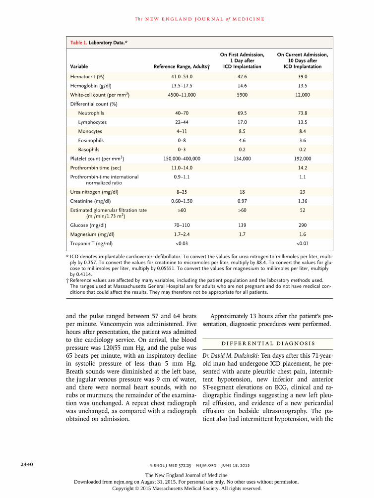
On examination, the temperature was 36.1°C, the blood pressure 130/70 mm Hg, the pulse 78 beats per minute and irregular, the respiratory rate 20 breaths per minute, and the oxygen satu-ration 96% while the patient was breathing am-bient air. The skin was cool, with no inflammation or hematoma over the ICD placement site. Adhe-sive strips over the incision were intact. The first and second heart sounds were normal, with no murmur, and the breath sounds were slightly di-minished at the bases. The jugular venous pressure was not recorded, and the remainder of the ex-amination was normal. Cardiac telemetry showed sinus rhythm with occasional premature ventricu-lar contractions. Blood levels of electrolytes, calci-um, and phosphorus were normal; other test results are shown in Table 1.
Dr. Anand M. Prabhakar: Posteroanterior and lateral chest radiographs (Fig. 1) showed a new left pleural effusion, with no other clinically sig-nificant changes, as compared with the chest radiograph that was obtained immediately after the procedure. The cardiomediastinal silhouette was within normal limits, and the ICD leads ap-peared to be in a position similar to that on the previous radiograph.
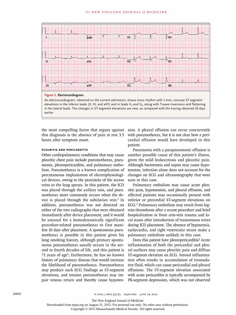
Dr. Clapham: Cardiac ultrasonography, per-formed at the bedside by emergency department personnel, revealed trace pericardial effusion and decreased ventricular contractile function. An electrocardiogram (ECG) showed sinus rhythm at a rate of 66 beats per minute, with new ST-seg-ment elevations in ECG leads II, III, aVF, V3, and V4 (Fig. 2). Aspirin, magnesium sulfate, and in-sulin were administered. Twenty-six minutes later, approximately 3 hours after the patient’s presen-tation, severe pain recurred. Fentanyl was admin-istered intravenously. Approximately 30 minutes after the onset of this pain, light-headedness, diz-ziness, diaphoresis, nausea, and transient hypo-tension (blood pressure, 67/44 mm Hg) occurred, with a pulse of 59 beats per minute. These symp-toms spontaneously improved within minutes; the systolic blood pressure rose to 92 mm Hg and subsequently ranged between 80 and 92 mm Hg,
The New England Journal of Medicine Downloaded from nejm.org on August 31, 2015. For personal use only. No other uses without permission.
Copyright © 2015 Massachusetts Medical Society. All rights reserved.
n engl j med 372;25 nejm.org June 18, 20152440
T h e n e w e ngl a nd j o u r na l o f m e dic i n e
and the pulse ranged between 57 and 64 beats per minute. Vancomycin was administered. Five hours after presentation, the patient was admitted to the cardiology service. On arrival, the blood pressure was 120/55 mm Hg, and the pulse was 65 beats per minute, with an inspiratory decline in systolic pressure of less than 5 mm Hg. Breath sounds were diminished at the left base, the jugular venous pressure was 9 cm of water, and there were normal heart sounds, with no rubs or murmurs; the remainder of the examina-tion was unchanged. A repeat chest radiograph was unchanged, as compared with a radiograph obtained on admission.
Approximately 13 hours after the patient’s pre-sentation, diagnostic procedures were performed.
Differ en ti a l Di agnosis
Dr. David M. Dudzinski: Ten days after this 71-year-old man had undergone ICD placement, he pre-sented with acute pleuritic chest pain, intermit-tent hypotension, new inferior and anterior ST-segment elevations on ECG, clinical and ra-diographic findings suggesting a new left pleu-ral effusion, and evidence of a new pericardial effusion on bedside ultrasonography. The pa-tient also had intermittent hypotension, with the
Variable Reference Range, Adults†
On First Admission, 1 Day after
ICD Implantation
On Current Admission, 10 Days after
ICD Implantation
Hematocrit (%) 41.0–53.0 42.6 39.0
Hemoglobin (g/dl) 13.5–17.5 14.6 13.5
White‑cell count (per mm3) 4500–11,000 5900 12,000
Differential count (%)
Neutrophils 40–70 69.5 73.8
Lymphocytes 22–44 17.0 13.5
Monocytes 4–11 8.5 8.4
Eosinophils 0–8 4.6 3.6
Basophils 0–3 0.2 0.2
Platelet count (per mm3) 150,000–400,000 134,000 192,000
Prothrombin time (sec) 11.0–14.0 14.2
Prothrombin‑time international normalized ratio
0.9–1.1 1.1
Urea nitrogen (mg/dl) 8–25 18 23
Creatinine (mg/dl) 0.60–1.50 0.97 1.36
Estimated glomerular filtration rate (ml/min/1.73 m2)
≥60 >60 52
Glucose (mg/dl) 70–110 139 290
Magnesium (mg/dl) 1.7–2.4 1.7 1.6
Troponin T (ng/ml) <0.03 <0.01
* ICD denotes implantable cardioverter–defibrillator. To convert the values for urea nitrogen to millimoles per liter, multi‑ply by 0.357. To convert the values for creatinine to micromoles per liter, multiply by 88.4. To convert the values for glu‑cose to millimoles per liter, multiply by 0.05551. To convert the values for magnesium to millimoles per liter, multiply by 0.4114.
† Reference values are affected by many variables, including the patient population and the laboratory methods used. The ranges used at Massachusetts General Hospital are for adults who are not pregnant and do not have medical con‑ditions that could affect the results. They may therefore not be appropriate for all patients.
Table 1. Laboratory Data.*
The New England Journal of Medicine Downloaded from nejm.org on August 31, 2015. For personal use only. No other uses without permission.
Copyright © 2015 Massachusetts Medical Society. All rights reserved.

n engl j med 372;25 nejm.org June 18, 2015 2441
Case Records of the Massachusetts Gener al Hospital
overall syndrome developing rapidly. A number of possibilities can explain this constellation of findings; I will consider these and highlight likely and potentially lethal diagnoses.
Myocardial Infarction
Given this patient’s history of ischemic heart dis-ease and multiple cardiac risk factors, the sudden onset of chest pain and new ST-segment eleva-tions raise concerns that he may have had an acute myocardial infarction. Hypotension can result from an anterior or inferior myocardial infarction due to incremental left ventricular systolic dys-function, a concomitant right ventricular infarc-tion in a patient with an inferior myocardial infarction, or a mechanical complication of a subacute infarction, such as papillary muscle rup-ture. A right ventricular infarction is unlikely in this case because no clinically significant ST-seg-ment elevations were present in ECG leads V1 or V4R and because Kussmaul’s sign (the paradoxi-cal rise in jugular venous pressure with inspira-tion)1 was not reported. The presence of new in-ferior and anterior ST-segment elevations, spanning multiple coronary territories, may itself argue against an acute ischemic event. It is important to note that, although the patient had a negative test for troponin T on presentation (3.5 hours after the onset of symptoms), this result does not necessarily rule out myocardial infarction. The pleuritic nature of this patient’s chest pain makes an acute coronary syndrome unlikely.2 This pa-tient also presented with maximal intensity of pain at onset; patients with ischemic pain clas-sically present with a crescendo temporal pattern of pain, whereas patients with aortic, pericarditic, or pleuritic pain are more likely to present with maximal intensity at onset.2
Acute Aortic Syndrome
An acute aortic syndrome of the ascending aorta can result in a dissection in the right coronary artery; this could produce the inferior ST-seg-ment elevations seen on ECG in this case and a hemorrhagic pericardial effusion, which could have caused the patient’s hypotension and pleu-ritic pain. It is unclear why this patient would have pleuritic pain due to a complication of dis-section but not the severe or tearing pain in the chest or back that is characteristic of aortic dis-
section. Nevertheless, the patient’s pulse pres-sure may have widened (from 110/70 mm Hg to 120/55 mm Hg), raising the possibility that he had acute aortic insufficiency, which may be seen in patients with dissection.3 The equal blood pressure in both arms, symmetric radial pulses, absence of murmur associated with aortic insuf-ficiency, and sharp and nonwidened mediastinum on radiography all weigh against a diagnosis of acute aortic syndrome in this patient. Perhaps
Figure 1. Chest Radiographs.
Posteroanterior and lateral chest radiographs (Panels A and B, respectively), obtained on the current admis‑sion, show a new left pleural effusion.
A
B
The New England Journal of Medicine Downloaded from nejm.org on August 31, 2015. For personal use only. No other uses without permission.
Copyright © 2015 Massachusetts Medical Society. All rights reserved.
n engl j med 372;25 nejm.org June 18, 20152442
T h e n e w e ngl a nd j o u r na l o f m e dic i n e
the most compelling factor that argues against this diagnosis is the absence of pain at rest 3.5 hours after symptom onset.
Pleuritis and Pericarditis
Other cardiopulmonary conditions that may cause pleuritic chest pain include pneumothorax, pneu-monia, pleuropericarditis, and pulmonary embo-lism. Pneumothorax is a known complication of percutaneous implantation of electrophysiologi-cal devices, owing to the proximity of the access veins to the lung apexes. In this patient, the ICD was placed through the axillary vein, and pneu-mothorax more commonly occurs when the de-vice is placed through the subclavian vein.4 In addition, pneumothorax was not detected on either of the two radiographs that were obtained immediately after device placement, and it would be unusual for a hemodynamically significant procedure-related pneumothorax to first mani-fest 10 days after placement. A spontaneous pneu-mothorax is possible in this patient given his long smoking history, although primary sponta-neous pneumothorax usually occurs in the sec-ond to fourth decades of life, and this patient is 71 years of age5; furthermore, he has no known history of pulmonary disease that would increase the likelihood of pneumothorax. Pneumothorax may produce such ECG findings as ST-segment elevations, and tension pneumothorax may im-pair venous return and thereby cause hypoten-
sion. A pleural effusion can occur concurrently with pneumothorax, but it is not clear how a peri-cardial effusion would have developed in this patient.
Pneumonia with a parapneumonic effusion is another possible cause of this patient’s illness, given the mild leukocytosis and pleuritic pain. Although bacteremia and sepsis may cause hypo-tension, infection alone does not account for the changes on ECG and ultrasonography that were seen in this case.
Pulmonary embolism may cause acute pleu-ritic pain, hypotension, and pleural effusion, and affected patients may occasionally present with inferior or precordial ST-segment elevations on ECG.6 Pulmonary embolism may result from leg-vein thrombosis after a recent procedure and brief hospitalization or from arm-vein trauma and lo-cal stasis after introduction of transvenous wires during ICD placement. The absence of hypoxemia, tachycardia, and right ventricular strain make a pulmonary embolism unlikely in this case.
Does this patient have pleuropericarditis? Acute inflammation of both the pericardial and pleu-ral surfaces may cause pleuritic pain and diffuse ST-segment elevation on ECG. Serosal inflamma-tion often results in accumulation of transuda-tive fluid, which can cause pericardial and pleural effusions. The ST-segment elevation associated with acute pericarditis is typically accompanied by PR-segment depression, which was not observed
Figure 2. Electrocardiogram.
An electrocardiogram, obtained on the current admission, shows sinus rhythm with 1‑mm, concave ST‑segment elevations in the inferior leads (II, III, and aVF) and in leads V3 and V4, along with T‑wave inversions and flattening in the lateral leads. The changes in ST‑segment elevations are new, as compared with the tracing obtained 10 days earlier.
I
II
III
aVR
aVL
aVF
V1
V2
V3
V4
V5
V6
The New England Journal of Medicine Downloaded from nejm.org on August 31, 2015. For personal use only. No other uses without permission.
Copyright © 2015 Massachusetts Medical Society. All rights reserved.

n engl j med 372;25 nejm.org June 18, 2015 2443
Case Records of the Massachusetts Gener al Hospital
in this case.7 It is unclear whether a new primary serositis would produce enough pleural fluid to be detectable on examination. In addition, most causes of diffuse serositis are associated with an indolent presentation, which is not consistent with this patient’s acute presentation.
Complications of ICD Implantation
Could the features of this patient’s presentation be attributed to the ICD placement? Complica-tions of ICD placement are well described, and ICD lead migration or dislodgment occurs within a few days after implantation in approximately 0.14 to 1.2% of patients8-11; the overall incidence of ICD lead dislodgment is highest in the first weeks after implantation, before myocardial fi-brosis occurs at the site of the tip of the ICD lead. In nearly 11% of patients with lead dislodg-ment, another related major adverse event (e.g., cardiac perforation and tamponade, pneumotho-rax, or cardiac arrest) or in-hospital death oc-curs.9 It is important to note that chest radiogra-phy is not very sensitive in the detection of lead migration. However, when we compare the chest radiographs obtained on the current admission with radiographs obtained 1 day after ICD im-plantation (not shown), there does appear to be a subtle difference in the positions of the leads with respect to the cardiac silhouette. Right ven-tricular myocardial perforation would generate inferior ST-segment elevations as a manifestation of direct cardiac injury. The immediate physio-logical result of free-wall perforation is hemo-pericardium, which would account for the find-ing of pericardial effusion on ultrasonography and the pleuritic pain. Hypotension could be the result of cardiac tamponade due to pericardial effusion, but this is unlikely in a patient who has a relatively normal jugular venous pressure, no pulsus paradoxus, and relative bradycardia (al-though this patient was receiving carvedilol, which may blunt compensatory tachycardia). Of note, tamponade physiology may be associated with large pleural effusions.12 Depending on the ana-tomical trajectory of the dislodged lead, intra-thoracic structures such as the pleura and lungs may be perforated, resulting in hemothorax with hypotension related to brisk blood loss; however, an increasing jugular venous pressure argues against a diagnosis of volume depletion.
During the diagnostic evaluation of a patient who has had any recent medical or surgical pro-
cedure, the clinician should consider and rule out periprocedural complications. The manifestations of ICD lead migration are protean and may be surprisingly subtle; in fact, both the overall ex-amination and initial bedside ultrasound exami-nation in this case may be deceptively interpreted as close to normal. However, the events after a lead perforation may evolve rapidly, and a nor-mal overall examination or ultrasound examina-tion at any one point in time cannot rule out a perforation. To make a diagnosis of lead perfo-ration, a high index of suspicion is required, and the diagnostic strategy must account for all these factors and expeditiously rule out other lethal possibilities, including aortic dissection and pul-monary embolism. I believe that the most likely diagnosis in this case is ICD lead migration and subsequent perforation of the myocardium and pleura, resulting in hemopericardium and hemo-thorax. This diagnosis neatly ties together all the signs and symptoms associated with this pa-tient’s presentation. In order to confirm the di-agnosis, I would perform axial computed to-mography (CT) to define the lead position, repeat device interrogation to confirm changes in lead impedance and capture threshold, and formal echocardiography to assess for pericar-dial effusion and evidence of tamponade physi-ology.13,14
Dr. Eric S. Rosenberg (Pathology): Dr. Ptaszek, what was your impression when you evaluated this patient?
Dr. Leon M. Ptaszek: Although there were nu-merous possible explanations for this patient’s chest pain, we were most concerned about the possibility of a procedural complication. In par-ticular, we were worried that the ICD lead had migrated from its implantation site and perfo-rated the myocardium, resulting in pericardial and pleural effusions. Lead perforation must be addressed promptly, because it can precipitate life-threatening cardiac tamponade within min-utes. Early clinical signs of lead perforation can be subtle and nonspecific, so a rapid and focused evaluation is required, even in the absence of signs of tamponade on physical examination.
Clinic a l Di agnosis
Myocardial perforation by an implantable car-dioverter–defibrillator lead, causing pericardial and pleural effusions.
The New England Journal of Medicine Downloaded from nejm.org on August 31, 2015. For personal use only. No other uses without permission.
Copyright © 2015 Massachusetts Medical Society. All rights reserved.

n engl j med 372;25 nejm.org June 18, 20152444
T h e n e w e ngl a nd j o u r na l o f m e dic i n e
Dr . Dav id M. Dudzinsk i’s Di agnosis
Migration of an implantable cardioverter–defibril-lator lead, resulting in myocardial perforation, hemopericardium, and hemothorax.
Im aging a nd Elec troph ysiol o gic a l S t udies
Dr. Rosenberg: Dr. Ptaszek, what did your evalua-tion show?
Dr. Ptaszek: Routine device interrogation that was performed the morning after ICD implanta-tion revealed that all lead measurements were within normal limits. Repeat device interrogation on the current admission revealed several abnor-mal measurements (Table 2). The voltage of the cardiac electrogram that was sensed by the elec-trode at the tip of the ventricular lead had fallen by nearly an order of magnitude since implanta-tion. In addition, the lead impedance had de-creased considerably since implantation. Pacing of the ventricle could not be achieved, even with maximum output. Together, these findings are consistent with compromised contact between the lead tip and the myocardium. The next step in the evaluation of this patient was to obtain a CT scan of the chest to determine whether the tip of the ventricular lead had migrated through the ventricular wall. Dr. Prabhakar, would you show us the CT scans?
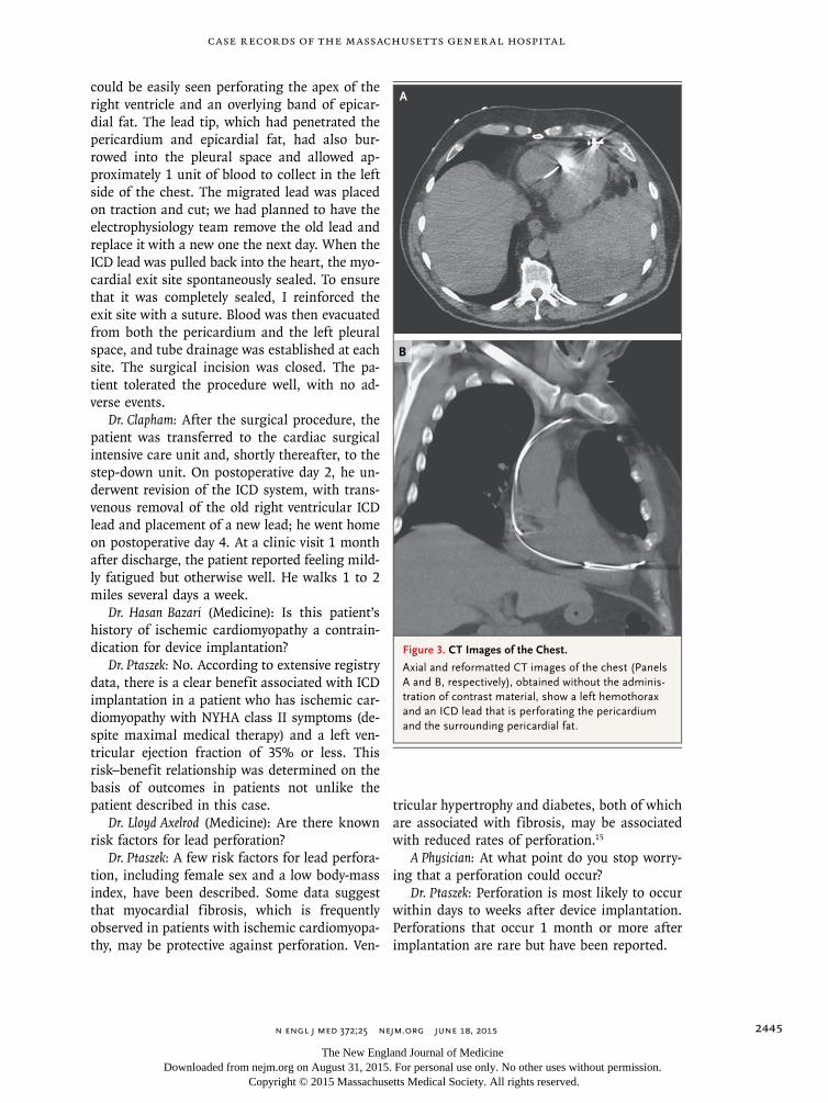
Dr. Prabhakar: Selected axial and reformatted CT images of the chest, obtained without the administration of contrast material, showed a high-density left pleural effusion, with an ICD lead perforating the pericardium and the adja-cent fat. The high-density left pleural effusion is consistent with a hemothorax (Fig. 3).
Dr. Ptaszek: Although the presence of the ven-tricular lead tip outside the myocardial wall was confirmed by the CT scan, lead migration can-not be managed on the basis of imaging find-ings alone. Careful correlation between the im-aging findings and repeat device interrogation is required before a treatment strategy can be formed. It is possible to see a lead tip located beyond the myocardial border on CT without finding any evidence of change in lead measurements or pericardial effusion; this is frequently termed an asymptomatic lead perforation and does not nec-essarily require revision of the lead.15 The changes in lead measurements that were seen in this case can also be observed if the lead detaches from the myocardium but remains inside the heart. It is also important to remember that changes in lead measurements can reveal lead migration even in the absence of definitive imaging findings.
Discussion of M a nagemen t
Dr. Gus J. Vlahakes: The key issue in the manage-ment of a lead perforation is to be prepared for decompensation or a disaster at the time the lead is extracted. Extraction of a migrated lead is performed in the operating room; the patient should receive general anesthesia and be moni-tored with transesophageal echocardiography. In the majority of cases, a lead associated with a perforation can be withdrawn without substan-tial bleeding into the pericardium. In this case, since the patient had an ICD lead with a large diameter and blood in the pericardium that need-ed to be evacuated, I prepared and draped the patient in case the sternum needed to be opened. I opened the subxiphoid part of the chest; with retraction of the diaphragm toward the feet and retraction of the sternum anteriorly, the ICD lead
Variable Normal
On First Admission, 1 Day after
ICD Implantation
On Current Admission, 10 Days after
ICD Implantation
Cardiac electrogram (mV) >4 11.2 1.5
Lead impedance (ohms) 300–1200 640 240
Capture threshold Capture (output, <1.5 V)
Capture (output, 0.5 V;
pulse width, 0.4 msec)
Failure to capture (output, 10 V;
pulse width, 1.5 msec)
Table 2. Measurements for Implantable Cardioverter–Defibrillator Leads.
The New England Journal of Medicine Downloaded from nejm.org on August 31, 2015. For personal use only. No other uses without permission.
Copyright © 2015 Massachusetts Medical Society. All rights reserved.
n engl j med 372;25 nejm.org June 18, 2015 2445
Case Records of the Massachusetts Gener al Hospital
could be easily seen perforating the apex of the right ventricle and an overlying band of epicar-dial fat. The lead tip, which had penetrated the pericardium and epicardial fat, had also bur-rowed into the pleural space and allowed ap-proximately 1 unit of blood to collect in the left side of the chest. The migrated lead was placed on traction and cut; we had planned to have the electrophysiology team remove the old lead and replace it with a new one the next day. When the ICD lead was pulled back into the heart, the myo-cardial exit site spontaneously sealed. To ensure that it was completely sealed, I reinforced the exit site with a suture. Blood was then evacuated from both the pericardium and the left pleural space, and tube drainage was established at each site. The surgical incision was closed. The pa-tient tolerated the procedure well, with no ad-verse events.
Dr. Clapham: After the surgical procedure, the patient was transferred to the cardiac surgical intensive care unit and, shortly thereafter, to the step-down unit. On postoperative day 2, he un-derwent revision of the ICD system, with trans-venous removal of the old right ventricular ICD lead and placement of a new lead; he went home on postoperative day 4. At a clinic visit 1 month after discharge, the patient reported feeling mild-ly fatigued but otherwise well. He walks 1 to 2 miles several days a week.
Dr. Hasan Bazari (Medicine): Is this patient’s history of ischemic cardiomyopathy a contrain-dication for device implantation?
Dr. Ptaszek: No. According to extensive registry data, there is a clear benefit associated with ICD implantation in a patient who has ischemic car-diomyopathy with NYHA class II symptoms (de-spite maximal medical therapy) and a left ven-tricular ejection fraction of 35% or less. This risk–benefit relationship was determined on the basis of outcomes in patients not unlike the patient described in this case.
Dr. Lloyd Axelrod (Medicine): Are there known risk factors for lead perforation?
Dr. Ptaszek: A few risk factors for lead perfora-tion, including female sex and a low body-mass index, have been described. Some data suggest that myocardial fibrosis, which is frequently observed in patients with ischemic cardiomyopa-thy, may be protective against perforation. Ven-
tricular hypertrophy and diabetes, both of which are associated with fibrosis, may be associated with reduced rates of perforation.15
A Physician: At what point do you stop worry-ing that a perforation could occur?
Dr. Ptaszek: Perforation is most likely to occur within days to weeks after device implantation. Perforations that occur 1 month or more after implantation are rare but have been reported.
Figure 3. CT Images of the Chest.
Axial and reformatted CT images of the chest (Panels A and B, respectively), obtained without the adminis‑tration of contrast material, show a left hemothorax and an ICD lead that is perforating the pericardium and the surrounding pericardial fat.
A
B
The New England Journal of Medicine Downloaded from nejm.org on August 31, 2015. For personal use only. No other uses without permission.
Copyright © 2015 Massachusetts Medical Society. All rights reserved.

n engl j med 372;25 nejm.org June 18, 20152446
Case Records of the Massachusetts Gener al Hospital
Fina l Di agnosis
Perforation of the right ventricular wall by an implantable cardioverter–defibrillator lead.
This case was presented at the Medical Case Conference.No potential conflict of interest relevant to this article was
reported.Disclosure forms provided by the authors are available with
the full text of this article at NEJM.org.
References1. Mansoor AM, Karlapudi SP. Kuss-maul’s sign. N Engl J Med 2015; 372(2): e3.2. Swap CJ, Nagurney JT. Value and limi-tations of chest pain history in the evalu-ation of patients with suspected acute coronary syndromes. JAMA 2005; 294: 2623-9.3. Hagan PG, Nienaber CA, Isselbacher EM, et al. The International Registry of Acute Aortic Dissection (IRAD): new in-sights into an old disease. JAMA 2000; 283: 897-903.4. van Rees JB, de Bie MK, Thijssen J, Borleffs CJ, Schalij MJ, van Erven L. Im-plantation-related complications of im-plantable cardioverter-defibrillators and cardiac resynchronization therapy devic-es: a systematic review of randomized clinical trials. J Am Coll Cardiol 2011; 58: 995-1000.5. Bintcliffe O, Maskell N. Spontaneous pneumothorax. BMJ 2014; 348: g2928.6. Wang K, Asinger RW, Marriott HJ. ST-segment elevation in conditions other than acute myocardial infarction. N Engl J Med 2003; 349: 2128-35.7. Dudzinski DM, Mak GS, Hung JW.
Pericardial diseases. Curr Probl Cardiol 2012; 37: 75-118.8. Lee DS, Krahn AD, Healey JS, et al. Evaluation of early complications related to De Novo cardioverter defibrillator im-plantation insights from the Ontario ICD database. J Am Coll Cardiol 2010; 55: 774-82.9. Cheng A, Wang Y, Curtis JP, Varosy PD. Acute lead dislodgements and in-hos-pital mortality in patients enrolled in the National Cardiovascular Data Registry implantable cardioverter defibrillator registry. J Am Coll Cardiol 2010; 56: 1651-6.10. Dewland TA, Pellegrini CN, Wang Y, Marcus GM, Keung E, Varosy PD. Dual-chamber implantable cardioverter-defi-brillator selection is associated with in-creased complication rates and mortality among patients enrolled in the NCDR implantable cardioverter-defibrillator registry. J Am Coll Cardiol 2011; 58: 1007-13.11. Hsu JC, Varosy PD, Bao H, Dewland TA, Curtis JP, Marcus GM. Cardiac perfo-ration from implantable cardioverter-defi-
brillator lead placement: insights from the national cardiovascular data registry. Circ Cardiovasc Qual Outcomes 2013; 6: 582-90.12. Chidambaram S, Sangareddi V, Gane-san G, et al. An echocardiographic as-sessment of cardiovascular hemodynam-ics in patients with large pleural effusion. Indian Heart J 2013; 65: 666-70.13. Henrikson CA, Leng CT, Yuh DD, Brinker JA. Computed tomography to as-sess possible cardiac lead perforation. Pacing Clin Electrophysiol 2006; 29: 509-11.14. Yavari A, Khawaja ZO, Krishnamoor-thy S, McWilliams ET. Perforation of right ventricular free wall by pacemaker lead detected by multidetector computed to-mography. Europace 2009; 11: 252-4.15. Pang BJ, Lui EH, Joshi SB, et al. Pacing and implantable cardioverter defibrillator lead perforation as assessed by multipla-nar reformatted ECG-gated cardiac com-puted tomography and clinical correlates. Pacing Clin Electrophysiol 2014; 37: 537-45.Copyright © 2015 Massachusetts Medical Society.
Lantern SLideS Updated: CompLete powerpoint SLide SetS from the CLiniCopathoLogiCaL ConferenCeS
Any reader of the Journal who uses the Case Records of the Massachusetts General Hospital as a teaching exercise or reference material is now eligible to receive a complete set of PowerPoint slides, including digital images, with identifying legends, shown at the live Clinicopathological Conference (CPC) that is the basis of the Case Record. This slide set contains all of the images from the CPC, not only those published in the Journal. Radiographic, neurologic, and cardiac studies, gross specimens, and photomicrographs, as well as unpublished text slides, tables, and diagrams, are included. Every year 40 sets are produced, averaging 50-60 slides per set. Each set is supplied on a compact disc and is mailed to coincide with the publication of the Case Record.
The cost of an annual subscription is $600, or individual sets may be purchased for $50 each. Application forms for the current subscription year, which began in January, may be obtained from the Lantern Slides Service, Department of Pathology, Massachusetts General Hospital, Boston, MA 02114 (telephone 617-726-2974) or e-mail [email protected].
The New England Journal of Medicine Downloaded from nejm.org on August 31, 2015. For personal use only. No other uses without permission.
Copyright © 2015 Massachusetts Medical Society. All rights reserved.