Embed Size (px)
Citation preview

http://ajs.sagepub.com/Medicine
The American Journal of Sports
http://ajs.sagepub.com/content/41/3/550The online version of this article can be found at:
DOI: 10.1177/0363546512473568
2013 41: 550 originally published online February 4, 2013Am J Sports MedAns Van Ginckel, Peter Verdonk, Jan Victor and Erik Witvrouw
Cartilage Status in Relation to Return to Sports After Anterior Cruciate Ligament Reconstruction
Published by:
http://www.sagepublications.com
On behalf of:
American Orthopaedic Society for Sports Medicine
can be found at:The American Journal of Sports MedicineAdditional services and information for
http://ajs.sagepub.com/cgi/alertsEmail Alerts:
http://ajs.sagepub.com/subscriptionsSubscriptions:
http://www.sagepub.com/journalsReprints.navReprints:
http://www.sagepub.com/journalsPermissions.navPermissions:
What is This?
- Feb 4, 2013OnlineFirst Version of Record
- Mar 1, 2013Version of Record >>
at Instituto Nacional de Educ on April 24, 2014ajs.sagepub.comDownloaded from at Instituto Nacional de Educ on April 24, 2014ajs.sagepub.comDownloaded from

Cartilage Status in Relation toReturn to Sports After AnteriorCruciate Ligament Reconstruction
Ans Van Ginckel,*yz PT, MSc, Peter Verdonk,§ MD, PhD,Jan Victor,§ MD, PhD, and Erik Witvrouw,z|| PT, PhDInvestigation performed at the Department of Rehabilitation Sciences and Physical Therapy,Ghent University, Ghent, Belgium
Background: Osteoarthritis after anterior cruciate ligament (ACL) reconstruction receives much attention in orthopaedic science.Anterior cruciate ligament reconstruction is related to increased joint fluid volumes, bone marrow edema, and cartilage biochem-ical and morphological changes believed to cause fragile joint conditions. These joint conditions may not be able to adequatelycounter the imposed loads during sports.
Hypothesis: At 6 months after surgery, knee cartilage displays inferior quality in ACL-reconstructed knees when compared withcontrols. This inferior quality is influenced by the time to return to sports and/or by the time to surgery.
Study Design: Cross-sectional study; Level of evidence, 3.
Methods: Fifteen patients treated with isolated ACL reconstruction were compared with 15 matched controls. In all participants, a 3-Tmagnetic resonance imaging cartilage evaluation was performed entailing quantitative morphological characteristics (3-dimensionalvolume/thickness), biochemical composition (T2/T2* mapping), and function (after a 30-minute run: in vivo deformation including recov-ery). Nonparametric statistics were executed reporting median (95% CI).
Results: No volume and thickness between-group differences existed. In patients, medial femur (FM) T2 was higher (45.44 ms [95%CI, 40.64-51.49] vs 37.19 ms [95% CI, 34.67-40.39]; P = .028), whereas T2* was lower in the FM (21.81 ms [95% CI, 19.89-22.74] vs24.29 ms [95% CI, 22.70-26.26]; P = .004), medial tibia (TM) (13.81 ms [95% CI, 10.26-16.78] vs 17.98 ms [95% CI, 15.95-18.90]; P =.016), and lateral tibia (TL) (14.69 ms [95% CI, 11.71-16.72] vs 18.62 ms [95% CI, 17.85-22.04]; P \ .001). Patients showed dimin-ished recovery at 30 minutes after a 30-minute run in the FM (–1.60% [95% CI, –4.82 to –0.13] vs 0.01% [95% CI, –0.34 to 1.23]; P =.040) and at 30 (–3.76% [95% CI, –9.29 to –1.78] vs 0.04% [95% CI, –1.52 to –0.72]; P = .004) and 45 minutes after exercise (–1.86%[95% CI, –4.66 to –0.40] vs 0.43% [95% CI, –0.91 to 0.77]; P = .024) in the TL. Eight patients returned to sports at 6 months or earlier.Return before 5 months (3/8 patients) was associated with increased cartilage thickness (in TM, TL, and lateral femur [FL]), defor-mation (in FL), and delayed recovery after running (in FL and FM). Median surgical delay was 10 weeks (range, 5-17 weeks). Surgerywithin 10 weeks (9/15 patients) was also associated with delayed cartilage recovery (in FL and FM). For the other parameters, nosignificant relationships could be established for either return to sports or surgical delay.
Conclusion: At 6 months after surgery, cartilage in patients with ACL reconstruction shows diminished quality and in vivo resil-iency compared with controls. Caution is advised in an early return to sports especially when dealing with patients who receivedprompt surgery. Possibly, high impacts on this qualitatively diminished cartilage might play a role in the development of osteo-arthritis in ACL reconstruction. Replication in larger samples and follow-up are warranted.
Keywords: anterior cruciate ligament; MRI; cartilage; return to sports
Osteoarthritis (OA) after anterior cruciate ligament (ACL)reconstruction is a comorbidity receiving much attention inorthopaedic science. Pivoting sports are considered ‘‘highrisk’’ for the incidence of ACL ruptures.18 According toa review by Myklebust and Bahr,18 treatment with ACLreconstruction effects a return to sports in 65% to 88% ofathletes compared with 19% to 82% enrolled in nonopera-tive treatments.
Postoperative sports participation, however, might not betailored to ongoing structural remodeling of the traumatizedknee. Surgery within 6 weeks after injury showed increasedjoint fluid volumes combined with slowly resorbing bone mar-row lesions (BMLs) and cartilage morphological changes dur-ing the first year. Hence, these signs depict precarious jointconditions that might not be able to counter the excessive tor-sional loads that the knee would be subjected to when return-ing to strenuous activities.8,9 Interestingly, in a 6-yearprospective study, Keays et al14 found that type or level ofpostoperative sports played was not a predictor for the inci-dence of radiographic OA after ACL reconstruction. In casesof no further sustained chondral or meniscal damage
The American Journal of Sports Medicine, Vol. 41, No. 3DOI: 10.1177/0363546512473568� 2013 The Author(s)
550 at Instituto Nacional de Educ on April 24, 2014ajs.sagepub.comDownloaded from

requiring meniscectomy, pursuing athletic activities was sug-gested as not accelerating joint deterioration. Surgical delay(mean, 40.59 months), however, was in this particular studymuch longer and related to OA development, whereas thetime to return to play was not reported or accounted for inthe analysis. In cases of an average 3-month surgical delay,Hoffelner et al11 concluded that patients, who all returnedto competitive sports within 8 months after isolated ACL sur-gery, showed no increased OA risk at 10 years’ follow-up.Osteoarthritis was detected in 36% of operated knees; how-ever, no significant differences could be attained aftercross-sectional within-patient comparison.
To date, it is agreed that surgical delay determines thepresence of concomitant injury on arthroscopic evaluation,affecting the risk for OA progression.10 Surgery is proposedto be performed within the first year, whereas a delay ofover 6 months was even put forward as a risk factor forOA development.5,10,23 In athletes wishing to return to piv-oting sports, reconstruction is advised to be performedwithin 4 to 8 weeks in cases of absent joint swelling andfull range of motion.18 Also, in cases of surgery within 6weeks, ACL-reconstructed knees were suggested to benefitfrom a longer recovery in view of the morphological sequel-lae that the joint seemed to endure after surgery.8,9 Conse-quently, the impact of return to sports and surgical delayon future joint deterioration remains enigmatic.
In clinical practice, most patients are usually dischargedfrom rehabilitation and attempt a full return to leisure orathletic activities at an average of 6 months after surgery.16
Therefore, the purpose of this study was 2-fold. First, a com-prehensive evaluation of cartilage status was undertaken inACL-reconstructed patients 6 months after surgery and com-pared with matched control patients. Using 3-T magneticresonance imaging (MRI), the cartilage evaluation entailedin vivo quantitative morphological characteristics, biochemi-cal composition, and function (ie, tissue resiliency). Second,within patients, the role of time to return to sports and/orsurgical delay on cartilage status was explored. It washypothesized that knee cartilage would display inferior qual-ity when compared with controls. An early return to sportsand/or surgery (ie, too soon8,9) was hypothesized to contrib-ute to a worse cartilage outcome at 6 months after surgery.These results may have implications on fine-tuning rehabili-tation strategies and surgical planning.
MATERIALS AND METHODS
Patients
Fifteen patients (8 men, 7 women) who underwent ACLreconstructive arthroscopic surgery were recruited from
an experienced orthopaedic surgeon’s practice. All patientsreceived surgery between April 2010 and April 2011. Eligi-bility to participate was evaluated based on arthroscopicevaluation and standard questionnaires. Inclusion criteriawere (1) 20 to 40 years of age to reduce the risk of cartilagedegeneration associated with ageing, (2) body mass index(BMI) of 20 to 30, (3) regular sports participation beforeinjury, (4) isolated ACL reconstruction using hamstringtendon autografts, and (5) being able to run 30 minutes.Exclusion criteria were (1) any cartilage lesion presenton arthroscopic evaluation or diagnosed before injury asthe latter might have evolved into degenerative changesregardless of the incident, (2) concomitant meniscectomyor meniscal sutures, (3) concomitant collateral ligamentinjuries �grade 2, (4) history of knee injury and/or surgery,(5) MRI contraindications, and (6) other joint and/or bonelesions. In all patients, anatomic ACL reconstruction wasperformed using a double semitendinosus and gracilisautograft with a cortical suspension system (EndobuttonCL, Smith & Nephew, Andover, Massachusetts) on thefemoral side and use of a metal interference screw (RCI,Smith & Nephew) and staple on the tibial side. Briefly,anatomic position was achieved with the tibial tunnel inthe center of the ACL remnant and through the anterome-dial portal for the femoral tunnel. The grafts were fixedwith the knee in 30� of flexion. After surgery, all patientsenrolled in a structured 4- to 6-month rehabilitationprogram.
Fifteen control participants were recruited from thecommunity or university campus. Eligibility was verifiedusing standard questionnaires. Control participants werematched to the patients by sex, age, BMI, sports participa-tion (ie, type of sports and training volume as determinedby the Baecke sport index2), and dominance of the limbto be investigated. Additionally, all controls were able torun 30 minutes. Exclusion criteria were (1) a history ofknee injury including cartilage defects and/or surgery, (2)known bone and/or joint pathological abnormality includ-ing clinical or MRI-related signs of OA at the time of thestudy, and (3) MRI contraindications.
Informed consent forms ratified by the local ethics com-mittee were obtained from all patients. Patient demo-graphics are listed in Table 1.
Experimental Procedures
As the patients were evaluated 6 months after surgery,experimental procedures took place from October 2010until November 2011. All patients were instructed not topractice sports the day before testing or on the testingday and to avoid running, lifting heavy weights, and tak-ing stairs 4 hours preceding the actual experimental
*Address correspondence to Ans Van Ginckel, PT, MSc, Department of Rehabilitation Sciences and Physical Therapy, Ghent University Hospital Cam-pus, 3B3, De Pintelaan 185, BE-9000 Ghent, Belgium (e-mail: [email protected]).
yResearch Foundation–Flanders, Brussels, Belgium.zDepartment of Rehabilitation Sciences and Physical Therapy, Ghent University, Ghent, Belgium.§Department of Physical Medicine and Orthopedic Surgery, Ghent University, Ghent, Belgium.||Department of Physiotherapy, Aspetar, Doha, Qatar.
One or more of the authors has declared the following potential conflict of interest or source of funding: This study was funded by Research Foundation–Flanders.
Vol. 41, No. 3, 2013 Cartilage Quality After ACL Reconstruction and Return to Sports 551
at Instituto Nacional de Educ on April 24, 2014ajs.sagepub.comDownloaded from
procedures. The procedures were performed on campusand occurred at the same time of day for all patients.27
The protocol comprised (1) MRI evaluation of cartilage sta-tus including in vivo quantitative morphological character-istics (ie, 3-dimensional [3D] volume/thickness),biochemical composition (ie, T2/T2* mapping), and func-tion (ie, deformational behavior including recovery); (2)evaluation of lower limb function and knee laxity; and (3)questionnaires.
MRI Evaluation of Cartilage Status: 3D Morphology,Biochemical Composition, and Function. High-resolutionimages of knee cartilage morphology (ie, sagittal 3D doubleecho steady state sequence with fat suppression by waterexcitation [DESS WE]) and baseline biochemical T2/T2*maps (T2/T2* MapIt, Siemens Medical Solutions, Erlan-gen, Germany) were obtained using a dedicated 8-channelknee coil on a 3-T Trio Tim magnet (Siemens MedicalSolutions). Cartilage deformation and recovery were regis-tered by means of monitoring the changes in cartilage mor-phological characteristics before and after an in vivoweightbearing exercise.27 Upon diagnosis of the ACLrupture and at 6 months after surgery, an Intermediate(IM)-weighted fat-saturated turbo spin echo (TSE)sequence was included to determine the presence of BMLs.
The sagittal 3D DESS WE sequence implemented a par-tition thickness of 0.69 mm, echo time/repetition time [TE/TR] of 4.7/16.44 milliseconds, flip angle [FA] of 25�, field of
view [FOV] of 140 mm, matrix of 384 pixels (in-plane res-olution: 0.36 3 0.36), and acquisition time (TA) of 60160 0.The T2 map, a sagittal multiecho spin echo sequence, cen-tered on the medial and lateral knee compartment imple-mented a partition thickness of 3 mm; TE/TR of 13.8,27.6, 41.4, 55.2, and 69.0/1000 milliseconds; FA of 180�;FOV of 159 mm; matrix of 384 pixels (in-plane resolution:0.42 3 0.42); and TA of 30280 0. The T2* map, a sagittal mul-tiecho gradient echo sequence, centered on the medial andlateral knee compartment implemented a partition thick-ness of 3 mm; TE/TR of 4.18, 11.32, 18.46, 25.60, and 32.47/422 milliseconds; FA of 60�; FOV of 159 mm; matrix of 384(interpolated 768 3 768) pixels (in-plane resolution: 0.21 3
0.21); and TA of 204200. The IM-weighted TSE sequenceimplemented a partition thickness of 3 mm, TE/TR of 44/3140 milliseconds, FA of 160�, FOV of 134 3 179 mm,matrix of 448 3 218 pixels (in-plane resolution: 0.40 3
0.40), and TA of 20340 0.To reduce interference from residual deformation pre-
ceding the experiment, the MRI protocol started with1 hour of physical rest during which the participantswere positioned supine.15,27 After the patient had rested,baseline scans (ie, tpre: sagittal 3D DESS WE, T2 andT2* maps, IM-weighted TSE) were performed followed bythe weightbearing exercise under study. Within a maxi-mum of 2 minutes after exercise cessation,15 the first post-scans (ie, tpostt0: sagittal 3D DESS WE) were started and
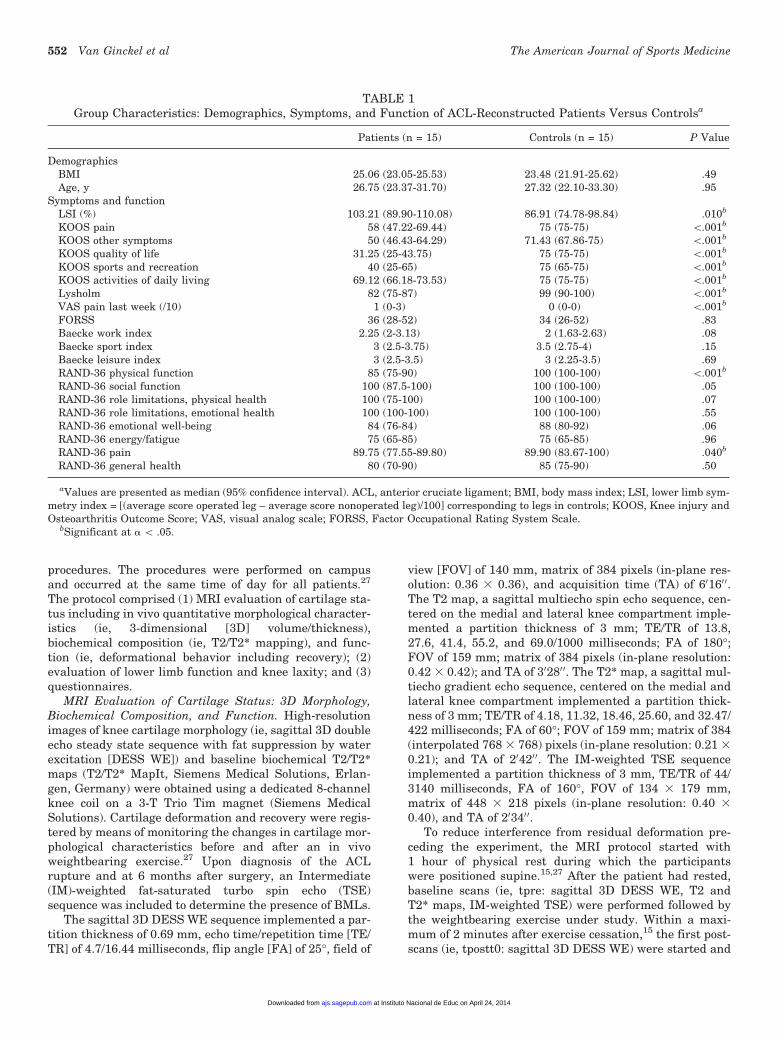
TABLE 1Group Characteristics: Demographics, Symptoms, and Function of ACL-Reconstructed Patients Versus Controlsa
Patients (n = 15) Controls (n = 15) P Value
DemographicsBMI 25.06 (23.05-25.53) 23.48 (21.91-25.62) .49Age, y 26.75 (23.37-31.70) 27.32 (22.10-33.30) .95
Symptoms and functionLSI (%) 103.21 (89.90-110.08) 86.91 (74.78-98.84) .010b
KOOS pain 58 (47.22-69.44) 75 (75-75) \.001b
KOOS other symptoms 50 (46.43-64.29) 71.43 (67.86-75) \.001b
KOOS quality of life 31.25 (25-43.75) 75 (75-75) \.001b
KOOS sports and recreation 40 (25-65) 75 (65-75) \.001b
KOOS activities of daily living 69.12 (66.18-73.53) 75 (75-75) \.001b
Lysholm 82 (75-87) 99 (90-100) \.001b
VAS pain last week (/10) 1 (0-3) 0 (0-0) \.001b
FORSS 36 (28-52) 34 (26-52) .83Baecke work index 2.25 (2-3.13) 2 (1.63-2.63) .08Baecke sport index 3 (2.5-3.75) 3.5 (2.75-4) .15Baecke leisure index 3 (2.5-3.5) 3 (2.25-3.5) .69RAND-36 physical function 85 (75-90) 100 (100-100) \.001b
RAND-36 social function 100 (87.5-100) 100 (100-100) .05RAND-36 role limitations, physical health 100 (75-100) 100 (100-100) .07RAND-36 role limitations, emotional health 100 (100-100) 100 (100-100) .55RAND-36 emotional well-being 84 (76-84) 88 (80-92) .06RAND-36 energy/fatigue 75 (65-85) 75 (65-85) .96RAND-36 pain 89.75 (77.55-89.80) 89.90 (83.67-100) .040b
RAND-36 general health 80 (70-90) 85 (75-90) .50
aValues are presented as median (95% confidence interval). ACL, anterior cruciate ligament; BMI, body mass index; LSI, lower limb sym-metry index = [(average score operated leg – average score nonoperated leg)/100] corresponding to legs in controls; KOOS, Knee injury andOsteoarthritis Outcome Score; VAS, visual analog scale; FORSS, Factor Occupational Rating System Scale.
bSignificant at a \ .05.
552 Van Ginckel et al The American Journal of Sports Medicine
at Instituto Nacional de Educ on April 24, 2014ajs.sagepub.comDownloaded from

repeated with 15-minute intervals up to 45 minutes afterthe exercise (ie, tpostt15, tpostt30, and tpostt45: sagittal3D DESS WE) (total of 4 scans).27 ‘‘Deformation’’ isexpressed as the morphological change measured attpostt0 relative to baseline (ie, [(3D morphology tpostt0 –3D morphology tpre)/3D morphology tpre 3 100]). Morpho-logical changes measured at tpostt15, tpostt30, andtpostt45 relative to baseline are attributed to ‘‘recovery.’’27
The sequence of events is displayed in Figure 1.The weightbearing exercise consisted of a 30-minute
run on a predefined track on campus. Participants ran ata self-selected comfortable pace during which runningspeed (km/h), distance (km), and step count were recorded.To standardize the cushioning properties of footwear, allpatients wore the same type of neutral running shoe dur-ing the experiment (Ekiden 50; Kalenji, Villeneuve D’ascq,France). After running, all patients provided a visual ana-log scale (VAS) score for knee pain experienced during theexercise (on a 10-cm scale: 0 cm representing ‘‘no pain atall’’ and 10 cm representing ‘‘extremely painful’’). In viewof the active patient population under study, runningwas preferred as it is a basic component in (late) postoper-ative rehabilitation and in many sports activities. A30-minute run was adequate to allow for cartilage changesto be detected using MRI.21,25 Tibial plates have beenshown to recover within 60 minutes after a 20-km run, pro-viding a rationale to repeat postscans up to tpostt45.15
Evaluation of Lower Limb Function and Knee Laxity.Functional performance was evaluated using the single-legged hop test for distance, and lower limb symmetryindex (LSI) values were calculated. Anteroposterior kneelaxity and side-to-side differences were quantified usingthe Genourob device (Genourob, Laval, France) withincreasing loads (67, 89, 134, 150, and 250 N).
Questionnaires. All patients completed a Baecke ques-tionnaire quantifying general physical activity scoresbased on a work index, sport index, and leisure index2;Tegner scale for current activity level; Factor OccupationalRating System Scale (FORSS)19; Knee injury and Osteoar-thritis Outcome Score (KOOS); Lysholm knee score; andRAND 36-Item Health Survey. Within the patient group,additional data were collected concerning the injury andoperation date, injury event (ie, activity during incident),VAS score for the amount of pain experienced during thelast week (0 representing ‘‘no pain at all’’ and 10 represent-ing ‘‘extremely painful’’), and return to sports at 6 monthsafter surgery (If ‘‘yes’’: [1] What kind of sports is practicedat what weekly frequency? [2] When did you return tosports?). According to Keays et al,14 postsurgery sportswere allocated to 3 categories: ‘‘safe,’’ ‘‘low risk,’’ and‘‘high risk.’’ Reasons for no return were also collected.
Data Analysis
Image Analysis: 3D Morphology and T2/T2* Quantifi-cation. Three-dimensional reconstruction, volume calcula-tion, surface area calculation, and model registrationwere performed using a commercial modeling softwarepackage (Mimics, version 14.0, Materialise NV, Leuven,Belgium).27
Three-dimensional DESS image stacks were subse-quently segmented to generate a 3D reconstruction oflateral/medial femoral/tibial cartilage (lateral femur [FL],medial femur [FM], lateral tibia [TL], medial tibia [TM]).A semiautomatic segmentation procedure was imple-mented based on a 3D LiveWire algorithm and a slice-by-slice manual correction to digitize cartilage plates by mask-ing. Manual correction was preceded by a region growingalgorithm to dispose of abundant voxels. Subsequently,3D cartilage plates were reconstructed, and absolute 3Dvolumes (in mm3) were calculated for baseline and post-scans.27 Normalized 3D cartilage volumes (ie, normalizedto cartilage-bone interface area: [absolute 3D volume(mm3)/cartilage-bone interface area (mm2)]) were deter-mined. Cartilage-bone interface area was defined as thearea of the underlying bone surface in contact with the car-tilage plate. By means of eroding morphological operations,surface areas were extracted from 3D reconstructions ofthe underlying bone and determined by means of surfacetriangulation.27 Normalized 3D volumes are consideredan equivalent measurement of cartilage thickness for thecartilage areas under study13 and are referred to as ‘‘thick-ness (in mm)’’ in this article.
The T2/T2* relaxation times were derived from onlinereconstructed maps centered on the medial/lateral com-partment using a pixel-wise, mono-exponential, nonnega-tive least squares fit analysis (MapIt, Siemens MedicalSolutions). Regions of interest covering full-thickness car-tilage were segmented, delineating the entire layers to cal-culate the global T2/T2*. Next to cartilage tissue, posteriorhorns of the menisci were segmented. Although casestreated with meniscal procedures were excluded, subclini-cal meniscal degeneration was considered important in
1 hour rest
15’
30-minute run
Post-exercise scans: 3D DESS
B
B Baseline scans: T2/T2∗ MapIt, 3D DESS, TSE
1 2 3 4
1- 4
15’ 15’
Figure 1. Schematic overview of the sequence of eventsduring the magnetic resonance imaging experimental proto-col. Adapted from Van Ginckel et al.27 B, baseline scans(tpre); 1-4, postexercise scans (tpostt0, tpostt15, tpostt30,and tpostt45: postexercise scans started within a maximumof 2 minutes after cessation of exercise [tpostt0] andrepeated at 15, 30, and 45 minutes after onset of tpostt0,respectively).
Vol. 41, No. 3, 2013 Cartilage Quality After ACL Reconstruction and Return to Sports 553
at Instituto Nacional de Educ on April 24, 2014ajs.sagepub.comDownloaded from
view of cartilage health outcomes.28,29 The 3D DESS WEimages served as visual guidance.
All image analyses were performed by a singleresearcher with 3 years of experience at the time of analy-sis and who was blinded to the time sequence of scanning.
Intratester reliability, precision errors for volume, andT2/T2* quantification were determined (see the Appendix,available online at http://ajs.sagepub.com/supplemental/).
Statistical Analysis. The Shapiro-Wilk test revealed anoverall nonparametric distribution (P \ .05). Hence, non-parametric statistics were executed, and descriptive statis-tics were reported as median (95% confidence interval[CI]).
To investigate differences between groups, the Mann-Whitney U or Kruskal-Wallis test was applied. To testwhether cartilage morphology for all plates changed overtime, the Friedman test for repeated measures was imple-mented. Post hoc pairwise comparisons were conductedusing Wilcoxon signed-rank tests. P values were adjustedfor multiple comparisons of main effects (‘‘time’’ or ‘‘carti-lage plate’’) by applying Bonferroni corrections. Spearmanr coefficients were calculated to investigate all correla-tions. Significant correlation coefficients of �0.5 were con-sidered. Level of significance was set at a \ .05; SPSS(version 20, IBM Statistics, Armonk, New York) wasused for all analyses.
RESULTS
Group Characteristics: Demographics, Symptoms,Function, Preinjury Sports Participation,and Injury Event
No significant between-group differences were present forBMI (P = .49); age (P = .95); Baecke work, sport, and lei-sure index (P = .08, P = .15, and P = .69, respectively);FORSS (P = .83); and all RAND 36 items except physicalfunction and pain (Table 1). The latter were significantlydecreased in the patients (P \ .001 and P = .04, respec-tively). Additionally, in patients, Lysholm knee scores(P \ .001), KOOS values (all subitems, P \ .001), andLSI values (P = .010) were significantly decreased whencompared with controls. On VAS scores, patients reportedno to mild knee pain during the last week before the study(median, 1 [95% CI, 0-3]). Median (95% CI) and P valuesare tabulated in Table 1.
Sports practiced before injury were soccer (5/15), out-door running (5/15), climbing (1/15), horseback riding(1/15), inline skating (1/15), rugby (1/15), and volleyball(1/15). Injury events were soccer (6/15), alpine skiing(4/15), rugby (1/15), volleyball (1/15), and other outdooractivities (3/15).
Side-to-side differences in knee laxity in patientsranged from 1.60 mm (95% CI, –0.24 to 2.10), 1.64 mm(95% CI, –0.74 to 2.30), 1.60 mm (95% CI, –0.46 to 2.70),and 1.70 mm (95% CI, –0.57 to 2.90) to 2.36 mm (95% CI,–0.93 to 3.51) with increasing loads (67, 89, 134, 150, and250 N, respectively).
Cartilage Status: 3D Morphological Characteristics,Biochemical Composition, and Function
No significant between-group differences could be estab-lished for baseline 3D volume and thickness in all plates(Table 2). Global T2 relaxation times in the FM were signif-icantly higher in patients when compared with controls(45.44 ms [95% CI, 40.64-51.49] vs 37.19 ms [95% CI,34.67-40.39], respectively; P = .028). No other significantbetween-group differences could be revealed. Global T2*relaxation times were significantly lower in patientswhen compared with controls in the FM (21.81 ms [95%CI, 19.89-22.74] vs 24.29 ms [95% CI, 22.70-26.26], respec-tively; P = .004), TM (13.81 ms [95% CI, 10.26-16.78] vs17.98 ms [95% CI, 15.95-18.90], respectively; P = .016),and TL (14.69 ms [95% CI, 11.71-16.72] vs 18.62 ms [95%CI, 17.85-22.04], respectively; P \ .001). No significantbetween-group differences could be shown for the FL. T2/T2* relaxation times did not differ between groups forthe meniscal posterior horn in the medial and lateral com-partment. Median (95% CI) T2 and T2* relaxation timesand P values for all cartilage plates and menisci are dis-played in Table 2.
Regarding cartilage function, none of the plates showeda significant between-group difference at deformation(tpostt0: P = .80, P = 1.00, P = 1.00, and P = 1.00 for FM,FL, TM, and TL, respectively). During recovery, thepatient group’s FM continued to exhibit significantlylarger volume decreases at tpostt15 when compared withcontrols (–1.60% [95% CI, –4.82 to –0.13] vs 0.01% [95%CI, –0.34 to 1.23], respectively; P = .040). In the TL, thepatients showed significantly larger volume decreases attpostt30 (–3.76% [95% CI, –9.29 to –1.78] vs 0.04% [95%CI, –1.52 to –0.72], respectively; P = .004) and tpostt45(–1.86% [95% CI, –4.66 to –0.40] vs 0.43% [95% CI,–0.91 to 0.77], respectively; P = .024). During recovery,no other significant between-group differences were found(Table 3).
Within controls, none of the morphological changes dif-fered significantly in the FM (P = .73, P = 1.00, P = .27, andP = .27, respectively) and TM (P = .10, P = .28, P = 1.00, andP = 1.00, respectively) when compared with baseline. Inthe FL (P = .020), only changes at tpostt0 differed signifi-cantly from baseline. In the TL, decreases at both tpostt0(P = .005) and tpostt15 (P = .035) were significant.
Within patients, morphological changes measured atboth tpostt0 and tpostt15 differed significantly in the FM(P = .005 and P = .030, respectively) and FL (P = .005and P = .030, respectively) when compared with baseline.In the TL, decreases at tpostt0 (P = .005), tpostt15 (P =.005), tpostt30 (P = .005), and tpostt45 (P = .025) remainedsignificant when compared with baseline. In the TM, noneof the changes differed significantly from baseline (P = .15,P = .26, P = .46, and P = .82, respectively).
No significant between-group differences in speed (P =.72), distance (P = .62), and step count (P = .51) wereexposed during running. In patients, median speed was10.00 km/h (95% CI, 7.20-11), distance was 4.82 km(95% CI, 3.75-5.38), and step count was 4680 (95% CI,
554 Van Ginckel et al The American Journal of Sports Medicine
at Instituto Nacional de Educ on April 24, 2014ajs.sagepub.comDownloaded from

TABLE 3Cartilage Function, Deformation, and Recovery: 3-Dimensional Morphological Changes
After Exercise Relative to Baseline Within and Between Groupsa
Patients (n = 15) Controls (n = 15) P Value
FMChange 1 (at tpostt0) 23.37 (–9.97 to –1.6)b 21.69 (–4.1 to –0.88) .80Change 2 (at tpostt15) 21.60 (–4.82 to –0.13)b 0.01 (–0.34 to 1.23) .040c
Change 3 (at tpostt30) 20.40 (–3.77 to 0.99) 0.49 (0 to 1.23) .42Change 4 (at tpostt45) 20.06 (–2.03 to 1.06) 0.49 (0.01 to 1.23) .93
FLChange 1 (at tpostt0) 23.74 (–6.70 to –2.06)b 23.28 (–6.59 to –0.55)b 1.00Change 2 (at tpostt15) 21.83 (–3.82 to –0.09)b 0.28 (–1.21 to 1.30) .11Change 3 (at tpostt30) 20.44 (–2.45 to 0.39) 0.49 (0.12 to 1.53) .92Change 4 (at tpostt45) 0.25 (–0.74 to 0.76) 0.49 (0.15 to 1.53) .93
TMChange 1 (at tpostt0) 28.61 (–10.96 to –2.90) 26.20 (–9.52 to –0.42) 1.00Change 2 (at tpostt15) 26.53 (–9.25 to –1.59) 21.82 (–4.54 to 0.54) .50Change 3 (at tpostt30) 24.63 (–8.34 to 0.78) 0.26 (–1.94 to 1.16) .35Change 4 (at tpostt45) 21.45 (–5.81 to 0.78) 0.54 (–1.64 to 1.61) .64
TLChange 1 (at tpostt0) 26.63 (–11.07 to –4.32)b 25.95 (–8.70 to –2.48)b 1.00Change 2 (at tpostt15) 26.53 (–11.07 to –2.65)b 22.37 (–5.50 to 0)b .06Change 3 (at tpostt30) 23.76 (–9.29 to –1.78)b 0.036 (–1.52 to 0.72) .004c
Change 4 (at tpostt45) 21.86 (–4.66 to –0.40)b 0.43 (–0.91 to 0.77) .024c
aValues are presented as median (95% confidence interval). FM, medial femur; FL, lateral femur; TM, medial tibia; TL, lateral tibia.tpostt0, tpostt15, tpostt30, and tpostt45: morphology measured within a maximum of 2 minutes after cessation of exercise (tpostt0) andat 15, 30, and 45 minutes after onset of tpostt0, respectively.
bSignificant difference relative to baseline within groups at a \ .05.cSignificant difference between groups at a \ .05.
TABLE 2Cartilage and Meniscal Status: Baseline Quantitative 3-Dimensional Morphological Characteristics
and Biochemical Composition in Patients Versus Controlsa
Patients (n = 15) Controls (n = 15) P Value
Volume, mm3
FM 5025.76 (4310.52-5963.46) 4669.68 (3624.92-5694.33) 1.00FL 5565.77 (4632.14-6890.84) 5285.34 (4144.82-6832.41) 1.00TM 1991.77 (1687-2126.26) 1906.99 (1455.70-2295.19) 1.00TL 2598.78 (1778.36-2877.70) 2236.29 (1865.06-2744.33) 1.00
Thickness, mmFM 1.94 (1.6-2.13) 1.76 (1.6-1.93) .49FL 1.96 (1.65-2.17) 1.96 (1.56-2.16) 1.00TM 1.90 (1.57-1.94) 1.78 (1.48-1.9) 1.00TL 2.24 (2.14-2.41) 2.09 (1.95-2.24) .80
Cartilage T2, msFM 45.44 (40.64-51.49) 37.19 (34.67-40.39) .028b
FL 35.54 (33.19-42.56) 38.63 (35.83-41.58) 1.00TM 28.65 (26.10-36.88) 29.74 (27.08-33.16) 1.00TL 31.30 (25.57-41.73) 30.95 (25.93-36.60) 1.00
Cartilage T2*, msFM 21.81 (19.89-22.74) 24.29 (22.7-26.26) .004b
FL 21.65 (20.62-23.63) 23.38 (20.87-24.81) .94TM 13.81 (10.26-16.78) 17.98 (15.95-18.9) .016b
TL 14.69 (11.71-16.72) 18.62 (17.85-22.04) \.001b
Meniscus T2, msMedial compartment 19.41 (16.34-26.07) 23.08 (18.32-28.36) .28Lateral compartment 18.83 (17.46-24.58) 17.93 (15.23-21.8) .23
Meniscus T2*, msMedial compartment 7.95 (7-8.33) 7.45 (6.98-9.31) .72Lateral compartment 7.56 (5.8-7.75) 7.48 (6.33-7.74) .80
aValues are presented as median (95% confidence interval). FM, medial femur; FL, lateral femur; TM, medial tibia; TL, lateral tibia.bSignificant at a \ .05.
Vol. 41, No. 3, 2013 Cartilage Quality After ACL Reconstruction and Return to Sports 555
at Instituto Nacional de Educ on April 24, 2014ajs.sagepub.comDownloaded from

4577-4835). In controls, median speed was 9.80 km/h (95%CI, 9.00-10.70), distance was 4.94 km (95% CI, 4.64-5.30),and step count was 4742 (95% CI, 4737-4753). The patientsreported no to mild knee pain during running (VAS score:median, 1.60 [95% CI, 0.00-2.90]). For all plates at all timepoints, percentage changes and P values are tabulated inTable 3 for both groups.
Upon diagnosis, BMLs were present on MRI in 10 of 15cases (1 or multiple locations/patient: 12.5% posterior TM,12.5% weightbearing third FL, 25% anterolateral FL, 50%posterior TL), there was no BML present in 2 of 15, andthere were no baseline MRI scans available in 3 of 15. At6 months after surgery, in all but 1 MRI scan, no BMLwas detected.
Return to Sports and Surgical Delay
At 6 months after surgery, 8 of 15 patients had returned tosports. In these cases of return to sports, only 1 patientpracticed the preinjury sport again (ie, outdoor running).The remainder engaged in another safe or low-risk sport:outdoor running (1/8), fitness (4/8), cycling (1/8), and swim-ming (1/8). Median weekly frequency was 2.5 h/wk (95%CI, 1.5-4.5). Tegner scores were significantly lower inpatients when compared with controls (3 [95% CI, 2-4] vs6 [95% CI, 3-8], respectively; P = .025). Whether a patientreturned to sports or not did not correlate with symptom-atic and/or functional status in this sample (for relevantvariables in Table 1, all rs \ 0.4 and P . .05). Reasonsfor a restrained return were intrinsic motivation (3/7),withdrawing from facility (2/7), fear of reinjury (1/7), and‘‘not yet allowed’’ (1/7).
Comparison between controls and either patients whoreturned to sports or those who did not revealed no signif-icant differences for all cartilage status parameters exceptfor function of the TL during recovery. More specifically, attpostt30, the TL showed significantly larger volumedecreases in those who returned to sports compared withthose who did not and compared with controls (–6.15%[95% CI, –12.10 to –0.96], –2.96% [95% CI, –12.11 to0.22], and 0.04% [95% CI, –1.52 to 0.72], respectively; P =.012 between ‘‘returned to sports’’ and ‘‘controls’’).
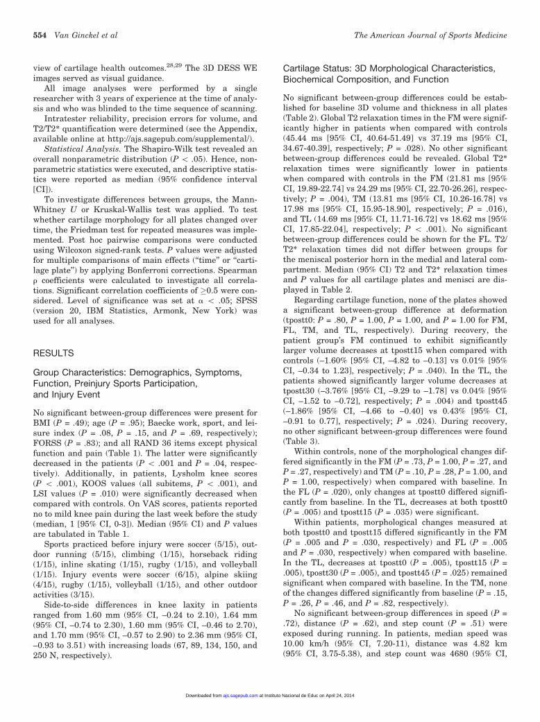
Among patients who returned to sports, timing ofreturn was recorded the latest in the sixth month andthe soonest in the fourth month after surgery. Spearmanr revealed significant and strong positive correlations (ie,the sooner the return, the larger the baseline morphologi-cal characteristics) between time to return to sports andbaseline volumes for the TM and TL (both rs = 0.76, P =.049) and thickness for the FL (rs = 0.76, P = .046) andTM (rs = 0.76, P = .049). With regard to function, signifi-cant and strong negative correlations (ie, the sooner thereturn, the larger the morphological decrease) were shownfor deformation in the FL (tpostt0: rs = –0.95, P = .001) andfor recovery at tpostt15 in the FL (rs = –0.79, P = .033) andFM (rs = –0.78, P = .041) (Figure 2). All other parametersinvestigating cartilage status did not correlate with timeto return to sports (all rs \ 0.5, P . .05).
Median surgical delay was 10 weeks (95% CI, 5-17). Theshortest delay recorded was 3 weeks, and the longest was
Figure 2. Scatterplots of the relationship between time toreturn to sports and cartilage function compared with a refer-ence median value of control participants (dashed line) forthe specific time points in the medial and lateral femur.Data points below the reference line represent morphologicaldecreases that lag behind the control recovery process andhence display a delay in recovery. Note that the correlationoutcome is primarily driven by the patients returning to sportssooner than 5 months after anterior cruciate ligamentsurgery.
556 Van Ginckel et al The American Journal of Sports Medicine
at Instituto Nacional de Educ on April 24, 2014ajs.sagepub.comDownloaded from

24 weeks. With regard to recovery, significant and moder-ate to strong positive correlations (ie, the shorter the delay,the larger the morphological decrease) were found in theFM at tpostt30 (rs = 0.60, P = .019) and in the FL attpostt45 (rs = 0.58, P = .025) (Figure 3). All other parame-ters investigating cartilage status did not correlate withsurgical delay (all rs \ 0.4, P . .05). No correlation existedbetween time to return to sports and surgical delay (rs =–0.03, P = .93).
DISCUSSION
The most important finding of this study revealed thatalthough no differences in cartilage volume and thickness
could be shown between ACL-reconstructed knees andhealthy matched controls, differences in biochemical com-position were apparent at 6 months after surgery. Basedon 4 measurements up to 45 minutes after exercise cessa-tion, equal cartilage deformation after a 30-minute runamong patients and controls was noted. Equal deformationbetween groups preceded significantly slower recovery ofcartilage morphological characteristics in patients. Addi-tionally, early postoperative sports participation wasrelated to trends toward increased cartilage volume, thick-ness, and deformation and to slower recovery of cartilagemorphological characteristics after running. Similarly,although all patients were operated on before 6 monthsafter injury, a shorter surgical delay was associated withslower cartilage recovery after running.
Knee cartilage volume and thickness did not differbetween patients and controls. In this regard, interindivid-ual differences in cartilage morphology are suggested asbeing larger than side-to-side differences, potentiallyaccounting for the present lack of significance.7 The ratio-nale for including distinct control patients, however, was 3-fold. (1) After injury and during rehabilitation, overstrain-ing of the nonreconstructed knee might occur as a compen-satory overuse phenomenon to guard the operated side.11
(2) Biochemical marker concentrations were reported tobe elevated also in the contralateral joint possibly becauseof altered gait patterns and/or release of matrix fragmentsor cytokines into circulation.6,26 (3) Cartilage defects,meniscal injuries, and previous ACL injuries are possiblysustained in the opposite limb. Excluding those patientswould have further impeded recruitment. Nonetheless,cross-sectional designs do not adequately reflect cartilageadaptations over time. Frobell et al9 monitored cartilagechanges after surgery, revealing overall small changesthat could only attain significance after 2 years. Hence,substantial changes might not yet have occurred at 6months’ follow-up.
When compared with controls, T2 prolongation wasdetected in patients’ FM, and T2* decreases were regis-tered in the FM, TM, and TL. T2/T2* mapping techniquesare reported to target water content and collagen matrix.As opposed to increasing T2, T2* tends to decrease incase of degeneration.3 To the best of our knowledge,this is the first study reporting T2/T2* values in ACL-reconstructed knees as soon as 6 months after surgery.One year after reconstruction, Li et al17 reported T2 eleva-tions of 1% to 12.2%. Potter et al22 substantiated progres-sive prolongation of T2 in the FL from 1 year onward.Although the capacity of T2* in early disease detectionwarrants further investigation,3 overall, biochemical map-ping results suggest diminished cartilage quality in ACL-reconstructed knees at 6 months after surgery.
Regarding cartilage function, deformation appearedequal among patients and controls. However, between-group differences revealed significantly slower recoveryof cartilage deformation after running in patients for theFM and TL. Additionally, differences in significant timeeffects within groups suggest potential differences betweengroups that could not be statistically proven because ofsample size. As such, a tendency is noted toward slower
Figure 3. Scatterplots of the relationship between timing ofsurgical delay and cartilage function compared with a refer-ence median value of control participants (dashed line) forthe specific time points in the medial and lateral femur.Data points below the reference line represent morphologicaldecreases that lag behind the control recovery process andhence display a delay in recovery. Note that the correlationoutcome is primarily driven by the patients receiving surgeryless than 10 weeks after anterior cruciate ligament injury.
Vol. 41, No. 3, 2013 Cartilage Quality After ACL Reconstruction and Return to Sports 557
at Instituto Nacional de Educ on April 24, 2014ajs.sagepub.comDownloaded from

recovery in the FL and more deformation in the FM. Ourresults are in agreement with those of Hosseini et al,12
who reported, during a single-legged quasistatic lunge atlower flexion angles (0�-15�), increased contact deforma-tion in overlapping tibiofemoral cartilage layers in bothmedial and lateral compartments of isolated ACL-reconstructed knees at 6 months after surgery. Investiga-tion of in vivo cartilage deformational behavior providesa means to encompass tissue resiliency, determining inpart its vulnerability to degeneration.27 Collagen disrup-tion, endorsed by the aberrant T2 values, causes loss of col-lagen tensile strength, which possibly accounts for thedelayed recovery observed. Delayed recovery might inducea state of maintained deformation and dehydration as com-pared with healthy joints. Enduring dehydration may havedeleterious effects on chondrocyte metabolism.24 In thisrespect, because of the fast and repetitive (high) impactloads to be encountered during sports, delayed cartilagerecovery may be potentially deleterious, eliciting a negativevicious circle toward degeneration.
The results of our study are the first to associate anearly return to sports with trends toward increased carti-lage deformation and diminished cartilage function at 6months after surgery. Although not significantly differentfrom controls, a tendency toward increasing volume/thickness in the patients may be suggestive signs of OAonset. During early OA, collagen disruption causes wateraccumulation, resulting in swollen tissue recognized bythe present abnormal T2. In support, in early OA, next tothinning, cartilage thickening associated with swellingwas previously described.4,8 Frobell et al8,9 similarlyreported central FM volume increases up to 2 years afterACL surgery. Notwithstanding the limited sample (Figure2), return to sports from 5 months onward (ie, 5/8 returners)seemed to be associated with deformation and recovery sim-ilar to the control group. As such, postponing sports this farmay be more suitable for knee cartilage to counter excessiverepetitive loads. One might argue that baseline measure-ments are needed to attribute the worse cartilage outcomesto timing of return to sports. This study did not includebaseline testing because current rehabilitation guidelinesadvise against 30-minute runs being implemented in theacute postinjury or postreconstruction phase.16 Recent lon-gitudinal MRI studies in ACL-reconstructed knees, how-ever, describe baseline increases in T2/T1r along withdecreases in delayed gadolinium-enhanced MRI of cartilage(dGEMRIC), which were attributed to blunt trauma. Subse-quently, these markers of decreased tissue quality weredescribed to monitor an attempt of restitution during thefirst year(s).17,20 Hence, in support of the current correlationoutcomes, it is likely to suggest that the earlier the return tosport during the first year, the more fragile the cartilageand thus the more deleterious the effect of resuming sports.In agreement with the recent epidemiological literature1
and the rehabilitation guidelines,16 the return rate at 6months was relatively low. Fifty-three percent practicedsome sort of sports, of which 12.5% reported a return tothe preinjury sport. Actual return to the same sport doesnot affect the main outcome of this study. In fact, the cur-rent notion of cartilage fragility supports the advice to
consider a delayed return to sports. In this regard, investi-gation of later time points would be an interesting lead forfuture research.
Interestingly, the sooner surgery was performed, themore cartilaginous functional decline was apparent at 6months after surgery. In view of the body of literature,5,10,23
in this sample, with a median surgical delay of 2.5 months,all patients received early surgery, which accounted for theabsence of substantial collateral damage on baseline arthro-scopic surgery (as intended). Considering limited samplesizes, the data in Figure 3 suggest that surgery beforeapproximately 10 weeks (ie, 9/15 patients) was associatedwith delayed cartilage recovery after running. Surgery per-formed within a 2.5- to 6-month window led to recovery com-parable with the control group, suggesting adequate tissueresiliency. During an ACL rupture, considerable impact iscushioned, accounting for inflammatory processes and post-traumatic BMLs to present. On top of primary-phase BMLs,secondary-phase BMLs are introduced by surgery itself,accompanied by prolonged knee effusion.9 Surgery per-formed promptly potentially endangers the joint’s possibil-ity to allow the primary-phase effect to be adequatelyprocessed. Hence, potential biological side effects of surgeryon cartilage homeostasis26 might be amplified whenpatients enroll in rehabilitation. In case of the need for sur-gery, early reconstruction (ie, within the first 6 months or 1year at the latest 5,10,23) is encouraged to avoid progressivecartilage and/or meniscal damage. However, based on theresults of this study, proper balance with return to sportsis suggested for consideration.
The nature of the protocol led to a relatively limitedsample size. Although suggested outcomes agree withresults from longitudinal studies using larger sam-ples8,9,17,20 and significance is attained for the mainresults, replication remains warranted for the sake of gen-eralizability. Second, selection based on ‘‘clear arthroscopicevaluation’’ (ie, absence of concomitant visible cartilagelesions, meniscus injuries, or other grade 2 ligament inju-ries) was pursued to include a homogeneous sample, whichfacilitates the investigation into the effect of the recon-struction itself. It should be mentioned that arthroscopicsurgery cannot preclude the presence of ultrastructuraldamage in the cartilage or menisci at baseline. With regardto the latter, next to the exclusion of cases treated withconcomitant meniscal procedures at baseline, it is interest-ing to note that biochemical mapping did not revealbetween-group differences for the menisci in the presentanalysis. Third, comparison with nonoperatively treatedACL tears and/or patient baseline conditions would be aninteresting expansion of the protocol. However, healthymatched controls were our first option as the former 2groups cannot serve as healthy references. Fourth, asthis is a retrospective case control design, prospects inview of long-term outcome cannot be drawn.
CONCLUSION
This experimental study reports MRI signs suggesting car-tilage fragility at 6 months after isolated ACL
558 Van Ginckel et al The American Journal of Sports Medicine
at Instituto Nacional de Educ on April 24, 2014ajs.sagepub.comDownloaded from

reconstruction in a young, active population comparedwith matched control patients. Although no macromorpho-logical differences existed, ultrastructural MRI changessuggested early degeneration, corresponding with declin-ing in vivo tissue resiliency. Diminished cartilage cushion-ing properties are proposed to be a potential danger in viewof a full return to sports. Based on the results of this study,caution is advised for an early return to sports (ie, before 5months after surgery), especially when dealing withpatients who received early surgery (ie, within 10 weeksafter injury).
ACKNOWLEDGMENT
The authors gratefully acknowledge Greta Vandemaele,PhD, Siemens MRI application specialist, for her helpand expertise in parameter implementation for the sequen-ces used in this study.
REFERENCES
1. Ardern CL, Webster KE, Taylor NF, Feller JA. Return to the pre-injury
level of competitive sport after anterior cruciate ligament reconstruc-
tion surgery: two-thirds of patients have not returned by 12 months
after surgery. Am J Sports Med. 2011;39(3):538-543.
2. Baecke JA, Burema J, Frijters JE. A short questionnaire for the mea-
surement of habitual physical activity in epidemiological studies. Am
J Clin Nutr. 1982;36(5):936-942.
3. Bittersohl B, Miese FR, Hosalkar HS, et al. T2* mapping of hip joint
cartilage in various histological grades of degeneration. Osteoarthritis
Cartilage. 2012;20(7):653-660.
4. Buck RJ, Wyman BT, Le Graverand MP, et al. Osteoarthritis may not
be a one-way-road of cartilage loss: comparison of spatial patterns
of cartilage change between osteoarthritic and healthy knees. Osteo-
arthritis Cartilage. 2010;18(3):329-335.
5. Church S, Keating JF. Reconstruction of the anterior cruciate liga-
ment: timing of surgery and the incidence of meniscal tears and
degenerative change. J Bone Joint Surg Br. 2005;87(12):1639-1642.
6. Dahlberg L, Roos H, Saxne T, et al. Cartilage metabolism in the
injured and uninjured knee of the same patient. Ann Rheum Dis.
1994;53(12):823-827.
7. Eckstein F, Muller S, Faber SC, Englmeier KH, Reiser M, Putz R. Side
differences of knee joint cartilage volume, thickness, and surface
area, and correlation with lower limb dominance: an MRI-based
study. Osteoarthritis Cartilage. 2002;10(12):914-921.
8. Frobell RB. Change in cartilage thickness, posttraumatic bone mar-
row lesions and joint fluid volumes after acute ACL disruption:
a two-year prospective MRI study of sixty-one subjects. J Bone Joint
Surg Am. 2011;93:1096-1103.
9. Frobell RB, Le Graverand MP, Buck R, et al. The acutely ACL injured
knee assessed by MRI: changes in joint fluid, bone marrow lesions,
and cartilage during the first year. Osteoarthritis Cartilage.
2009;17:161-167.
10. Granan LP, Bahr R, Lie SA, Engebretsen L. Timing of anterior cruci-
ate ligament reconstructive surgery and risk of cartilage lesions and
meniscal tears: a cohort study based on the Norwegian National
Knee Ligament Registry. Am J Sports Med. 2009;37(5):955-961.
11. Hoffelner T, Resch H, Moroder P, et al. No increased occurrence of
osteoarthritis after anterior cruciate ligament reconstruction after
isolated anterior cruciate ligament injury in athletes. Arthroscopy.
2012;28(4):517-525.
12. Hosseini A, Van de Velde S, Gill TJ, Li G. Tibiofemoral cartilage con-
tact biomechanics in patients after reconstruction of a ruptured ante-
rior cruciate ligament. J Orthop Res. 2012;30(11):1781-1788.
13. Hunter DJ, Li L, Zhang YQ, et al. Region of interest analysis: by
selecting regions with denuded areas can we detect greater amounts
of change? Osteoarthritis Cartilage. 2010;18(2):175-183.
14. Keays SL, Newcombe PA, Bullock-Saxton JE, Bullock ML, Keays
AC. Factors involved in the development of osteoarthritis after ante-
rior cruciate ligament surgery. Am J Sports Med. 2010;38(3):455-463.
15. Kessler MA, Glaser C, Tittel S, Reiser M, Imhoff AB. Recovery of the
menisci and articular cartilage of runners after cessation of exercise:
additional aspects of in vivo investigation based on 3-dimensional
magnetic resonance imaging. Am J Sports Med. 2008;36(5):966-970.
16. Kvist J. Rehabilitation following anterior cruciate ligament injury: cur-
rent recommendations for sports participation. Sports Med.
2004;34(4):269-280.
17. Li X, Kuo D, Theologis A, et al. Cartilage in anterior cruciate ligament-
reconstructed knees: MR imaging T1r and T2-initial experience with
1-year follow-up. Radiology. 2011;258(2):505-514.
18. Myklebust G, Bahr R. Return to play guidelines after anterior cruciate
ligament surgery. Br J Sports Med. 2005;39(3):127-131.
19. Neeb TB, Aufdemkampe G, Wagener JHD, Mastenbroek L. Assess-
ing anterior cruciate ligament injuries: the association and differential
value of questionnaires, clinical tests and functional tests. J Orthop
Sports Phys Ther. 1997;26(6):324-331.
20. Neuman P, Tjornstrand J, Svensson J, et al. Longitudinal assessment
of femoral knee cartilage quality using contrast enhanced MRI
(dGEMRIC) in patients with anterior cruciate ligament injury: compar-
ison with asymptomatic volunteers. Osteoarthritis Cartilage. 2011;
19(8):977-983.
21. Niefhoff A, Muller M, Bruggeman L, et al. Deformational behaviour of
knee cartilage and changes in serum cartilage oligomeric matrix pro-
tein (COMP) after running and drop landing. Osteoarthritis Cartilage.
2011;19(8):1003-1010.
22. Potter HG, Jain SK, Ma Y, Black BR, Fung S, Lyman S. Cartilage
injury after acute, isolated anterior cruciate ligament tear: immediate
and longitudinal effect with clinical/MRI follow-up. Am J Sports Med.
2012;40(2):276-285.
23. Seon JK, Song EK, Park SJ. Osteoarthritis after anterior cruciate lig-
ament reconstruction using a patellar tendon autograft. Int Orthop.
2006;30(2):94-98.
24. Song Y, Greve JM, Carter DR, Giori NJ. Meniscectomy alters the
dynamic deformational behavior and cumulative strain of tibial artic-
ular cartilage in knee joints subjected to cyclic loads. Osteoarthritis
Cartilage. 2008;16(12):1545-1554.
25. Subburaj K, Kumar D, Souza RB, et al. The acute effects of running on
knee articular cartilage and meniscus magnetic resonance relaxation
times in young healthy adults. Am J Sports Med. 2012;40(9):2134-2141.
26. Taskiran E, Taskiran D, Duran T, Lok V. Articular cartilage homeosta-
sis after anterior cruciate ligament reconstruction. Knee Surg Sports
Traumatol Arthrosc. 1998;6:93-98.
27. Van Ginckel A, Roosen P, Almqvist KF, Verstraete K, Witvrouw E.
Effects of in vivo exercise on ankle cartilage deformation and recov-
ery in healthy volunteers: an experimental study. Osteoarthritis Carti-
lage. 2011;19(9):1123-1131.
28. Williams A, Qian Y, Golla S, Chu CR. UTE-T2* mapping detects sub-
clinical meniscus injury after anterior cruciate ligament tear. Osteoar-
thritis Cartilage. 2012;20(6):486-494.
29. Zarins ZA, Bolbos RI, Pialat JB, et al. Cartilage and meniscus assess-
ment using T1rho and T2 measurements in healthy subjects and
patients with osteoarthritis. Osteoarthritis Cartilage. 2010;
18(11):1408-1416.
For reprints and permission queries, please visit SAGE’s Web site at http://www.sagepub.com/journalsPermissions.nav
Vol. 41, No. 3, 2013 Cartilage Quality After ACL Reconstruction and Return to Sports 559
at Instituto Nacional de Educ on April 24, 2014ajs.sagepub.comDownloaded from


