Embed Size (px)
Citation preview

CARTILAGE

CARTILAGE
Cartilage is a form of connective tissue composed of cells called chondrocytes and a highly specialized extracellular matrix.
Types of CARTILAGE:• HYALINE CARTILAGE is characterized by a
homogeneous amorphous matrix• ELASTIC CARTILAGE, whose matrix contains elastic
fibers and elastic lamellae• FIBROCARTILAGE, whose matrix contains large
bundles of type 1 collagen


STRUCTURAL PLAN OF CARTILAGE
PERICHONDRIUM• outer fibrous layer• inner cell layer (chondrogenic cells + chondroblasts)
CHONDROCYTES (isogenous groups of cells located in lacunas) with integrin (anchoring glycoprotein of plasma membrane)
CARTILAGE MATRIX• type II collagen fibers or elastic fibers• ground substance consisting
- aggrecan (proteoglycan rich in hyaluronic acid + chondroitin sulfate + keratan sulfate)
- chondronectin (anchoring glycoprotein)


LOCALIZATION OF DIFFERENT KINDS OF CARTILAGE
• HYALINE CARTILAGE is found as the structural framework for the larynx, trachea and bronchi; on the anterior ends of the ribs; on the surfaces of synovial joints. Hyaline cartilage constitutes much of the fetal skeleton.
• ELASTIC CARTILAGE is found in the auricle of the external ear, in the auditory tube, in the epiglottis, and in part of the larynx.
• FIBROCARTILAGE is found at the intervertebral discs, the symphysis pubis , and some joints.


BONE

BONES AND BONE (OSSEOUS) TISSUE
Bone is a type of connective tissue characterized by a highly mineralized extracellular matrix (65% of calcium hydroxyapatite crystals) + 35% of type I collagen fibers. Non-calcified intercellular substance is called Osteoid
• Bone tissue is CLASSIFIED as: - Primary (Woven) bone - Secondary (Lamellar) bone
- Compact (Dense) bone- Spongy (Cancellous) bone
• Bones are covered by Periosteum: a sheath of dense connective tissue
• Bone cavities are lined by Endosteum: a delicate layer of connective tissue cells and fibers

CELLS OF BONE TISSUE
• Osteoprogenitor cell is a resting cell that can transform into an osteoblast
• Osteoblast is the differentiated bone-forming cell that secretes bone matrix
• Osteocyte, the mature bone cell, residing in lacuna, is enclosed by bone matrix that is previously secreted by an osteoblast
• Osteoclast is a large multinucleated cell whose function is resorption of bone

Light micrograph of intramembranous ossification with different cell types

EM of bone-forming cells


Osteoclast structure and function (scheme)

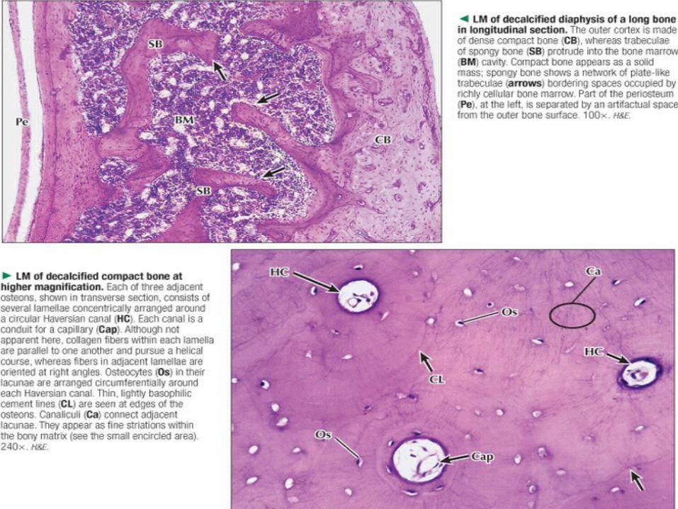
STRUCTURE OF ADULT LONG BONE
Anatomy: Diaphysis + 2 Epiphyses + 2 MetaphysesUnits of diaphyseal bone structure are called OSTEONS.
Each osteon includes:- concentric lamellae – collagen fibers arranged in parallel manner- Haversian canal with blood vessel, osteoblasts and osteoprogenitor cells inside- osteocytes located in lacunas forvarding their processes into the bony canaliculi
Besides osteons diaphysis consists of:• Interstitial lamellae• Circumferential lamellae• Perforating (Volkmann’s) canals• Periosteum• Endosteum


Primary (intramembranous) bone formation


STAGES OF ENDOCHONDRAL OSSIFICATION
• Formation of a hyaline cartilage template (model)• Appearance of a bony collar around the cartilage model• Calcification of cartilage matrix• Migration of periosteal cells into the cavity with growing
blood vessels
Structure of epiphyseal cartilage throughout the growth period:
• Zone of reserve cartilage• Zone of proliferation • Zone of maturation and hypertrophy• Zone of cartilage calcification• Zone of ossification (cartilage resorption)


Anatomy of diarthrodial joint

Events in bone fracture repair