Embed Size (px)
Citation preview

1
Connective Tissue
Dr. Heba KalbounehAssistant Professor of Anatomy and Histology
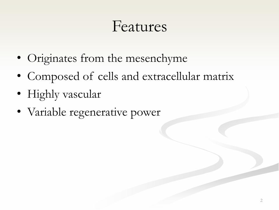
Features
• Originates from the mesenchyme
• Composed of cells and extracellular matrix
• Highly vascular
• Variable regenerative power
2

3

Functions
• Support:
• Defense and protection:
• Storage:
• Transport:
4
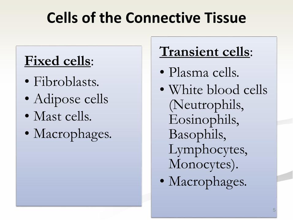
Cells of the Connective Tissue
Fixed cells:
• Fibroblasts.
• Adipose cells
• Mast cells.
• Macrophages.
Transient cells:
• Plasma cells.
• White blood cells (Neutrophils, Eosinophils, Basophils, Lymphocytes, Monocytes).
• Macrophages.
5
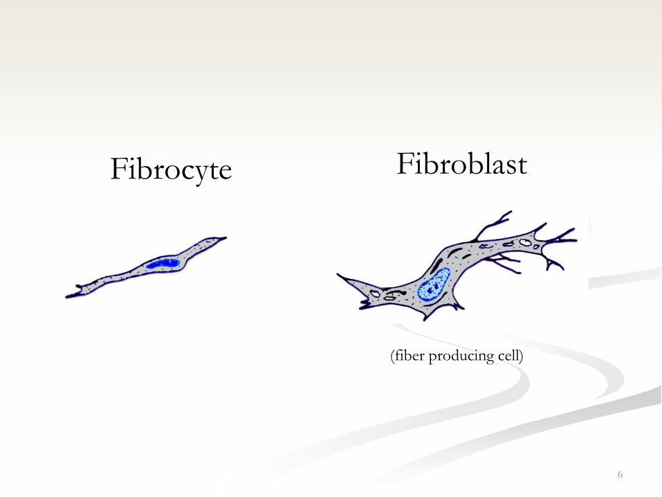
6
(fiber producing cell)
FibroblastFibrocyte

7
Fibroblast
• The most numerous cells of connective tissue.
• Occur in active and inactive forms (fibrocyte).
• Originate from undifferentiated mesenchymal
cells.
• Capable of some movement.
• Rarely undergo division (in adults).
9

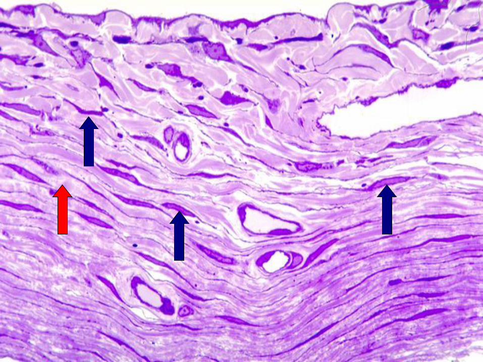
Fibroblasts/ histological features
• Closely associated with collagen bundles.
• Elongated, fusiform, and have many processes.
• Cytoplasm is pale and difficult to be
differentiated from near by tissue.
• Nucleus is large (ovoid) euchromatic, prominent
nucleolus
• E.M: prominent Golgi, mitochondria, rER, actin
and myosin.
10
Fibrocytes/histological features
• Smaller and spindle-shaped.
• The nucleus is smaller and darker (heterochromatic).
• Few processes.
• E.M: few rER.
• When stimulated, it may revert to fibroblast.
11

Myofibroblasts
• Has features of both smooth muscles and
fibroblasts.
• Their contraction is responsible for wound contraction.
12
13
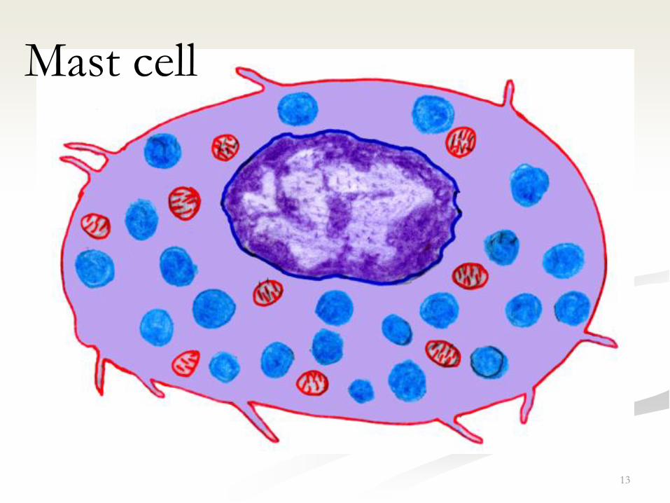
Mast cell
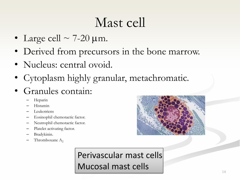
Mast cell • Large cell ~ 7-20 m.
• Derived from precursors in the bone marrow.
• Nucleus: central ovoid.
• Cytoplasm highly granular, metachromatic.
• Granules contain:– Heparin
– Histamin
– Leukotriens
– Eosinophil chemotactic factor.
– Neutrophil chemotactic factor.
– Platelet activating factor.
– Bradykinin.
– Thromboxane A2
14
Perivascular mast cells Mucosal mast cells

15
16
Plasma cell
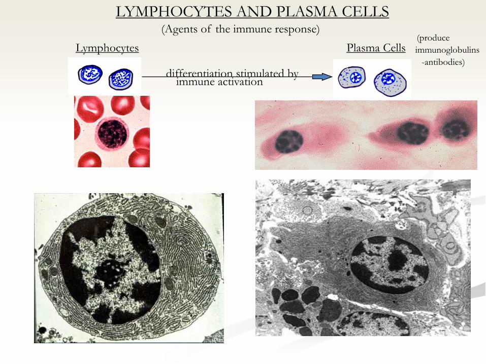
LYMPHOCYTES AND PLASMA CELLS(Agents of the immune response)
differentiation stimulated byimmune activation
Lymphocytes Plasma Cells(produce
immunoglobulins
-antibodies)

Plasma cell
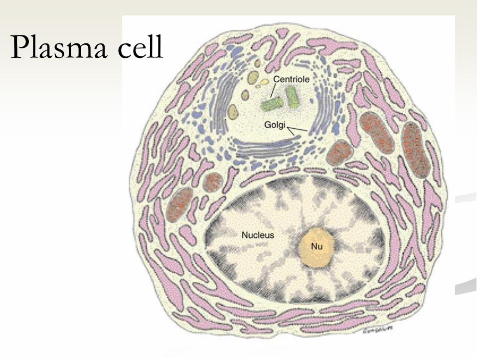
• Derived from B lymphocytes following exposure to an antigen.
• Present at portal of entry of organisms and sites of chronic inflammation.
• Life span ~ 10-20 days.
• Large ovoid cells ~ 20 m.
• Nucleus: eccentric with clusters of heterochromatin cart-wheel or clock-face nucleus.
18
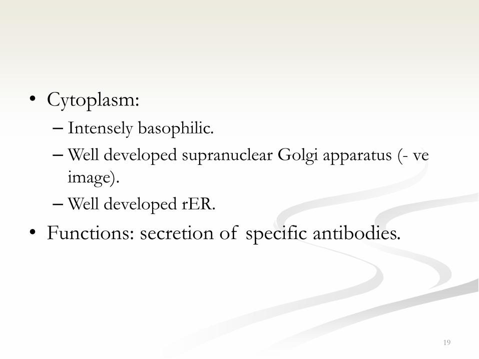
• Cytoplasm:
– Intensely basophilic.
– Well developed supranuclear Golgi apparatus (- ve
image).
– Well developed rER.
• Functions: secretion of specific antibodies.
19

21
Macrophage

Macrophage
• Derived from monocyte.
• Large cells ~10-30 m.
• Surface shows many projections.
• Nucleus: eccentric, oval or indented (kidney-shaped).
• Cytoplasm: well developed Golgi, prominent rER,
many lysosomes.
• They are part of the MPS.
• Multinuclear giant cells
22
23
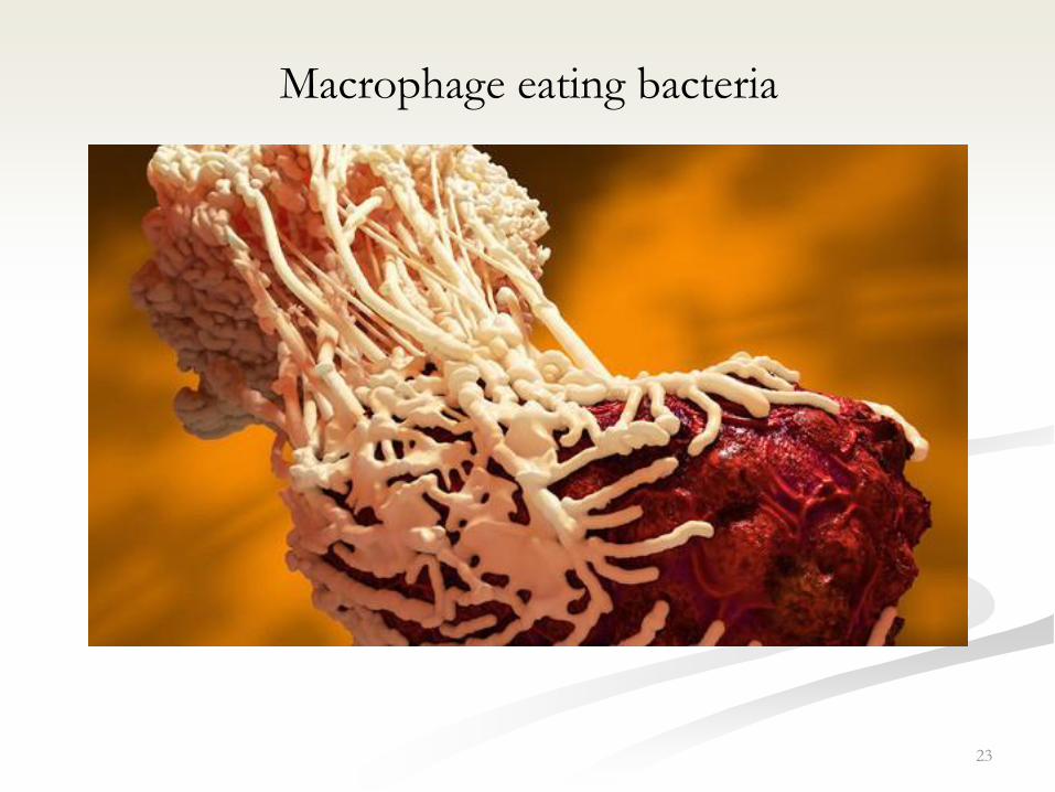
Macrophage eating bacteria

24
Monocytes and macrophages are the same
cells at different stages of maturation
25
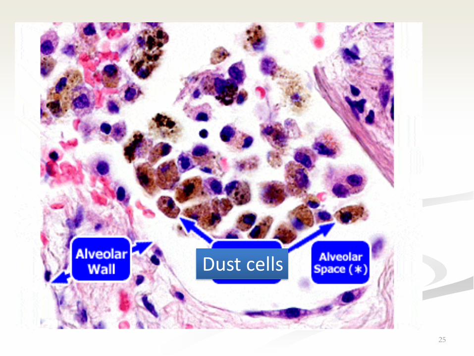
Dust cells

Mononuclear phagocyte system
• Is a part of the immune system that consists of
the phagocytic cells
• The macrophage like cells have been given
different names in different organs
• also called Reticuloendothelial System or
Macrophage System
26
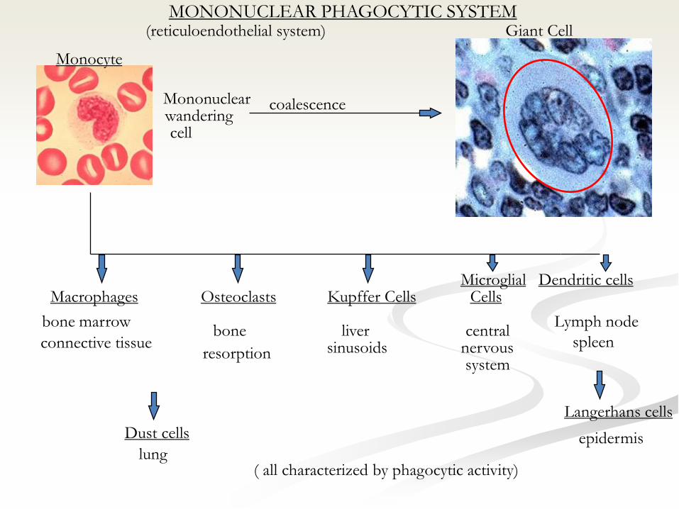
MONONUCLEAR PHAGOCYTIC SYSTEM
Mononuclearwanderingcell
coalescence
Giant Cell
Macrophages Osteoclasts Kupffer CellsMicroglial
CellsDendritic cells
bone marrow
connective tissue
lung
bone
resorption
liversinusoids
centralnervoussystem
Lymph node
spleen
Monocyte
(reticuloendothelial system)
( all characterized by phagocytic activity)
Langerhans cells
epidermisDust cells

Extracellular Matrix
• Extracellular Matrix = ground substance +
fibers.
– Resists compression and stretching forces.
– The water content allows rapid exchange of
metabolites.
28
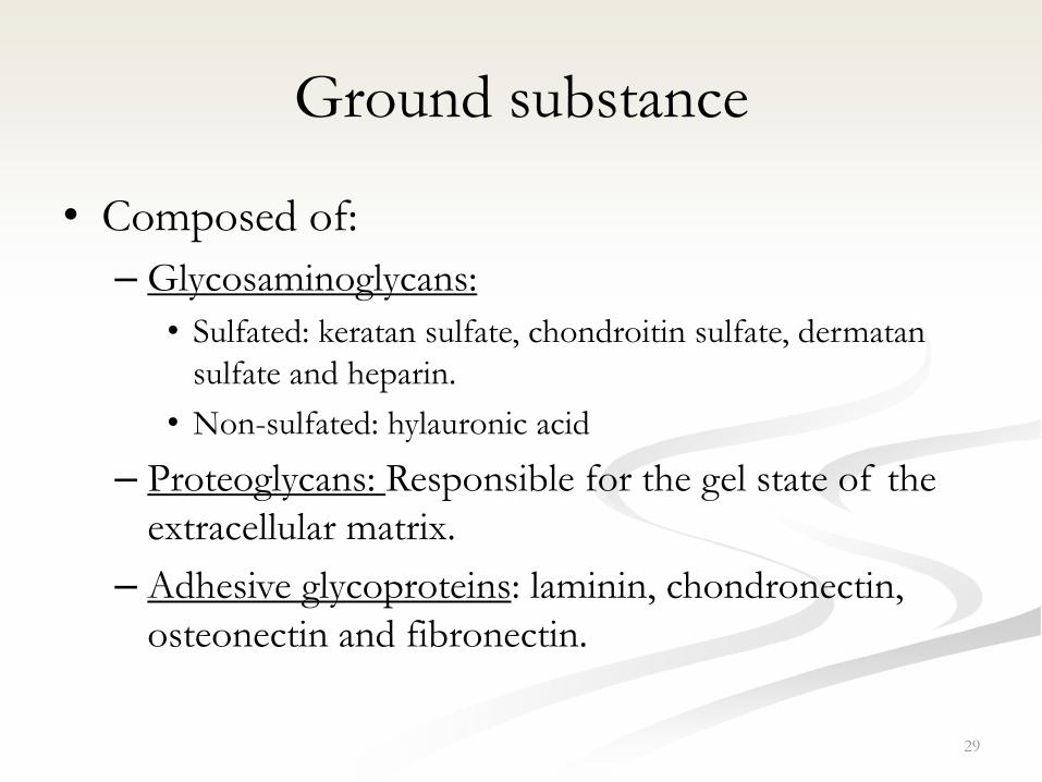
Ground substance
• Composed of:
– Glycosaminoglycans:
• Sulfated: keratan sulfate, chondroitin sulfate, dermatan
sulfate and heparin.
• Non-sulfated: hylauronic acid
– Proteoglycans: Responsible for the gel state of the
extracellular matrix.
– Adhesive glycoproteins: laminin, chondronectin,
osteonectin and fibronectin.
29

30
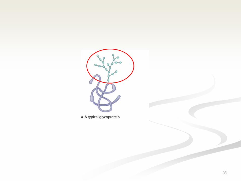
Proteoglycan
GAG
Protein core

31hyaluronidase
33

Functions of proteoglycans
• Resistance of compression.
• Retardation of movement of microorganisms.
• Act as a filter.
34
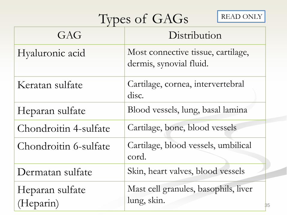
Types of GAGs
35
DistributionGAG
Most connective tissue, cartilage,
dermis, synovial fluid.Hyaluronic acid
Cartilage, cornea, intervertebral
disc.Keratan sulfate
Blood vessels, lung, basal laminaHeparan sulfate
Cartilage, bone, blood vesselsChondroitin 4-sulfate
Cartilage, blood vessels, umbilical
cord.Chondroitin 6-sulfate
Skin, heart valves, blood vesselsDermatan sulfate
Mast cell granules, basophils, liver
lung, skin.Heparan sulfate
(Heparin)
READ ONLY

Connective Tissue Fibers
• Collagen
• Elastic
• Reticular
36
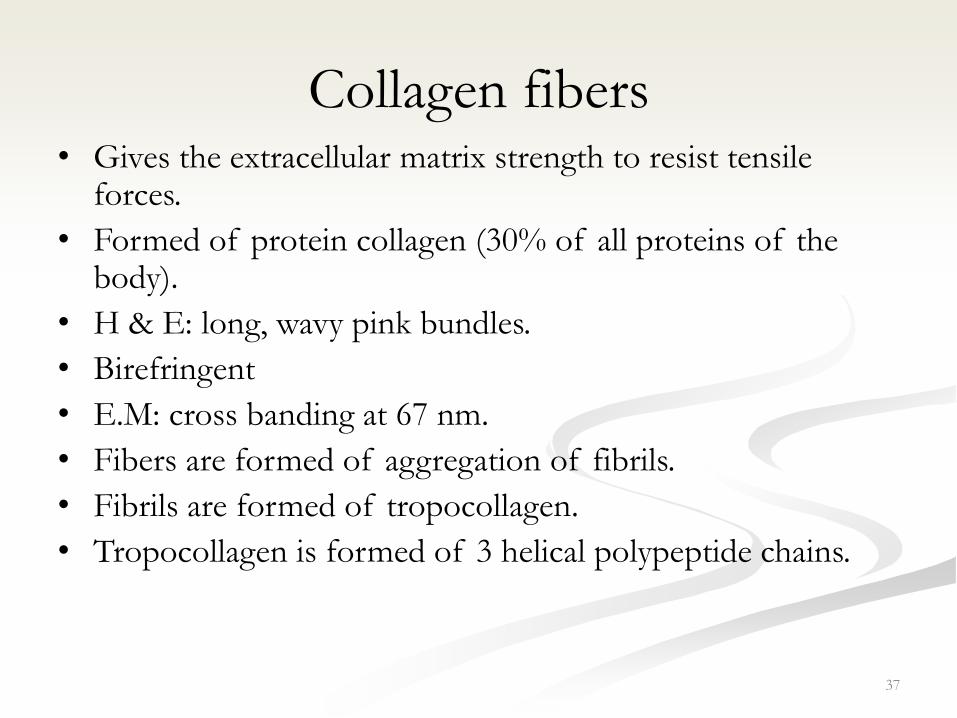
Collagen fibers• Gives the extracellular matrix strength to resist tensile
forces.
• Formed of protein collagen (30% of all proteins of the body).
• H & E: long, wavy pink bundles.
• Birefringent
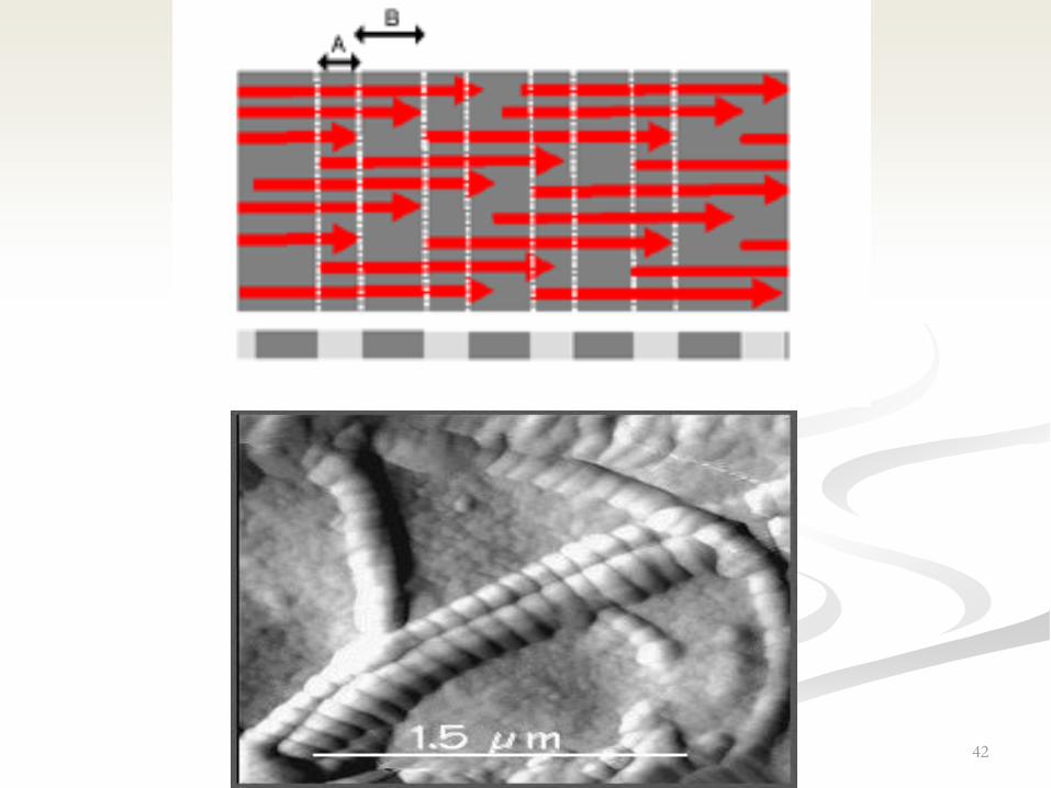
• E.M: cross banding at 67 nm.
• Fibers are formed of aggregation of fibrils.
• Fibrils are formed of tropocollagen.
• Tropocollagen is formed of 3 helical polypeptide chains.
37

• α-chains possess 1000 amino acids.
• Every 3rd amino acid is glycine.
– Other amino acids: proline, hydroxyproline,
hydroxylysine.
• The sequence of aminoacids determines the
type of collagen.
– There are 28 types of collagen.
38

LocationFunctionSynthesizing cellType
Dermis, tendons,
ligaments, capsules,
bone, dentin,
cementum
Resist tensionFibroblast, osteoblast,
odontoblast, cementoblast
I
CartilageResists pressurechondroblastsII
Reticuloendothelial
system, lung, skin
Form structural
framework of organs
Fibroblasts, reticular cells,
smooth muscle, hepatocytes
III
Basal laminaMeshwork of the
lamina densa
Epithelium, muscle, Schwann
cells
IV
As in type I and
placenta
Associated with type I.Fibroblasts, mesenchymal
cells
V
Derma-epidermal
junction
Anchoring fibrils
between the lamina
densa and reticularis
Epidermal cellsVII
39
Major Types of Collagen
40

41
42

Collagen bundle
Fibers
Fibrils
Tropocollagen
3 Helical polypeptide chains, α-chains.
43
44
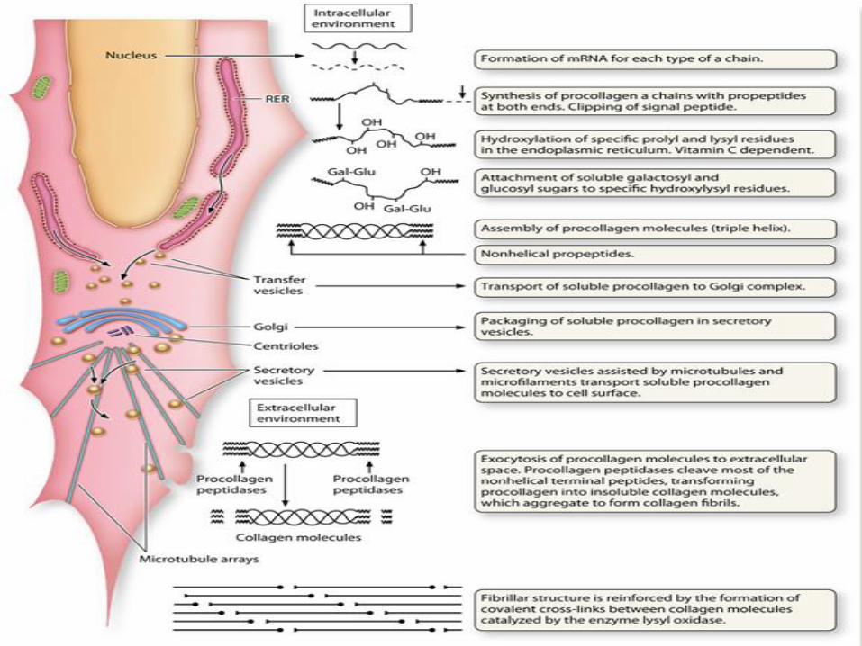
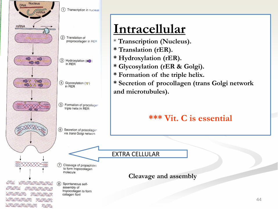
EXTRA CELLULAR
Cleavage and assembly
Intracellular* Transcription (Nucleus).
* Translation (rER).
* Hydroxylation (rER).
* Glycosylation (rER & Golgi).
* Formation of the triple helix.
* Secretion of procollagen (trans Golgi network
and microtubules).
*** Vit. C is essential

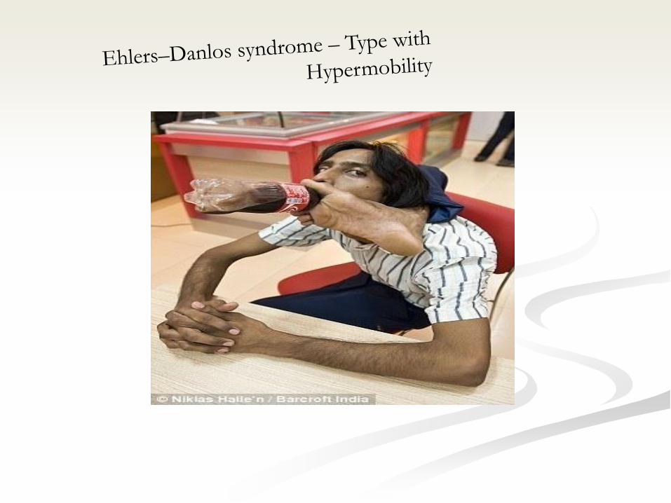
Clinical applications
• Keloid
• Vitamin C deficiency (Scurvy)
• Ehlers–Danlos syndrome
45
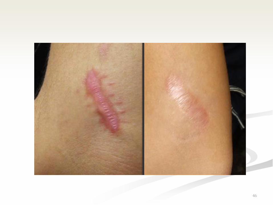
46
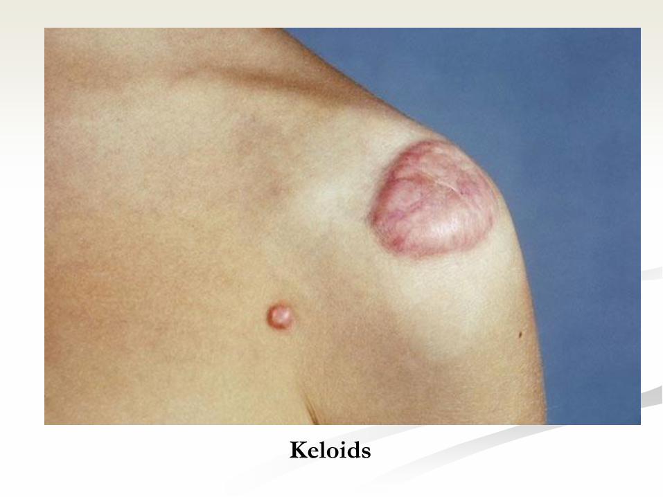
Keloids

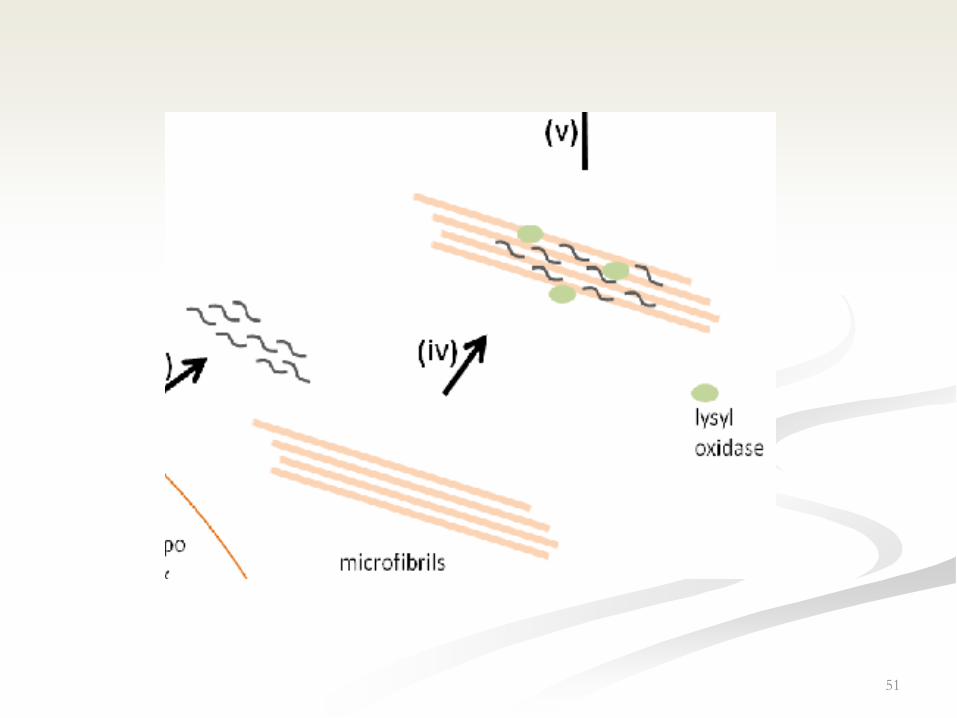
Elastic fibers
• Composed of:
1- Elastin
2- Fibrillin
• Elasticity is due to elastin.
• Stability is due to fibrillin microfibrils
(resistant to boiling).
– Appears yellow in fresh tissue (if
large amount is present)
• Digested by pancreatic enzyme elastase
50
51

52
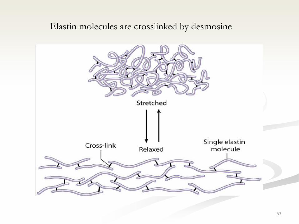
Elastic fibers consist of individual microfibrils(fibrillin) which are
embedded in an amorphous matrix (90% of the fiber and composed of
elastin)
Elastic material is found in certain ligaments (elastic ligaments), some
cartilage (called elastic cartilage) and in large arteries (elastic arteries).
53
Elastin molecules are crosslinked by desmosine

Reticular fibers
• Consist mainly type III collagen.
• Short, thin and branching.
• High sugar content
• Give PAS +ve reaction.
• Stain with Silver Nitrate (Argyrophylic).
• Found mainly in reticular lamina of basement
membrane, RES organs (supporting stroma)
54
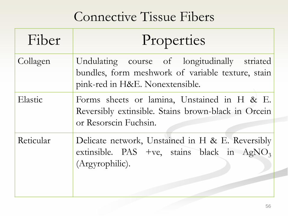
Connective Tissue Fibers
56
Fiber Properties
Collagen Undulating course of longitudinally striated
bundles, form meshwork of variable texture, stain
pink-red in H&E. Nonextensible.
Elastic Forms sheets or lamina, Unstained in H & E.
Reversibly extinsible. Stains brown-black in Orcein
or Resorscin Fuchsin.
Reticular Delicate network, Unstained in H & E. Reversibly
extinsible. PAS +ve, stains black in AgNO3
(Argyrophilic).
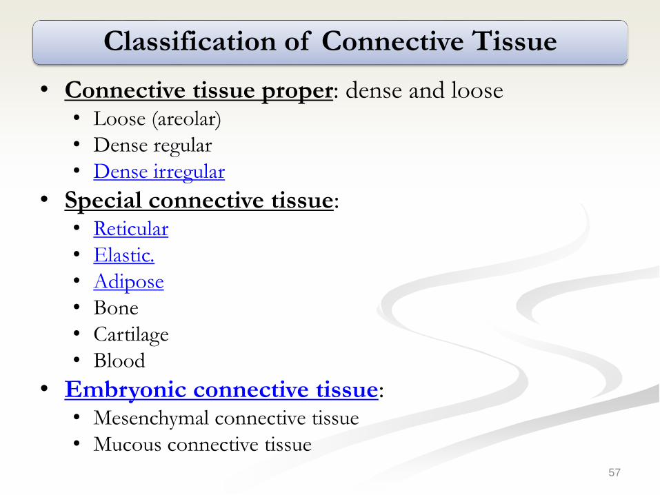
Classification of Connective Tissue
• Connective tissue proper: dense and loose• Loose (areolar)
• Dense regular
• Dense irregular
• Special connective tissue:• Reticular
• Elastic.
• Adipose
• Bone
• Cartilage
• Blood
• Embryonic connective tissue:• Mesenchymal connective tissue
• Mucous connective tissue
57
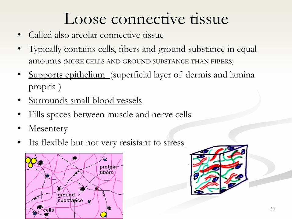
Loose connective tissue• Called also areolar connective tissue
• Typically contains cells, fibers and ground substance in equal
amounts (MORE CELLS AND GROUND SUBSTANCE THAN FIBERS)
• Supports epithelium (superficial layer of dermis and lamina
propria )
• Surrounds small blood vessels
• Fills spaces between muscle and nerve cells
• Mesentery
• Its flexible but not very resistant to stress
58

Dense irregular connective tissue
• Bundles of collagen fibers are randomly
interwoven with no definite orientation
• Provides resistance to stress from all directions
• Dermis of skin, organ capsules, submucosa of
digestive tract
59

Dense regular connective tissue
• Parallel Bundles of collagen fibers with few fibrocytes
aligned with collagen and separated by very little
ground substance
• Provides resistance to prolonged or repeated stresses
exerted in the same direction
• Ligaments, tendons, aponeuroses
• Tendons are poorly vascularized and repair of damaged
tendons is very slow
60
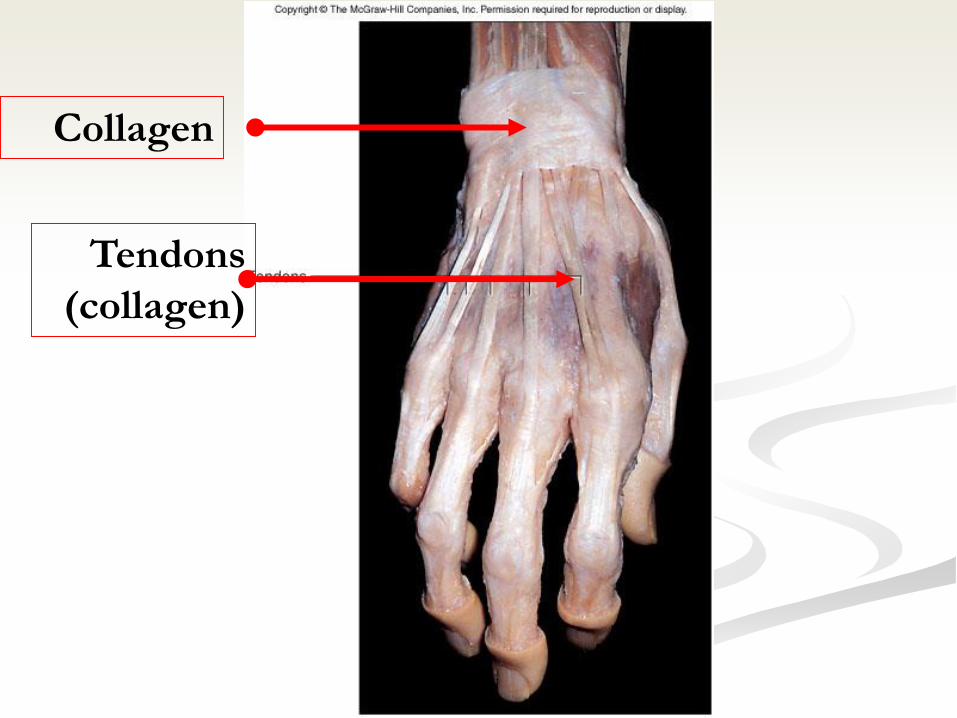
Tendons
(collagen)
Collagen

62
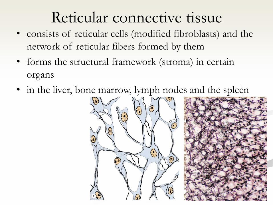
Reticular connective tissue • consists of reticular cells (modified fibroblasts) and the
network of reticular fibers formed by them
• forms the structural framework (stroma) in certain
organs
• in the liver, bone marrow, lymph nodes and the spleen
63

Mesenchymal connective tissue
• Mesenchyme forms the undifferentiated "filling" of the early embryo.
• It consists of mesenchymal cells with slender cell processes.
• Mesenchymal cells have stem cell properties, i.e. they are able give rise to other cell and tissues types.
• The extracellular matrix is mainly ground substance, which can be stained with dyes that also stain mucin- hence the alternative name of this tissue type: mucoid connective tissue
64

65
Mesenchymal connective tissue
Mucoid connective tissue also forms a compliant cushion around the
vessels of the umbilical cord, where it is also called Wharton's jelly.
In adult humans, mesenchymal connective tissue is only found in the
dental pulp.


![Cartilage - facultymembers.sbu.ac.irfacultymembers.sbu.ac.ir/rajabi/ppt toPDF/Cartilage [Compatibility Mode].pdfFibrocartilage • Fibrous Cartilage • is a form of connective tissue](https://img.dokumen.tips/doc/110x75/6012989a4318862a0e5813ae/cartilage-topdfcartilage-compatibility-modepdf-fibrocartilage-a-fibrous.jpg)