Embed Size (px)
Citation preview

T h e n e w e ngl a nd j o u r na l o f m e dic i n e
n engl j med 373;3 nejm.org July 16, 2015252
Clinical Practice
A 64-year-old woman who is hospitalized with endocarditis and whose condition is clinically stable while she is receiving intravenous antibiotic agents has had a de-crease in platelet count from 161,000 per cubic millimeter on day 7 of hospitalization to 60,000 per cubic millimeter on day 9. She has been receiving low-molecular-weight heparin at a dose of 40 mg per day since admission. How should her case be further evaluated and treated?
The Clinic a l Problem
In contrast to other conditions caused by enhanced consumption, impaired production, or destruction of platelets, which lead to bleeding com-plications, immune-mediated heparin-induced thrombocytopenia (HIT) does
not induce bleeding but rather results in a paradoxical prothrombotic state.1 This prothrombotic action makes the early recognition of HIT very important. HIT oc-curs in approximately 1 in 5000 hospitalized patients, with a large variability among patient populations. Patients who receive unfractionated heparin for 7 to 10 days are at the highest risk2; incidence rates of 1 to 3% have been reported after cardiac surgery. Thromboembolic complications develop in approximately 50% of patients with confirmed HIT. Venous thrombosis of the large vessels of the lower limbs and pulmonary embolism are the most frequent complications, followed by peripheral arterial thrombosis and then stroke; myocardial infarction is uncommon.3 Thrombotic complications may also affect other vessels, including the cerebral sinus or splanchnic veins.
The onset of HIT characteristically occurs between 5 and 10 days after heparin is started,4 both in patients who receive heparin for the first time and in patients with reexposure. However, there are exceptions. For one, major surgery resets the clock (i.e., the window of 5 to 10 days restarts), even if the patient has recently received heparin (e.g., HIT can develop 5 to 10 days after surgery in patients who have been undergoing hemodialysis for a long time).5 Second, in persons who have received heparin within the previous 90 days (especially, ≤30 days), there may be persistent circulating anti–platelet factor 4 (PF4)–heparin antibodies, and HIT can start abruptly on reexposure to heparin (rapid-onset HIT); in this case, HIT is sometimes complicated by an anaphylactoid reaction within 30 minutes after a heparin bolus.6 Otherwise, once the typically transient antibodies have disap-peared (median, 50 to 85 days, depending on the assay used),4 the regeneration of antibodies requires at least 5 days (no earlier anamnestic response).7,8 In some patients, HIT develops or worsens after heparin has been discontinued (delayed-onset HIT). These patients can present with thrombosis up to 3 weeks after the
From Institut für Immunologie und Transfusionsmedizin, Universitätsmedizin Greifswald, Greifswald, Germany. Address reprint requests to Dr. Greinacher at Institut für Immunologie und Transfusionsmedizin, Sauerbruchstr., 17475 Greifswald, Germany, or at greinach@ unigreifswald . de.
N Engl J Med 2015;373:252-61.DOI: 10.1056/NEJMcp1411910Copyright © 2015 Massachusetts Medical Society.
Caren G. Solomon, M.D., Editor
Heparin-Induced ThrombocytopeniaAndreas Greinacher, M.D.
This Journal feature begins with a case vignette highlighting a common clinical problem. Evidence supporting various strategies is then presented, followed by a review of formal guidelines,
when they exist. The article ends with the author’s clinical recommendations.
An audio version of this article is available at
NEJM.org
The New England Journal of Medicine Downloaded from nejm.org at ADVOCATE LIBRARY NETWORK on February 12, 2016. For personal use only. No other uses without permission.
Copyright © 2015 Massachusetts Medical Society. All rights reserved.
n engl j med 373;3 nejm.org July 16, 2015 253
Clinical Pr actice
253
start of heparin exposure.9 Note that in some cases, a single heparin bolus is sufficient to in-duce the syndrome, so the start of heparin is the only fixed time point.
A rare but often catastrophic form of HIT is spontaneous10,11 or autoimmune HIT, which de-velops in the absence of heparin exposure, most often after major surgery (especially knee re-placement) or recent infection. In contrast to typical HIT, in which the platelet count increases within 2 to 5 days after the start of an alterna-tive anticoagulant, autoimmune HIT may persist for weeks.10
Patho genesis
HIT is induced by IgG antibodies recognizing neoepitopes on the positively charged chemo-kine PF4 within PF4–polyanion complexes (Fig. 1).12-14 The resulting immune complexes cross-link Fcγ receptors on platelets (Fcγ RIIa)15 and monocytes (Fcγ RI),16-18 thus activating them. Further enhanced by the alteration of en-dothelial cells,19 the activation of platelets and monocytes increases thrombin generation. In-creased thrombin, not thrombocytopenia, causes the clinical problems.
In addition to binding heparin, PF4 binds other polyanions, such as nucleic acids20 and lipo-polysaccharides on bacteria.21 This phenomenon may explain cases of spontaneous HIT after major surgery (causing DNA, RNA, or glycos-aminoglycan release) or bacterial infection. An interesting concept22 is that conformationally changed23 PF4 in complex with nonheparin poly-
anions (e.g., on the bacterial surface) induces pri-mary immunization. These PF4–polyanion com-plexes serve as a danger signal and result in the rapid generation of IgG antibodies, which facili-tate opsonization and phagocytosis of PF4-coated bacteria, even against PF4-labeled pathogens that the immune system has not encountered before. This mechanism, however, results in HIT when it is misdirected22 during heparin treat-ment as a secondary immune reaction toward platelets coated with PF4–heparin complexes, which results in early production (between day 5 and day 14) of high-titer IgG antibodies. Anti–PF4–heparin antibodies are produced by B cells (probably marginal-zone B cells),24 which can mediate a transient antibody response.
S tr ategies a nd E v idence
Risk of HIT
The risk of HIT depends on the type of heparin and the patient population. The incidence is up to 10 times as high among patients receiving unfractionated heparin as it is among those re-ceiving low-molecular-weight heparin,25 and HIT occurs more frequently among patients who have had major surgery than among those who have had minor surgery26 or are receiving medi-cal therapy.27 HIT is rare in obstetrical patients, although in contexts other than pregnancy, women are at slightly higher risk than men.27
Diagnosis
The diagnosis of HIT is based on a decrease in the platelet count of more than 50% or throm-
Key Clinical Points
Heparin-Induced Thrombocytopenia
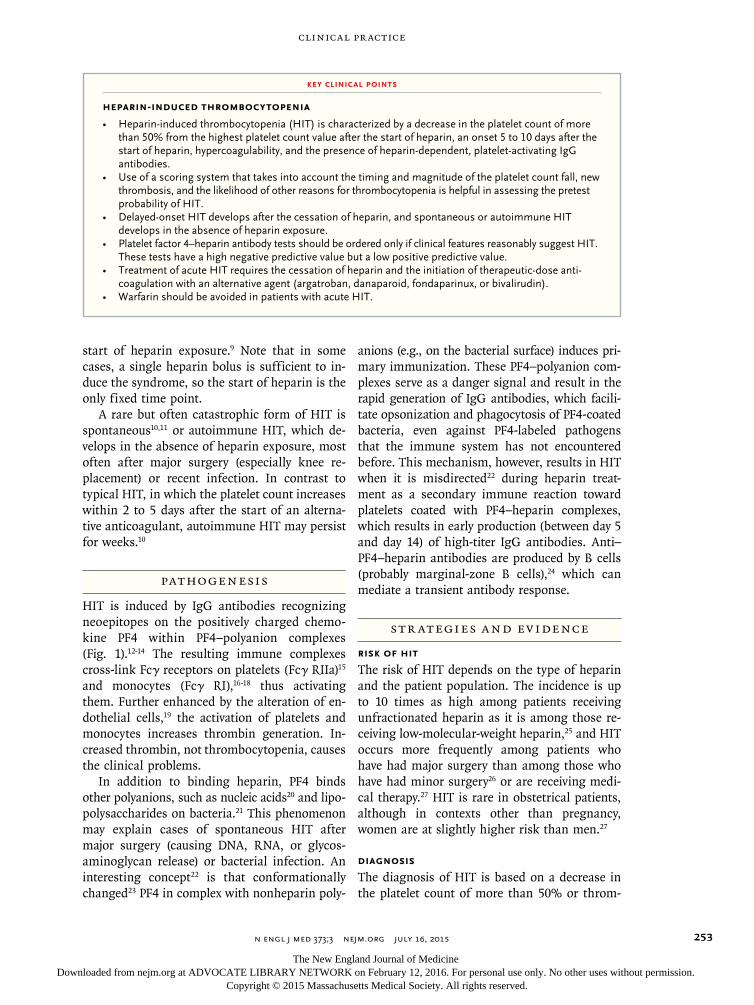
• Heparininduced thrombocytopenia (HIT) is characterized by a decrease in the platelet count of more than 50% from the highest platelet count value after the start of heparin, an onset 5 to 10 days after the start of heparin, hypercoagulability, and the presence of heparindependent, plateletactivating IgG antibodies.
• Use of a scoring system that takes into account the timing and magnitude of the platelet count fall, new thrombosis, and the likelihood of other reasons for thrombocytopenia is helpful in assessing the pretest probability of HIT.
• Delayedonset HIT develops after the cessation of heparin, and spontaneous or autoimmune HIT develops in the absence of heparin exposure.
• Platelet factor 4–heparin antibody tests should be ordered only if clinical features reasonably suggest HIT. These tests have a high negative predictive value but a low positive predictive value.
• Treatment of acute HIT requires the cessation of heparin and the initiation of therapeuticdose anticoagulation with an alternative agent (argatroban, danaparoid, fondaparinux, or bivalirudin).
• Warfarin should be avoided in patients with acute HIT.
The New England Journal of Medicine Downloaded from nejm.org at ADVOCATE LIBRARY NETWORK on February 12, 2016. For personal use only. No other uses without permission.
Copyright © 2015 Massachusetts Medical Society. All rights reserved.
n engl j med 373;3 nejm.org July 16, 2015254
T h e n e w e ngl a nd j o u r na l o f m e dic i n e
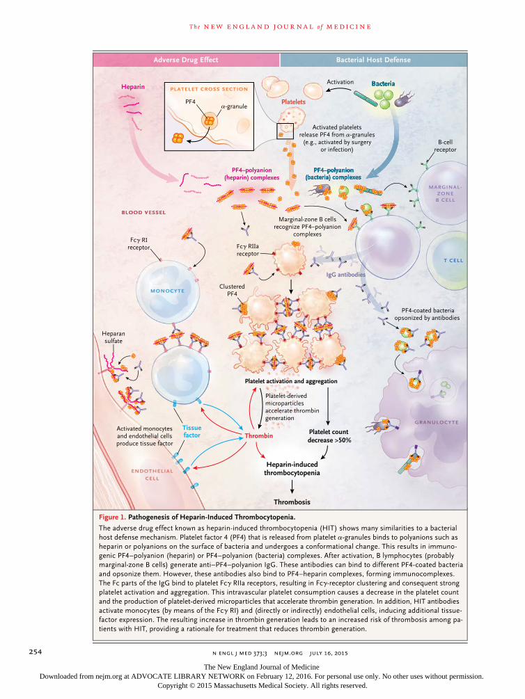
Figure 1. Pathogenesis of Heparin-Induced Thrombocytopenia.
The adverse drug effect known as heparininduced thrombocytopenia (HIT) shows many similarities to a bacterial host defense mechanism. Platelet factor 4 (PF4) that is released from platelet αgranules binds to polyanions such as heparin or polyanions on the surface of bacteria and undergoes a conformational change. This results in immunogenic PF4–polyanion (heparin) or PF4–polyanion (bacteria) complexes. After activation, B lymphocytes (probably marginalzone B cells) generate anti–PF4–polyanion IgG. These antibodies can bind to different PF4coated bacteria and opsonize them. However, these antibodies also bind to PF4–heparin complexes, forming immunocomplexes. The Fc parts of the IgG bind to platelet Fcγ RIIa receptors, resulting in Fcγreceptor clustering and consequent strong platelet activation and aggregation. This intravascular platelet consumption causes a decrease in the platelet count and the production of plateletderived microparticles that accelerate thrombin generation. In addition, HIT antibodies activate monocytes (by means of the Fcγ RI) and (directly or indirectly) endothelial cells, inducing additional tissuefactor expression. The resulting increase in thrombin generation leads to an increased risk of thrombosis among patients with HIT, providing a rationale for treatment that reduces thrombin generation.
GRANULOCYTE
T CELL
Platelets
Fcγ RIIareceptor
B-cellreceptor
Fcγ RIreceptor
PF4–polyanion(heparin) complexes
PF4-coated bacteriaopsonized by antibodies
ClusteredPF4
Activated plateletsrelease PF4 from α-granules
(e.g., activated by surgeryor infection)
Activation BacteriaBacteria
Heparin-inducedthrombocytopenia
Thrombosis
Platelet count decrease >50%
Heparin
PF4
PLATELET CROSS SECTION
α-granule
BLOOD VESSEL
MONOCYTE
Heparansulfate
ENDOTHELIAL
CELL
Tissuefactor
Activated monocytesand endothelial cellsproduce tissue factor
Marginal-zone B cellsrecognize PF4–polyanion
complexes
PF4–polyanion(bacteria) complexes
Thrombin
MARGINAL-ZONE
B CELL
IgG antibodies
Platelet activation and aggregation
Platelet-derivedmicroparticlesaccelerate thrombingeneration
Adverse Drug Effect Bacterial Host Defense
The New England Journal of Medicine Downloaded from nejm.org at ADVOCATE LIBRARY NETWORK on February 12, 2016. For personal use only. No other uses without permission.
Copyright © 2015 Massachusetts Medical Society. All rights reserved.

n engl j med 373;3 nejm.org July 16, 2015 255
Clinical Pr actice
bosis beginning 5 to 10 days after the start of heparin, in association with the appearance of platelet-activating HIT antibodies, as shown by means of a functional assay or inferred by means of a strong positive immunoassay. The fall in platelet count in HIT occurs rapidly (over a period of 1 to 3 days) and is assessed relative to the highest platelet count after the start of heparin. The typical nadir is 40,000 to 80,000 platelets per cubic millimeter, but the count may remain in the normal range (e.g., a decline from 500,000 to 200,000 per cubic millimeter). In less than 10% of patients, the decrease in platelet count is less pronounced (30 to 50% of the high-est preceding value). Rarely, the platelet count may fall below 20,000 per cubic millimeter, es-pecially when HIT is associated with other causes of thrombocytopenia, such as consump-tive coagulopathy.1
Although monitoring of platelet counts facili-tates the recognition of HIT, it is difficult to justify in many patients, especially outpatients. Monitoring should be considered when the risk of HIT is relatively high (>1%), such as among patients who have undergone cardiac surgery and those receiving unfractionated heparin after major surgery28 (other than heparin received for intraoperative flushes or catheter-related flush-es). After major surgery, patients typically have a reactive platelet count increase that exceeds the baseline value (i.e., the value before the receipt of heparin) after the first postoperative week. Given the typical time window of HIT,4 platelet count monitoring on days 5, 7, and 9 allows for the early recognition of HIT in the majority of patients.29 Even if HIT manifests thereafter, the platelet count on day 9 is close to the peak post-surgery platelet count. Monitoring platelet counts on these days facilitates later recognition of a fall in the platelet count of more than 50%, which can be missed when only the lower pre-surgery baseline value is considered. Even with platelet count monitoring, however, the first thrombotic complication may not be prevent-able, because in approximately 20% of patients it occurs shortly before, or concomitant with, the platelet count decrease (Fig. 2).30
Scoring systems can be helpful in estimating the probability of HIT.31,32 A widely used scoring system is the 4T score,31 which evaluates four indicators: the relative platelet count fall, the timing of the onset of the platelet count fall, the presence or absence of thrombosis, and the like-
lihood of another cause, with scores on the in-dividual components ranging from 0 to 2 and higher scores indicating a higher likelihood of HIT. A total score of less than 4 points has a very high negative predictive value (97 to 99%) (Table 1),31,33 whereas the positive predictive val-ue is low (10 to 20% for an intermediate score [4 or 5 points] and 40 to 80% for a high score [6
Figure 2. Timing of HIT and Rationale for Platelet Count Monitoring at Various Time Points.
Guidelines from the American College of Chest Physicians (ACCP)28 recommend platelet count monitoring in patients with a risk of HIT that is higher than 1% (e.g., patients undergoing cardiac surgery, those receiving unfractionated heparin at either a prophylactic or therapeutic dose, and those with cancer). The shaded curve shows the median (black line) ±2 SD of the platelet count in 452 patients who underwent trauma surgery.26 After major surgery, the platelet count (black line) reaches its nadir between day 2 and day 4, followed by a reactive increase that exceeds the baseline value. Because HIT typically manifests between day 5 and day 10, platelet count monitoring before day 1 and on days 5, 7, and 9 (purple arrows) is appropriate to identify the majority of patients with HIT. Comparing the platelet count at the onset of a HITrelated new thrombosis with the presurgery baseline platelet count will often not reveal the 50% decrease without documented preceding platelet counts. This situation is exemplified by an individual patient’s platelet count course (blue line). At the time that HITrelated thrombosis became evident, comparison of the actual platelet count (lower dashed line) with the presurgery platelet count (upper dashed line) shows only a 30% decrease (right red arrow), whereas comparison with the platelet count peak at days 6 and 7 shows the fall in the platelet count of more than 50% (left red arrow), which is indicative of HIT. Patients who are receiving medical therapy do not have this reactive increase in the platelet count, and it is sufficient to compare the platelet count at the time of clinical suspicion of HIT with the baseline platelet count (before the administration of heparin). LMWH denotes lowmolecularweight heparin.
Plat
elet
Cou
nt (x
10–3
per
mm
3 )
500
300
400
200
100
50
01 2 3 4 5 6 7 8 9 10
Days after Start of Heparin or after Surgery If Heparin Was Also Given before Surgery
Daily Heparin or LMWH Prophylaxis
Majorsurgery
Monitoring for HITnot necessary
Platelet countmonitoring
50%
30% Deep-veinthrombosis
The New England Journal of Medicine Downloaded from nejm.org at ADVOCATE LIBRARY NETWORK on February 12, 2016. For personal use only. No other uses without permission.
Copyright © 2015 Massachusetts Medical Society. All rights reserved.

n engl j med 373;3 nejm.org July 16, 2015256
T h e n e w e ngl a nd j o u r na l o f m e dic i n e
to 8 points]).32 A falsely low score may result from missing platelet count values or coexisting conditions that may also underlie thrombocyto-penia. For patients with missing values or coex-isting conditions and for those whose score is intermediate or high, laboratory tests are needed to rule out HIT.
Additional Laboratory Testing
Anti–PF4–heparin enzyme immunoassays have an excellent negative predictive value (98 to 99%)34 but a low positive predictive value, owing to the detection of clinically insignificant anti–PF4–heparin antibodies. In systematic sero-surveillance studies, clinically evident HIT de-veloped in only a minority (2 to 15%) of heparin-treated patients who had anti–PF4–heparin antibodies detected by means of enzyme immunoassay.35,36 In patients with thrombocyto-penia who have a negative test, repeat testing is generally not indicated in the absence of a new decrease in the platelet count or a thrombotic event. Anti–PF4–heparin antibodies are always present before the platelet count begins to de-cline,37 and seroconversion after an initially negative test for anti–PF4–heparin antibodies nearly always detects coincidental, clinically ir-relevant antibodies. Overdiagnosis and associ-
ated overtreatment of HIT are probably more common than underrecognition, given the high frequency of thrombocytopenia among early postoperative and critically ill patients and the low specificity of the assays.
Several strategies can be used to increase the specificity of PF4–heparin enzyme immunoas-says. One strategy is the restriction of the assay to IgG antibodies, because only IgG activates platelets and monocytes by means of Fcγ recep-tors, yet some commercial assays detect com-bined IgG, IgA, and IgM. Also, the magnitude of anti–PF4–heparin reactivity on enzyme immu-noassay should be considered, because greater reactivity correlates with a greater likelihood of HIT; an optical density of less than 1.0 on en-zyme immunoassay is rarely associated with clinically relevant anti–PF4–heparin antibod-ies.38 However, optical-density values are arbi-trary units and may vary among laboratories.39 Inhibition of the enzyme immunoassay by high concentrations of heparin also increases speci-ficity, but the most relevant, very strongly react-ing antibodies may not be inhibited.34
Diagnostic accuracy for HIT is improved with the use of both an anti–PF4–heparin enzyme immunoassay and a functional test (e.g., a platelet-activation assay). In particular, platelet-
Variable Score
2 1 0
Acute thrombocytopenia Platelet count decrease of >50% and nadir ≥20,000/mm3
Platelet count decrease of 30–50% or nadir 10,000–19,000/mm3
Platelet count decrease of <30% or nadir ≤10,000/mm3
Timing of onset Day 5–10, or day 1 if recent heparin exposure
>Day 10 or unclear exposure ≤Day 4 with no recent heparin exposure
Thrombosis New thrombosis or anaphy lactoid reaction after heparin bolus
Progressive or recurrent thrombosis
None
Other cause of thrombocytopenia
None Possible Definite
Total score 6–8, indicating high score 4 or 5, indicating intermediate score
0–3, indicating low score
* Adapted from Lo et al.31 A low 4T score (0 to 3 points) has a high negative predictive value. The day that heparin was started is considered as day 0. The onset of heparininduced thrombocytopenia (HIT) is defined as the day that the platelet count begins to decrease. Patients in whom the score is difficult to apply, owing to missing platelet count values or coexisting conditions causing thrombocytopenia, and those with an intermediate or high score require further evaluation. This score can be included on ordering forms for HIT laboratory testing (e.g., www2 . medizin . unigreifswald . de/ transfus/ fileadmin/ user_upload/ doku_thrombo_gerinnung/ platelet_lab_request_form . pdf).
Table 1. 4T Scoring System for Evaluating the Pretest Probability of Heparin-Induced Thrombocytopenia.*
The New England Journal of Medicine Downloaded from nejm.org at ADVOCATE LIBRARY NETWORK on February 12, 2016. For personal use only. No other uses without permission.
Copyright © 2015 Massachusetts Medical Society. All rights reserved.

n engl j med 373;3 nejm.org July 16, 2015 257
Clinical Pr actice
activation assays with the use of washed plate-lets34 (e.g., serotonin-release assay and heparin-induced platelet-activation test, both with the use of high heparin inhibition) are much more specific than enzyme immunoassays for clini-cally relevant antibodies and also detect the rare antibodies with other specificities.34 A negative functional assay essentially rules out HIT. These assays are restricted to specialized laboratories and are usually applied as second-line tests in the diagnostic workup of HIT.
HIT assays should not be used to screen as-ymptomatic patients and should be interpreted only in the context of the pretest probability of HIT.33,34 A low or intermediate 4T score together with a negative antigen test rules out HIT, whereas an intermediate or high score together with a positive functional assay makes HIT very likely (Fig. 3).
In a subgroup of patients, anti–PF4–heparin antibodies show very high optical densities (>2.0) and strongly activate platelets even in the absence of heparin. First recognized in delayed-onset HIT,9 these autoreactive antibodies medi-ate spontaneous HIT. They may also be tran-siently present during the first 5 to 7 days in typical HIT, without affecting treatment.40,41
In patients with strongly suspected or con-firmed HIT, duplex ultrasonography can rule out subclinical deep-vein thrombosis, which may affect the duration of treatment.1 In patients with HIT who have abdominal pain or hypoten-sion, bilateral adrenal hemorrhage associated with adrenal-vein thrombosis should be consid-ered; severe headache should prompt the consid-eration of cavernous sinus thrombosis.
Treatment
Key interventions in patients with highly sus-pected or confirmed acute HIT are the prompt cessation of heparin (if still being administered) and the initiation of an alternative anticoagulant at a therapeutic dose. Prophylactic-dose antico-agulation is insufficient42 to compensate for massive thrombin generation, even if the patient has no apparent thrombosis. Vitamin K antago-nists (e.g., warfarin and phenprocoumon) should not be given until HIT has abated (e.g., the plate-let count has increased to >150,000 per cubic millimeter at a stable plateau for 2 consecutive days), because they increase the risk of venous limb gangrene and limb loss by decreasing the
level of protein C. When vitamin K antagonists are initiated, overlap with an alternative antico-agulant is needed.29
Two drugs are approved for the treatment of HIT — the direct thrombin inhibitor argatroban (in the United States, Canada, the European Union, and Australia) and the antithrombin-de-pendent factor Xa inhibitor danaparoid (in Can-ada, the European Union, and Australia).29 An analysis of prospective cohorts showed a re-duced risk of the composite outcome of new thrombosis, death due to thrombosis, or ampu-tation related to thrombosis in patients treated with argatroban, as compared with historical controls (hazard ratio for the composite out-come among patients with HIT without throm-bosis, 0.33; 95% confidence interval [CI], 0.20 to 0.54; hazard ratio for the composite outcome among patients with HIT with thrombosis, 0.30; 95% CI, 0.25 to 0.62).43 An analysis of outcomes with danaparoid29 that was provided on a com-passionate-use basis showed a rate of treatment success (platelet count recovery without new thrombosis and absence of major adverse events requiring drug cessation) that was higher than 90%.44 In a small, open-label, randomized trial comparing danaparoid with dextran 70 for the treatment of HIT with thrombosis, the rates of recovery from thromboembolism were signifi-cantly higher with danaparoid.45 Fondaparinux and bivalirudin are also used in this context, although they have not been approved by the Food and Drug Administration for this indica-tion. Case series have shown good outcomes in patients with HIT treated with fondaparinux29,46 or bivalirudin.28,47
Argatroban is frequently used in critically ill patients. It has a relatively short half-life, which is independent of renal function, but it requires intravenous administration. Because argatroban affects the international normalized ratio, the transition to warfarin has to follow a special protocol.28 An underrecognized issue is that the activated partial-thromboplastin time may be falsely high when argatroban is given to patients who have additional coagulopathies (e.g., con-sumptive coagulopathy or impaired liver func-tion) or to patients who have received pretreat-ment with warfarin. This situation may lead to underdosing of argatroban, with the risk of progressive microvascular thrombosis and ische-mic limb loss. This limitation may be overcome
The New England Journal of Medicine Downloaded from nejm.org at ADVOCATE LIBRARY NETWORK on February 12, 2016. For personal use only. No other uses without permission.
Copyright © 2015 Massachusetts Medical Society. All rights reserved.

n engl j med 373;3 nejm.org July 16, 2015258
T h e n e w e ngl a nd j o u r na l o f m e dic i n e
Figure 3. Diagnosis of HIT.
The flowchart provides a guide to decision making regarding a patient who is suspected to have HIT. Antigen assays for PF4–heparin antibodies are widely available, whereas functional assays with the use of washed platelets, such as the serotoninrelease assay (SRA) or the heparininduced plateletactivation (HIPA) test, are restricted to specialized laboratories. In centers where a functional assay is unavailable or cannot be obtained promptly, other options include the use of high reactivity of the antigen test (e.g., optical density [OD], >1.0) as a surrogate marker for clinically relevant antibodies or incorporating the 4T score in interpretation (dashed lines), although the overdiagnosis of HIT remains possible. The 4T scoring system evaluates four indicators (the relative platelet count fall, the timing of the onset of the platelet count fall, the presence or absence of thrombosis, and the likelihood of another cause), with scores on the individual components ranging from 0 to 2 and higher scores indicating a greater likelihood of HIT. In the case of a high 4T score and a negative result on the PF4–heparin IgG antibody immunoassay, consider that laboratory error may be a cause of a false negative result. Even if shortterm treatment decisions are made without confirmation of the presence of plateletactivating, heparindependent antibodies, efforts should be made to rule out or confirm the presence of these antibodies to guide the future treatment of the patient.
Evaluate clinical probability of HIT by a systemicscoring system (e.g., 4T score)
Thrombocytopenia or thrombosisduring heparin use
Intermediate probability (4T score, 4 or 5) High probability (4T score, 6–8)
Replace heparin and initiate laboratory investigations
Low probability (4T score, ≤3)
Score difficult to apply (owing to missing platelet counts or coexisting condition
causing thrombocytopenia)
PF4–heparin IgG immunoassayYesNo
PositiveNegative
HIT verylikely
HITunlikely
PositiveStrongly positive
OD >1.0
Functional assay with washed platelets(e.g., SRA or HIPA test)
HIT ruled outContinue or restart heparin
Therapeutic-dose anticoagulation(e.g., argatroban, danaparoid, bivali-rudin, or fondaparinux)
Rule out deep-vein thrombosis
Negative
The New England Journal of Medicine Downloaded from nejm.org at ADVOCATE LIBRARY NETWORK on February 12, 2016. For personal use only. No other uses without permission.
Copyright © 2015 Massachusetts Medical Society. All rights reserved.

n engl j med 373;3 nejm.org July 16, 2015 259
Clinical Pr actice
by the use of an ecarin-based clotting assay (which has limited availability) or the plasma-diluted thrombin time assay (for which an in-house standard for argatroban is required).
Danaparoid can be administered intravenous-ly or subcutaneously, whereas fondaparinux is given only subcutaneously. These two drugs can be reliably monitored by anti–factor Xa assays, but they have long half-lives and in patients with renal insufficiency, a dose adjustment will be necessary. Subcutaneous injection makes these drugs easier than argatroban to use outside the intensive care unit. In addition, danaparoid in-terferes with the immune mechanism of HIT by disrupting PF4–heparin complexes.48 Exacerba-tions of HIT have been reported infrequently with the use of either danaparoid or fondaparinux. Although most cases of exacerbations are attrib-utable to autoimmune HIT antibodies and are independent of these drugs, a small percentage of patients have true in vivo and in vitro cross-reactivity. If manifestations of HIT worsen de-spite sufficient levels of anti–factor Xa activity in patients taking danaparoid or fondaparinux, treat-ment should be switched (e.g., to argatroban).
Prophylactic platelet transfusions should be avoided in patients with HIT. The risk of bleed-ing is very low, and such transfusions can in-crease the risk of thrombosis.49
A r e a s of Uncerta in t y
Whereas in vitro data suggest that dabigatran, rivaroxaban, and apixaban might also be used to treat patients with HIT,50 more data are needed before these drugs can be recommended in the context of acute HIT. A concern is whether trough levels are sufficient to prevent thrombin generation by strongly reactive anti–PF4–heparin antibodies.
Patients who have HIT with thrombosis re-quire therapeutic-dose anticoagulation for at least 3 months. However, in patients who have HIT without thrombosis, the duration of therapeutic-dose anticoagulation after the platelet count has reached a stable plateau (ideally >150,000 per cubic millimeter)28 is unresolved.
High-dose intravenous immune globulin G (e.g., at a dose of 2 g per kilogram of body weight over a 2-day period) interferes with HIT by blocking platelet Fcγ receptors. Limited data suggest that this drug may be an option (along
with anticoagulation) in patients at high risk for thrombosis and bleeding (e.g., those who are preg-nant or have sinus-vein thrombosis complicating HIT) or in patients who have autoimmune HIT.51
PF4 forms complexes with negatively charged nucleic acids and aptamers,20 which cross-react with anti–PF4–heparin antibodies. Aptamers and other nucleic acid–based drugs are entering clinical application, and it is unclear whether they can induce HIT.
In patients with a history of HIT who require cardiac surgery, postponing surgery until plate-let-activating anti–PF4–heparin antibodies dis-appear and then using heparin intraoperatively is a safe approach.8,28,52 Another option in urgent situations is the removal of platelet-activating anti–PF4–heparin antibodies by plasmapheresis, as described in anecdotal reports.53 Otherwise, bivalirudin is a compatible anticoagulant for cardiac surgery if platelet-activating anti–PF4–heparin antibodies are present. However, its use requires special approaches to avoiding stagna-tion of blood (which results in degradation of bivalirudin).28
Guidelines
The guidelines of the American College of Chest Physicians (ACCP)28 and national European guide-lines54-56 address HIT. In the absence of data from randomized trials, most recommendations are supported by low-grade evidence. All these guidelines recommend the use of a scoring sys-tem to determine the probability of HIT before testing is performed and note the need for therapeutic-dose anticoagulation in cases of acute HIT. Guidelines differ with each other regarding specific recommendations about which patients should undergo routine platelet count monitor-ing and the frequency of monitoring. The ACCP guidelines recommend assessing patients at high risk (>1%) for HIT every 2 to 3 days between day 4 and day 14.28
Conclusions a nd R ecommendations
The patient described in the vignette had a marked decrease in the platelet count after several days of therapy with low-molecular-weight hepa-rin, which raises concern about HIT. Calculation of the 4T score is recommended to determine
The New England Journal of Medicine Downloaded from nejm.org at ADVOCATE LIBRARY NETWORK on February 12, 2016. For personal use only. No other uses without permission.
Copyright © 2015 Massachusetts Medical Society. All rights reserved.

n engl j med 373;3 nejm.org July 16, 2015260
T h e n e w e ngl a nd j o u r na l o f m e dic i n e
her risk of HIT. Her score of 5 points (decrease in platelet count, 2; timing, 2; thrombosis, 0; and likelihood of other reasons, 1, since her endocar-ditis is stable and the platelet count is too high for antibiotic-induced immune thrombocytopenia) places her at intermediate risk. Although routine screening for PF4–heparin antibodies is strongly discouraged, patients at intermediate or high risk should undergo this testing. A positive anti–PF4–heparin IgG enzyme immunoassay is necessary for the diagnosis of HIT but is nonspecific. A strongly positive test (optical density, >1.5) or positive platelet-activation assay would strongly support the diagnosis of HIT. Treatment involves the prompt cessation of heparin and the initiation
of an alternative anticoagulant (argatroban or danaparoid, both of which are approved for this indication, or fondaparinux or bivalirudin, with use of these agents supported by case series).
Dr. Greinacher reports receiving fees for serving on an advi-sory board from Instrumentation Laboratory; consulting fees from Bayer HealthCare, Macopharma, Merck Sharp & Dohme, and W.L. Gore and Associates; travel support from Boehringer Ingelheim; fees for the planning of and participation in a Con-tinuing Medical Education program from Bristol-Myers Squibb; grant support from Macopharma; and institutional fees to his university from Boehringer Ingelheim. He also reports an agree-ment in development with BioMarin Nederland and Nanjing King-friend Biochemical Pharmaceutical, which will provide institutional but no personal fees. No other potential conflict of interest relevant to this article was reported.
Disclosure forms provided by the author are available with the full text of this article at NEJM.org.
References1. Greinacher A, Warkentin TE, Chong BH. Heparin-induced thrombocytopenia. In: Michelson AD, ed. Platelets. 3rd ed. Oxford, United Kingdom: Elsevier’s Sci-ence and Technology, 2012: 851-82.2. Warkentin TE, Levine MN, Hirsh J, et al. Heparin-induced thrombocytopenia in patients treated with low-molecular-weight heparin or unfractionated heparin. N Engl J Med 1995; 332: 1330-5.3. Warkentin TE, Kelton JG. A 14-year study of heparin-induced thrombocytope-nia. Am J Med 1996; 101: 502-7.4. Warkentin TE, Kelton JG. Temporal aspects of heparin-induced thrombocyto-penia. N Engl J Med 2001; 344: 1286-92.5. Tholl U, Greinacher A, Overdick K, Anlauf M. Life-threatening anaphylactic reaction following parathyroidectomy in a dialysis patient with heparin-induced thrombocytopenia. Nephrol Dial Trans-plant 1997; 12: 2750-5.6. Warkentin TE, Greinacher A. Heparin-induced anaphylactic and anaphylactoid reactions: two distinct but overlapping syndromes. Expert Opin Drug Saf 2009; 8: 129-44.7. Pötzsch B, Klövekorn WP, Madlener K. Use of heparin during cardiopulmonary bypass in patients with a history of hepa-rin-induced thrombocytopenia. N Engl J Med 2000; 343: 515.8. Warkentin TE, Sheppard JA. Serologi-cal investigation of patients with a previ-ous history of heparin-induced thrombo-cytopenia who are reexposed to heparin. Blood 2014; 123: 2485-93.9. Warkentin TE, Kelton JG. Delayed-onset heparin-induced thrombocytopenia and thrombosis. Ann Intern Med 2001; 135: 502-6.10. Warkentin TE, Basciano PA, Knop-man J, Bernstein RA. Spontaneous hepa-rin-induced thrombocytopenia syndrome: 2 new cases and a proposal for defining this disorder. Blood 2014; 123: 3651-4.
11. Greinacher A. Me or not me? The dan-ger of spontaneity. Blood 2014; 123: 3536-8.12. Brandt S, Krauel K, Gottschalk KE, et al. Characterisation of the conforma-tional changes in platelet factor 4 induced by polyanions: towards in vitro prediction of antigenicity. Thromb Haemost 2014; 112: 53-64.13. Ziporen L, Li ZQ, Park KS, et al. De-fining an antigenic epitope on platelet factor 4 associated with heparin-induced thrombocytopenia. Blood 1998; 92: 3250-9.14. Li ZQ, Liu W, Park KS, et al. Defining a second epitope for heparin-induced thrombocytopenia/thrombosis antibodies using KKO, a murine HIT-like monoclo-nal antibody. Blood 2002; 99: 1230-6.15. Kelton JG, Sheridan D, Santos A, et al. Heparin-induced thrombocytopenia: lab-oratory studies. Blood 1988; 72: 925-30.16. Gruel Y, Pouplard C, Lasne D, Magde-laine-Beuzelin C, Charroing C, Watier H. The homozygous FcgammaRIIIa-158V gen-otype is a risk factor for heparin-induced thrombocytopenia in patients with anti-bodies to heparin-platelet factor 4 com-plexes. Blood 2004; 104: 2791-3.17. Kasthuri RS, Glover SL, Jonas W, et al. PF4/heparin-antibody complex induces monocyte tissue factor expression and re-lease of tissue factor positive microparti-cles by activation of FcγRI. Blood 2012; 119: 5285-93.18. Rauova L, Hirsch JD, Greene TK, et al. Monocyte-bound PF4 in the pathogenesis of heparin-induced thrombocytopenia. Blood 2010; 116: 5021-31.19. Cines DB, Tomaski A, Tannenbaum S. Immune endothelial-cell injury in heparin-associated thrombocytopenia. N Engl J Med 1987; 316: 581-9.20. Jaax ME, Krauel K, Marschall T, et al. Complex formation with nucleic acids and aptamers alters the antigenic properties of platelet factor 4. Blood 2013; 122: 272-81.21. Krauel K, Weber C, Brandt S, et al.
Platelet factor 4 binding to lipid A of Gram-negative bacteria exposes PF4/heparin-like epitopes. Blood 2012; 120: 3345-52.22. Krauel K, Pötschke C, Weber C, et al. Platelet factor 4 binds to bacteria, induc-ing antibodies cross-reacting with the major antigen in heparin-induced throm-bocytopenia. Blood 2011; 117: 1370-8.23. Kreimann M, Brandt S, Krauel K, et al. Binding of anti-platelet factor 4/heparin antibodies depends on the thermodynam-ics of conformational changes in platelet factor 4. Blood 2014; 124: 2442-9.24. Zheng Y, Yu M, Podd A, et al. Critical role for mouse marginal zone B cells in PF4/heparin antibody production. Blood 2013; 121: 3484-92.25. Martel N, Lee J, Wells PS. Risk for heparin-induced thrombocytopenia with unfractionated and low-molecular-weight heparin thromboprophylaxis: a meta-analysis. Blood 2005; 106: 2710-5.26. Lubenow N, Hinz P, Thomaschewski S, et al. The severity of trauma determines the immune response to PF4/heparin and the frequency of heparin-induced throm-bocytopenia. Blood 2010; 115: 1797-803.27. Greinacher A, Warkentin TE. Risk of heparin-induced thrombocytopenia in pa-tients receiving thromboprophylaxis. Ex-pert Rev Hematol 2008; 1: 75-85.28. Linkins LA, Dans AL, Moores LK, et al. Treatment and prevention of heparin-induced thrombocytopenia: antithrombot-ic therapy and prevention of thrombosis, 9th ed: American College of Chest Phy-sicians evidence-based clinical practice guidelines. Chest 2012; 141: 2 Suppl: e495S-e530S.29. Warkentin TE, Greinacher A, Koster A, Lincoff AM. Treatment and prevention of heparin-induced thrombocytopenia: Amer-ican College of Chest Physicians evidence-based clinical practice guidelines (8th edition). Chest 2008; 133: 6 Suppl: 340S-380S.
The New England Journal of Medicine Downloaded from nejm.org at ADVOCATE LIBRARY NETWORK on February 12, 2016. For personal use only. No other uses without permission.
Copyright © 2015 Massachusetts Medical Society. All rights reserved.

n engl j med 373;3 nejm.org July 16, 2015 261
Clinical Pr actice
30. Greinacher A, Farner B, Kroll H, Kohlmann T, Warkentin TE, Eichler P. Clinical features of heparin-induced thrombocytopenia including risk factors for thrombosis: a retrospective analysis of 408 patients. Thromb Haemost 2005; 94: 132-5.31. Lo GK, Juhl D, Warkentin TE, Sigouin CS, Eichler P, Greinacher A. Evaluation of pretest clinical score (4 T’s) for the diag-nosis of heparin-induced thrombocytope-nia in two clinical settings. J Thromb Haemost 2006; 4: 759-65.32. Cuker A, Arepally G, Crowther MA, et al. The HIT Expert Probability (HEP) Score: a novel pre-test probability model for heparin-induced thrombocytopenia based on broad expert opinion. J Thromb Haemost 2010; 8: 2642-50.33. Cuker A, Gimotty PA, Crowther MA, Warkentin TE. Predictive value of the 4Ts scoring system for heparin-induced throm-bocytopenia: a systematic review and meta-analysis. Blood 2012; 120: 4160-7.34. Warkentin TE, Greinacher A. Labora-tory testing for heparin-induced thrombo-cytopenia. In: Warkentin TE, Greinacher A, eds. Heparin-induced thrombocytopenia. 5th ed. New York: Informa Healthcare, 2013: 272-314.35. Warkentin TE, Sheppard JA, Horse-wood P, Simpson PJ, Moore JC, Kelton JG. Impact of the patient population on the risk for heparin-induced thrombocytope-nia. Blood 2000; 96: 1703-8.36. Pouplard C, May MA, Regina S, Marchand M, Fusciardi J, Gruel Y. Changes in platelet count after cardiac surgery can effectively predict the development of pathogenic heparin-dependent antibodies. Br J Haematol 2005; 128: 837-41.37. Warkentin TE, Sheppard JA, Moore JC, Cook RJ, Kelton JG. Studies of the im-mune response in heparin-induced throm-bocytopenia. Blood 2009; 113: 4963-9.38. Warkentin TE, Sheppard JI, Moore JC, Sigouin CS, Kelton JG. Quantitative inter-pretation of optical density measurements using PF4-dependent enzyme-immunoas-says. J Thromb Haemost 2008; 6: 1304-12.39. Greinacher A, Ittermann T, Bagemühl J, et al. Heparin-induced thrombocytope-
nia: towards standardization of platelet factor 4/heparin antigen tests. J Thromb Haemost 2010; 8: 2025-31.40. Socher I, Kroll H, Jorks S, Santoso S, Sachs UJ. Heparin-independent activation of platelets by heparin-induced thrombo-cytopenia antibodies: a common occur-rence. J Thromb Haemost 2008; 6: 197-200.41. Prechel MM, McDonald MK, Jeske WP, Messmore HL, Walenga JM. Activation of platelets by heparin-induced thrombo-cytopenia antibodies in the serotonin re-lease assay is not dependent on the pres-ence of heparin. J Thromb Haemost 2005; 3: 2168-75.42. Farner B, Eichler P, Kroll H, Grein-acher A. A comparison of danaparoid and lepirudin in heparin-induced thrombocy-topenia. Thromb Haemost 2001; 85: 950-7.43. Lewis BE, Wallis DE, Hursting MJ, Levine RL, Leya F. Effects of argatroban therapy, demographic variables, and plate-let count on thrombotic risks in heparin-induced thrombocytopenia. Chest 2006; 129: 1407-16.44. Chong BH, Magnani H. Danaparoid for the treatment of heparin-induced thrombocytopenia. In: Warkentin TE, Greinacher A, eds. Heparin-induced throm-bocytopenia. 5th ed. New York: Informa Healthcare, 2013: 466-88.45. Chong BH, Gallus AS, Cade JF, et al. Prospective randomised open-label com-parison of danaparoid with dextran 70 in the treatment of heparin-induced throm-bocytopaenia with thrombosis: a clinical outcome study. Thromb Haemost 2001; 86: 1170-5.46. Kang M, Alahmadi M, Sawh S, Kovacs MJ, Lazo-Langner A. Fondaparinux for the treatment of suspected heparin-induced thrombocytopenia: a propensity score-matched study. Blood 2015; 125: 924-9.47. Bartholomew JR, Prats J. Bivalirudin for the treatment of heparin-induced thrombocytopenia. In: Warkentin TE, Greinacher A, eds. Heparin-induced throm-bocytopenia. 5th ed. New York: Informa Healthcare, 2013: 429-65.48. Krauel K, Fürll B, Warkentin TE, et al. Heparin-induced thrombocytopenia — therapeutic concentrations of danaparoid,
unlike fondaparinux and direct thrombin inhibitors, inhibit formation of platelet factor 4-heparin complexes. J Thromb Haemost 2008; 6: 2160-7.49. Goel R, Ness PM, Takemoto CM, Krishnamurti L, King KE, Tobian AA. Platelet transfusions in platelet consump-tive disorders are associated with arterial thrombosis and in-hospital mortality. Blood 2015; 125: 1470-6.50. Krauel K, Hackbarth C, Fürll B, Grei-nacher A. Heparin-induced thrombocyto-penia: in vitro studies on the interaction of dabigatran, rivaroxaban, and low-sul-fated heparin, with platelet factor 4 and anti-PF4/heparin antibodies. Blood 2012; 119: 1248-55.51. Tvito A, Bakchoul T, Rowe JM, Grein-acher A, Ganzel C. Severe and persistent heparin-induced thrombocytopenia de-spite fondaparinux treatment. Am J He-matol 2015; 90: 675-8.52. Selleng S, Haneya A, Hirt S, Selleng K, Schmid C, Greinacher A. Management of anticoagulation in patients with subacute heparin-induced thrombocytopenia sched-uled for heart transplantation. Blood 2008; 112: 4024-7.53. Warkentin TE, Sheppard JA, Chu FV, Kapoor A, Crowther MA, Gangji A. Plas-ma exchange to remove HIT antibodies: dissociation between enzyme-immunoas-say and platelet activation test reactivities. Blood 2015; 125: 195-8.54. Watson H, Davidson S, Keeling D; Haemostasis and Thrombosis Task Force of the British Committee for Standards in Haematology. Guidelines on the diagno-sis and management of heparin-induced thrombocytopenia: second edition. Br J Haematol 2012; 159: 528-40.55. The Association of the Scientific Med-ical Societies of Germany. German guide-lines for thrombosis prophylaxis (http://www .awmf .org/ leitlinien/ detail/ ll/ 003-001 .html).56. The Association of the Scientific Med-ical Societies of Germany. German guide-lines for thrombosis therapy (http://www .awmf .org/ leitlinien/ detail/ ll/ 065-002 .html).Copyright © 2015 Massachusetts Medical Society.
images in clinical medicine
The Journal welcomes consideration of new submissions for Images in Clinical Medicine. Instructions for authors and procedures for submissions can be found on the Journal’s website at NEJM.org. At the discretion of the editor, images that
are accepted for publication may appear in the print version of the Journal, the electronic version, or both.
The New England Journal of Medicine Downloaded from nejm.org at ADVOCATE LIBRARY NETWORK on February 12, 2016. For personal use only. No other uses without permission.
Copyright © 2015 Massachusetts Medical Society. All rights reserved.