Embed Size (px)
Citation preview

BioOne sees sustainable scholarly publishing as an inherently collaborative enterprise connecting authors, nonprofit publishers, academic institutions, researchlibraries, and research funders in the common goal of maximizing access to critical research.
Cancer-Prone Mice Expressing the Ki-rasG12C Gene Show Increased LungCarcinogenesis after CT Screening ExposuresAuthor(s): Michael T. Munley, Joseph E. Moore, Matthew C. Walb, Scott P. Isom, John D. Olson, J.Gregory Zora, Nancy D. Kock, Kenneth T. Wheeler and Mark Steven MillerSource: Radiation Research, 176(6):842-848. 2011.Published By: Radiation Research SocietyDOI: http://dx.doi.org/10.1667/RR2649.1URL: http://www.bioone.org/doi/full/10.1667/RR2649.1
BioOne (www.bioone.org) is a nonprofit, online aggregation of core research in the biological, ecological, andenvironmental sciences. BioOne provides a sustainable online platform for over 170 journals and books publishedby nonprofit societies, associations, museums, institutions, and presses.
Your use of this PDF, the BioOne Web site, and all posted and associated content indicates your acceptance ofBioOne’s Terms of Use, available at www.bioone.org/page/terms_of_use.
Usage of BioOne content is strictly limited to personal, educational, and non-commercial use. Commercial inquiriesor rights and permissions requests should be directed to the individual publisher as copyright holder.

RADIATION RESEARCH 176, 842–848 (2011)0033-7587/11 $15.00�2011 by Radiation Research Society.All rights of reproduction in any form reserved.DOI: 10.1667/RR2649.1
SHORT COMMUNICATION
Cancer-Prone Mice Expressing the Ki-rasG12C Gene Show Increased LungCarcinogenesis after CT Screening Exposures
Michael T. Munley,a,g,1 Joseph E. Moore,b Matthew C. Walb,a Scott P. Isom,c,g John D. Olson,d J. Gregory Zora,b
Nancy D. Kock,e Kenneth T. Wheelerf,g and Mark Steven Millerb,g
Departments of a Radiation Oncology, b Cancer Biology, c Public Health Sciences, d Center for Biomolecular Imaging, e Pathology,f Radiology and the g Comprehensive Cancer Center of Wake Forest University, Wake Forest School of Medicine, Medical Center Boulevard,
Winston-Salem, North Carolina 27157
Munley, M. T., Moore, J. E., Walb, M. C., Isom, S. P., Olson,J. D., Zora, J. G., Kock, N. D., Wheeler, K. T. and Miller, M. S.Cancer-Prone Mice Expressing the Ki-rasG12C Gene ShowIncreased Lung Carcinogenesis after CT Screening Expo-sures. Radiat. Res. 176, 842–848 (2011).
A .20-fold increase in X-ray computed tomography (CT)use during the last 30 years has caused considerable concernbecause of the potential carcinogenic risk from these CTexposures. Estimating the carcinogenic risk from high-energy,single high-dose exposures obtained from atomic bombsurvivors and extrapolating these data to multiple low-energy,low-dose CT exposures using the Linear No-Threshold (LNT)model may not give an accurate assessment of actual cancerrisk. Recently, the National Lung Cancer Screening Trial(NLST) reported that annual CT scans of current and formerheavy smokers reduced lung cancer mortality by 20%,highlighting the need to better define the carcinogenic riskassociated with these annual CT screening exposures. In thisstudy, we used the bitransgenic CCSP-rtTA/Ki-ras mousemodel that conditionally expresses the human mutant Ki-rasG12C gene in a doxycycline-inducible and lung-specificmanner to measure the carcinogenic risk of exposure tomultiple whole-body CT doses that approximate the annualNLST screening protocol. Irradiated mice expressing the Ki-rasG12C gene in their lungs had a significant (P ¼ 0.01) 43%increase in the number of tumors/mouse (24.1 6 1.9)compared to unirradiated mice (16.8 6 1.3). Irradiatedfemales had significantly (P , 0.005) more excess tumorsthan irradiated males. No tumor size difference or doseresponse was observed over the total dose range of 80–160mGy for either sex. Irradiated bitransgenic mice that did notexpress the Ki-rasG12C gene had a low tumor incidence (�0.1/mouse) that was not affected by exposure to CT radiation.These results suggest that (i) estimating the carcinogenic riskof multiple CT exposures from high-dose carcinogenesis datausing the LNT model may be inappropriate for current and
former smokers and (ii) any increased carcinogenic risk afterexposure to fractionated low-dose CT-radiation may berestricted to only those individuals expressing cancer suscep-tibility genes in their tissues at the time of exposure. � 2011 by
Radiation Research Society
INTRODUCTION
A .20-fold increase in X-ray computed tomography (CT)use during the last 30 years has caused considerable concern
because of the potential carcinogenic risk from these CTexposures (1–7). It has been reported that as many as15,000–45,000 excess tumors are caused by exposure to CTradiation in the U.S. each year (8, 9). However, theseestimates are derived from mathematical models that
assume that (i) everyone has the same carcinogenic riskafter a specific dose of CT radiation, (ii) the carcinogenicrisk from multiple low-energy, low-dose (,50 mGy) CTexposures is identical to that of the Japanese atomic bomb
survivors, who were exposed predominantly to high-energy,single doses (,2 Gy) of mixed-beam radiation, and (iii) thecarcinogenic risk from high-energy, high single-dose datacan be extrapolated to multiple low-energy, low-dose CTexposures by the Linear No-Threshold (LNT) model (10,11). All of these assumptions are required because there areno carcinogenic risk data from low-dose CT exposures totest these assumptions.
Recently, the National Lung Screening Trial (NLST)reported that annual CT scans of 55–74-year-old current and
former heavy (�30 pack years) smokers reduced lungcancer mortality by 20% (12, 13). This result creates achallenge for those responsible for making screeningrecommendations. On the one hand, annual CT screeningof older current and former heavy smokers has the potential
to significantly reduce mortality by identifying patients withearly-stage localized lung tumors who have approximately a50% probability of surviving 5 years (14). At present,
1 Address for correspondence: Department of Radiation Oncology,Wake Forest School of Medicine, Medical Center Boulevard,Winston-Salem, NC 27157; e-mail: [email protected].
842

;80% of all lung tumors are diagnosed with regional and/ordistant spread of disease; these patients have an averageprobability of ,10% of surviving 5 years (14). Alterna-tively, annual CT scans of older current and former heavysmokers carry the risk of either initiating normal cells tobecome tumor cells or promoting the growth of alreadyinitiated cells into tumors. This conundrum highlights theneed to better define the carcinogenic risk associated withannual CT screening of current and former smokers. Thisrequires testing the assumptions of the mathematical modelsoutlined above by directly measuring excess tumorproduction after multiple CT exposures in an animal modelof current and former smokers.
Adenocarcinoma, a form of non-small cell lung cancer(NSCLC), is the most common histological variant, and itsincidence has been increasing (14, 15). In the study reportedhere, we used a transgenic mouse model developed in ourlaboratory (16) that contains a mutant form of the human Ki-ras gene. Mutations in Ki-ras have been implicated as one ofthe key genetic alterations that drive tumorigenesis inapproximately 30% of human lung adenocarcinomas (17–20). The mutant form of the human Ki-ras gene was insertedin the mouse genome to create a transgenic mouse in whichthe mutant human gene is expressed in an inducible andlung-specific manner. This system makes use of theinducible ‘‘tet-on’’ system (21) and involves crossing twodifferent transgenic mice. One transgenic mouse contains areverse tetracycline (tet) trans-activator (rtTA) proteinlinked to the Clara cell secretory protein (CCSP) promoter.These mice constitutively express the rtTA gene productspecifically in the lungs (22). The CCSP-rtTA transgenicmice are crossed with a second transgenic mouse thatcontains the cDNA of Ki-rasG12C cloned into the tetO7-CMVplasmid, placing the mutant Ki-rasG12C transgene down-stream of a tet-inducible promoter (16). When these twotransgenic mice are crossed and treated with doxycycline(DOX), lung-specific expression of the mutant Ki-rastransgene product occurs, resulting in the formation ofsmall, benign, hyperplastic foci and very early-stage
adenomas that do not normally progress to more severetumor types even after a year of continuous DOX treatment(16, 23). We have shown that exposure of DOX-treated miceto lung tumor promoters will result in increased tumormultiplicity and the development of adenocarcinomas (24).Thus our bitransgenic mouse model recapitulates the earlieststages of lung tumor formation and would be representativeof asymptomatic smokers and ex-smokers who harborgenetic damage in their lung epithelial cells and containsmall, undetectable and relatively benign early-stage lesions.Consequently, this study was designed to evaluate thecarcinogenic risk of whole-body multiple CT exposures toasymptomatic smokers and ex-smokers using a protocol thatapproximates the annual NLST screening protocol (12, 13).
MATERIALS AND METHODS
Mouse Model
All procedures in this study were approved by the institutionalanimal care and use committee. The bitransgenic CCSP-rtTA/Ki-rasmice used in these experiments were generated on an FVB/Nbackground and expressed the Ki-rasG12C gene in a doxycycline(DOX)-inducible and lung-specific manner (16). Mice treated with500 lg/ml of DOX in their drinking water have extensive epithelialhyperplasia of the alveolar region of the lungs and have an average ofapproximately 20 well-differentiated adenomas/mouse by 9–12months (16). The lung morphology of CCSP-rtTA/Ki-ras mice thatdo not receive DOX and do not express the transgene is normal. All ofthe mice were maintained on an AIN-76 diet.
CT Dosimetry and Irradiation Procedures
Mice were irradiated using a clinical multi-detector row (8-slice)helical CT scanner (Fig. 1). This unit is routinely used for radiationtherapy treatment planning and has quality assurance performed dailyto verify geometric and image intensity accuracy. Dosimetrymeasurements were made with an ion chamber calibrated at severalenergies ranging from 30–150 kVp. This ion chamber was placedinside tissue-equivalent material having a diameter of 2.8 cm toapproximate the diameter of a mouse. A phantom mouse made oftissue-equivalent material was positioned in the CT scanner next to theion chamber using the localization lasers (red crosshairs, Fig. 1). Thisaccounted for the scatter dose that occurred when two mice were
FIG. 1. Irradiation setup and parameters. Panel A: Position of one mouse and the phantom prior to starting irradiation. Panel B: CT irradiationparameters. The three doses were obtained by adjusting the tube current. The irradiation time was ;30 s for all doses.
SHORT COMMUNICATION 843

irradiated at the same time. The doses (5, 15 or 25 mGy/fraction) weredelivered to the whole body of each mouse using the CT screeningparameters in Fig. 1 and adjusting the tube current to obtain thedesired doses.
Experimental Design
Eight groups of 12 CCSP-rtTA/Ki-ras mice were entered into theexperiment as follows: Group 1: No DOX, sham irradiation; Group 2:No DOX þ 5 mGy/fraction; Group 3: No DOX þ 15 mGy/fraction;Group 4: No DOX þ 25 mGy/fraction; Group 5: DOX þ 5 mGy/fraction; Group 6: DOX þ 15 mGy/fraction; Group 7: DOX þ 25mGy/fraction; Group 8: DOX þ sham irradiation. Each group hadapproximately the same number of males and females. The timelinefor the experiment is shown in Fig. 2.
At 8 weeks of age, the mice in Groups 5–8 began receiving 500 lg/ml of DOX in their drinking water. Beginning at 9 weeks of age, micewere lightly anesthetized with 90 mg/kg of ketamine/10 mg/kg ofxylazine and whole-body irradiated in the CT scanner once/week for 4weeks. Usually two mice were irradiated at a time, but if only onemouse was irradiated, the phantom was placed next to the mouse toensure that the scatter dose was similar to the two-mouse setup (Fig.1A). The weekly irradiation schedule was selected to approximate theannual lung screening protocol of the NLST in a short-lived mousemodel.
At 3, 6 and 9 months after the last radiation fraction, tumor growthwas monitored by CT imaging (30 mGy/exposure) (25–27). At 9months after the last radiation fraction, the ;1-year-old mice wereeuthanized, the lungs were removed, and the numbers of tumors andtheir sizes were determined (16, 23, 24). The lungs were then fixed inMethacarn (methanol/chloroform/acetic acid, 6:3:1) for 48 h,embedded in paraffin, serially sectioned, stained with hematoxylin/eosin, and classified by a blinded board-certified veterinary pathol-ogist using standard murine pulmonary tumor characteristics (28).
Statistical Analysis
Generalized Estimating Equations were used to model the effects ofradiation and sex on the number of tumors. The variance of the tumorcounts was substantially larger than the mean, leading to the use of thenegative binomial distribution with a log link to model the tumorcounts. Model-adjusted means were then produced to describe thedifferences found between the control group and the irradiated groups.The analysis of tumor size was performed using a log transformationto normalize the size values. Statistical significance was set at P ,
0.05.
RESULTS
Monitoring the effect of multiple CT exposures on thetumor latency and growth required that tumor size bemeasured noninvasively over the 9-month postirradiationperiod. When this study began, CT imaging was the onlyvalidated technique available at our institution to accom-plish this. Given that the 9-month CT imaging procedurewas performed just before the mice were euthanized, onlythe imaging procedures at 3 and 6 months postirradiationcould affect the tumor number and size measured at 9months postirradiation. The four weekly whole-bodyexposures of 5, 15 or 25 mGy plus the lung imagingexposures of 30 mGy at 3 and 6 months resulted in totallung doses of 80 mGy (Groups 2, 5), 120 mGy (Groups 3,6), and 160 mGy (Groups 4, 7). Consequently, these totaldose values, rather than the sum of the weekly exposures,were used to describe these data.
In this mouse model, two or more tumors can growtogether to form a single large lesion defined as a coalescedtumor. In this study, ,8% of the total tumors werecoalesced in any single group, and their frequency did notdiffer between groups or genders. Thus all coalesced tumorswere censored before performing the statistical analyses.
In the DOX alone group, there was no significantdifference (P . 0.3) in the number of tumors found inmales and females. Combining all of the data for irradiatedmales and females revealed an increase in the number oftumors at each of the total doses investigated (Fig. 3A). Nosignificant (P . 0.1) dose response was observed, so all ofthe radiation data were combined for the subsequentanalyses. The number of tumors/mouse (24.1 6 1.9) inirradiated mice expressing the Ki-rasG12C gene in their lungswas 43% greater than the number of tumors/mouse (16.8 6
1.3) in the unirradiated mice expressing the Ki-rasG12C genein their lungs (Fig. 3B; P ¼ 0.01). In contrast, the tumorincidence in irradiated (4 tumors in 38 mice) andunirradiated (1 tumor in 12 mice) mice that did not expressthe Ki-rasG12C transgene was identical (Fig. 3B). Thus theincreased carcinogenic risk from CT exposures appears torequire expression of the Ki-rasG12C gene.
For those mice expressing the Ki-rasG12C gene, irradiatedfemales had significantly (P , 0.005) more tumors/mouse(25.8 6 2.2) than irradiated males (18.3 6 1.7; Fig. 4). Thisobservation is consistent with other data showing that femalesare often more radiosensitive than males (2, 4, 29, 30).
No significant difference (P . 0.3) in tumor size wasobserved between irradiated and unirradiated mice expressingthe Ki-rasG12C gene in their lungs (Fig. 5). There was also noevidence to suggest that exposure to low-dose CT radiationaffected the tumor growth rate (volume doubling time: sham-irradiated ¼ 2.1 months, irradiated ¼ 2.2 months) ormorphology (Fig. 6). Although initiation cannot be com-pletely ruled out as contributing to the observed excess tumorformation, these data suggest that (i) the increased number oflung tumors found in irradiated mice expressing the Ki-
FIG. 2. Experimental timeline. At 8 weeks of age, groups of 12bitransgenic CCSP-rtTA/Ki-ras mice were entered into the experi-ment. Half of the groups were given 500 lg/ml of doxycycline (DOX)in their drinking water; the other half were given normal drinkingwater. One week after initiating the DOX treatment, DOX or No DOXmice were lightly anesthetized and either sham-irradiated or irradiatedonce each week for 4 weeks with 5, 15 or 25 mGy of 100 kVp X raysfrom a clinical helical CT scanner. At 3, 6 and 9 months after the lastweekly fraction, the lungs were imaged with CT (30 mGy/image) tononinvasively determine the size of the tumors. At 12 months of agethe mice were euthanized, the lungs were excised, and the tumors werecounted and sized.
844 SHORT COMMUNICATION

rasG12C gene is likely due to promotion, and (ii) estimating the
carcinogenic risk of multiple CT exposures from high-dose
carcinogenesis data using the LNT model may be inappro-
priate for current and former smokers.
DISCUSSION
The most recent evaluation of the carcinogenic risks
associated with low doses of ionizing radiation was
undertaken by the National Academies’ 7th committee on
the Biological Effects of Ionizing Radiation. Their task was
to update risk estimates for exposures to low doses (,100
mGy) of low-LET radiation such as CT X rays. In their
BEIR VII report, the relative risk of solid cancers, averaged
over the sexes, 30 years after exposure at 30 years of age
was adequately fitted by the LNT model (10). This
conclusion is consistent with ionizing radiation being aninitiator where the carcinogenic risk is the result ofstochastic events. However, the committee also cautionedthat genetic variation in a population is potentially animportant factor in estimating cancer risk. In an at-riskpopulation, radiation may act as a promoter whereby oncepromotion has occurred, additional doses of radiation wouldnot necessarily increase the risk (31–33). In this case, theLNT model would not predict the carcinogenic risk fromlow-dose exposures (4, 34).
Since BEIR VII, several estimates of the annual excesscancers produced by CT exposures have been published (8,9). Consistent with the BEIR VII guidelines, these estimatesof 15,000–45,000 excess cancers annually have beenderived with mathematical models that (i) extrapolate highsingle-dose data to low-dose CT exposures using the LNTmodel and (ii) do not account for the potential impact of sexand genetic variation on these risk estimates (10). However,the recent results from the NLST where chest X-rayscreening did not reduce lung cancer mortality, while CTscreening reduced it by 20%, require a re-examination ofthese assumptions (12, 13). When compared to thepopulations receiving CT scans to diagnose injuries ordiseases other than cancer, the population being screenedfor lung cancer has several unique characteristics that coulddrastically alter their carcinogenic risk estimates.
The lungs of 55–74-year-old current and former heavysmokers likely have many initiated cells that are progressingor are ready to progress to premalignant or malignantlesions. Thus promotion could have a far greater impact onthe carcinogenic risk estimates after screening this popula-tion with CT. It has been estimated that ;30% of humanlung adenocarcinomas have a mutant Ki-ras gene. In ourstudy, the bitransgenic CCSP-rtTA/Ki-ras mouse has thehuman mutant Ki-rasG12C gene conditionally expressed onlyin the lungs, as might be expected in current and formerheavy smokers. Although the total CT doses ranging from
FIG. 3. Tumor formation with and without CT irradiation. Panel A: Mean number (6 SEM) of lung tumors/mouse for each total dose level.There was a significant radiation effect that was independent of dose. *P , 0.05 compared to unirradiated mice expressing the Ki-rasG12C gene.Panel B: Comparison of the mean number of lung tumors/mouse for the combined data for irradiated mice. IR¼ ionizing radiation. White bar:unirradiated mice expressing the Ki-rasG12C gene; gray bar: irradiated mice expressing the Ki-rasG12C gene. *P¼ 0.01 compared to unirradiatedmice expressing the Ki-rasG12C gene. Black bar: irradiated mice not expressing the Ki-rasG12C gene.
FIG. 4. Comparison of the number of lung tumors/mouse (mean 6SEM) for irradiated female and male mice expressing the Ki-rasG12C
gene after combining all of the irradiated groups (*P , 0.005).
SHORT COMMUNICATION 845

80–160 mGy resulted in a 43% increase in the number oftumors/mouse (Fig. 3B), (i) the absence of a dose response(Fig. 3A), (ii) no increase in tumorigenesis in the group of
mice not treated with DOX that lack upregulation of themutant Ki-ras gene, and (iii) no tumor size differencebetween the irradiated and unirradiated mice at 9 months
after the last weekly fraction (Fig. 5) suggest thatpromotion, rather than initiation, is the likely mechanismpredominantly responsible for the observed increase in
tumor number in the DOX-treated mice.
If initiation was the predominant mechanism responsiblefor the tumor increase in the irradiated mice, we would haveexpected that many of the tumors observed at 9 months after
the last weekly fraction would have been produced by the30-mGy CT imaging doses at 3 and 6 months postirradi-ation. If many of the tumors were initiated at 3 and 6 months
postirradiation, the average tumor size for the irradiatedmice should have been less at 9 months than that for theunirradiated mice, which is not what was observed (Fig. 5).
Thus our results are consistent with the hypothesis that (i)promotion is the primary mechanism responsible for the
increased tumor number in irradiated mice expressing theKi-rasG12C gene and (ii) most, if not all, of this promotionoccurred during the four weekly exposures (total doses of
20–100 mGy).
The critical role of inflammation in mediating cancerprogression has been documented in a number ofepidemiological and animal studies (35–37). Several studies
have shown that pulmonary inflammation is associated withelevated levels of lung tumorigenesis in both murine andhuman tumors (38–41). Activation of the NF-jB pathway
and its downstream effector molecules enhances theproduction of reactive oxygen species (ROS) and stimulates
signal transduction pathways that regulate cell proliferation,cell survival, immune surveillance and angiogenesis (42,43), thus causing additional genetic and/or epigenetic
alterations in postinitiated cells that lead to malignant tumorformation. However, the steps leading from an initiated cell
to visible neoplasia still have not been clearly elucidated. Inthe present study, inflammation may have played a role in
the observed radiation-induced increase in lung tumorigen-esis; however, that role will need to be defined in futurestudies.
Interpretation of these results for lung cancer CTscreening programs must be tempered by the limitations
of our study. First, we have studied only one geneassociated with human lung cancer, and that gene is foundin only ;30% of human lung adenocarcinomas. Similar
studies with other genetic models of human cancer shouldbe performed to confirm these results. Second, the use of
CT imaging at 3, 6 and 9 months postirradiation to monitortumor latency and growth complicated interpretation of thedata. We can now perform 7T small animal magnetic
resonance imaging (MRI) and have confirmed that tumorsizes measured at 9 months postirradiation by electronic
calipers, CT and MRI are identical. Thus MRI can be usedin subsequent experiments to avoid the complication of CT
imaging. Third, although no dose response was observed,the dose range of 80–160 mGy was relatively small, 38–75% of the total dose came from the imaging procedures,
and the dose at which the putative threshold begins was notdefined. Fourth, although the difference between the male
and female response was highly significant (P , 0.005), thenumbers were relatively small in each irradiated group.
Recently we initiated an experiment using larger numbers ofmale and female CCSP-rtTA/Ki-ras mice, single CT doses
FIG. 5. Comparison of the mean tumor size (6 95% CI) for female and male mice expressing the Ki-rasG12C gene with and without exposure toCT radiation at 1 year of age (9 months postirradiation). P . 0.3 for all comparisons.
846 SHORT COMMUNICATION
of 1.25–80 mGy, and MRI to more definitively assess
whether (i) a threshold for tumor formation occurs in this
model and (ii) CT radiation affects tumor latency and
growth. Finally, it should also be pointed out that our CT
screening studies were carried out in young adult mice
while middle-aged and older individuals are the likely
subjects for lung tumor screening in the clinic. Future
studies will be designed to assess the effect of age on
radiation-induced tumor formation in this animal model.
Within the limitations described above, our data suggestthat (i) individuals expressing one or more cancersusceptibility genes have a higher carcinogenic risk fromCT exposures, (ii) individuals not expressing a cancersusceptibility gene have little or no carcinogenic risk fromCT exposures, (iii) the increased carcinogenic risk from CTexposures is likely due to promotion rather than initiation,and (iv) estimates of the carcinogenic risk from CT imagingthat extrapolate Japanese atomic bomb survivor data (likelyinitiation) to low-dose CT exposures (likely promotion)using the LNT model should be viewed with caution. Untilmore definitive data are available, it may be prudent forcancer CT screening programs to use MRI procedures forindividuals expressing cancer susceptibility genes or forindividuals who need follow-up imaging of suspiciouslesions identified by an initial CT examination. Finally,these data suggest that the carcinogenic risk from whole-body backscatter scanners presently being used to screenpassengers at airports may be underestimated for at-riskpopulations (44, 45).
ACKNOWLEDGMENTS
This study was supported by National Cancer Institute grant R01-
CA136910 (to MTM), the Wake Forest University Cancer Center Support
Grant P30-CA12197, and a Partner Grant from the Comprehensive Cancer
Center and the Department of Radiation Oncology at the Wake Forest
University School of Medicine. MCW was partially supported by the
Dalton L. McMichael, Sr. Fund in Cancer Research. The authors thank
Michael E. Robbins, Ph.D., for his critical review of this manuscript.
Received: April 26, 2011; accepted September 5, 2011; published online:
September 30, 2011
REFERENCES
1. Brenner DJ, Hall EJ. Computed tomography—an increasingsource of radiation exposure. N Engl J Med 2007; 357:2277–84.
2. Einstein AJ, Henzlova MJ, Rajagopalan S. Estimating risk ofcancer associated with radiation exposure from 64-slice computedtomography coronary angiography. JAMA 2007; 298:317–23.
3. Dauer LT, Brooks AL, Hoel DG, Morgan WF, Stram D, Tran P.Review and evaluation of updated research on the health effectsassociated with low-dose ionising radiation. Radiat Prot Dosimetry2010; 140:103–36.
4. Shuryak I, Sachs RK, Brenner DJ. Cancer risks after radiationexposure in middle age. J Natl Cancer Inst 2010; 102:1628–36.
5. Mossman KL. Economic and policy considerations drive the LNTdebate. Radiat Res 2008; 169:245–7.
6. Leonard BE. Common sense about the linear no-thresholdcontroversy—give the general public a break. Radiat Res 2008;169:245–6.
7. Feinendegen LE, Paretzke H, Neumann RD. Two principalconsiderations are needed after low doses of ionizing radiation.Radiat Res 2008; 169:247–8.
8. Brenner DJ. Radiation risks potentially associated with low-doseCT screening of adult smokers for lung cancer. Radiology 2004;231:440–5.
9. Berrington de Gonzalez A, Mahesh M, Kim KP, Bhargavan M,Lewis R, Mettler F, et al. Projected cancer risks from computedtomographic scans performed in the United States in 2007. ArchIntern Med 2009; 169:2071–7.
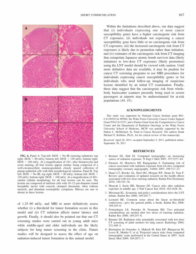
FIG. 6. Panel A: Top left: DOX þ No IR (ionizing radiation); topright: DOX þ 80 mGy; bottom left: DOX þ 120 mGy; bottom right:DOX þ 160 mGy. At a magnification of 103, after hematoxylin andeosin staining, all four lesions appear similar, being composed of awell-circumscribed, nonencapsulated, closely spaced collection ofplump epithelial cells with little morphological variation. Panel B: Topleft: DOX þ No IR; top right: DOX þ 80 mGy; bottom left: DOX þ120 mGy; bottom right: DOX þ 160 mGy. At a magnification of 403,similar cellular morphology for all four lesions can be seen. Thelesions are composed of uniform cells with 10–12-lm-diameter roundbasophilic nuclei with coarsely clumped chromatin, often withoutnucleoli, and abundant eosinophilic cytoplasm. Mitoses are rare toabsent in these lesions.
SHORT COMMUNICATION 847

10. National Research Council Committee to Assess Health Risksfrom Exposure to Low Levels of Ionizing Radiation. Health risksfrom exposure to low levels of ionizing radiation: BEIR VII Phase2. Washington, DC: National Academies Press; 2006.
11. Brenner DJ, Doll R, Goodhead DT, Hall EJ, Land CE, Little JB, etal. Cancer risks attributable to low doses of ionizing radiation:assessing what we really know. Proc Natl Acad Sci U S A 2003;100:13761–6.
12. National Lung Screening Trial Research Team. The National LungCancer Screening Trial: Overview and Study Design. NationalLung Screening Trial Research Team. Radiology 2011; 258:243–53.
13. National Lung Screening Trial: Questions and Answers. NCICancer Bulletin 2010. [updated 2010 Nov 26, cited 2011 Apr 13]Available from: http://www.cancer.gov/newscenter/qa/2002/nlstqaQA.
14. Jemal A, Siegel R, Xu J, Ward E. Cancer statistics, 2010. CACancer J Clin 2010; 60:277–300.
15. Alberg AJ, Samet JM. Epidemiology of lung cancer. In: Kane MA,Bunn PA Jr, editors. Biology of lung cancer. New York: MarcelDekker; 1998. p. 11–51.
16. Floyd HS, Farnsworth CL, Kock ND, Mizesko MC, Little JL,Dance ST, et al. Conditional expression of the mutant Ki-rasG12C
allele results in formation of benign lung adenomas: Developmentof a novel mouse lung tumor model. Carcinogenesis 2005;26:2196–206.
17. Li ZH, Zheng J, Weiss LM, Shibata D. c-K-ras and p53 mutationsoccur very early in adenocarcinoma of the lung. Am J Pathol 1994;144:303–9.
18. Westra WH, Slebos RJ, Offerhaus GJ, Goodman SN, Evers SG,Kensler TW, et al. K-ras oncogene activation in lung adenocar-cinomas from former smokers. Evidence that K-ras mutations arean early and irreversible event in the development of adenocar-cinoma of the lung. Cancer 1993; 72:432–8.
19. Westra WH, Baas IO, Hruban RH, Askin FB, Wilson K, OfferhausGJ, et al. K-ras oncogene activation in atypical alveolarhyperplasias of the human lung. Cancer Res 1996; 56:2224–8.
20. Reynolds SH, Wiest JS, Devereux TR, Anderson MW, You M.Protooncogene activation in spontaneously occurring and chem-ically induced rodent and human lung tumors. In: Klein-SzantoAJP, Anderson MW, Barrett JC, Slaga TJ, editors. Comparativemolecular carcinogenesis. New York: Wiley-Liss; 1992. p. 303–20.
21. Shockett PE, Schatz DG. Diverse strategies for tetracycline-regulated inducible gene expression. Proc Natl Acad Sci U S A1996; 93:5173–6.
22. Hayashida S, Harrod KS, Whitsett JA. Regulation and function ofCCSP during pulmonary Pseudomonas aeruginosa infection invivo. Am J Physiol Lung Cell Mol Physiol 2000; 279:L452–9.
23. Dance-Barnes ST, Kock ND, Floyd HS, Moore JE, Mosley LJ,D’Agostino RB Jr, et al. Effects of mutant human Ki-ras(G12C)gene dosage on murine lung tumorigenesis and signaling to itsdownstream effectors. Toxicol Appl Pharmacol 2008; 231:77–84.
24. Dance-Barnes ST, Kock ND, Moore JE, Lin EY, Mosley LJ,D’Agostino RB Jr, et al. Lung tumor promotion by curcumin.Carcinogenesis 2009; 30:1016–1023.
25. Schuster DP, Kovacs A, Garbow J, Piwnica-Worms D. Recentadvances in imaging the lungs of intact small animals. Am J RespirCell Mol Biol 2004; 30:129–38.
26. Jennings-Gee JE, Moore JE, Xu M, Dance ST, Kock ND, McCoy
TP, et al. Strain-specific induction of murine lung tumorsfollowing in utero exposure to 3-methylcholanthrene. MolCarcinog 2006; 45:676–84.
27. Kennel SJ, Davis IA, Branning J, Pan H, Kabalka GW, Paulus MJ.High resolution computed tomography and MRI for monitoringlung tumor growth in mice undergoing radioimmunotherapy:correlation with histology. Med Phys 2000; 27:1101–7.
28. Nikitin AY, Alcaraz A, Anver MR, Bronson RT, Cardiff RD,Dixon D, et al. Classification of proliferative pulmonary lesions ofthe mouse: recommendations of the mouse models of humancancers consortium. Cancer Res 2004; 64:2307–16.
29. Goldsby RE, Liu Q, Nathan PC, Bowers DC, Yeaton-Massey A,Raber SH, et al. Late-occurring neurologic sequelae in adultsurvivors of childhood acute lymphoblastic leukemia: a reportfrom the Childhood Cancer Survivor Study. J Clin Oncol 2010;28:324–31.
30. Kaste SC, Waszilycsak GL, McCarville MB, Daw NC. Estimationof potential excess cancer incidence in pediatric 201Tl imaging.AJR Am J Roentgenol 2010; 194:245–9.
31. Shuryak I, Ullrich RL, Sachs RK, Brenner DJ. The balancebetween initiation and promotion in radiation-induced murinecarcinogenesis. Radiat Res 2010; 174:357–66.
32. Curtis SB, Luebeck EG, Hazelton WD, Moolgavkar SH. The roleof promotion in carcinogenesis from protracted high-LETexposure. Phys Med 2001;17 Suppl 1:157–60.
33. Curtis SB, Hazelton WD, Luebeck EG, Moolgavkar SH. Frommechanisms to risk estimation—bridging the chasm. Adv SpaceRes 2004; 34:1404–9.
34. Scott BR. Low-dose radiation risk extrapolation fallacy associatedwith the linear-no-threshold model. Hum Exp Toxicol 2008;27:163–8.
35. Hussain SP, Hofseth LJ, Harris CC. Radical causes of cancer. NatRev Cancer 2003; 3:276–85.
36. Klaunig JE, Kamendulis LM. The role of oxidative stress incarcinogenesis. Annu Rev Pharmacol Toxicol 2004; 44:239–67.
37. Malkinson AM. Role of inflammation in mouse lung tumorigen-esis: a review. Exp Lung Res 2005; 31:57–82.
38. Witschi H, Williamson D, Lock S. Enhancement of urethantumorigenesis in mouse lung by butylated hydroxytoluene. J NatlCancer Inst 1977; 58:301–5.
39. Cohen BH, Diamond EL, Graves CG, Kreiss P, Levy DA, MenkesHA, et al. A common familial component in lung cancer andchronic obstructive pulmonary disease. Lancet 1977; 2:523–6.
40. Tockman MS, Anthonisen NR, Wright EC, Donithan MG.Airways obstruction and the risk for lung cancer. Ann InternMed 1987; 106:512–8.
41. Keith RL, Miller YE, Hoshikawa Y, Moore MD, Gesell TL, GaoB, et al. Manipulation of pulmonary prostacyclin synthaseexpression prevents murine lung cancer. Cancer Res 2002;62:734–40.
42. Karin M. NF-kappaB and cancer: Mechanisms and targets. MolCarcinog 2006; 45:355–61.
43. Lu H, Ouyang W, Huang C. Inflammation, a key event in cancerdevelopment. Mol Cancer Res 2006; 4:221–33.
44. Hupe O, Ankerhold U. X-ray security scanners for personnel andvehicle control: dose quantities and dose values. Eur J Radiol2007; 63:237–41.
45. Rez P, Metzger RL, Mossman KL. The dose from Comptonbackscatter screening. Radiat Prot Dosimetry 2011; 145:75–81.
848 SHORT COMMUNICATION