Embed Size (px)
Citation preview

ChemicalScience
EDGE ARTICLE
Ope
n A
cces
s A
rtic
le. P
ublis
hed
on 3
0 A
pril
2020
. Dow
nloa
ded
on 1
2/1/
2021
2:1
1:57
AM
. T
his
artic
le is
lice
nsed
und
er a
Cre
ativ
e C
omm
ons
Attr
ibut
ion
3.0
Unp
orte
d L
icen
ce.
View Article OnlineView Journal | View Issue
Cancer cell discr
aDepartment of Chemistry, Hong Kong B
Research Center for Tissue Restoration an
Study, Department of Chemical and Bi
University of Science and Technology, C
999077, China. E-mail: [email protected]; tanbHKUST-Shenzhen Research Institute, No. 9
Nanshan, Shenzhen 518057, ChinacCenter of Bio and Micro/Nano Functional M
Materials, Shandong University, Jinan 2501dChimie ParisTech, PSL University Paris, CN
75005 Paris, France. E-mail: min-hui.li@cheCenter for Aggregation-Induced Emission, SC
Key Laboratory of Luminescent Materials
Technology, Guangzhou 510640, China
† Electronic supplementary information (crystallographic data; photophysical dcalculations; synthetic details, NMR specFor ESI and crystallographic data in CI10.1039/d0sc01213k
Cite this: Chem. Sci., 2020, 11, 7676
All publication charges for this articlehave been paid for by the Royal Societyof Chemistry
Received 28th February 2020Accepted 29th April 2020
DOI: 10.1039/d0sc01213k
rsc.li/chemical-science
7676 | Chem. Sci., 2020, 11, 7676–76
imination and dynamic viabilitymonitoring through wash-free bioimaging usingAIEgens†
Ruoyao Zhang,ab Guangle Niu, abc Qing Lu,c Xiaolin Huang, ab Joe H. C. Chau,a
Ryan T. K. Kwok,ab Xiaoqiang Yu, c Min-Hui Li, *d Jacky W. Y. Lam*ab
and Ben Zhong Tang *abe
Cancer cell discrimination and cellular viability monitoring are closely related to human health. A universal
and convenient fluorescence system with a dual function of wide-spectrum cancer cell discrimination and
dynamic cellular viability monitoring is desperately needed, and is still extremely challenging. Herein we
present a series of aggregation-induced emission luminogens (AIEgens) (denoted as IVP) which can
allow accurate discrimination between cancer and normal cells and dynamic monitoring of cellular
viability through mitochondria–nucleolus migration. By regulating the lengths and positions of alkyl
chains in IVP molecules, we systematically studied the discrimination behavior of these AIEgens between
cancer cells and normal cells and further investigated how they can migrate between the mitochondria
and nucleolus based on the change of mitochondrial membrane potential (DJm). Using IVP-02 as
a model molecule, wash-free bioimaging, excellent two-photon properties, and low cytotoxicity were
demonstrated. This present work proves that these designed IVP AIEgens show great potential for cancer
identification and metastasis monitoring, as well as activity evaluation and screening of drugs.
Introduction
Cancer is one of the greatest enemies of humanity. Earlydetection of cancer before its metastasis is very important toincrease the survival rate of patients.1 Different from normalcells, cancer cells overexpress some receptors, which usuallyserve as targets for identifying cancer cells.2 Currently, immu-nouorescence and aptamer-based uorescence systems arecommon tools for cancer detection.3–10 In cancer detection with
ranch of Chinese National Engineering
d Reconstruction, Institute for Advanced
ological Engineering, The Hong Kong
lear Water Bay, Kowloon, Hong Kong
Yuexing 1st RD, South Area, Hi-tech Park,
aterials, State Key Laboratory of Crystal
00, China
RS, Institut de Recherche de Chimie Paris,
imieparistech.psl.eu
UT-HKUST Joint Research Institute, State
and Devices, South China University of
ESI) available: Materials and methods;ata; bioimaging; RNA titration andtra and HRMS spectra. CCDC 1986367.F or other electronic format see DOI:
84
immunouorescence, antibodies conjugated with uorescentdyes are highly specic to the overexpressed receptors,achieving high specicity to cancer cells.3–6 However, in theearly stages of cancer, the receptors are less expressed, whichincreases the difficulty of early cancer detection. Moreover, thepreparation of specic recognition elements against cancer cellreceptors is complex and costly, and the conjugation of uo-rescent dyes to antibodies sometimes affects antibody activity.In cancer detection with aptamer-based uorescence systems,screening the aptamers specic to cancer cells and conjugationof uorescent dyes to aptamers are also complicatedprocesses.7–10 Although these specic ligands can efficientlyidentify cancer cell types, they are limited by the availabletumour cell species, especially from unknown cancers, whichmakes it hard to achieve wide-spectrum cancer screening.Therefore, there is still much room for improvement of cancerdetection. A universal and convenient method is stronglyneeded for wide-spectrum cancer cell detection.
The viability of autologous cells is closely related to humanhealth. Monitoring cell viability is important for human health,sub-health, and disease detection. In particular, in terms ofdrug screening including drug development and efficacy eval-uation, cell viability monitoring also plays an important role.11
The standard method commonly used for detecting cell viabilityis the MTT assay.12 Due to reduction by mitochondrial reduc-tase, MTT with yellow color will turn into formazan with deeppurple color. Then by measuring the absorbance of formazan at
This journal is © The Royal Society of Chemistry 2020

Fig. 1 (A) Chemical structure of IVPI-2 and the CLSM images of liveHeLa cells with normal DJm and decreased DJm stained with IVPI-2.(B) Chemical structure of IVP-02 and its single-molecule configurationin single crystal cells with atoms labelled in color. C, gray; H, white; N,blue; P, purple; F, green.
Edge Article Chemical Science
Ope
n A
cces
s A
rtic
le. P
ublis
hed
on 3
0 A
pril
2020
. Dow
nloa
ded
on 1
2/1/
2021
2:1
1:57
AM
. T
his
artic
le is
lice
nsed
und
er a
Cre
ativ
e C
omm
ons
Attr
ibut
ion
3.0
Unp
orte
d L
icen
ce.
View Article Online
570 nm, the cell viability can be obtained. However, MTT assayneeds a long testing time, and detailed information such as thecell morphology cannot be visualized. Fluorescence microscopyis a powerful tool for in situ real-time detection and monitoringof biosamples in vivo.13–20 Researchers developed some uores-cent probes for cell viability detection.21–26 For example, calceinAM is nonuorescent in dead cells but exhibits strong uores-cence in live cells.24 Fluorescein labelled annexin V is used forthe detection of phosphatidylserine expression in earlyapoptotic cells.25 Propidium iodide (PI) can only stain lateapoptotic and dead cells, but cannot enter live cells.26 However,it is hard for these probes to monitor cell viability in real time.New systems for fast and in situ real-time monitoring of cellviability are highly desirable but are still extremely challenging.
In response to these challenges, a variety of organic uo-rophores have been developed for biosample imaging.27–32
Traditional aromatic and planar uorophores have poor solu-bility under aqueous conditions due to the inherent hydro-phobicity. Increasing the amount would lead to aggregation-caused quenching (ACQ),33 while in very dilute solutions, theuorescence is too weak to be detected and easy to bleach byirradiation. Generally, a physical effect is oen positively relatedto the amount of the added substance. As the membranepermeability and mitochondrial membrane potential (DJm) ofcancer cells is higher than those of normal cells,34,35 theoreti-cally more uorescent molecules would enter cancer cells thannormal cells, providing an opportunity for cancer cell discrim-ination. For traditional uorophores with the ACQ effect, fewermolecules entering normal cells lead to relatively weak uo-rescence signals. However, more molecules entering cancercells in turn lead to uorescence decrease resulting from ACQ,thus attenuating the uorescence signal difference betweencancer and normal cells. Therefore, it is actually difficult todistinguish cancer cells from normal cells using traditionaluorophores due to the low contrast between them.
In this work, we designed and synthesized a battery ofunique aggregation-induced emission luminogens (AIEgens,denoted as IVP) for cancer cell discrimination and cellularviability monitoring. Different from the uorophores with theACQ effect, AIEgens are highly emissive at high concentration.33
As cancer cells possess higher membrane permeability andDJm than normal cells, more AIEgens would enter cancer cellswhile fewer AIEgens enter normal cells. With higher concen-tration in cancer cells, AIEgens would emit obviously strongeruorescence than in normal cells with fewer AIEgens. Thus, theconcentration effect would amplify the uorescence differencebetween cancer and normal cells, achieving cancer celldiscrimination. As is known, some important positions ormolecules are negatively charged inside a cell, such as mito-chondria and nucleic acid in the nucleus.36,37 Lipophilic cationsare inclined to target these negatively charged positions ormolecules through electrostatic interaction.38 Optimizing thecharge and lipophilicity of AIEgens would realize the change ofdyeing position, according to variation of the cell viability. Herewe designed and synthesized a series of AIEgens which couldselectively stain cancer cells as well as monitor cell viabilitythrough mitochondria–nucleolus migration. Simultaneously,
This journal is © The Royal Society of Chemistry 2020
the relevant mechanism is carefully studied by changing thechemical structures of AIEgens.
Results and discussionDesign and synthesis
In our previous work shown in Fig. 1A, we found that IVPI-2could stain mitochondria in live cancer cells. When DJm
decreased, it would migrate into the nucleolus.39 Since IVPI-2 istarget-changeable according to the change of mitochondrialphysiology, we tried to modify IVPI-2 carefully. The iodide ion isa well-known effective uorescence quencher due to its heavy-atom effect.40 Consequently, the iodide ion was replaced byhexauorophosphate and IVP-02 was obtained as shown inFig. 1B. The quantum yield of IVP-02 in the solid state is 4.3%,which is about 3 times that of IVPI-2 (1.3%). The syntheticroutes to IVP-02 are depicted in Scheme S1.† The chemicalstructure of IVP-02 was fully characterized by 1H NMR, 13CNMR, 19F NMR andHRMS as shown in the ESI.† In addition, thestructure of IVP-02 was further conrmed by single-crystal X-raydiffraction analysis (CCDC 1986367,† Fig. 1B). The details of theexperimental conditions, unit cell data and renement data aresummarized in Table S1.†
Photophysical properties
The absorption and one-photon excited uorescence (FL) spectraof IVP-02 in different solvents are shown in Fig. 2A. IVP-02 showedstrong absorbance from 400 to 450 nm and intense emissionfrom 500 to 550 nm. IVP-02 possesses a distinct donor–p–acceptor structure. It showed a bathochromic shi in the FLspectra with the increase of solvent polarity, due to intra-molecular charge transfer effect. Addition of THF and EtOH to thesolution of IVP-02 in water failed to make the dye moleculesaggregate probably due to their amphiphilic nature. Similarresults were reported by others.41–43 Then the emission of IVP-02in the increasingly viscous environment and in the solid state wascarefully measured to study whether IVP-02 possessed AIEactivity. In Fig. 2B and C, IVP-02 shows weak emission in purewater solution. With the increase of the glycerol volume content
Chem. Sci., 2020, 11, 7676–7684 | 7677

Fig. 2 (A) Normalized UV (dashed line) and FL (solid line) spectra ofIVP-02 in different solvents. (B) FL spectra of IVP-02 in H2O and H2O/glycerol mixtures with different glycerol fractions (fGly). (C) Changes inthe FL peak intensities (I) of the solutions of IVP-02 with the glycerolcontent in the H2O/glycerol mixtures. I0 is the intensity in pure H2O.(D) FL spectra of IVP-02 in the H2O/glycerol mixture with 90% glycerolat 25 and �20 �C. (E) FL spectrum of IVP-02 in the solid state. lex ¼440 nm. Inset: fluorescence photo of IVP-02 solid obtained under365 nm UV irradiation using a handheld UV lamp. (F) TPEF spectra ofIVP-02 in DMSO excited by 780, 800, 820, 840, 860, 880, and 900 nm,respectively. Concentration: 10 mM. The top view (G) and side view (H)of the crystal structure of IVP-02.
Fig. 3 CLSM images of live cancer cells (A549 and HeLa) co-culturedwith normal cells (COS7 and HLF) (A) and the images of A549 and HLFcells seeded on different cover glasses (B) stained with 2 mM IVP-02 for30min, respectively. lex¼ 488 nm, lem¼ 500–650 nm. Scale bar¼ 20mm (A); scale bar ¼ 50 mm (B).
Chemical Science Edge Article
Ope
n A
cces
s A
rtic
le. P
ublis
hed
on 3
0 A
pril
2020
. Dow
nloa
ded
on 1
2/1/
2021
2:1
1:57
AM
. T
his
artic
le is
lice
nsed
und
er a
Cre
ativ
e C
omm
ons
Attr
ibut
ion
3.0
Unp
orte
d L
icen
ce.
View Article Online
accompanied by the increasing viscosity, the FL intensityincreased gradually. In addition to a water/glycerol system,similar experiments were carried out in the MeOH/glycerolsystem in Fig. S1A and B.† The results also showed that IVP-02was highly emissive in a high viscosity environment. Moreover,when the solution temperature decreased from 25 �C to �20 �C,the FL intensity also increased obviously as shown in Fig. 2D andS1C.† These phenomena occur because high viscosity and lowtemperature could hamper intramolecular motion, leading to theclosure of the nonradiative decay channel and thus enhanced FLemission.33 Furthermore, we added some RNA in a PBS solutionof IVP-02. IVP-02 showed weak emission in PBS solution, but withthe increase of RNA concentration, the FL intensity increasedobviously as shown in Fig. S1D.† Based on the calculation resultsin Fig. 9, IVP-02 located in the minor grooves of RNA, where theintramolecular motion of IVP-02 was also hampered. Theseresults indicated that the restriction of intramolecular motion(RIM) is the main reason that makes the dye highly emissive,which is also the luminescence mechanism of AIEgens. IVP-02also exhibited strong uorescence around 575 nm in the solidstate and its powder emitted bright yellow light (Fig. 2E). There-fore, based on the above results, IVP-02 possesses AIE activity.
IVP-02 shows weak emission in aqueous solution, butemits strong uorescence under high-viscosity conditions;thus it is greatly favourable for wash-free bioimaging. Inaddition, IVP-02 emits redder uorescence in the solid state
7678 | Chem. Sci., 2020, 11, 7676–7684
than in solution. To conrm the mechanism of the red shi inthe solid state of IVP-02, its crystal is analyzed as shown inFig. 2G and H. The molecules of IVP-02 are anti-parallellystacked and form multimers in the crystalline state. Theshort intermolecular stacking distances of the multimers are3.636 A, 3.455 A, 3.394 A, 3.375 A, 3.466 A, and 3.465 A,indicating strong intermolecular interactions inside themultimers. So the red-shi emission in the solid state shouldbe attributed to the intermolecular p–p interactions inducedby the short contact between the molecules. Generally,organic dyes with a donor–acceptor structure exhibit goodtwo-photon absorption (TPA) and two-photon excited uo-rescence (TPEF).44 The TPEF spectra of IVP-02 excited atdifferent pulse wavelengths (780–900 nm) in DMSO are shownin Fig. 2F. Using uorescein as the standard, the two-photonabsorption cross section (d) of IVP-02 was calculated and isshown in Table S2.† The highest d was 287 GM excited at800 nm. Such a high d value is benecial for two-photonimaging in live cells and deep tissues.
Cancer cell discrimination
The bioimaging properties of IVP-02 in live cells were investi-gated by confocal laser scanning microscopy (CLSM). Cancer
This journal is © The Royal Society of Chemistry 2020

Edge Article Chemical Science
Ope
n A
cces
s A
rtic
le. P
ublis
hed
on 3
0 A
pril
2020
. Dow
nloa
ded
on 1
2/1/
2021
2:1
1:57
AM
. T
his
artic
le is
lice
nsed
und
er a
Cre
ativ
e C
omm
ons
Attr
ibut
ion
3.0
Unp
orte
d L
icen
ce.
View Article Online
cells (A549 and HeLa) and normal cells (COS7 and HLF) werestained with IVP-02. In Fig. S2,† the uorescence intensity innormal cells wasmuch weaker than that in cancer cells under thesame staining and imaging conditions. So IVP-02 has a highpotential to differentiate cancer and normal cells. To verify thisspeculation, cancer and normal cells were co-cultured andstained with IVP-02. In Fig. 3A, it could be seen that only cancercells (A549 andHeLa) were highly illuminated, while normal cells(COS7 and HLF) showed almost no uorescence. To preciselyconrm the selectivity of IVP-02 to cancer cells, cancer cells A549and HeLa and normal cells COS7 and HLF were seeded ondifferent cover glasses, respectively. Then two cover glasses withcancer and normal cells, respectively, were placed in the samedish and stainedwith IVP-02 at the same time. In Fig. 3B and S3,†only cancer cells (A549 and HeLa) on the upper glass are illu-minated, while normal cells (COS7 and HLF) on the lower glassare not illuminated. These results indicated that IVP-02 couldselectively differentiate cancer cells from normal cells.
In the above pictures, lamentous structures in the cyto-plasm of cancer cells were observed, which are the typicalmorphology of mitochondria. Then co-staining experimentswith the commercial mitochondrial probe MitoTracker DeepRed FM (MTDR) were carried out (Fig. S4†). The co-localizationcoefficient of IVP-02 and MTDR was around 0.9, demonstratingthe localization of IVP-02 in mitochondria in cancer cells.
Fig. 4 (A) Chemical structures of IVP-02, 04, 06, 22, 42, and 62. (B) CLSMfor 30 min, respectively. lex ¼ 488 nm, lem ¼ 500–650 nm. (C) CLSM imamMMTDR, respectively. IVP-04, 06, 22, 42, and 62: lex ¼ 488 nm, lem ¼ 5mm.
This journal is © The Royal Society of Chemistry 2020
Mechanism study
Then we carefully studied why IVP-02 could selectively staincancer cells over normal cells. Some previous work indicatedthe possible reason that the DJm of cancer cells is much higherthan that of normal cells.45 However, it is a fact that somecommercial mitochondrial probes and reported ones, alsosensitive to DJm, can stain both cancer and normal cells.46 SoDJm is not the only reason that enables dyes to distinguishcancer and normal cells. The chemical structure of the dye itselfalso plays an important role.
We speculated that the membrane permeability of the probewas a key factor in its selectivity to cancer cells, since the plasmamembrane of cancer cells was reported to be more permeablethan that of normal cells. Then we tried to modify IVP-02 bylengthening the alkyl chain on the pyridine salt side and indoleside, respectively, to tune the membrane permeability.47 Fivenew molecules IVP-04, IVP-06, IVP-22, IVP-42, and IVP-62 wereobtained as shown in Fig. 4A. The synthetic routes to these newIVP molecules are depicted in Scheme S1.† Their chemicalstructures were fully characterized by 1H NMR, 13C NMR, and19F NMR as shown in the ESI.† The FL spectra of IVP-04, IVP-06,IVP-22, IVP-42, and IVP-62 in water and glycerol are shown inFig. S5.† It could be seen that all of the ve IVP moleculesshowed weak emission in aqueous solution, but exhibitedstrong uorescence in glycerol with high viscosity. Thus they
images of live A549 cells stained with 2 mM IVP-04, 06, 22, 42, and 62ges of A549 cells stained with 2 mM IVP-04, 06, 22, 42, and 62 and 0.200–650 nm; MTDR: lex ¼ 640 nm, lem ¼ 650–700 nm. Scale bar¼ 20
Chem. Sci., 2020, 11, 7676–7684 | 7679

Fig. 6 (A) Equation of Born energy. (B) Schematic diagram of a lipo-philic cation passing through the membrane.
Chemical Science Edge Article
Ope
n A
cces
s A
rtic
le. P
ublis
hed
on 3
0 A
pril
2020
. Dow
nloa
ded
on 1
2/1/
2021
2:1
1:57
AM
. T
his
artic
le is
lice
nsed
und
er a
Cre
ativ
e C
omm
ons
Attr
ibut
ion
3.0
Unp
orte
d L
icen
ce.
View Article Online
could be used in wash-free bioimaging. Then cancer cells A549were stained with the ve new molecules separately. In Fig. 4B,the uorescence pattern showed that all these moleculesstained mitochondria. The co-staining experiments with MTDRalso conrmed their location in mitochondria in A549 cells inFig. 4C. In addition to A549 cells, the same experiments werealso carried out in HeLa cells (Fig. S6 and S7†). The results weresimilar to those in A549 cells. So IVP-04, 06, 22, 42, and 62 canselectively stain mitochondria in cancer cells.
Cancer cells A549 co-cultured with normal cells COS7 werestained with these molecules separately. As shown in Fig. 5A,IVP-02, 22, 42, and 62 can only stain A549 cells, indicating thatthey can distinctly differentiate cancer and normal cells.Intriguingly, IVP-04 and 06 can stain both A549 and COS7 cells,which means that they cannot distinguish cancer and normalcells. Then HeLa cells co-cultured with COS7 cells (Fig. S8†) andA549 cells co-cultured with HLF cells (Fig. S9†) were also stainedwith these molecules; the results were similar to those in A549cells co-cultured with COS7 cells. In addition to co-culturing,A549 cells and HLF cells were also seeded on different coverglasses and stained with these molecules. The imaging results(Fig. 5B) were also similar to the co-culturing results in whichIVP-04 and IVP-06 could stain both A549 and HLF cells, whilethe other four molecules only stain A549 cells. Based on theimaging results above, preliminary conclusions could be drawnthat the selectivity of these IVP molecules to cancer cells is alsobased on the length of the alkyl chain on the pyridinium saltside. Lengthening the alkyl chain on the pyridinium salt sidewill eliminate the selectivity to cancer cells.
Fig. 5 CLSM images of live cancer cells (A549) co-cultured withnormal cells (COS7) (A) and the images of A549 and HLF cells seededon different cover glasses (B) stained with 2 mM IVP-02, 04, 06, 22, 42,and 62 for 30 min, respectively (the first column in (B) is the same asFig. 3B). lex¼ 488 nm, lem¼ 500–650 nm. Scale bar¼ 20 mm (A); scalebar ¼ 50 mm (B).
7680 | Chem. Sci., 2020, 11, 7676–7684
We further investigated the role of the alkyl chain on thepyridinium salt side. The way in which these IVP molecules enterthe cells was rst investigated. Cancer cells A549 were incubatedwith the IVP molecules at 4 �C for 20 min. In Fig. S10,† at lowtemperature, obvious uorescence signals inside the cells couldstill be observed. This result indicated that these IVP moleculesentered the cell by diffusion. In some reported studies,researchers found that lipophilic cations cross membranes verywell.38,48 The activation energy formoving a lipophilic cation fromthe aqueous medium to the hydrophobic core of a membrane ismainly from electrostatic interactions. The main electrostaticenergy component, Born energy (WB, Fig. 6A), is due to theenthalpy input required to remove water molecules from thecation upon transfer from the aqueous environment to the lipidcore of the membrane.48 With lowerWB, the lipophilic cation canpass through the membrane more easily. The Born energy isgiven by the equation in Fig. 6A, in which Z is the cation chargeand r is the ionic radius. From the equation, WB is inverselyproportional to the ionic radius. For these IVP molecules, theionic radius is the average distance from the molecule charge tothe water molecules around. Fig. 6B shows a schematic diagramof a lipophilic cation passing through the membrane. Removingwater molecules around the lipophilic cation is the rst step. Forthe lipophilic cation, the larger the ionic radius, the weaker theinteraction between the cation and water molecule and the lowerthe WB; as a result, the molecule passes more easily through themembrane.
Regarding the chemical structures of these IVP molecules,the pyridinium side of all these molecules is positively charged.This side is more hydrophilic, meaning that more water mole-cules are enriched on this side, so this side determines the ionicradius. Moreover, the similarity of IVP-02, 22, 42, and 62 on thepyridinium side is the 2-carbon alkyl chain. The differencebetween IVP-02, 22, 42, and 62 and IVP-04 and 06 on the pyr-idinium side is the length of the alkyl chain. IVP-04 and 06 havelonger alkyl chains on the pyridinium side, so that their ionicradius is larger than that of IVP-02, 22, 42, and 62, implying thatthe interaction between the IVP-04 and 06 and water moleculesis weaker. So the Born energy values of IVP-04 and 06 are lowerthan those of IVP-02, 22, 42, and 62. As such, they pass through
This journal is © The Royal Society of Chemistry 2020
Edge Article Chemical Science
Ope
n A
cces
s A
rtic
le. P
ublis
hed
on 3
0 A
pril
2020
. Dow
nloa
ded
on 1
2/1/
2021
2:1
1:57
AM
. T
his
artic
le is
lice
nsed
und
er a
Cre
ativ
e C
omm
ons
Attr
ibut
ion
3.0
Unp
orte
d L
icen
ce.
View Article Online
the membrane more easily. When staining normal cells, IVP-04and 06more easily penetrate cytomembrane than IVP-02, 22, 42,and 62.
Cellular viability monitoring
Monitoring cell viability is a highly valuable task for funda-mental research in biology, pathology, and medicine.11 Weinvestigated whether IVP-02 could monitor cell viability. DJm isa very important indicator to characterize cell viability.49–51 Withthe loss of cell viability, DJm would decrease, while with therecovery of cell viability, DJm would also recover to the normallevel. Hence visualizing the change of DJm is an effective way tomonitor cell viability. Carbonyl cyanide m-chlorophenyl hydra-zone (CCCP) is a type of protonophore and can cause rapidacidication of mitochondria by decreasing DJm.52 Cancer cellsA549 were pre-stained with IVP-02 for 30 min. In Fig. 7A, in thebeginning, IVP-02 stained mitochondria clearly. With theaddition of CCCP, the uorescence intensity in mitochondriadecreased, while that in the nucleolus increased, meaning thatIVP-02 was released from mitochondria and migrated to thenucleolus. Then aer removal of CCCP with the recovery ofDJm, the uorescence in the nucleolus disappeared, while theuorescence in mitochondria recovered gradually, indicatingthat IVP-02 migrated back to the mitochondria. In addition toA549 cells, similar results were also observed in HeLa cells asshown in Fig. S11.† Hydrogen peroxide (H2O2) can inhibit theoxidative respiratory chain which would cause the loss of cellviability.53 Then live A549 and HeLa cells were pre-stained withIVP-02 and treated with 10 mMH2O2. In Fig. S12,† it can be seenthat aer adding H2O2 with the cell viability decreasing, stronguorescence was observed in the nucleolus. These results
Fig. 7 Live A549 cells (A), live A549 co-cultured with COS 7 cells, andlive HeLa co-cultured with COS7 cells (B) were pre-stained with 2 mMIVP-02 for 30min and treated with 20 mMCCCP for 5 min. Then CCCPwas removed and fresh culture medium was added for another 5 min.lex ¼ 488 nm, lem ¼ 500–650 nm. Scale bar ¼ 20 mm.
This journal is © The Royal Society of Chemistry 2020
indicated that IVP-02 could monitor cancer cell viabilitythrough mitochondria–nucleolus migration.
To test whether IVP-02 could selectively stain cancer cellsand monitor their viability, cancer cells and normal cells wereco-cultured and stained with IVP-02. As we predicted, only themitochondrial morphology in A549 cells is clearly shown inFig. 7B. Aer CCCP was added, IVP-02 was released frommitochondria and migrated to the nucleolus. Aer CCCP wasremoved, the uorescence in the nucleolus disappeared, whilethe uorescence in mitochondria recovered gradually. In thewhole process, little uorescence was observed in normal cellsCOS7. The same experiments were also carried out in HeLa cellsco-cultured with COS7 cells as shown in Fig. 7B and similarphenomena were observed. These results demonstrated thatIVP-02 could selectively stain cancer cells and monitor theirviability through mitochondria–nucleolus migration in coex-isting cancer cells and normal cells.
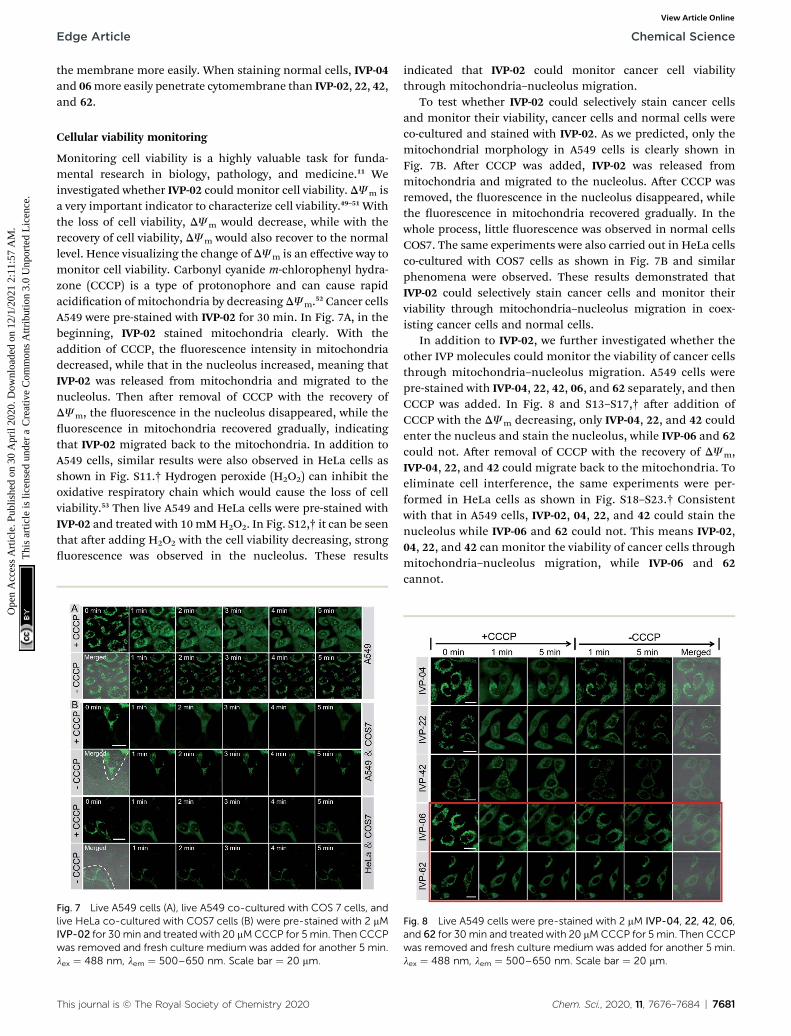
In addition to IVP-02, we further investigated whether theother IVP molecules could monitor the viability of cancer cellsthrough mitochondria–nucleolus migration. A549 cells werepre-stained with IVP-04, 22, 42, 06, and 62 separately, and thenCCCP was added. In Fig. 8 and S13–S17,† aer addition ofCCCP with the DJm decreasing, only IVP-04, 22, and 42 couldenter the nucleus and stain the nucleolus, while IVP-06 and 62could not. Aer removal of CCCP with the recovery of DJm,IVP-04, 22, and 42 could migrate back to the mitochondria. Toeliminate cell interference, the same experiments were per-formed in HeLa cells as shown in Fig. S18–S23.† Consistentwith that in A549 cells, IVP-02, 04, 22, and 42 could stain thenucleolus while IVP-06 and 62 could not. This means IVP-02,04, 22, and 42 can monitor the viability of cancer cells throughmitochondria–nucleolus migration, while IVP-06 and 62cannot.
Fig. 8 Live A549 cells were pre-stained with 2 mM IVP-04, 22, 42, 06,and 62 for 30min and treated with 20 mMCCCP for 5 min. Then CCCPwas removed and fresh culture medium was added for another 5 min.lex ¼ 488 nm, lem ¼ 500–650 nm. Scale bar ¼ 20 mm.
Chem. Sci., 2020, 11, 7676–7684 | 7681

Fig. 10 (A) Two-photon microscopy images of live HeLa cells stainedwith 2 mM IVP-02 for 30 min. lex ¼ 800 nm, lem ¼ 495–540 nm. Scalebar¼ 20 mm. (B) Viability of A549 cells after incubationwith IVP-02, 04,06, 22, 42, and 62 at different concentrations for 24 h.
Chemical Science Edge Article
Ope
n A
cces
s A
rtic
le. P
ublis
hed
on 3
0 A
pril
2020
. Dow
nloa
ded
on 1
2/1/
2021
2:1
1:57
AM
. T
his
artic
le is
lice
nsed
und
er a
Cre
ativ
e C
omm
ons
Attr
ibut
ion
3.0
Unp
orte
d L
icen
ce.
View Article Online
We further studied the reason that IVP-06 and 62 cannotstain the nucleolus in cancer cells. On one hand, when IVPmolecules are located in the mitochondria, in addition to theelectrostatic interaction between the cation and negativelycharged inner membrane of mitochondria, hydrophobic inter-action between the alkyl chain and phospholipids also existed.When DJm decreased, although the electrostatic interactionweakened, the hydrophobic interaction still remained. IVP-06and 62 have longer alkyl chains than the other four IVP mole-cules, so the hydrophobic interaction between IVP-06 and 62and phospholipids is stronger than that of the other four IVPmolecules. Therefore, IVP-06 and 62 are more inclined to stay inthe mitochondria, while IVP-02, 04, 22, and 42 more easilyescape from the mitochondria. On the other hand, the affinityof IVP molecules to RNA should also be considered as thenucleolus is rich in RNA. Thus RNA titration experiments wererst performed. In Fig. 9A and S24,† with the increase of RNAconcentration, the uorescence intensity of all the moleculesincreases. Based on the Scatchard equation,54 the bindingconstant (k) of these molecules to RNA was calculated and issummarized in Fig. 9C. It could be seen that the bindingconstant of IVP-06 and 62 is lower than that of IVP-02, 04, 22,and 42. Moreover, molecular docking calculations based on thestructure of IVP molecules and RNA have also been performed.As shown in Fig. 9B and S24,† IVP molecules were bound to theminor grooves of RNA, and the binding energy (E) was calcu-lated and is summarized in Fig. 9C. The calculated bindingenergy of IVP-02, 04, 22, and 42 is also higher than that of IVP-06and 62, indicating that IVP-02, 04, 22, and 42 have strongeraffinity to RNA than IVP-06, 62.
Two-photon imaging and cytotoxicity
Given the high two-photon absorption cross sectional values ofIVP-02 excited by 800 nm, we performed in vitro two-photonimaging of IVP-02 in live HeLa cells. As displayed in Fig. 10A,bright two-photon uorescence with high delity from la-mentous structures of mitochondria in the cytoplasm could beclearly collected, demonstrating that IVP-02 has great potentialin two-photon imaging.
Fig. 9 (A) Fluorescence titration (left) of IVP-02 with RNA and thefitted curve (right) according to the Scatchard equation. (B) Thebinding mode of IVP-02 to RNA. (C) Binding constant (k) and bindingenergy (E) of IVP molecules to RNA.
7682 | Chem. Sci., 2020, 11, 7676–7684
The potential long-term cytotoxicity of bioprobes should becarefully considered for imaging in live cells. Thus we studiedthe cytotoxicity of these IVP molecules in live A549 cells by thestandard MTT assay. In Fig. 10B, it's clearly seen that theviability of A549 cells was higher than 80% aer incubation withIVP molecules at a concentration less than 5 mM for 24 h,exhibiting very low cytotoxicity. When the incubation concen-tration was 10 mM, the cell viability was between 60% and 80%,showing certain cytotoxicity. Therefore, all cell imaging experi-ments in this work were conducted at a low concentration of 2mM, which is reasonable and acceptable.
Conclusions
To summarize, we have successfully synthesized a series of IVPmolecules bearing different length alkyl chains. We mainlyfocus on two aspects. One is cancer cell discrimination. IVP-02,22, 42, and 62 can distinguish cancer cells from normal cellsbecause the membrane permeability and mitochondrialmembrane potential of cancer cells is higher than those ofnormal cells. Compared with IVP-02, 22, 42, and 62, more IVP-04 and 06 molecules enter normal cells. The obvious differencebetween IVP-02, 22, 42, and 62, and IVP-04 and 06 is the lengthof the alkyl chain on the pyridinium salt side. The length of thealkyl chain on the pyridinium salt side determines the ionicradius. The longer the alkyl chain, the larger the ionic radiusand the more easily it passes into the cell. IVP-04 and 06 canstain cancer and normal cells simultaneously. The brightness incancer cells is higher than that in normal cells because the
This journal is © The Royal Society of Chemistry 2020

Edge Article Chemical Science
Ope
n A
cces
s A
rtic
le. P
ublis
hed
on 3
0 A
pril
2020
. Dow
nloa
ded
on 1
2/1/
2021
2:1
1:57
AM
. T
his
artic
le is
lice
nsed
und
er a
Cre
ativ
e C
omm
ons
Attr
ibut
ion
3.0
Unp
orte
d L
icen
ce.
View Article Online
mitochondrial membrane potential of cancer cells is higherthan that of normal cells. The other aspect is cellular viabilitymonitoring. IVP-02, 04, 22, and 42 can monitor the viability ofcancer cells through mitochondria–nucleolus migration, whileIVP-06 and 62 cannot due to the stronger hydrophobic inter-action between IVP-06 and 62 and phospholipids and loweraffinity to RNA. Finally, IVP-02 has good two-photon propertiesand low cytotoxicity. These IVP molecules show excellentperformance in cancer cell detection and cancer cell metastasismonitoring, and they show great potential in evaluating theactivity and efficacy of drugs for cancer therapy. This workprovides a theoretical and experimental basis for the design ofother uorescent probes to realize cancer cell discriminationand dynamic viability monitoring.
Conflicts of interest
There are no conicts to declare.
Acknowledgements
This work was partially supported by the National NaturalScience Foundation of China (21788102), the Research GrantsCouncil of Hong Kong (N-HKUST609/19, A-HKUST 605/16 andC6009-17G), the Innovation and Technology Commission (ITC-CNERC14SC01), the Science and Technology Plan of Shenzhen(JCYJ20170818113851132, JCYJ20170818113840164 andJCYJ20180507183832744), and the Special Fund of TaishanScholars Project of Shandong Province, China (tsqn201909012).
Notes and references
1 S. Mousa, Nanotechnol., Sci. Appl., 2010, 1.2 L. C. Hartmann, G. L. Keeney, W. L. Lingle,T. J. H. Christianson, B. Varghese, D. Hillman, A. L. Obergand P. S. Low, Int. J. Cancer, 2007, 121, 938–942.
3 H. Nakagawa, S. Liyanarachchi, R. V. Davuluri, H. Auer,E. W. Martin, A. de la Chapelle and W. L. Frankel,Oncogene, 2004, 23, 7366–7377.
4 Y. Wang, K. Zhou, G. Huang, C. Hensley, X. Huang, X. Ma,T. Zhao, B. D. Sumer, R. J. DeBerardinis and J. Gao, Nat.Mater., 2014, 13, 204–212.
5 X. Wu, H. Liu, J. Liu, K. N. Haley, J. A. Treadway, J. P. Larson,N. Ge, F. Peale and M. P. Bruchez, Nat. Biotechnol., 2003, 21,41–46.
6 S. E. Cross, Y.-S. Jin, J. Rao and J. K. Gimzewski, Nat.Nanotechnol., 2007, 2, 780–783.
7 T. Zamay, G. Zamay, O. Kolovskaya, R. Zukov, M. Petrova,A. Gargaun, M. Berezovski and A. Kichkailo, Cancers, 2017,9, 155.
8 R. Peng, X. Zheng, Y. Lyu, L. Xu, X. Zhang, G. Ke, Q. Liu,C. You, S. Huan and W. Tan, J. Am. Chem. Soc., 2018, 140,9793–9796.
9 J. R. Kanwar, K. Roy and R. K. Kanwar, Crit. Rev. Biochem.Mol. Biol., 2011, 46, 459–477.
10 L. Cerchia and V. de Franciscis, Trends Biotechnol., 2010, 28,517–525.
This journal is © The Royal Society of Chemistry 2020
11 C. N. Ramirez, C. Antczak and H. Djaballah, Expert Opin.Drug Discovery, 2010, 5, 223–233.
12 P. Kumar, A. Nagarajan and P. D. Uchil, Cold Spring Harb.Protoc., 2018, 2018, pdb.prot095505.
13 A. Colom, E. Derivery, S. Soleimanpour, C. Tomba,M. D. Molin, N. Sakai, M. Gonzalez-Gaitan, S. Matile andA. Roux, Nat. Chem., 2018, 10, 1118–1125.
14 Z. Yang, A. Sharma, J. Qi, X. Peng, D. Y. Lee, R. Hu, D. Lin,J. Qu and J. S. Kim, Chem. Soc. Rev., 2016, 45, 4651–4667.
15 H. Zhu, J. Fan, J. Du and X. Peng, Acc. Chem. Res., 2016, 49,2115–2126.
16 H. Bai, H. Lu, X. Fu, E. Zhang, F. Lv, L. Liu and S. Wang,Biomacromolecules, 2018, 19, 2117–2122.
17 A. S. Klymchenko, Acc. Chem. Res., 2017, 50, 366–375.18 P. Gao, W. Pan, N. Li and B. Tang, Chem. Sci., 2019, 10, 6035–
6071.19 R. Jia, W. Tian, H. Bai, J. Zhang, S. Wang and J. Zhang, Nat.
Commun., 2019, 10, 795.20 W. Xu, Z. Zeng, J.-H. Jiang, Y.-T. Chang and L. Yuan, Angew.
Chem., Int. Ed., 2016, 55, 13658–13699.21 M. Tian, Y. Ma and W. Lin, Acc. Chem. Res., 2019, 52, 2147–
2157.22 Y. Wang, L. Feng and S. Wang, Adv. Funct. Mater., 2019, 29,
1806818.23 M. Tian, J. Sun, Y. Tang, B. Dong and W. Lin, Anal. Chem.,
2018, 90, 998–1005.24 D. Bratosin, L. Mitrofan, C. Palii, J. Estaquier and
J. Montreuil, Cytometry, Part A, 2005, 66A, 78–84.25 I. Vermes, C. Haanen, H. Steffens-Nakken and
C. Reutellingsperger, J. Immunol. Methods, 1995, 184, 39–51.26 L. C. Crowley, A. P. Scott, B. J. Marfell, J. A. Boughaba,
G. Chojnowski and N. J. Waterhouse, Cold Spring Harb.Protoc., 2016, 2016, pdb.prot087163.
27 Z. He, P. Liu, S. Zhang, J. Yan, M. Wang, Z. Cai, J. Wang andY. Dong, Angew. Chem., Int. Ed., 2019, 58, 3834–3837.
28 C. Chen, Z. Song, X. Zheng, Z. He, B. Liu, X. Huang, D. Kong,D. Ding and B. Z. Tang, Chem. Sci., 2017, 8, 2191–2198.
29 Q. Hu, M. Gao, G. Feng and B. Liu, Angew. Chem., Int. Ed.,2014, 53, 14225–14229.
30 F. Xia, J. Wu, X. Wu, Q. Hu, J. Dai and X. Lou, Acc. Chem. Res.,2019, 52, 3064–3074.
31 K. Gu, W. Qiu, Z. Guo, C. Yan, S. Zhu, D. Yao, P. Shi, H. Tianand W.-H. Zhu, Chem. Sci., 2019, 10, 398–405.
32 H. Bai, H. Chen, R. Hu, M. Li, F. Lv, L. Liu and S. Wang, ACSAppl. Mater. Interfaces, 2016, 8, 31550–31557.
33 J. Mei, N. L. C. Leung, R. T. K. Kwok, J. W. Y. Lam andB. Z. Tang, Chem. Rev., 2015, 115, 11718–11940.
34 S. Zalba and T. L. M. ten Hagen, Cancer Treat. Rev., 2017, 52,48–57.
35 J. S. Modica-Napolitano and J. R. Aprille, Adv. Drug DeliveryRev., 2001, 49, 63–70.
36 J. D. Ly, D. R. Grubb and A. Lawen, Apoptosis, 2003, 8, 115–128.
37 L. Pollack, Annu. Rev. Biophys., 2011, 40, 225–242.38 M. P. Murphy, Biochim. Biophys. Acta, Bioenerg., 2008, 1777,
1028–1031.
Chem. Sci., 2020, 11, 7676–7684 | 7683

Chemical Science Edge Article
Ope
n A
cces
s A
rtic
le. P
ublis
hed
on 3
0 A
pril
2020
. Dow
nloa
ded
on 1
2/1/
2021
2:1
1:57
AM
. T
his
artic
le is
lice
nsed
und
er a
Cre
ativ
e C
omm
ons
Attr
ibut
ion
3.0
Unp
orte
d L
icen
ce.
View Article Online
39 R. Zhang, G. Niu, X. Li, L. Guo, H. Zhang, R. Yang, Y. Chen,X. Yu and B. Z. Tang, Chem. Sci., 2019, 10, 1994–2000.
40 P. Xue, P. Wang, P. Chen, B. Yao, P. Gong, J. Sun, Z. Zhangand R. Lu, Chem. Sci., 2017, 8, 6060–6065.
41 H. Tong, Y. Hong, Y. Dong, M. Haußler, J. W. Y. Lam, Z. Li,Z. Guo, Z. Guo and B. Z. Tang, Chem. Commun., 2006, 3705–3707.
42 L. Wang, L. Yang and D. Cao, Sens. Actuators, B, 2015, 221,155–166.
43 H. Lu, B. Xu, Y. Dong, F. Chen, Y. Li, Z. Li, J. He, H. Li andW. Tian, Langmuir, 2010, 26, 6838–6844.
44 M. Pawlicki, H. A. Collins, R. G. Denning andH. L. Anderson, Angew. Chem., Int. Ed., 2009, 48, 3244–3266.
45 C. Gui, E. Zhao, R. T. K. Kwok, A. C. S. Leung, J. W. Y. Lam,M. Jiang, H. Deng, Y. Cai, W. Zhang, H. Su and B. Z. Tang,Chem. Sci., 2017, 8, 1822–1830.
46 N. Jiang, J. Fan, F. Xu, X. Peng, H. Mu, J. Wang and X. Xiong,Angew. Chem., Int. Ed., 2015, 54, 2510–2514.
7684 | Chem. Sci., 2020, 11, 7676–7684
47 L. Guo, C. Li, H. Shang, R. Zhang, X. Li, Q. Lu, X. Cheng,Z. Liu, J. Z. Sun and X. Yu, Chem. Sci., 2020, 11, 661–670.
48 M. F. Ross, G. F. Kelso, F. H. Blaikie, A. M. James,H. M. Cocheme, A. Filipovska, T. Da Ros, T. R. Hurd,R. A. J. Smith and M. P. Murphy, Biochemistry, 2005, 70,222–230.
49 J. Nunnari and A. Suomalainen, Cell, 2012, 148, 1145–1159.50 R. S. Balaban, S. Nemoto and T. Finkel, Cell, 2005, 120, 483–
495.51 X. Li, M. Tian, G. Zhang, R. Zhang, R. Feng, L. Guo, X. Yu,
N. Zhao and X. He, Anal. Chem., 2017, 89, 3335–3344.52 M. L. R. Lim, T. Minamikawa and P. Nagley, FEBS Lett., 2001,
503, 69–74.53 X. Zhang, M. D. Lee, C. Wilson and J. G. McCarron, Cell
Calcium, 2019, 84, 102108.54 M. Tian, J. Sun, B. Dong and W. Lin, Angew. Chem., Int. Ed.,
2018, 57, 16506–16510.
This journal is © The Royal Society of Chemistry 2020