Embed Size (px)
Citation preview
REVIEW
Cancer cachexia—pathophysiology and management
Hajime Suzuki • Akihiro Asakawa •
Haruka Amitani • Norifumi Nakamura •
Akio Inui
Received: 15 February 2013 / Accepted: 20 February 2013 / Published online: 20 March 2013
� Springer Japan 2013
Abstract About half of all cancer patients show a syn-
drome of cachexia, characterized by anorexia and loss of
adipose tissue and skeletal muscle mass. Cachexia can have
a profound impact on quality of life, symptom burden, and
a patient’s sense of dignity. It is a very serious complica-
tion, as weight loss during cancer treatment is associated
with more chemotherapy-related side effects, fewer com-
pleted cycles of chemotherapy, and decreased survival
rates. Numerous cytokines have been postulated to play a
role in the etiology of cancer cachexia. Cytokines can elicit
effects that mimic leptin signaling and suppress orexigenic
ghrelin and neuropeptide Y (NPY) signaling, inducing
sustained anorexia and cachexia not accompanied by the
usual compensatory response. Furthermore, cytokines have
been implicated in the induction of cancer-related muscle
wasting. Cytokine-induced skeletal muscle wasting is
probably a multifactorial process, which involves a protein
synthesis inhibition, an increase in protein degradation, or a
combination of both. The best treatment of the cachectic
syndrome is a multifactorial approach. Many drugs
including appetite stimulants, thalidomide, cytokine
inhibitors, steroids, nonsteroidal anti-inflammatory drugs,
branched-chain amino acids, eicosapentaenoic acid, and
antiserotoninergic drugs have been proposed and used in
clinical trials, while others are still under investigation
using experimental animals. There is a growing awareness
of the positive impact of supportive care measures and
development of promising novel pharmaceutical agents for
cachexia. While there has been great progress in under-
standing the underlying biological mechanisms of
cachexia, health care providers must also recognize the
psychosocial and biomedical impact cachexia can have.
Keywords Cachexia � Anorexia � Cytokine �Skeletal muscle � Palliative care
Pathophysiology
Hormones and mediators
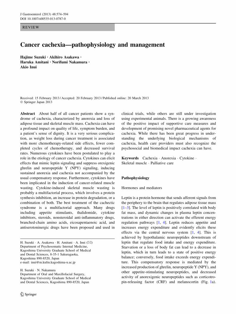
Leptin is a protein hormone that sends afferent signals from
the periphery to the brain that regulates adipose tissue mass
[1–3]. The level of leptin is positively correlated with body
fat mass, and dynamic changes in plasma leptin concen-
trations in either direction can activate the efferent energy
regulation pathways [1, 4]. Leptin reduces appetite and
increases energy expenditure and evidently elicits these
effects via the central nervous system [1, 4]. This is
achieved by hypothalamic neuropeptides downstream of
leptin that regulate food intake and energy expenditure.
Starvation or a loss of body fat can lead to a decrease in
leptin, which in turn leads to a state of positive energy
balance; conversely, food intake exceeds energy expendi-
ture. This compensatory response is mediated by the
increased production of ghrelin, neuropeptide Y (NPY), and
other appetite-stimulating neuropeptides, and decreased
activity of anorexigenic neuropeptides such as corticotro-
pin-releasing factor (CRF) and melanocortin (Fig. 1a).
H. Suzuki � A. Asakawa � H. Amitani � A. Inui (&)
Department of Psychosomatic Internal Medicine,
Kagoshima University Graduate School of Medical
and Dental Sciences, 8-35-1 Sakuragaoka,
Kagoshima 890-8520, Japan
e-mail: [email protected]
H. Suzuki � N. Nakamura
Department of Oral and Maxillofacial Surgery,
Kagoshima University Graduate School of Medical
and Dental Sciences, Kagoshima 890-8520, Japan
123
J Gastroenterol (2013) 48:574–594
DOI 10.1007/s00535-013-0787-0

Thus, if a disease process such as cancer was to produce
factors that induce or mimic the hypothalamic effect of
excess negative feedback signaling from leptin, the expec-
ted outcome would be sustained anorexia (lack of appetite)
and cachexia (muscle wasting and uncontrolled weight
loss), without the usual compensatory response [5]. In fact,
in tumor-bearing states, cachectic factors such as cytokines
can elicit effects on energy homeostasis that mimic leptin
and suppress orexigenic ghrelin and NPY signaling. Con-
sequently, the increases and decreases in hypothalamic
actions caused by these mediators induce anorexia and
unopposed weight loss (Fig. 1b).
Serotonin (5-HT) may also play a role in the develop-
ment of cancer-induced anorexia. This is because increased
levels of plasma and brain tryptophan, the precursor of
5-HT, and interleukin (IL)-1 may underlie the increased
serotonergic activity seen in the cancer cachexia. In addi-
tion, cisplatin-induced anorexia has become problematic in
clinical settings. Cisplatin is a widely used and effective
anti-cancer chemotherapy drug, however, the undesirable
gastrointestinal side effects associated with it, such
as nausea, vomiting, and anorexia, markedly decrease
patients’ quality of life, rendering continuation of chemo-
therapy difficult [6]. Cisplatin-induced gastrointestinal tract
Fig. 1 A simplified model of the hypothalamic neuropeptide cir-
cuitry in response to starvation (a) and cancer cachexia (b). Full linearrows indicate the activation of the process, and broken line arrowsindicate the inhibition of the process. Under normal conditions,
energy intake is determined by the hypothalamic integration of
peripheral signals conveying inputs on adiposity status, digestive
processes, and metabolic profile. Some of these signals such as
adipocyte-derived leptin inhibit energy intake, while other signals
such as stomach-derived ghrelin stimulate energy intake. In the
hypothalamus, the arcuate nucleus (ARC) receives information from
the periphery and integrates these inputs to modulate food intake via
second-order neurons. According to the information conveyed to the
brain, peripheral signals may differentially activate or inhibit POMC/
CART and NPY/AgRP neurons. When an energy deficit (e.g.,
starvation) is signaled, orexigenic NPY/AgRP neurons are activated
and anorexigenic POMC/CART neurons are inhibited, resulting in
increased energy intake. When an energy excess is signaled, NPY/
AgRP neurons are inhibited and POMC/CART neurons are activated.
During cancer, cachectic factors such as cytokines elicit effects on
energy homeostasis that mimic leptin in some respects and suppress
orexigenic Ghrelin-NPY/AgRP signaling. Increased brain cytokine
expression disrupts hypothalamic neurochemistry, particularly in the
ARC where cytokines activate POMC/CART neurons, while inacti-
vate NPY/AgRP neurons. The anorexia and unopposed weight loss in
cachexia could be accomplished through persistent inhibition of the
NPY orexigenic network and stimulation of anorexigenic neuropep-
tides, although the hypothalamic pathways participating in this
response remain to be determined. AgRP Agouti-related peptide,
MCH melanin-concentrating hormone, CART cocaine- and
amphetamine-related transcript, NPY neuropeptide Y, POMC pro-
opiomelanocortin, CRH corticotropin-releasing hormone, MC4Rmelanocortin-4 receptor, PVN paraventricular nucleus. LHA lateral
hypothalamic area. Source: (5) with modification
J Gastroenterol (2013) 48:574–594 575
123
disorders are thought to be due to the release of large
amounts of 5-HT from enterochromaffin cells, which then
bind to 5-HT receptors [6]. 5-HT activates various sero-
tonin receptor subtypes in the gastrointestinal tract and
ganglia, exerting a range of biological and physiological
effects [6]. It has been reported that a significant increase in
5-HT concentrations in the hypothalamus of cisplatin-
treated rats [7]. Accumulated findings suggest that seroto-
nin 2C (5-HT2C) receptor subtypes are involved in appetite
regulation [8, 9]. The 5-HT2C receptor subtype is expres-
sed in proopiomelanocortin neurons in the hypothalamus,
which is the major site of its anorexigenic action [6]. In the
present clinical setting, nausea and vomiting can be con-
trolled by administering 5-HT3 receptor antagonists toge-
ther with anticancer agents [6]. However, 5-HT3 receptor
antagonists may not be sufficiently controlled in cisplatin-
induced anorexia [6]. Recent studies have reported that
cisplatin-induced anorexia is mediated through reduced
gastric and hypothalamic ghrelin secretion, and peripheral
5-HT2B and cerebral 5-HT2C receptor activation are
responsible for the phenomenon [6, 10, 11]. Facilitating the
gastric and hypothalamic ghrelin secretion through
5-HT2C receptor inhibition can be a useful therapeutic
approach for cisplatin-induced anorexia.
Cytokines
Cytokines are protein molecules released by lymphocytes
and/or monocyte macrophages [5]. They are released into the
circulation and transported to the brain through the blood–
brain barrier (BBB) and circumventricular organs (i.e.,
‘leaky’ areas in the BBB) [12–17]. Peripheral cytokines may
influence the brain via neural pathways or second messen-
gers such as nitric oxide (NO) and prostanoids [5]. Cytokines
are also produced by neurons and glial cells within the brain,
partly in response to peripheral cytokines [12–17]. Although
the site of cytokine synthesis within the brain is dependent on
the nature of the stimulus, systemic disease seems to pre-
dominantly influence expression in the hypothalamus, the
area with the highest densities of receptors [16].
Numerous cytokines, including tumor necrosis factor-
alpha (TNF-a), interleukin-1 (IL-1), interleukin-6 (IL-6),
and interferon-gamma (IFN-c), have been postulated
to play a role in the etiology of cancer cachexia [12, 13,
18–21]. It is not certain whether the cytokine production is
primarily from tumour or host inflammatory cells. It has
been hypothesised that either tumour cell production of
proinflammatory cytokines or the host inflammatory cell
response to tumour cells is the source of the acute phase
protein response (APPR) seen in many malignancies and in
cachexia [22].
High serum levels of TNF-a, IL-6, and IL-1 have been
found in some cancer patients, and the levels of these
cytokines seem to correlate with the progression of some
tumors [23–25]. Chronic administration of these cyto-
kines, either alone or in combination, is capable of
reducing food intake and inducing cancer cachexia
[18, 23–26]. The role of TNF-a in mediating cancer
cachexia is supported by evidence that intraperitoneal
injection of a soluble recombinant human TNF-receptor
antagonist improved anorexia in tumor-bearing animals
[27]. In humans, IL-1 appears to play a significant role in
mediating cachexia, as megestrol acetate has been shown
to exert its effects via reduced expression of IL-1 by
mononuclear cells beyond its influence on hypothalamic
NPY concentrations, which shows orexigenic effect [28].
Interestingly, anorexigenic neurons, such as proopiomel-
anocortin (POMC)/cocaine and amphetamine-regulated
transcript (CART) neurons in the arcuate nucleus of the
hypothalamus express the type 1 IL-1 receptor, and
intracerebroventricular injection of IL-1 increases the
frequency of action potentials of POMC/CART neurons
and stimulates the release of alpha-melanocyte-stimulating
hormone (a-MSH), which shows anorexigenic effect as
well [29].
TNF-a, IL-1, IL-6, and IFN-c have been implicated in
the induction of cancer-related muscle wasting [30]. There
is growing evidence that the accelerated muscle proteolysis
seen during malignant tumor growth is mediated by the
activation of the non-lysosomal adenosine triphosphate-
dependent (ATP-dependent) ubiquitin proteasome pathway
[31, 32]. In addition, inflammatory cytokines influence the
expression of functionally relevant enzymes in cardiac
cachexia [30]. It has been demonstrated that TNF-a, IFN-c,
and IL-1b are potent activators of inducible nitric oxide
synthase (iNOS) expression [30], which in turn produces
toxic levels of NO high enough to inhibit the key enzymes
of oxidative phosphorylation [30]. It has also been shown
in vitro that NO is able to impair the contractile perfor-
mance of skeletal muscle [33].
More direct evidence of cytokine involvement comes
from experiments in which specific neutralization of
cytokines can relieve anorexia and cachexia in experi-
mental animal models [18, 24, 25, 34]. Examples of anti-
bodies that have been shown to successfully relieve
anorexia and cachexia when administered include the anti-
TNF-a, anti-IL-6, anti-IL-1, and anti-IFN-c antibodies,
although no single antibody has been proven to reverse all
of the features of wasting seen in cancer cachexia [24].
These studies revealed that cachexia can rarely be attrib-
uted to any one cytokine but rather is associated with a set
of cytokines and other cachectic factors that work in con-
cert [5]. Recent studies include the use of anti-IL-6
humanized monoclonal antibody, which appears to inhibit
cancer cachexia in murine models [35] and may be of
clinical significance in cancer patients [35].
576 J Gastroenterol (2013) 48:574–594
123

The problem with ascribing specific tissue responses to
individual cytokines is that considerable overlap and
redundancy exists in the cytokine network [14–17, 21].
Administration of either TNF-a or IL-1 will induce the
synthesis of a variety of other proinflammatory cytokines
such as IL-6 [5]. Thus, studies that use pharmacological
administration of recombinant cytokines may not discrim-
inate between biological responses induced directly by the
administered cytokine and those induced secondarily by
other stimulated cytokines [5]. Systemic disease such as
cancer and inflammation may elicit a cytokine cascade in
which several cytokines are induced simultaneously [21].
Systemic changes in response to inflammation are
denoted the acute phase response [36]. Up to 50 % of
patients with solid epithelial cancers may have an elevated
APPR [37]. APPR is correlated with elevated resting
energy expenditure and reduced energy intake [38]. Other
longitudinal studies have found a poorer prognosis in
patients displaying this response, independent of weight
loss [39].
C-reactive protein
C-reactive protein (CRP) is the most common method used
to assess the magnitude of the systemic inflammatory
response [36]. The modified Glasgow prognostic score
combines CRP and plasma albumin concentrations to cre-
ate a simple scoring system that serves as a prognostic
factor that is independent of stage and treatment and that
predicts survival [40, 41] (Table 1).
Raised CRP concentrations at the time of admission to
hospital is indicative of an increased risk for all-cause
mortality; there is a 22.8-fold increase in cancer mortality
in patients with highly elevated CRP concentrations
([80 mg/L) [42]. It has been shown that patients with
inoperable non-small cell lung cancer had at least 5 %
weight loss and almost 80 % an elevated CRP levels [43].
In patients without weight loss, those who displayed evi-
dence of a systemic inflammatory response reported more
fatigue (P \ 0.05) [43]. In another study of patients with
gastroesophageal cancer, the rate of weight loss was also
correlated with elevated CRP serum concentrations [44].
Negative nitrogen balance
In adults, muscle mass remains fairly constant in the
absence of stimuli (e.g., exercise) and thus protein syn-
thesis and degradation generally remain in balance [45].
However, in cachexia, muscle atrophy occurs, which
results from a decrease in protein synthesis, an increase in
protein degradation, or a combination of both [45]. In
recent years, it has become evident that specific regulating
molecules are upregulated (e.g., members of the ubiquitin–
proteasome system, myostatin, and apoptosis inducing
factors), whereas other factors (e.g., insulin-like growth
factor 1) are down-regulated in cachexia muscle wasting
[30]. A major barrier to the effective management of
skeletal muscle wasting is the inadequate understanding of
its underlying biological mechanisms [30]. The most evi-
dent metabolic explanation for muscle decline is an
imbalance between protein catabolism and anabolism [30].
In addition to an increase in catabolism, a reduction in
anabolism has been shown to occur in cancer cachexia
[30]. Skeletal muscle wasting in cancer cachexia can be
mediated by multiple factors derived from tumor and host
cells [46].
At least four major proteolytic pathways (lysosomal,
Ca2?-dependent, caspase-dependent, and ubiquitin–pro-
teasome-dependent) operate in skeletal muscle and may be
altered during muscle cachexia [30]. Aside from these four
distinct pathways, the autophagic/lysosomal pathway must
also be considered [30]. In this pathway, portions of the
cytoplasm and cell organelles are sequestered into auto-
phagosomes, which subsequently fuse with lysosomes,
where the proteins are digested [47].
When dissecting the molecular regulation of the ubiq-
uitin–proteasome-dependent system (UPS) and autophagy,
it became evident that forkhead box O (FoxO) transcription
factors play a central role [30]. FoxO transcription factors,
which are normally phosphorylated and inactivated by
phosphatidylinositol 3-kinase (PI3K)-Akt/PKB, translocate
into the cell nucleus and induce the transcription of the
skeletal muscle-specific E3 ubiquitin ligases, muscle
RING-finger protein-1 (MuRF1), and atrogin-1/muscle
atrophy F-box (MAFbx) [48], as well as autophagy-related
genes such as LC3 and Bnip3 [49]. Upstream of PI3K-Akt,
several factors including reactive oxygen species (ROS),
TNF-a, tumor-released proteolysis-inducing factor (PIF),
peroxisome proliferator-activated receptor gamma coacti-
vator 1 alpha (PGC-1a), and IGF-1 have been shown to
influence this regulatory system [48, 50–52]. In contrast,
protein anabolic factors such as IGF-1 counteract muscle
atrophy [30]. Aside from inhibiting autophagy and the
UPS, IGF-1 activates protein synthesis via the Akt-mam-
malian target of rapamycin (mTOR)–p70 S6 kinase
(p70S6K) signaling pathway [53, 54].
Table 1 Modified Glasgow Prognostic Score (mGPS): an inflam-
mation-based prognostic score
Biochemical measure Score
C-reactive protein B10 mg/L ? albumin C35 g/L 0
C-reactive protein B10 mg/L ? albumin \35 g/L 0
C-reactive protein [10 mg/L 1
C-reactive protein [10 mg/L ? albumin \35 g/L 2
Source: [41]
J Gastroenterol (2013) 48:574–594 577
123

The UPS is a major intracellular system that regulates
skeletal muscle wasting in response to tumor factors and
inflammatory cytokines [46]. In cancer cachexia, the
decrease in skeletal muscle protein synthesis is partly
related to the increased serum levels of PIF [30]. Intrave-
nous administration of PIF to normal mice produced a
rapid decrease in body weight that was accompanied by
increased mRNA levels of ubiquitin in the gastrocnemius
muscle [51]. There were also increased protein levels of the
20 S proteasome core and the 19 S regulatory subunit,
suggesting activation of the ATP–ubiquitin-dependent
proteolytic pathway [30]. Recent evidence suggests that
PIF decreases protein synthesis by inhibiting protein
translation initiation through phosphorylation of the
eukaryotic initiation factor 2 (eIF2-alpha) [55].
Myostatin
Myostatin is an extracellular cytokine that is mostly
expressed in skeletal muscles and is known to play a cru-
cial role in the negative regulation of muscle mass [56].
Upon binding to the activin type IIB receptor, myostatin
can initiate several different signaling cascades, resulting
in decreased muscle growth and differentiation [56].
Muscle size is regulated via a complex interplay of
myostatin signaling with the IGF-1/PI3K/Akt pathway,
which is responsible for increased protein synthesis in
muscle [56]. Therefore, the regulation of muscle weight is
a process in which myostatin plays a central role, but the
mechanism of its action and the role of the signaling cas-
cades involved are not fully understood [56]. Myostatin
upregulation was observed in the pathogenesis of muscle
wasting during cancer cachexia [56].
Data are available that demonstrate a beneficial effect of
myostatin inhibition in cancer cachexia [57], but conflicting
study results have also been reported [58]. With respect to
apoptosis, several reports demonstrated an increase in
apoptosis or apoptosis-related proteins in skeletal muscle
after the induction of cachexia [30]. The skeletal muscle of
cachectic tumor-bearing animals reveals the presence of
DNA fragmentation, a hallmark of apoptosis [59]. In
addition to DNA fragmentation, a significant up-regulation
of caspase-1, -3, -6, -8, and -9 activity was also documented
in the gastrocnemius muscles of tumor-bearing mice [60].
Insulin-like growth factor-1
One of the main positive regulators of muscle growth is IGF-1
[56]. Under normal conditions, IGF-1 signaling seems to be
dominant and blocks the myostatin pathway [61]. However,
Fig. 2 An abbreviated diagram of skeletal muscle in cancer cachexia.
In adults, muscle mass remains fairly constant in the absence of
stimuli (e.g., exercise) and thus protein synthesis and degradation
generally remain in balance. However, in cachectic situation, the
balance of skeletal muscle has been shifted towards protein break-
down, finally leading to the weight loss, weakness, and fatigue that
characterize cancer cachexia. In recent years, it has become evident
that catabolic factors are up-regulated (e.g., cytokines, myostatin and
members of the ubiquitin–proteasome system), whereas anabolic
factors (e.g., insulin-like growth factor 1) are down-regulated in
cachexia muscle wasting. IGF-1 Insulin-like growth factor 1, FoxOforkhead box O, UPS ubiquitin–proteasome system, ROS reactive
oxygen species, NF-jBPIF tumor-released proteolysis-inducing fac-
tor, mTOR mammalian target of rapamycin, p70S6K p70 S6 kinase
578 J Gastroenterol (2013) 48:574–594
123

an inhibition of IGF-1 can occur when myostatin is overex-
pressed [62, 63]. IGF-1 can prevent TGF-a family-mediated
apoptosis [64], and it was shown that in the absence of IGF-1,
the level of apoptosis in C2C12 cells treated with myostatin
increased [56]. The mechanism by which IGF-1 regulates
myostatin signaling includes the inhibition of transcription
factors responsible for the induction of atrogenes via phos-
phorylation through the PI3K/Akt pathway [56]. Akt plays a
significant role in different metabolic processes in the cell,
particularly in the hypertrophic response to insulin and IGF-1
[65, 66]. Akt is the ‘crossing point’ between the IGF-1 and
myostatin pathways [56]. It is likely that under conditions of
muscle wasting, myostatin can reverse the Akt/mTOR path-
way, which is normally responsible for protein synthesis, to
inhibit protein synthesis via FoxO, GSK-3b, or other unknown
patterns, leading to the loss of muscle mass [56] (Fig. 2).
Another factor that may contribute to decreased anab-
olism is angiotensin II [30]. In an animal model of con-
tinuously administered angiotensin II, markedly reduced
plasma IGF-1 levels occurred [67]. Compared with a sham
treatment, angiotensin II-infused hypertensive rats lost
18–26 % of their body weight within a week, an effect that
was completely reversed by losartan (an angiotensin II
receptor type 1 receptor antagonist) [67].
Experimental data suggest that local IGF-1 may act as a
regenerative agent, promoting the recruitment of stem cells
to sites of muscle injury [68]. Because IGF-1 is reduced in
experimental models of cachexia [69], it is reasonable to
assume that under conditions of cachexia, the function of
satellite cells is impaired [30].
Oxidative stress
There is a wealth of evidence suggesting that oxidative
stress is associated with chronic diseases and it is assumed
that an increase in ROS directs muscle cells into a catabolic
state that leads to muscle wasting [30, 70, 71]. In cachexia,
ROS are regarded as crucial players for muscle protein
catabolism via their stimulation of the UPS [30]. Reaction
products are measured as indirect markers of oxidative
stress [30]. In cachexia, malondialdehyde (MDA) is
regarded as one such indirect marker [30].
In addition, experimental cancer cachexia appears to be
mediated by increased nitrosative stress secondary to
increased nitric oxide formation. Indeed, protein tyrosine
nitration is markedly increased in the muscles of tumor-
bearing rats with advanced cachexia, due to lower levels of
antioxidant enzymes [72, 73].
Anabolic hormones
There is a relative deficiency or resistance to anabolic
hormones in cachectic states. Up to 50 % of men with
metastatic cancer present with low concentrations of tes-
tosterone prior to chemotherapy [74]. A reduction in tes-
tosterone might lead to reduced bone mass, muscle
strength, and sexual function in both men and women [75,
76]. Low concentrations of testosterone and other anabolic
hormones are major contributors to cachexia-related
wasting of skeletal muscle [77]. However, with respect to a
correlation between body composition (including muscle
mass) and the concentration of anabolic hormones, con-
flicting results have been reported in the current literature
[30, 74, 78, 79].
Effects on antineoplastic therapy
Catabolic drivers
Antineoplastic therapies such as surgery, radiotherapy, and
chemotherapy are known to have a negative impact on a
patient’s nutritional intake through the development of
systemic inflammation, exacerbation of already-reduced
energy, and, particularly, on swallowing difficulties and
anorexia due to nausea [22, 80]. Additionally, surgical
patients may be fasted for prolonged periods periopera-
tively and both chemotherapy and radiotherapy can induce
side-effects such as anorexia, nausea, vomiting, mucositis,
taste change, or lethargy [80]. Consequently, antineoplastic
therapies interfere with the maintenance of the nutritional
state [81] (Tables 2, 3, 4).
Symptoms will depend on the nature and course of the
chemotherapeutic drugs being used and the location, vol-
ume, and dose of radiotherapy [80]. Some cytotoxic drugs
may even generate their own cachexia-like side effects
[82]. For example, treatment with antitubulin taxanes
reduces body weight in tumor-bearing mice more than
healthy mice, even when the agents significantly reduce
tumor growth [82]. However, the complex relationship
between cancer cachexia and the effects of antineoplastic
drugs remains to be fully elucidated [82].
C-reactive protein
A key (but often variable) component of cachexia is
hypercatabolism that is directly caused by tumor metabo-
lism, systemic inflammation, or other tumor-mediated
effects. The most widely accepted index of systemic
inflammation is serum CRP [83]. CRP plasma values are
positively correlated with weight loss, the occurence of
cachexia, and recurrence in advanced cancer [84]. Its role
as a predictor of survival has been shown in multiple
myeloma, melanoma, lymphoma, ovarian, renal, pancre-
atic, and gastrointestinal tumors [84, 85].
Recent studies suggest that CRP is much more than a
mere marker of the body’s inflammatory load [86, 87]. In
J Gastroenterol (2013) 48:574–594 579
123

Table 2 Nutritional
consequences of radical
resection of alimentary tract
organs
Source: [81]
Nutritional consequences
Tongue or pharynx Need for nutrition by tube (dysphagia)
Thoracic oesophagus Gastric stasis (due to vagotomy), malabsorption of fats (due to
vagotomy)
Stomach Dumping syndrome, anaemia, malabsorption of fats, iron,
calcium and vitamins
Duodenum Biliary-pancreatic deficiency
Jejunum (up to 120 cm) Reduced absorption of glucose, fats, protein, folic acid, vitamin
B12, etc.
Ileum (60 cm) or ileocaecal valve Malabsorption of vitamin B12, biliary salts and fats
Small intestine (75 %) Malabsorption of fats, glucose, protein, folic acid, vitamin
B12, etc., diarrhea
Jejunum and ileum Complete malabsorption
Colon (subtotal or total resection) Water and electrolyte loss
Pancreas Malabsorption and diabetes
Liver Transient hypoalbuminaemia
Table 3 Nutritional
complications associated with
radiotherapy
Source: [81]
Region
irradiated
Early effects Late effects
Head and neck Odynophagia, xerostomia,
mucositis, anorexia, dysosmia,
hypogeusia
Ulceration, xerostomia, dental caries,
osteoradionecrosis, trismus,
hypogeusia
Thorax Dysphagia Fibrosis, stenosis, fistula
Abdomen and pelvis Anorexia, nausea, vomiting,
diarrhea, acute enteritis, acute
colitis
Ulceration, malabsorption, diarrhea,
chronic enteritis, chronic colitis
Table 4 Effects of
chemotherapeutic drugs
Source: [81]
Drug Severity and duration
Chemotherapeutic drugs commonly associated with severe nausea and vomiting
Nitrogen mustard (mustine hydrochloride;
mechlorethamine hydrochloride USP)
Occurs in virtually all patients. May be severe, but usually
subsides within 24 h
Chloroethyl nitrosoureas, streptozotoci
(streptozocin)
Variable, but may be severe. Occurs in nearly all patients.
Tolerance improves with each successive dose given on a
5-day schedule
Cis-platinum (cisplatin) May be very severe. Tolerance improves with intravenous
hydration and continuous 5-day infusion. Nausea may
persist for several days
Imidazole carboxamide (DTIC;
dacarbazine)
Occurs in virtually all patients. Tolerance improves with
each successive dose given on a 5-day schedule
Chemotherapeutic drugs commonly associated with mucositis
Methotrexate May be quite severe with prolonged infusions or if renal
function is compromised. Severity is enhanced by
irradiation. May be prevented with administration of
adequate citrovorum rescue factor (folinic acid;
leucovorin)
5-Fluorouracil (fluorouracil USP) Severity increase with higher doses, frequency of cycles,
and arterial infusions
Actinomycin D (dactinomycin USP) Very common; may prevent oral alimentation. Severity
enhanced by irradiation
Adriamycin (doxorubicin) May be severe and ulcerative. Increased in presence of
liver disease. Severity enhanced by irradiation
Bleomycin May be severe and ulcerative
Vinblastine Frequently ulcerative
580 J Gastroenterol (2013) 48:574–594
123

cultured human umbilical vein endothelial cells, CRP was
shown to activate endothelial cells, which, in turn, express
Intracellular Adhesion Molecule-1 (ICAM-1) [86, 88].
CRP also induces other adhesion molecules in endothelial
cells such as vascular-cell adhesion molecule-1 (VCAM-1)
and E-selectin [86]. These molecules are involved in leu-
kocyte-binding to the endothelial layer. CRP also activates
the expression of monocyte chemotactic protein-1 (MCP-1)
[87]. In addition, circulating factors, such as lipid-mobil-
ising factors (LMF), and proteolysis-inducing factor (PIF)
may play a role in the development of cancer anorexia and
cachexia [88]. These are tumor-derived catabolic factors
acting directly on adipose tissue and skeletal muscle,
without affecting food intake [88].
However cachexia can exist without overt systemic
inflammation, and thus indirect indices reflecting the cat-
abolic drive such as responsiveness to chemotherapy and
the rate of progression should also be assessed [83]. No
consensus was reached about the usefulness of other factors
contributing to catabolism [83]. These include insulin
resistance, prolonged high-dose corticosteroid therapy,
hypogonadism, and increased resting energy expenditure
[83].
Increased nuclear factor-jB activity
Nuclear factor-jB (NF-jB), a nuclear transition activator
factor, plays a major role in upregulating inflammatory
gene expression, including expression of COX-2, nitric
oxide synthase, TNF-a, IL-1, and IL-6 [89]. The proin-
flammatory response occurs particularly with the formation
of p65–p50 dimers, which act as the central control to an
inflammatory response. NF-jB is one of the principal
transcription factors to transduce TNF-a signals into the
cells. Moreover, it also activates gene transcription of
cytokines, acute-phase response proteins, and cell adhesion
molecules [90, 91].
Activation of NF-jB accelerates inflammation, increa-
ses cellular proliferation of tumors, and prevents apoptosis
[90]. Inhibition of NF-jB therefore is both antineoplastic
and can reduce cachexia. Inhibition of NF-jB also sensi-
tizes tumors to chemotherapy and radiation, something that
is the subject of multiple research trials [92].
In fact, genetic overexpression of IjB blocks NF-jB-
dependent processes. Two studies have shown that gluco-
corticoid administration induces transcription of IjB gene
[93, 94]. Increased levels of IjB gene trap NF-jB in
inactive cytoplasmic complexes, hence inhibiting its ability
to induce transcription of inflammatory cytokines. More-
over, fumar acid, which blocks the nuclear translocation of
NF-jB, has a high anti-inflammatory capacity [93]. More
recently, activation of NF-jB by overexpression of a IjB
phosphorylating kinase has been shown sufficiently to
block myogenesis, thus illustrating the link between NF-jB
and cachexia development [94]. However, complete inhi-
bition of NF-jB has proven detrimental; knockout studies
targeting the major subunits of NF-jB show severe
immunodeficiency in mice, which was lethal in some cases
[95]. Resolution of inflammation also requires NF-jB
expression and complete inhibition of NF-jB can lead to
severe cellular apoptotic damage in critical illness [96].
Managing cancer cachexia
Treatment goals in current standard of care
The European Palliative Care Research Collaboration
(EPCRC) has developed evidence-based recommendations
for the classification and treatment of cachexia in advanced
cancer patients [97]. These treatment guidelines focus on
patients with advanced cancer that are likely to suffer from
refractory cachexia. Many of these patients are receiving
palliative care, and life expectancy often is short. Only
little cachexia-specific research has been done on this
patient group, and the EPCRC treatment guidelines had to
consider whether research results taken from other disease
stages could be applicable for patients with advanced and
incurable disease with refractory cachexia [97].
Management of cachexia must take into account the
patient’s prognosis [97], as it may take several weeks for
patients to respond to anti-cachectic treatment [97]. For
patients with a short life expectancy, treatment options for
cachexia may add to the disease burden without offering
adequate symptom relief and thus may not be appropriate
[97]. Health care professionals should discuss all treatment
options with the patient and ensure that they are well-
informed about available treatments and expected treat-
ment outcomes [97]. All patients should have equal access
to appropriate assessment and management of cachexia,
whether they are receiving home care, day care, or are
hospital inpatients [97].
The best way to treat cancer cachexia is to cure the
cancer, but unfortunately this remains an infrequent
achievement among adults with advanced solid tumors [98,
99]. Therefore, the treatment goal for cachexia should be
the reversal of the loss of body weight and muscle mass
with a variety of pharmacological agents (Fig. 3) [99]. As a
minimal goal, body weight should be maintained and fur-
ther loss prevented [97]. The treatment approach should be
multimodal and similar to treatment used in patients with
pre-cachexia [97]. This includes detailed assessment and
repeated monitoring, vigorous nutritional support, anti-
inflammatory treatment, treatment of secondary gastroin-
testinal symptoms and other causes for decreased oral
nutritional intake as well as evaluation of anti-neoplastic
J Gastroenterol (2013) 48:574–594 581
123

options to reduce the catabolic drive of the cancer [97].
However, for refractory cachexia, the primary treatment
goal should not be reversal of weight loss, but the allevi-
ation of cachexia-related symptoms and an overall increase
of well-being [97].
Pharmacological treatments
Appetite stimulants
Reversing the effects of cancer cachexia does not appear to
be influenced by stimulating the appetite [100]. Thus, the
decision to use an orexigenic drug should be based on
tolerance of the side effects, cost effectiveness, and treat-
ment burden [100]. Current studies are investigating an
approach of drug combinations to reverse cancer cachexia
[101, 102]. A recent study with 332 patients comparing
medroxyprogesterone, megestrol acetate, oral supplemen-
tation with eicosapentaenoic acid, L-carnitine, and thalid-
omide found that the combination therapy was superior to
any of the other treatment arms with single drug treatment
[102]. Combination therapy led to increased lean body
mass, decreased resting energy expenditure, and improved
appetite [102]. Until an effective intervention for reversing
Fig. 3 The potential modalities
of pharmacological intervention
of cancer anorexia-cachexia
syndrome. Agents were
classified as those established
(first-line) or those unproven/
investigational (second-line),
depending on their site or
mechanism of actions. �,
inhibitors of production/release
of cytokines and other factors;
`, gastroprokinetic agents with
or without antinausea effect; ´,
blockers of Cori cycle; ˆ ˜,
blockers of fat and muscle tissue
wasting; Þ, appetite stimulants
with or without antinausea
effect; and þ, anti-anxiety/
depressant drugs. These agents
should be selected on an
individual basis according to the
cause of cachexia or the state of
the patient. *The precise actions
of statins on skeletal muscle still
remain controversial. First-line
treatments: glucocorticoids �
Þ, progesterones � Þ. Second-
line treatments: cannabinoids
Þ, cyproheptadine Þ,
branched-chain amino acids ˜
Þ, metoclopramide ` Þ,
eicosapentanoic acid � ˆ ˜, 50-deoxy-5-fluorouridine �,
melatonin �, thalidomide �,
b2-adrenoceptor agonists ˜,
non-steroidal anti-inflammatory
drugs � Þ, others anabolic
steroids ˜, pentoxifylline �,
hydrazine sulfate ´, statin �
˜*, angiotensin-converting-
enzyme inhibitor inhibitor ˜,
selective androgen receptor
modulator ˜. Source: [99] with
modification
582 J Gastroenterol (2013) 48:574–594
123
cancer cachexia is developed, early intervention with
nutritional support and prevention of treatment-related
morbidities (e.g., nausea, vomiting, diarrhea, dysphagia,
pain, or depression) is advised [102, 103].
Progestational drugs, cannabinoids, and cyproheptadine
are used in the clinic as appetite stimulatus in the therapy
of the cancer-induced anorexia and cachexia syndrome
[99]. These drugs have been shown to be partially effective
in reversing or maintaining the symptom of body weight
loss in patients with chronic illness [99].
Cannabinoids are highly liquid-soluble substances with
delta-9-tetrahydrocannabinol (THC) as an active ingredient
that work synergistically, additively, or even antagonisti-
cally when ingested together (e.g., by smoking marijuana).
Appetite stimulation and body weight gain are well-rec-
ognized effects of using marijuana and its derivatives [99].
This may have significant implications for the clinical
usefulness of marijuana or its individual compounds in
treating cachexia.
Dronabinol is the synthetic oral form of THC, which is
the active ingredient responsible for the appetite-stimulat-
ing effect [99, 104–106]. Dronabinol and marinol (in the
United States) and nabilone (in Canada) have been used as
antiemetics in cancer, with many studies demonstrating
their efficiency in treating chemotherapy-induced nausea
and vomiting [99]. Several studies of THC in advanced
cancer-associated anorexia have shown some improvement
in mood and appetite, with either no or some improvement
in body weight [107, 108]. However, randomized, con-
trolled trials are needed to better determine the efficacy and
usefulness of THC in cancer cachexia.
The effects of cannabinoids are mediated via specific
receptors. Two types of cannabinoid receptors, CB1 and
CB2 have been detected. However, the precise mechanism
by which cannabinoids exert their effect has yet to be
clarified. It has been shown that almost 20 percent of the
cancer patients receiving chemotherapy along with dro-
nabinol as an antiemetic experienced side effects, such as
euphoria, dizziness, somnolence, and confusion resulting in
a dose reduction or less frequently in withdrawal of the
treatment [106]. It has been suggested that the drug could
be taken at bedtime to avoid some psychotomimetic effects
and that it might produce long-lasting appetite stimulation
for 24-h period following ingestion [104].
Cyproheptadine is an antiserotoninergic drug with
antihistaminic properties that has been shown to have a
slight appetite-stimulant effect in a number of human
conditions [109]. A randomized, controlled trial found mild
appetite stimulation in patients with advanced cancer,
although it did not prevent progressive weight loss [110].
Considerable evidence, both in humans and experimental
animals, suggests that anorexia may be mediated by
increased serotonergic activity in the brain. Its blockade,
therefore, might be beneficial in reducing symptoms [111,
112]. Cyproheptadine also appeared to stimulate appetite
and decrease diarrhea in patients with advanced carcinoid
tumors [113]. Studies on the effects of cyproheptadine in
progressive weight loss in patients with cancer or other
causes of cachexia suggest that cyproheptadine has a
beneficial effect on appetite stimulation but only slight
effects on weight gain [110, 114, 115]. 5-hytroxytrypta-
mine type 3 (5HT3) receptor antagonists, such as ondan-
setron and granisetron, have entered widespread clinical
use as antiemetics for cancer chemotherapy [99].
Progestins
Megestrol acetate (MA) and medroxyprogesterone acetate
(MPA) are synthetic, orally active progestational agents. In
several randomized controlled studies, these compounds
have been found to improve appetite, caloric intake, and
nutritional status in patients with non-hormone responsive
tumors and cancer anorexia-cachexia syndrome [104–106,
116–122].
MA has demonstrated a dose-related beneficial effect, in
a dose range from 160 mg to 1600 mg/day on appetite,
caloric intake, body weight gain (mainly fat), and sensation
of well-being (with an optimal dosage of 800 mg daily)
[118]. Increasing MA dosages from 160 mg to 800 mg/day
improves response to a level beyond which no further
improvement occurs [118]. It is recommended that a
patient is started on the lowest dosage (i.e, 160 mg/day)
and that the dose is uptitrated according to clinical response
[105, 109].
MPA has similarly been shown to increase appetite and
food intake with a stabilization of body weight at a dose of
1000 mg (i.e., 500 mg twice daily) [109]. Although the
drug is safe at doses of 500–4000 mg daily, side effects
have been shown to increase above oral doses of 1000 mg
[104]. At present, there is considerable evidence for the
effect of synthetic progestins on appetite and body weight
in patients with cancer anorexia and cachexia [123].
However, further issues regarding the optimal treatment
duration, the best time to start treatment during the natural
history of the disease, and the eventual impact on the
overall quality of life need to be clarified [123]. Moreover,
optimal dose regimens for MA in different indications,
such as appetite improvement, patients’ sense of well-
being, weight gain, are still to be identified.
The following adverse events have been reported with
MPA: thromboembolic phenomena, breakthrough uterine
bleeding, peripheral edema, hyperglycemia, hypertension,
adrenal suppression, and adrenal insufficiency if the drug is
abruptly discontinued [104–106, 116–120, 124]. Although
patients rarely need to stop taking these drugs because of
adverse effects, these drugs should not be prescribed in
J Gastroenterol (2013) 48:574–594 583
123
cases of thromboembolic/thrombotic disease, heart disease,
or for patients at risk for serious fluid retention [104].
Although the mechanism of weight gain of progesta-
tional drugs in presently uncertain, it might be related to
glucocorticoid activity [105]. MA may induce appetite via
stimulation of NPY, a potent central appetite stimulant in
the hypothalamus, modulation of calcium channels in the
ventromedial hypothalamus (VMH)—a well known satiety
center [1, 4, 125–133] which reduces the firing tone of
VMH neurons. On the other hand, MPA has been shown to
inhibit the activity of proinflammatory cytokines such as
IL-1, IL-6, and TNF-a [28, 109, 134]. Serum levels of such
cytokines were reported to be decreased in cancer patients
after MA or MPA treatment [109]. More studies are needed
to finally clarify the pharmacologic effects of MA and
MPA drugs and to confirm the anti-inflammatory effects.
Several other drugs have been evaluated as agents to
ameliorate cancer anorexia-cachexia. Corticosteroids are
frequently used in clinical practice for appetite stimulation
in patients with advanced malignancies and randomized
clinical trials showed that corticosteroid medications may
stimulate appetites in patients with advanced cancer [135].
However, these studies were not able to show any sub-
stantial non-fluid weight gain in treated patients [135].
Efforts are also ongoing to evaluate both anabolic steroids
and hydrazine sulfate as drugs for the treatment of patients
with cancer cachexia [135].
Hydrazine is a substance that inhibits the enzyme
phosphoenol pyruvate carboxykinase (PEP-CK) and inter-
feres with gluconeogenesis [135]. However, hydrazine and
hydrazine sulfate might be a human carcinogen based on
evidence for carcinogenicity in animal studies [135, 136].
The preliminary nature of these investigations, however,
precludes recommendations for the use of these drugs in
routine clinical practice [135].
Other orexigenic agents
The orexigenic mediator ghrelin has been reported as
having a key role in increasing appetite and, therefore, food
intake. Ghrelin is an endogenous ligand for the growth
hormone secretagogue receptors [137, 138]. It is synthe-
sized principally in the stomach and is released in response
to fasting [138].
Ghrelin strongly stimulates GH secretion in humans
[139–142] and does so more potently than GHRH by
several fold under similar circumstances [143]. Further-
more, ghrelin and GHRH synergistically increases GH
release [141]. GH regulates IGF-1 levels and increases
muscle strength [144, 145], whereas GH enhances lipoly-
sis, IGF-1 stimulates protein synthesis, myoblast differen-
tiation, and muscle growth [143]. Evidence that ghrelin
exerts anti-inflammatory actions has been accumulating
[143]. Ghrelin induces the anti-inflammatory cytokine
IL-10 [146, 147], suppressing the production of proin-
flammatory cytokines, including IL-1b, IL-6, and TNF-aboth in vitro [148, 149], and in vivo [146, 150, 151].
Additionally, ghrelin inhibits the activation of NF-jB,
which controls the production of multiple proinflammatory
cytokines during inflammatory insults [147, 149, 150].
Although the molecular mechanisms and cellular targets
mediating ghrelin inhibition of NF-jB activation remain to
be determined, the vagus nerve may play an important role
in the ghrelin-mediated inhibition of proinflammatory
cytokine release [150, 152]. MuRF1 and MAFbx are
upregulated under cachectic catabolic conditions, and NF-
jB activation may regulate skeletal muscle proteasome
expression and protein degradation [143]. The elevations in
MuRF1 and MAFbx expression seen in skeletal muscle
after thermal injury, arthritis, and dexamethasone admin-
istration were normalized, attenuated, and prevented,
respectively, by ghrelin or GHS administration [153–156].
IGF-1 prevents the expression of MuRF1 and MAFbx by
inhibiting FoxO transcription factors via stimulation of the
PI3K/Akt pathway [143]. The IGF-1 receptor triggers
activation of several intracellular kinases, including PI3K
[156]. Thus, the effects of ghrelin on NF-jB activation and
IGF-1 synthesis are favorable for minimizing inflammatory
responses and skeletal muscle wasting in patients with
cachexia [143].
In addition to increasing food intake, an experimental
study has shown that repeated administration of ghrelin
improves cardiac structure and function, and attenuates the
development of cardiac cachexia in chronic heart failure
(CHF). These results suggest that ghrelin has cardiovas-
cular effects and regulates energy metabolism through
growth hormone-dependent and -independent mechanisms
[157]. Thus, administration of ghrelin may be a new ther-
apeutic strategy for the treatment of severe CHF [157].
At present, a phase II randomized, placebo-controlled,
double-blind study, using an oral ghrelin mimetic, demon-
strated an improvement in lean body mass, total body mass
and hand grip strength in cachectic cancer patients [158].
Several clinical trials with ghrelin are currently on going.
Herbal medicine; translational aspects, particularly
for cancer cachexia
There have been a number of published cases of cancer
patients treated with Kampo, a form of Japanese traditional
herbal medical practice, who reportedly experienced sig-
nificant clinical benefits [159]. As an example, rikkunshito,
a Kampo formula, has been shown to be useful in clinical
practice for cachectic cancer patients.
Rikkunshito has been used to treat gastrointestinal tract
disorders such as functional dyspepsia [160–164] and
584 J Gastroenterol (2013) 48:574–594
123

gastroesophageal reflux [165]. A recent study regarding the
underlying mechanisms of rikkunshito has shown that
rikkunshito and its component 10-gingerol may inhibit the
degradation of acyl-ghrelin by inhibiting the circulating
ghrelin degrading enzyme [166]. Another study has shown
that administration of rikkunshito reversed the decrease in
hypothalamic ghrelin secretion and food intake 24 h after
cisplatin treatment [6]. A most recent study showed that
rikkunshito improved anorexia, gastrointestinal dysmotili-
ty, muscle wasting, and anxiety-related behavior [10]. In
this study, rikkunshito in tumor-bearing rats was effective
not only against anorexia–cachexia, but also for promoting
survival, particularly in combination with chemotherapy
[10]. Moreover, median survival of pancreatic cancer
patients with ascites who were treated with gemcita-
bine was significantly prolonged by administration of
rikkunshito [10]. Active components of rikkunshito, hes-
peridin and atractylodin, potentiated ghrelin secretion and
receptor signaling, respectively, and atractylodin pro-
longed survival in tumor-bearing rats [10]. The physio-
logical functions of endogenous ghrelin are enhanced by
the dual actions of rikkunshito; which involve the stimu-
lation of ghrelin secretion and the activation of GHS-R
activity, possibly due to allosteric changes in the receptor
[10]. These studies suggest that rikkunshito may be useful
in clinical practice for cachectic cancer patients.
Because Japanese Kampo has long been used, the
potential risks and benefits of its use are well recognized
despite the relative paucity of the mechanistic insights
[159]. Unconventional therapies such as herbs and minerals
that have been used in ancient medical traditions have led
to the identification of active anticancer agents [159].
Although the working mechanisms of some of the herbs
and minerals are unclear and remain to be elucidated, they
are worth further studying as newly potential therapy
agents for cancer treatment [167].
Non pharmacological treatments
Diet modification
Because cancer cachexia differs from starvation, to date,
single modality therapies with traditional nutritional regi-
mens have failed to demonstrate efficacy in improving
weight gain, including a gain in lean body mass, in patients
diagnosed with cancer cachexia [168]. The average caloric
deficit in weight-losing patients with cancer cachexia is
approximately 250–400 kcals/day [168]. An average sup-
plementation of 1 calorie/mL has not been shown to
improve the nutritional status of patients receiving che-
motherapy [169, 170]. However, recent studies using a
more calorie- protein-dense supplementation have sug-
gested that weight stabilization can be achieved; however,
improvements in lean body mass has not yet been observed
[171].
Patients with cancer cachexia undergoing aggressive re-
feeding are at risk for ‘re-feeding syndrome’ during the
first 2–3 weeks of treatment [172]. This potentially lethal
condition is characterized by severe electrolyte and fluid
shifts due to metabolic abnormalities and bears a signifi-
cant risk for morbidity and mortality [172]. The clinical
features include fluid-balance disturbances, abnormal glu-
cose metabolism, hypophosphatemia, hypomagnesiemia,
and hypokalemia [173].
Before starting the re-feeding process, electrolyte dis-
orders should be corrected and circulatory volume should
be carefully restored. This may delay the administration of
complete nutrition but is usually accomplished within
12–24 h. Caloric repletion should be at a slow rate of
approximately 20 kcal/kg per day (or 1000 kcal per day)
initially. However this rate may not meet the patients’ fluid,
sodium, potassium, protein, or vitamin requirements unless
these are specifically addressed.
Gradual introduction of calories, particularly over the
first week of re-feeding, should be prudent until the patient
is metabolically stable [174]. Hypophosphatemia has to be
treated if the serum level is less than 0.30 mmol/l or the
patient is symptomatic. Supplementation of phosphate
should be given intravenously at 40–80 mmol/day, toge-
ther with magnesium (8–16 mmol/day) and potassium
(80–120 mmol per day). These dosages should be adjusted
according to monitored serum levels [175].
Exercise
Physical exercise may be beneficial in the treatment of
cancer cachexia, as it increases insulin sensitivity, protein
synthesis rate, and anti-oxidative enzyme activity [97]. It
also may lead to a suppression of the inflammatory
response and an enhancement of immune function [176].
All of these mechanisms can help to curb the pathophysi-
ological changes underlying cachexia.
There is significant evidence that endurance exercise
(e.g., high number of repetitions performed over extended
time periods against relatively low resistance) ameliorates
cancer-related fatigue [177]. By contrast, resistance exer-
cise (lower number of repetitions against higher resistance)
attenuates muscle wasting in different catabolic conditions
[97]. Physical therapy is also advised during periods of bed
rest, as reduced fitness, strength, and loss of lean body mass
may occur [178]. Physical therapy can help to counteract
fatigue and depression, as well as maintain strength and
range of motion [97].
Although it is increasingly recognized that exercise
training seems to be a polypill against the dramatic changes
in the skeletal muscle in cachexia, scientific proof is scarce
J Gastroenterol (2013) 48:574–594 585
123
[97]. As a matter of fact, there are only very few clinical
trials investigating the impact of exercise training in
cachexia [97]. A few small studies have shown that exer-
cise training leads to changes in body composition [97].
Investigations with larger cohorts and hard end points are
still missing [97]. Most of the research concerning exercise
training and cachexia has been done in the field of cancer
cachexia, preferably with animal models [97]. It is still
under discussion as to which patients with refractory
cachexia might profit from mild physical activity inter-
vention [97]. Counseling patients on cancer-related fatigue
can encourage patients to maintain a minimal form of
activity and slow down the decrease in physical function
and quality of life [97].
Nutritional counseling
The management of cachexia in advanced cancer patients
should focus on maximizing oral intake by allowing the
patient flexibility in type, quantity, and timing of meals
[99]. Nutritional counseling has been reported to improve
nutritional intake in patients undergoing chemotherapy
[179]. Moreover, it has also been shown to improve quality
of life in patients undergoing radiotherapy [180]. However,
the influence of counseling on reducing psychological
distress in patients with a palliative care setting remains to
be established [97].
Adequate education and counseling should also address the
concerns of family members who may worry that their relative
appears to be ‘starving to death’ by underscoring the differ-
ences between starvation and cachexia [97]. The appropriate
provision of counseling, for example dietetic consultation or
information sheet has not been established for patients with
refractory cachexia [97]. Professional health care teams of
oncology physicians, nurses, and dietitians can diagnose spe-
cific needs and plan individualized treatment for improved
nutritional health with patients and their families [97]. Coun-
seling, which any member of the health care team may provide,
is an effective and inexpensive intervention and should be
combined with other nutritional interventions [181]. Nursing
interventions to counteract cachexia should be aimed at min-
imizing the negative factors of nausea, vomiting, diarrhea,
pain, fatigue, changes in taste or food preferences that may
influence appetite [99].
Even if there is no evidence that nutritional counseling
improves overall quality of life or physical functioning in
patients with refractory cancer cachexia, there is a strong
support by experts that nutritional counseling can aid
cancer patients and family members to understand the
changes, and to differentiate what they can improve and
where the limitations of nutrition [97, 181]. However this
requires advanced psychological and nutritional knowledge
on the part of the counselors [97].
Palliative care and mental health support
The health care team should ensure that patients’ physical
symptoms (e.g., pain, fatigue, breathlessness) are being
assessed and managed effectively, as this may improve
appetite, ability to take up food, and general well-being
[97]. Psychological distress and psychiatric disorders are
common among patients with cancer and have a prevalence
ranging from 10 to 79 % [99]. These problems are also as
common among the family members of people with cancer
[99]. Anorexia and cachexia may result in secondary
depression, or depression itself may be a prime contributor
to anorexia and subsequent weight loss. Benzodiazepines
can be helpful for persistent fear and anxiety, and antide-
pressant drugs are increasingly used in patients with cancer
with comorbid depression [99].
The use of psychological and behavioral interventions
(e.g., relaxation, hypnosis, and short-term group psycho-
therapy) in cancer is increasing and recent studies have
suggested that some of these techniques may affect quality
of life and, perhaps, survival rates [99]. However, there is
no evidence that psychotherapeutic interventions have an
effect on nutritional status [97]. Moreover, for refractory
cachexia, reduced performance status and short prognosis
may preclude this intervention [97].
Caring for a person with advanced disease can be
physically and emotionally stressful [97]. Caregivers often
note that when friction occurs between themselves and the
individual for whom they are caring, it often occurs over
the issue of eating [99]. These caregivers report that they
find it hard to cope with the patient who relentlessly loses
weight and strength and yet persistently refuses adequate
food intake [99]. Effective communication with patients
and their families is essential and is an important compo-
nent of treatment [99].
Managing side effects
Many cancer interventions will exacerbate already reduced
energy and nutrient intake [80]. Surgical patients may be
fasted for prolonged periods peri-operatively, and both
chemotherapy and radiotherapy can induce side-effects
such as anorexia, nausea, vomiting, mucositis, taste
change, or lethargy [80]. Symptoms will depend on the
nature and course of the chemotherapeutic drugs being
used and the location, volume, and dose of radiotherapy
[80]. Some cytotoxic drugs may even generate their own
cachexia-like side-effects [82]. For example, antitubulin
taxanes induce greater loss of body weight in tumor-bear-
ing mice than in healthy mice, even when the agents sig-
nificantly reduce tumor growth [80]. The complex
interaction between nutrition, cachexia, and chemotherapy
still requires elucidation [80, 182, 183].
586 J Gastroenterol (2013) 48:574–594
123

Adverse Effects of Chemotherapy and Radiation
Although chemotherapy and radiation treatments are usu-
ally directed by a subspecialist, the physician must be
aware of potential adverse effects and, in some practice
settings, may be called on to manage them [184].
Approximately 70–80 % of patients treated with che-
motherapy experience nausea and vomiting [185], which
may be acute (occurring within a few hours after chemo-
therapy), delayed (occurring 24 or more hours after che-
motherapy), breakthrough or refractory (occurring despite
prophylactic treatment), or anticipatory (occurring before
chemotherapy treatment). The emetogenic (vomit-induc-
ing) potential of chemotherapeutic agents varies from mild
to severe [186]. Drug dose, schedule and route of admin-
istration, and patient variability are also factors [184].
Antiemetic therapy is most effective if given before
chemotherapy and maintained while the emetic potential of
the agent continues. Oral formulations are as effective as
parenteral or rectal routes if the patient is able to swallow
and digest tablets. Lorazepam, metoclopramide, and pro-
chlorperazine often are used for moderate- to low emetic-
risk chemotherapy and for breakthrough nausea.
Currently, 5-HT antagonists (ondansetron, granisetron,
dolasetron and palonosetron) are most widely used in
practice for patients given chemotherapy with a moderate-
to-high risk of gastrointestinal side effects. Trials with
these agents indicate that they are highly effective in
controlling acute nausea and vomiting associated with
chemotherapy and have minimal adverse effects [187–
189]. They are equally effective for acute nausea [190], but
palonosetron, which has a much higher affinity for the
5-HT receptor and a longer half-life than the other 5-HT
antagonists, is more effective than dolasetron in preventing
delayed emesis [191]. The co-administration of dexa-
methasone improves the effectiveness of 5-HT antagonists
in controlling acute emesis. However, one study found that
adding a 5-HT antagonist to dexamethasone for the treat-
ment of delayed nausea and vomiting did not result in an
improved antiemetic effect over dexamethasone alone
[184, 192]. Aprepitant, the first neurokinin-1 receptor
antagonist, augments the activity of 5-HT antagonists and
dexamethasone to inhibit acute and delayed emesis induced
by cisplatin [184, 193, 194].
Nausea and vomiting can also occur following radiation
treatment and are most likely in patients undergoing whole
body or upper abdominal radiation [184]. Higher total dose
of radiation, larger amount of tissue radiated, and a higher
daily fraction of radiation are also factors in the severity of
nausea and vomiting [184].
Fever and neutropenia in a patient undergoing chemo-
therapy are also common and should be treated promptly
[184]. Fever in a patient undergoing chemotherapy is
common and worrisome [184]. In the guidelines developed
by the Infectious Diseases Society of America (IDSA)
[195], fever is defined as a single oral temperature higher
than 100.9 �F (38.3 �C) or an oral temperature of 100.4 �F
(38.0 �C) or higher for more than 1 h.
An absolute neutrophil count less than 500 per mm3
(0.5 9 109 per L) is defined as severe neutropenia. The
severity of infection is inversely related to the neutrophil
count, with the greatest risk of bacteremia at absolute neu-
trophil levels lower than 100 per mm3 (0.1 9 109 per L)
[196]. Evaluation of the patient with neutropenia includes
physical examination (with attention to indwelling vascular
access devices), laboratory data, radiographs, and blood and
urine cultures.
No single antibiotic or antibiotic combination can be
uniformly recommended for all febrile neutropenic patients
[184]. Initial therapy is selected after considering the most
likely potential infecting organism, site of infection, organ
function (e.g., kidney, liver), medication allergies, and
recent antibiotic treatment [184].
The most widely used outpatient antibiotic choice is an
oral fluoroquinolone or amoxicillin/clavulanate [184].
Commonly used empiric intravenous antibiotic monother-
apies include carbapenems (e.g., imipenem/cilastatin, me-
ropenem), and extended-spectrum antipseudomonal
cephalosporins (e.g., ceftazidime, cefepime). Dual therapy
agents include an aminoglycoside with antipseudomonal
penicillin (with or without a betalactamase inhibitor) or an
extended-spectrum antipseudomonal cephalosporin; and
ciprofloxacin with antipseudomonal penicillin [184].
According to IDSA and National Comprehensive
Cancer Network guidelines, diagnostic reassessment
should occur if fever does not improve in 3–4 days [195].
Although most patients with cancer-related febrile neutro-
penia will recover without major complications, involve-
ment of a subspecialist should be considered when the
patient’s fever does not improve after 3 or 4 days of
appropriate antimicrobial treatment or when the patient has
septic shock, methicillin-resistant Staphylococcus aureus
infection, or signs and symptoms of invasive fungal
infection [184].
Cancer cachexia in special populations
Elderly
The management of cancer in the older person is an
increasingly common problem, as 60 % of all neoplasms
occur in individuals age 65 and older [197]. Cachexia is
one of the major causes of weight loss in the elderly and
numerous studies have shown that weight loss is associated
with an increase in mortality [198–201]. Although body
weight is easily measured, the evaluation of unintended
J Gastroenterol (2013) 48:574–594 587
123

weight loss in long-term care facilities is difficult [202].
Whether anorexia and weight loss are reversible or
unavoidable requires a careful clinical evaluation in the
individual patient [203]. A structured approach to the dif-
ferential diagnosis of malnutrition in long-term care was
developed by the Council for Nutritional Clinical Strate-
gies in Long- Term Care [203].
Additionally, muscle mass loss is characteristic of
physical frailty and sarcopenia (age-related loss of muscle
mass). Physical frailty has been characterized as a condi-
tion that results from reduced strength, reduced gait
velocity, reduced physical activity, weight loss, and
exhaustion. Thus, sarcopenia and frailty could be classified
as cachectic conditions because they are associated with
muscle mass loss.
Treating weight loss in the elderly can ameliorate many
medical conditions. For example, rehabilitation time fol-
lowing post-hip fractures has been shown to decrease with
nutritional supplementation [204]. In hospitalised geriatric
patients, nutritional supplementation resulted in improve-
ment in serum protein and, nutritional status, and decreased
mortality [205]. In a subset of geriatric inpatients, low
serum albumin with weight loss predicts those patients at
highest risk for dying during the subsequent 2 years [206].
Moreover, in elderly patients with cachexia, medical,
cognitive, and psychiatric disorders may diminish self-
sufficiency in activities of daily living (e.g., grooming,
ambulation), thus reducing health-related quality of life and
increasing the frequency of secondary procedures, hospi-
talizations, and need for skilled nursing care [198, 199].
Increased understanding of the pathophysiology of geriatric
cachexia in geriatric patients has resulted in effective and
safe nutritional measures [206]. In particular, a better
understanding of the role of proinflammatory cytokines
(e.g., increased levels of negative regulatory cytokines) in
cancer cachexia in the elderly may lead to pharmacological
treatment targeted for this population [207].
The potential involvement of IL-6, TNF-a, IL-1, sero-
tonin, PGE2 and other cytokines (e.g., IL-10, IL-4, IL-15)
in the pathophysiology of aging, chronic diseases, and
wasting calls for additional research on ways to suppress
the secretion, dysregulation, or downstream effects of the
pharmacotherapy for the treatment of cachexia in elderly
[207]. Further investigation with specific nutritional
manipulations, and the administration of specific steroids,
neuropeptides, and peptide hormones is necessary [207].
Children
Anorexia and cachexia is commonly seen in pediatric
patients that receive cancer treatment. The most prominent
clinical feature of cachexia in children is growth failure
[97], and weight loss or decreased growth are valuable
indicators of malnutrition [208]. Growth is important for
children because it is an essential feature of their health
[208, 209]. However, criteria for weight loss or decreased
growth have seldom been used in the assessment of
nutritional status in children with cancer [208]. To date,
weight loss is mainly described in the literature concerning
failure to thrive [210, 211], but not for describing malnu-
trition [208].
Children appear to be at greater nutritional risk than
adults because of high protein and energy requirements and
limited caloric reserves [212]. Malnutrition is associated
with an increased rate of infection in children with
malignant neoplasms [212].
Given the increasing attention to evidence suggesting
the negative impact of cachexia on the quality of life of
children with cancer, it is necessary to develop a scale that
targets the concerns of pediatric patients with cancer that is
specific to anorexia and cachexia [212]. An appropriate
scale must have sound psychometric properties, be user
friendly, and monitor cachexia-related effects on quality of
life over time [212].
Conflict of interests The authors of this manuscript have no conflict
of interests to declare.
References
1. Friedman JM, Halaas JL. Leptin and the regulation of body
weight in mammals. Nature. 1998;395(6704):763–70.
2. Flier JS, Maratos-Flier E. Obesity and the hypothalamus: novel
peptides for new pathways. Cell. 1998;92(4):437–40.
3. Schwartz MW, Figlewicz DP, Baskin DG, Woods SC, Porte D.
Insulin in the brain: a hormonal regulator of energy balance.
Endocr Rev. 1992;13(3):387–414.
4. Inui A. Feeding and body-weight regulation by hypothalamic
neuropeptides—mediation of the actions of leptin. Trends
Neurosci. 1999;22(2):62–7.
5. Inui A. Cancer anorexia-cachexia syndrome: are neuropeptides
the key? Cancer Res. 1999;59(18):4493–501.
6. Yakabi K, Sadakane C, Noguchi M, Ohno S, Ro S, Chinen K,
et al. Reduced ghrelin secretion in the hypothalamus of rats due
to cisplatin-induced anorexia. Endocrinology [Internet]. 2010;
151(8):3773–82. Available from: http://www.ncbi.nlm.nih.gov/
pubmed/20534732.
7. Liu Y, Hamaue N, Endo T, Hirafuji M, Minami M.
5-hydroxytryptamine (5-HT) concentrations in the hippocam-
pus, the hypothalamus and the medulla oblongata related to
cisplatin-induced pica of rats. Res Commun Mol Pathol Phar-
macol. 2003;113–114:97–113.
8. De Vry J, Schreiber R. Effects of selected serotonin 5-HT1 and
5-HT2 receptor agonists on feeding behavior: possible mecha-
nisms of action. Neuroscience & Biobehavioral Reviews
[Internet]. 2000;24(3):341–53. Available from: http://www.
sciencedirect.com/science/article/pii/S0149763499000834.
9. Schreiber R, Selbach K, Asmussen M, Hesse D, De Vry J.
Effects of serotonin(1/2) receptor agonists on dark-phase food
and water intake in rats. Pharmacol Biochem Behav. 2000;
67(2):291–305.
588 J Gastroenterol (2013) 48:574–594
123

10. Fujitsuka N, Asakawa A, Uezono Y, Minami K, Yamaguchi T,
Niijima A, et al. Potentiation of ghrelin signaling attenuates
cancer anorexia–cachexia and prolongs survival. Transl Psy-
chiatry. 2011;1(7):e23.
11. Takeda H, Sadakane C, Hattori T, Katsurada T, Ohkawara T,
Nagai K, et al. Rikkunshito, an herbal medicine, suppresses
cisplatin-induced anorexia in rats via 5-HT2 receptor antago-
nism. Gastroenterology. 2008;134(7):2004–13.
12. Haslett PA. Anticytokine approaches to the treatment of
anorexia and cachexia. Semin Oncol. 1998;25(2 Suppl 6):
53–7.
13. Mantovani G, Maccio A, Lai P, Massa E. Cytokine activity in
cancer-related anorexia/cachexia: role of megestrol acetate and
medroxyprogesterone acetate. Semin Oncol. 1998;25:45–52.
14. Sternberg EM. Neural-immune interactions in health and dis-
ease. J Clin Invest. 1997;100(11):2641–7.
15. Licinio J, Wong ML. Pathways and mechanisms for cytokine
signaling of the central nervous system. J Clin Invest.
1997;100(12):2941–7.
16. Hopkins SJ, Rothwell NJ. Cytokines and the nervous system. I:
expression and recognition. Trends Neurosci. 1995;18(2):83–8.
17. Rothwell NJ, Hopkins SJ. Cytokines and the nervous system II:
actions and mechanisms of action. Trends Neurosci.
1995;18(3):130–6.
18. Tisdale M. Biology of cachexia. J Natl Cancer Inst. 1997;
89:1763–73.
19. Plata-Salaman CR. Immunoregulators in the nervous system.
Neurosci Biobehav Rev. 1991;15(2):185–215.
20. Plata-Salaman CR. Anorexia during acute and chronic disease.
Nutrition. 1996;12(2):69–78.
21. Moldawer LL, Copeland EM. Proinflammatory cytokines,
nutritional support, and the cachexia syndrome: interactions and
therapeutic options. Cancer. 1997;79(9):1828–39.
22. Donohoe CL, Ryan AM, Reynolds JV. Cancer cachexia:
mechanisms and clinical implications. Gastroenterol Res Pract.
2011;2011:601434.
23. Moldawer LL, Rogy MA, Lowry SF. The role of cytokines in
cancer cachexia. J Parenter Enteral Nutr. 1992;16(6 Suppl):43S–
9S.
24. Noguchi Y, Yoshikawa T, Matsumoto A, Svaninger GS, Gelin J.
Are cytokines possible mediators of cancer cachexia? Surg
Today. 1996;26(7):467–75.
25. Matthys P, Billiau A. Cytokines and cachexia. Nutrition. 1997;
13(9):763–70.
26. Gelin J, Moldawer L, Lonnroth C, Sherry B. Role of endogenous
tumor necrosis factor a and interleukin 1 for experimental tumor
growth and the development of cancer cachexia. Cancer Res.
1991;51:415–21.
27. Torelli G, Meguid M. Use of recombinant human soluble TNF
receptor in anorectic tumor-bearing rats. Am J Physiol Regul
Integr Comp Physiol. 1999;277:R850–5.
28. McCarthy HD, Crowder RE, Dryden S, Williams G. Megestrol
acetate stimulates food and water intake in the rat: effects on
regional hypothalamic neuropeptide Y concentrations. Eur J
Pharmacol. 1994;265(1–2):99–102.
29. Scarlett JM, Jobst EE, Enriori PJ, Bowe DD, Batra AK, Grant
WF, et al. Regulation of Central Melanocortin Signaling by
Interleukin-1b. Endocrinology. 2007;148(9):4217–25.
30. Lenk K, Schuler G, Adams V. Skeletal muscle wasting in
cachexia and sarcopenia: molecular pathophysiology and impact
of exercise training. J Cachex Sarcopenia Muscle. 2010;1(1):
9–21.
31. Williams A, Sun X, Fischer J. The expression of genes in the
ubiquitin-proteasome proteolytic pathway is increased in skel-
etal muscle from patients with cancer. Surgery. 1999;1999(126):
744–9.
32. Llovera M, Garcıa-Martınez C, Lopez-Soriano J, Carbo N, Agell
N, Lopez-Soriano FJ, et al. Role of TNF receptor 1 in protein
turnover during cancer cachexia using gene knockout mice. Mol
Cell Endocrinol. 1998;142(1–2):183–9.
33. Ungureanu-Longrois D, Balligand J. Myocardial contractile
dysfunction in the systematic inflammatory response syndrome:
role of a cytokine-inducible nitric oxide synthase in cardiac
myocytes. J Mol Cell Cardiol. 1995;1995(27):155–67.
34. Sherry B, Gelin J, Fong Y, Marano M, Wei H. Anticachectin/
tumor necrosis factor-alpha antibodies attenuate development of
cachexia in tumor models. FASEB J. 1989;3:1956–62.
35. Trikha M, Corringham R, Klein B. Targeted anti-interleukin-6
monoclonal antibody therapy for cancer. Clin Cancer Res.
2003;9:4653–65.
36. Gabay C, Kushner I. Acute-phase proteins and other systemic
responses to inflammation. N Engl J Med. 1999;340(6):448–54.
37. Falconer JS, Fearon KC, Ross JA, Elton R, Wigmore SJ, Garden
OJ, et al. Acute-phase protein response and survival duration of
patients with pancreatic cancer. Cancer. 1995;75(8):2077–82.
38. Falconer JS, Fearon KC, Plester CE, Ross JA, Carter DC.
Cytokines, the acute-phase response, and resting energy
expenditure in cachectic patients with pancreatic cancer. Ann
Surg. 1994;219(4):325–31.
39. O’Gorman P, McMillan DC, McArdle CS. Prognostic factors in
advanced gastrointestinal cancer patients with weight loss. Nutr
Cancer. 1999;37(1):36–40.
40. McMillan DC. An inflammation-based prognostic score and its
role in the nutrition-based management of patients with cancer.
Proc Nutr Soc. 2008;67(03):257–62.
41. McMillan DC. Systemic inflammation, nutritional status and
survival in patients with cancer. Curr Opin Clin Nutr Metab
Care. 2009;12(3):223–6.
42. Marsik C, Kazemi-Shirazi L, Schickbauer T, Winkler S, Jou-
khadar C, Wagner OF, et al. C-reactive protein and all-cause
mortality in a large hospital-based cohort. Clin Chem.
2008;54(2):343–9.
43. Scott HR, McMillan DC, Brown DJF, Forrest LM, McArdle CS,
Milroy R. A prospective study of the impact of weight loss and
the systemic inflammatory response on quality of life in patients
with inoperable non-small cell lung cancer. Lung Cancer.
2003;40(3):295–9.
44. Deans D, Tan B, Wigmore S, Ross J, de Beaux A, Paterson-
Brown S, et al. The influence of systemic inflammation, dietary
intake and stage of disease on rate of weight loss in patients with
gastro-oesophageal cancer. Br J Cancer. 2009;100(1):63–9.
45. Tisdale MJ. Mechanisms of Cancer Cachexia. Physiol Rev.
2009;89(2):381–410.
46. Guttridge DC. The biology of cancer cachexia and the role of
TNF-a. In: Caligiuri MA, Lotze MT, editors. Cytokines in the
genesis and treatment of cancer. Totowa, NJ: Humana Press;
2007. p. 285–303.
47. Lum JJ, DeBerardinis RJ, Thompson CB. Autophagy in meta-
zoans: cell survival in the land of plenty. Nat Rev Mol Cell Biol.
2005;6(6):439–48.
48. Sandri M, Sandri C, Gilbert A, Skurk C, Calabria E, Picard A,
et al. Foxo transcription factors induce the atrophy-related
ubiquitin ligase atrogin-1 and cause skeletal muscle atrophy.
Cell. 2004;117(3):399–412.
49. Mammucari C, Milan G, Romanello V, Masiero E, Rudolf R,
Del Piccolo P, et al. FoxO3 controls autophagy in skeletal
muscle in vivo. Cell Metab. 2007;6(6):458–71.
50. Dodd SL, Gagnon BJ, Senf SM, Hain BA, Judge AR.
Ros-mediated activation of NF-kappaB and Foxo during muscle
disuse. Muscle Nerve. 2010;41(1):110–3.
51. Lorite MJ, Smith HJ, Arnold JA, Morris A, Thompson MG,
Tisdale MJ. Activation of ATP-ubiquitin-dependent proteolysis
J Gastroenterol (2013) 48:574–594 589
123

in skeletal muscle in vivo and murine myoblasts in vitro by a
proteolysis-inducing factor (PIF). Br J Cancer. 2001;85(2):
297–302.
52. Sandri M, Lin J, Handschin C, Yang W, Arany ZP, Lecker SH,
et al. PGC-1alpha protects skeletal muscle from atrophy by
suppressing FoxO3 action and atrophy-specific gene transcrip-
tion. Proc Natl Acad Sci USA. 2006;103(44):16260–5.
53. Glass DJ. Signalling pathways that mediate skeletal muscle
hypertrophy and atrophy. Nat Cell Biol. 2003;5(2):87–90.
54. Guttridge DC. Signaling pathways weigh in on decisions to
make or break skeletal muscle. Curr Opin Clin Nutr Metab Care.
2004;7(4):443–50.
55. Eley HL, Russell ST, Tisdale MJ. Effect of branched-chain
amino acids on muscle atrophy in cancer cachexia. Biochem J.
2007;407(1):113–20.
56. Elkina Y, Haehling S, Anker SD, Springer J. The role of myo-
statin in muscle wasting: an overview. J Cachex Sarcopenia
Muscle. 2011;2(3):143–51.
57. Benny Klimek ME, Aydogdu T, Link MJ, Pons M, Koniaris LG,
Zimmers TA. Acute inhibition of myostatin-family proteins
preserves skeletal muscle in mouse models of cancer cachexia.
Biochem Biophys Res Commun. 2010;391(3):1548–54.
58. Bonetto A, Penna F, Minero VG, Reffo P, Bonelli G, Baccino
FM, et al. Deacetylase inhibitors modulate the myostatin/fol-
listatin axis without improving cachexia in tumor-bearing mice.
Curr Cancer Drug Targets. 2009;9(5):608–16.
59. van Royen M, Carbo N, Busquets S, Alvarez B, Quinn LS,
Lopez-Soriano FJ, et al. DNA fragmentation occurs in skeletal
muscle during tumor growth: a link with cancer cachexia?
Biochem Biophys Res Commun. 2000;270(2):533–7.
60. Belizario JE, Lorite MJ, Tisdale MJ. Cleavage of caspases-1, -3,
-6, -8 and -9 substrates by proteases in skeletal muscles from
mice undergoing cancer cachexia. Br J Cancer. 2001;84(8):
1135–40.
61. Trendelenburg AU, Meyer A, Rohner D, Boyle J, Hatakeyama
S, Glass DJ. Myostatin reduces Akt/TORC1/p70S6K signaling,
inhibiting myoblast differentiation and myotube size. Am J
Physiol Cell Physiol. 2009;296(6):C1258–70.
62. Amirouche A, Durieux A-C, Banzet S, Koulmann N, Bonnefoy
R, Mouret C, et al. Down-regulation of Akt/mammalian target of
rapamycin signaling pathway in response to myostatin overex-
pression in skeletal muscle. Endocrinology. 2009;150(1):
286–94.
63. Morissette MR, Cook SA, Buranasombati C, Rosenberg MA,
Rosenzweig A. Myostatin inhibits IGF-I-induced myotube
hypertrophy through Akt. Am J Physiol Cell Physiol. 2009;
297(5):C1124–32.
64. Chen RH, Su YH, Chuang RL, Chang TY. Suppression of
transforming growth factor-beta-induced apoptosis through a
phosphatidylinositol 3-kinase/Akt-dependent pathway. Onco-
gene. 1998;17(15):1959–68.
65. Bodine SC, Stitt TN, Gonzalez M, Kline WO, Stover GL,
Bauerlein R, et al. Akt/mTOR pathway is a crucial regulator of
skeletal muscle hypertrophy and can prevent muscle atrophy
in vivo. Nat Cell Biol. 2001;3(11):1014–9.
66. Zdychova J, Komers R. Emerging role of Akt kinase/protein
kinase B signaling in pathophysiology of diabetes and its
complications. Physiol Res. 2005;54(1):1–16.
67. Brink M, Wellen J, Delafontaine P. Angiotensin II causes
weight loss and decreases circulating insulin-like growth factor I
in rats through a pressor-independent mechanism. J Clin Invest.
1996;97(11):2509–16.
68. Musaro A, Giacinti C, Borsellino G, Dobrowolny G, Pelosi L,
Cairns L, et al. Stem cell-mediated muscle regeneration is
enhanced by local isoform of insulin-like growth factor 1. Proc
Natl Acad Sci USA. 2004;101(5):1206–10.
69. Costelli P, Muscaritoli M, Bossola M, Penna F, Reffo P, Bonetto
A, et al. IGF-1 is downregulated in experimental cancer
cachexia. Am J Physiol Regul Integr Comp Physiol. 2006;
291(3):R674–83.
70. Buck M, Chojkier M. Muscle wasting and dedifferentiation
induced by oxidative stress in a murine model of cachexia is
prevented by inhibitors of nitric oxide synthesis and antioxi-
dants. EMBO J. 1996;15(8):1753–65.
71. Laviano A, Meguid MM, Preziosa I, Fanelli FR. Oxidative stress
and wasting in cancer. Curr Opin Clin Nutr Metab Care.
2007;10(4):449–56.
72. Barreiro E, la Puente de B, Busquets S. Both oxidative and
nitrosative stress are associated with muscle wasting in tumour-
bearing rats. FEBS Lett. 2005;579:1646–52.
73. Mantovani G, Maccio A, Madeddu C, Mura L, Gramignano G,
Lusso MR, et al. Antioxidant agents are effective in inducing
lymphocyte progression through cell cycle in advanced cancer
patients: assessment of the most important laboratory indexes of
cachexia and oxidative stress. J Mol Med. 2003;81(10):
664–73.
74. Chlebowski RT, Heber D. Hypogonadism in male patients with
metastatic cancer prior to chemotherapy. Cancer Res. 1982;
42(6):2495–8.
75. Zitzmann M. Hormone substitution in male hypogonadism. Mol
Cell Endocrinol. 2000;2000(161):73–88.
76. Lobo RA. Androgens in postmenopausal women: production,
possible role, and replacement options. Obstet Gynecol Surv.
2001;56(6):361–76.
77. Evans WJ, Morley JE, Argiles J, Bales C, Baracos V, Guttridge
D, et al. Cachexia: a new definition. Clin Nutr. 2008;27(6):
793–9.
78. Hein L, Barsh G, Pratt R, Dzau V. Behavioural and cardiovas-
cular effects of disrupting the angiotensin II type-2 receptor
gene in mice. Nature. 1995;1995(377):744–7.
79. Garcia J, Li H, Mann D, Epner D, Hayes T. Hypogonadism in
male patients with cancer. Cancer. 2006;2006(106):2583–91.
80. Fearon K, Dahele M, Skipworth R. Citation information. In:
Nicholson J, Hofbauer K, Anker S, Inui A, editors. Pharmaco-
therapy of Cachexia. Boca Raton: CRC Press; 2009. p. 117–42.
81. Bozzetti F. Basics in clinical nutrition: nutritional support in
cancer. Eur J Clin Nutr Metab (e-SPEN). 5(3):e148–52.
82. Tohgo A, Kumazawa E, Akahane K, Asakawa A, Inui A.
Anticancer drugs that induce cancer-associated cachectic syn-
dromes. Expert Rev Anticancer Ther. 2002;2(1):121–9.
83. Fearon K, Strasser F, Anker SD, Bosaeus I, Bruera E, Fainsinger
RL, et al. Definition and classification of cancer cachexia: an
international consensus. Lancet Oncol. 2011;12(5):489–95.
84. Mahmoud FA, Rivera NI. The role of C-reactive protein as a
prognostic indicator in advanced cancer. Curr Oncol Rep.
2002;4(3):250–5.
85. Deans CC, Wigmore SJS. Systemic inflammation, cachexia and
prognosis in patients with cancer. Curr Opin Clin Nutr Metab
Care. 2005;8(3):265–9.
86. Pasceri V, Willerson JT, Yeh ET. Direct proinflammatory effect
of C-reactive protein on human endothelial cells. Circulation.
2000;102(18):2165–8.
87. Pasceri V, Cheng JS, Chang J. 5. Modulation of C-reactive
protein-mediated monocyte chemoattractant protein-1 induction
in human endothelial cells by anti-atherosclerosis drugs. Cir-
culation. 2001;103(21):2531–4.
88. Mantovani G. The Current Management of Cancer Cachexia.
Mantovani G, Anker SD, Inui A, Morley JE, Fanelli FR, Scevola
D, et al., editors. Cachexia and wasting: a modern approach.
link.springer.com. Milan: Springer; 2006. p. 563–79.
89. Straus DS, Pascual G, Li M, Welch JS, Ricote M, Hsiang CH,
et al. 15-deoxy-delta 12,14-prostaglandin J2 inhibits multiple
590 J Gastroenterol (2013) 48:574–594
123
steps in the NF-kappa B signaling pathway. Proc Natl Acad Sci
USA. 2000;97(9):4844–9.
90. Schwartz SA, Hernandez A, Mark Evers B. The role of NF-
kappaB/IkappaB proteins in cancer: implications for novel
treatment strategies. Surg Oncol. 1999;8(3):143–53.
91. Ghosh S, May MJ, Kopp EB. NF-kappa B and Rel proteins:
evolutionarily conserved mediators of immune responses. Annu
Rev Immunol. 1998;16:225–60.
92. Lawrence T, Gilroy DW, Colville-Nash PR, Willoughby DA.
Possible new role for NF-kappaB in the resolution of inflam-
mation. Nat Med. 2001;7(12):1291–7.
93. Loewe R, Holnthoner W, Groger M, Pillinger M, Gruber F,
Mechtcheriakova D, et al. Dimethylfumarate inhibits TNF-
induced nuclear entry of NF-kappa B/p65 in human endothelial
cells. J Immunol. 2002;168(9):4781–7.
94. Langen RCR, Schols AMA, Janssen-Heininger YMY. 5.
Inflammatory cytokines inhibit myogenic differentiation through
activation of nuclear factor-kappaB. FASEB J. 2001;15(7):
1169–80.
95. Fiorentino DF, Bond MW, Mosmann TR. Two types of mouse T
helper cell. IV. Th2 clones secrete a factor that inhibits cytokine
production by Th1 clones. J Exp Med. 1989;170(6):2081–95.
96. Groesdonk HV, Senftleben U. Modulation of inhibitor kappaB
kinase/nuclear factor kappaB signaling during critical illness: a
double-edged sword. Crit Care Med. 2004;32(5):1239–40.
97. Radbruch L, Elsner F, Trottenberg P, Strasser F, Fearon K.
Clinical practice guidelines on cancer cachexia in advanced
cancer patients. Aachen, Department of Palliative Medicinen/
European Palliative Care Research Collaborative. 2010.
98. Barber MD, Ross JA, Fearon KC. Cancer cachexia. Surg Oncol.
1999;8(3):133–41.
99. Inui A. Cancer anorexia-cachexia syndrome: current issues in
research and management. CA Cancer J Clin. 2002;52(2):72–91.
100. Del Fabbro E, Dalal S, Bruera E. Symptom control in palliative
care—Part II: cachexia/anorexia and fatigue. J Palliat Med.
2006;9(2):409–21.
101. Topkan E, Yavuz AA, Ozyilkan O. Cancer cachexia: patho-
physiologic aspects and treatment options. Asian Pac J Cancer
Prev. 2007;8(3):445–51.
102. Mantovani G, Maccio A, Madeddu C, Serpe R, Massa E, Dessı
M, et al. Randomized Phase III Clinical Trial of Five Different
Arms of Treatment in 332 Patients with Cancer Cachexia.
Oncologist. 2010;15(2):200–11.
103. Tazi E, Errihani H. Treatment of cachexia in oncology. Indian J
Palliat Care. 2010;16(3):129–37.
104. Nelson KA. The cancer anorexia-cachexia syndrome. Semin
Oncol. 2000;27(1):64–8.
105. Gagnon B, Bruera E. A review of the drug treatment of cachexia
associated with cancer. Drugs. 1998;55(5):675–88.
106. Argiles JM, Meijsing SH, Pallares-Trujillo J, Guirao X, Lopez-
Soriano FJ. Cancer cachexia: a therapeutic approach. Med Res
Rev. 2000;21(1):83–101.
107. Nelson K, Walsh D, Deeter P, Sheehan F. A phase II study of
delta-9-tetrahydrocannabinol for appetite stimulation in cancer-
associated anorexia. J Palliat Care. 1993;10(1):14–8.
108. Plasse TF, Gorter RW, Krasnow SH, Lane M, Shepard KV,
Wadleigh RG. Recent clinical experience with dronabinol.
Pharmacol Biochem Behav. 1991;40(3):695–700.
109. Mantovani G, Maccio A, Massa E, Madeddu C. Managing
cancer-related anorexia/cachexia. Drugs. 2001;61(4):499–514.
110. Kardinal CG, Loprinzi CL, Schaid DJ, Hass AC, Dose AM,
Athmann LM, et al. A controlled trial of cyproheptadine in
cancer patients with anorexia and/or cachexia. Cancer. 1990;
65(12):2657–62.
111. Meguid MM, Fetissov SO, Varma M, Sato T, Zhang L, Laviano
A, et al. Hypothalamic dopamine and serotonin in the regulation
of food intake. Nutrition. 2000;16(10):843–57.
112. Laviano A, Meguid MM, Yang ZJ, Gleason JR, Cangiano C,
Fanelli FR. Cracking the riddle of cancer anorexia. Nutrition.
1996;12(10):706–10.
113. Moertel CG, Kvols LK, Rubin J. A study of cyproheptadine in
the treatment of metastatic carcinoid tumor and the malignant
carcinoid syndrome. Cancer. 1990;67(1):33–6.
114. Daviss WB, Scott J. A chart review of cyproheptadine for
stimulant-induced weight loss. J Child Adolesc Psychopharma-
col. 2004;14(1):65–73.
115. Genazzani AD, Strucchi C, Malavasi B, Tortolani F, Vecchi F,
Luisi S, et al. Effects of cyproheptadine clorhydrate, a serotonin
receptor antagonist, on endocrine parameters in weight-loss
related amenorrhea. Gynecol Endocrinol. 2001;15(4):279–85.
116. Feliui J, Gontalez-Baron M, Berrocal A, Ordonez A, Baron-
Saura JM. Treatment of cancer anorexia with megestrol acetate:
which is the optimal dose? J Natl Cancer Inst. 1991;83(6):
449–50.
117. Loprinzi CL, Ellison NM, Schaid DJ, Krook JE, Athmann LM,
Dose AM, et al. Controlled trial of megestrol acetate for the
treatment of cancer anorexia and cachexia. J Natl Cancer Inst.
1990;82(13):1127–32.
118. Loprinzi CL, Michalak JC, Schaid DJ, Mailliard JA, Athmann
LM, Goldberg RM, et al. Phase III evaluation of four doses of
megestrol acetate as therapy for patients with cancer anorexia
and/or cachexia. J Clin Oncol. 1993;11(4):762–7.
119. Bruera E, Macmillan K, Kuehn N, Hanson J, MacDonald RN. A
controlled trial of megestrol acetate on appetite, caloric intake,
nutritional status, and other symptoms in patients with advanced
cancer. Cancer. 1990;66(6):1279–82.
120. Tchekmedyian NSN, Hickman MM, Siau JJ, Greco FAF, Keller
JJ, Browder HH, et al. Megestrol acetate in cancer anorexia andweight loss. Cancer. 1992;69(5):1268–74.
121. Loprinzi CL, Schaid DJ, Dose AM, Burnham NL, Jensen MD.
Body-composition changes in patients who gain weight while
receiving megestrol acetate. J Clin Oncol. 1993;11(1):152–4.
122. Rowland KM, Loprinzi CL, Shaw EG, Maksymiuk AW, Kuross
SA, Jung SH, et al. Randomized double-blind placebo-con-
trolled trial of cisplatin and etoposide plus megestrol acetate/
placebo in extensive-stage small-cell lung cancer: a North
Central Cancer Treatment Group study. J Clin Oncol. 1995;
14(1):135–41.
123. Maltoni M, Nanni O, Scarpi E, Rossi D, Serra P, Amadori D.
High-dose progestins for the treatment of cancer anorexia-
cachexia syndrome: a systematic review of randomised clinical
trials. Ann Oncol. 2001;12(3):289–300.
124. Bruera EE. Pharmacological treatment of cachexia: any pro-
gress? Support Care Cancer. 1998;6(2):109–13.
125. Schwartz M, Dallman M. Hypothalamic response to starvation:
implications for the study of wasting disorders. Am J Physiol.
1995;269:R949–57.
126. Elmquist JK, Maratos-Flier E, Saper CB, Flier JS. Unraveling
the central nervous system pathways underlying responses to
leptin. Nat Neurosci. 1998;1(6):445–50.
127. Woods SC, Seeley RJ, Porte D, Schwartz MW. Signals that
regulate food intake and energy homeostasis. Science. 1998;
280(5368):1378–83.
128. Bray G, York D. The MONA LISA hypothesis in the time of
leptin. Recent Prog Horm Res. 1998;53:95–117.
129. Kalra SP, Dube MG, Pu S, Xu B, Horvath TL, Kalra PS.
Interacting appetite-regulating pathways in the hypothalamic
regulation of body weight. Endocr Rev. 1999;20(1):68–100.
J Gastroenterol (2013) 48:574–594 591
123

130. Schwartz MW, Woods SC, Porte D, Seeley RJ, Baskin DG.
Central nervous system control of food intake. Nature. 2000;
404(6778):661–71.
131. Inui A. Transgenic approach to the study of body weight regu-
lation. Pharmacol Rev. 2000;52(1):35–61.
132. Inui A. Transgenic study of energy homeostasis equation:
implications and confounding influences. FASEB J. 2000;
14(14):2158–70.
133. Plata-Salaman C. Central nervous system mechanisms contrib-
uting to the cachexia–anorexia syndrome. Nutrition. 2000;16:
1009–12.
134. Costa AM, Spence KT, Plata-Salaman CR, ffrench-Mullen JM.
Residual Ca2? channel current modulation by megestrol acetate
via a G-protein alpha s-subunit in rat hypothalamic neurones.
J Physiol (Lond.). 1995;487 (Pt 2):291–303.
135. John M. Appetite stimulants. In: Nicholson JR, Hofbauer KG,
Anker SD, Inui A, editors. Pharmacotherapy of cachexia. Boca
Raton: CRC Press; 2005. p. 291–302. http://www.crcnetbase.
com/doi/abs/10.1201/9781420048957.ch16.
136. Kaegi E. Unconventional therapies for cancer: 4. Hydrazine
sulfate. Task force on alternative therapies of the Canadian
breast cancer research initiative. CMAJ. 1998;158(10):1327–30.
137. Kojima M, Hosoda H, Date Y, Nakazato M, Matsuo H, Kang-
awa K. Ghrelin is a growth-hormone-releasing acylated peptide
from stomach. Nature. 1999;402(6762):656–60.
138. Inui A. Ghrelin: an orexigenic and somatotrophic signal from
the stomach. Nat Rev Neurosci. 2001;2(8):551–60.
139. Takaya K, Ariyasu H, Kanamoto N, Iwakura H, Yoshimoto A,
Harada M, et al. Ghrelin strongly stimulates growth hormone
release in humans. J Clin Endocrinol Metab. 2000;85(12):
4908–11.
140. Arvat E, Maccario M, Di Vito L, Broglio F, Benso A, Gottero C,
et al. Endocrine activities of ghrelin, a natural growth hormone
secretagogue (GHS), in humans: comparison and interactions
with hexarelin, a nonnatural peptidyl GHS, and GH-releasing
hormone. J Clin Endocrinol Metab. 2001;86(3):1169–74.
141. Hataya Y, Akamizu T, Takaya K, Kanamoto N, Ariyasu H,
Saijo M, et al. A low dose of ghrelin stimulates growth hormone
(GH) release synergistically with GH-releasing hormone in
humans. J Clin Endocrinol Metab. 2001;86(9):4552.
142. Akamizu T, Takaya K, Irako T, Hosoda H, Teramukai S, Ma-
tsuyama A, et al. Pharmacokinetics, safety, and endocrine and
appetite effects of ghrelin administration in young healthy
subjects. Eur J Endocrinol. 2004;150(4):447–55.
143. Akamizu T, Kangawa K. Ghrelin for cachexia. J Cachex Sar-
copenia Muscle. 2010;1(2):169–76.
144. Gibney J, Healy ML, Sonksen PH. The growth hormone/insulin-
like growth factor-I Axis in exercise and sport. Endocr Rev.
2007;28(6):603–24.
145. Velloso CP. Regulation of muscle mass by growth hormone and
IGF-I. Br J Pharmacol. 2008;154(3):557–68.
146. Gonzalez-Rey E, Chorny A, Delgado M. Therapeutic action of
ghrelin in a mouse model of colitis. Gastroenterology. 2006;
130(6):1707–20.
147. Waseem T, Duxbury M, Ito H, Ashley SW, Robinson MK.
Exogenous ghrelin modulates release of pro-inflammatory and
anti-inflammatory cytokines in LPS-stimulated macrophages
through distinct signaling pathways. Surgery. 2008;143(3):
334–42.
148. Dixit VD, Schaffer EM, Pyle RS, Collins GD, Sakthivel SK,
Palaniappan R, et al. Ghrelin inhibits leptin- and activation-
induced proinflammatory cytokine expression by human
monocytes and T cells. J Clin Invest. 2004;114(1):57–66.
149. Li WG, Gavrila D, Liu X, Wang L, Gunnlaugsson S, Stoll LL,
et al. Ghrelin inhibits proinflammatory responses and nuclear
factor-jB activation in human endothelial cells. Circulation.
2004;109:2221–6.
150. Wu R, Dong W, Zhou M, Zhang F, Marini CP, Ravikumar TS,
et al. Ghrelin attenuates sepsis-induced acute lung injury and
mortality in rats. Am J Respir Crit Care Med. 2007;176(8):
805–13.
151. Theil M–M, Miyake S, Mizuno M, Tomi C, Croxford JL,
Hosoda H, et al. Suppression of experimental autoimmune
encephalomyelitis by ghrelin. J Immunol. 2009;183(4):2859–66.
152. Tracey KJ. Physiology and immunology of the cholinergic
antiinflammatory pathway. J Clin Invest. 2007;117(2):289–96.
153. Balasubramaniam A, Joshi R, Su C, Friend LA, Sheriff S, Kagan
RJ, et al. Ghrelin inhibits skeletal muscle protein breakdown in
rats with thermal injury through normalizing elevated expression
of E3 ubiquitin ligases MuRF1 and MAFbx. Am J Physiol Regul
Integr Comp Physiol. 2009;296(4):R893–901.
154. Sheriff S, Joshi R, Friend LA, James JH, Balasubramaniam A.
Ghrelin receptor agonist, GHRP-2, attenuates burn injury-
induced MuRF-1 and MAFbx expression and muscle proteolysis
in rats. Peptides. 2009;30(10):1909–13.
155. Yamamoto D, Ikeshita N, Matsubara T, Tasaki H, Herningtyas
EH, Toda K, et al. GHRP-2, a GHS-R agonist, directly acts on
myocytes to attenuate the dexamethasone-induced expressions
of muscle-specific ubiquitin ligases, Atrogin-1 and MuRF1. Life
Sci. 2008;82(9–10):460–6.
156. Stitt TN, Drujan D, Clarke BA, Panaro F, Timofeyva Y, Kline
WO, et al. The IGF-1/PI3K/Akt pathway prevents expression of
muscle atrophy-induced ubiquitin ligases by inhibiting FOXO
transcription factors 10.1016/S1097-2765(04), 00211–4: molec-
ular Cell | ScienceDirect.com. Mol Cell. 2004;14:395–403.
157. Nagaya N, Kojima M, Kangawa K. Ghrelin, a novel growth
hormone-releasing peptide, in the treatment of cardiopulmonary-
associated cachexia. Intern Med. 2005;45(3):127–34.
158. Garcia J, Boccia R, Graham C. A phase II randomized, placebo-
controlled, double-blind study of the efficacy and safety of RC-
1291 (RC) for the treatment of cancer cachexia. J Clin Oncol.
2007;25:9133.
159. Olaku O, White JD. Herbal therapy use by cancer patients: a
literature review on case reports. Eur J Cancer. 2011;47(4):
508–14.
160. Kusunoki H, Haruma K, Hata J, Ishii M, Kamada T, Yamashita
N, et al. Efficacy of Rikkunshito, a traditional Japanese medicine
(Kampo), in treating functional dyspepsia. Intern Med. 2010;
49(20):2195–202.
161. Takahashi T, Endo S, Nakajima K, Souma Y, Nishida T. Effect
of rikkunshito, a chinese herbal medicine, on stasis in patients
after pylorus-preserving gastrectomy. World J Surg. 2009;33(2):
296–302.
162. Oyachi N, Takano K, Hasuda N, Arai H. Effects of Rikkunshi-to
on infantile hypertrophic pyloric stenosis, refractory to atropine.
Pediatr Int. 2008;2008(50):581–3.
163. Oka T, Tamagawa Y, Hayashida S, Kaneda Y, Kodama N, Tsuji
S. Rikkunshi-to attenuates adverse gastrointestinal symptoms
induced by fluvoxamine. Biopsychosoc Med. 2007;1:21.
164. Yagi M, Homma S, Kubota M, Iinuma Y. The herbal medicine
Rikkunshi-to stimulates and coordinates the gastric myoelectric
activity in post-operative dyspeptic children after gastrointesti-
nal surgery. Pediatr Surg Int. 2004;2004(19):760–5.
165. Kawahara H, Okuyama H, Nose K, Nakai H, Yoneda A, Kubota
A, et al. Physiological and clinical characteristics of gastro-
esophageal reflux after congenital diaphragmatic hernia repair.
J Pediatr Surg. 2010;45(12):2346–50.
166. Sadakane C, Muto S, Nakagawa K, Ohnishi S, Saegusa Y,
Nahata M, et al. 10-Gingerol, a component of rikkunshito,
improves cisplatin-induced anorexia by inhibiting acylated
592 J Gastroenterol (2013) 48:574–594
123

ghrelin degradation. Biochem Biophys Res Commun. 2011;
412(3):506–11.
167. Huang C, Lin S, Liao P, Young S. The immunopharmaceutical
effects and mechanisms of herb medicine. Cell Mol Immunol.
2008;5:23–31.
168. Kumar NB, Kazi A, Smith T, Crocker T, Yu D, Reich RR, et al.
Cancer cachexia: traditional therapies and novel molecular
mechanism-based approaches to treatment. Curr Treat Options
Oncol. 2010;11(3–4):107–17.
169. Couch M, Lai V, Cannon T, Guttridge D, Zanation A, George J,
et al. Cancer cachexia syndrome in head and neck cancer
patients: part I. Diagnosis, impact on quality of life and survival,
and treatment. Head Neck. 2007;29(4):401–11.
170. Fearon KCH, Von Meyenfeldt MF, Moses AGW, Van Geenen
R, Roy A, Gouma DJ, et al. Effect of a protein and energy dense
N-3 fatty acid enriched oral supplement on loss of weight and
lean tissue in cancer cachexia: a randomised double blind trial.
Gut. 2003;52(10):1479–86.
171. Fearon KCH. Cancer cachexia: developing multimodal therapy
for a multidimensional problem. Eur J Cancer. 2008;44(8):
1124–32.
172. Palesty JA, Dudrick SJ. Cachexia, malnutrition, the refeeding
syndrome, and lessons from Goldilocks. Surg Clin North Am.
2011;91(3):653–73.
173. Khan LUR, Ahmed J, Khan S, Macfie J. Refeeding syndrome: a
literature review. Gastroenterol Res Pract. 2010;2011.
174. Solomon SM, Kirby DF. The refeeding syndrome: a review.
J Parenter Enteral Nutr. 1990;14(1):90–7.
175. Sobotka L. Refeeding syndrome. Basics in clinical nutrition. 3rd
ed. Prague: Galen; 2004. p. 288–91.
176. Ardies CM. Exercise, cachexia, and cancer therapy: a molecular
rationale. Nutr Cancer. 2001;42(2):143–57.
177. al-Majid S, McCarthy DO. Cancer-induced fatigue and skeletal
muscle wasting: the role of exercise. Biol Res Nurs. 2001;2(3):
186–97.
178. Doyle CC, Kushi LHL, Byers TT, Courneya KSK, Demark-
Wahnefried WW, Grant BB, et al. Nutrition and physical
activity during and after cancer treatment: an American Cancer
Society guide for informed choices. CA Cancer J Clin. 2005;
56:323–53.
179. Strasser F. Eating-related disorders in patients with advanced
cancer. Support Care Cancer. 2003;11(1):11–20.
180. Ravasco P, Monteiro Grillo I, Camilo M. Cancer wasting and
quality of life react to early individualized nutritional counsel-
ling! Clin Nutr. 2007;26(1):7–15.
181. Whitman MM. The starving patient: supportive care for people
with cancer. Clin J Oncol Nurs. 2000;4(3):121–5.
182. Samuels SE, Knowles AL, Tilignac T, Debiton E, Madelmont
JC, Attaix D. Protein metabolism in the small intestine during
cancer cachexia and chemotherapy in mice. Cancer Res.
2000;60(17):4968–74.
183. Nelson K, Walsh D, Sheehan F. Cancer and chemotherapy-
related upper gastrointestinal symptoms: the role of abnormal
gastric motor function and its evaluation in cancer patients.
Support Care Cancer. 2002;10(6):455–61.
184. SMITH GF, TOONEN TR. Primary care of the patient with
cancer. [Internet]. Am Fam Physician. 2007 [cited 2012 Mar 9].
pages 1207–14. Available from: http://www.aafp.org/afp/2007/
0415/p1207.html.
185. Jenns K. Importance of nausea. Cancer Nurs. 1994;17:488–93.
186. Hesketh PJ, Kris MG, Grunberg SM, Beck T, Hainsworth JD,
Harker G, et al. Proposal for classifying the acute emetoge-
nicity of cancer chemotherapy. J Clin Oncol. 1997;15(1):
103–9.
187. Hesketh PJ, Gandara DR. Serotonin antagonists: a new class of
antiemetic agents. J Natl Cancer Inst. 1991;83(9):613–20.
188. Koeller J, Aapro M, Gralla R, Grunberg S, Hesketh P, Kris M,
et al. Antiemetic guidelines: creating a more practical treatment
approach. Support Care Cancer. 2002;10(7):519–22.
189. Grunberg SM, Koeller JM. Palonosetron: a unique 5-HT3-
receptor antagonist for the prevention of chemotherapy-induced
emesis. Expert Opin Pharmacother. 2012;4(12):2297–303.
190. McNulty R. Are all 5-HT3 receptor antagonists the same? J Natl
Compr Canc Netw. 2007;5(1):35–43.
191. Eisenberg PP, Figueroa-Vadillo JJ, Zamora RR, Charu VV,
Hajdenberg JJ, Cartmell AA, et al. Improved prevention of
moderately emetogenic chemotherapy-induced nausea and
vomiting with palonosetron, a pharmacologically novel 5-HT3
receptor antagonist: results of a phase III, single-dose trial ver-
sus dolasetron. Cancer. 2003;98(11):2473–82.
192. Roila F, Tonato M, Cognetti F, Cortesi E, Favalli G, Marangolo
M, et al. Prevention of cisplatin-induced emesis: a double-blind
multicenter randomized crossover study comparing ondansetron
and ondansetron plus dexamethasone. J Clin Oncol. 1991;9(4):
675–8.
193. Hesketh PJ, Grunberg SM, Gralla RJ, Warr DG, Roila F, de Wit
R, et al. The oral neurokinin-1 antagonist aprepitant for the
prevention of chemotherapy-induced nausea and vomiting: a
multinational, randomized, double-blind, placebo-controlled
trial in patients receiving high-dose cisplatin–the Aprepitant
Protocol 052 Study Group. J Clin Oncol. 2003;21(22):4112–9.
194. de Wit R, Herrstedt J, Rapoport B, Carides AD, Guoguang-Ma J,
Elmer M, et al. The oral NK(1) antagonist, aprepitant, given
with standard antiemetics provides protection against nausea and
vomiting over multiple cycles of cisplatin-based chemotherapy:
a combined analysis of two randomised, placebo-controlled
phase III clinical trials. Eur J Cancer. 2004;40(3):403–10.
195. Hughes WT, Armstrong D, Bodey GP, Bow EJ, Brown AE,
Calandra T, et al. Guidelines for the use of antimicrobial agents
in neutropenic patients with cancer. Clin Infect Dis. 2002;
2002:730–51.
196. Bodey GP, Buckley M, Sathe YS, Freireich EJ. Quantitative
relationships between circulating leukocytes and infection in
patients with acute leukemia. Ann Intern Med. 1966;64(2):
328–40.
197. Balducci L, Hardy CL, Lyman GH. Hematopoietic growth
factors in the older cancer patient. Curr Opin Hematol. 2001;
8(3):170–87.
198. Sullivan DH, Walls RC. Impact of nutritional status on mor-
bidity in a population of geriatric rehabilitation patients. J Am
Geriatr Soc. 1994;42(5):471–7.
199. Sullivan DH. Impact of nutritional status on health outcomes of
nursing home residents. J Am Geriatr Soc. 1995;43(2):195–6.
200. Sullivan DH, Walls RC, Bopp MM. Protein-energy undernu-
trition and the risk of mortality within one year of hospital
discharge: a follow-up study. J Am Geriatr Soc. 1995;43(5):
507–12.
201. Sullivan DH. The role of nutrition in increased morbidity and
mortality. Clin Geriatr Med. 1995;11(4):661–74.
202. Omran ML, Morley JE. Assessment of protein energy malnu-
trition in older persons, part I: history, examination, body
composition, and screening tools. Nutrition. 2000;16(1):50–63.
203. Thomas DR, Ashmen W, Morley JE, Evans WJ. Nutritional
management in long-term care: development of a clinical
guideline. Council for Nutritional Strategies in Long-Term Care.
J Gerontol A Biol Sci Med Sci. 2000;55:M725–34.
204. Bastow M, Rawlings J, Allison S. Benefits of supplementary
tube feeding after fractured neck of femur: a randomised con-
trolled trial. Br Med J. 1983;287:1589–92.
205. Woo J, Ho SC, Mak YT, Law LK, Cheung A. Nutritional sta-
tus of elderly patients during recovery from chest infection
and the role of nutritional supplementation assessed by a
J Gastroenterol (2013) 48:574–594 593
123

prospective randomized single-blind trial. Age Ageing. 1994;
23(1):40–8.
206. McMurtry CT, Rosenthal A. Predictors of 2-year mortality
among older male veterans on a geriatric rehabilitation unit.
J Am Geriatr Soc. 1995;43(10):1123–6.
207. Yeh S-S, Schuster MW. Treatment of cachexia in the elderly.
In: Mantovani G, Anker SD, Inui A, Morley JE, Fanelli FR,
Scevola D, et al., editors. Cachexia and wasting: a modern
approach. link.springer.com. Milan: Springer; 2006. p. 701–
17.
208. Brinksma A, Huizinga G, Sulkers E, Kamps W, Roodbol P,
Tissing W. Malnutrition in childhood cancer patients: a review
on its prevalence and possible causes. Crit Rev Oncol Hematol.
2012;83:249–75.
209. Cole TJ. Assessment of growth. Best Pract Res Clin Endocrinol
Metab. 2002;16(3):383–98.
210. Wilcox WD, Nieburg P, Miller DS. Failure to thrive. A
continuing problem of definition. Clin Pediatr (Phila). 1989;
28(9):391–4.
211. Jolley CD. Failure to thrive. Curr Probl Pediatr Adolesc Health
Care. 2003;33(6):183–206.
212. Lai J-S, Cella D, Goldman S. 5. Anorexia/cachexia-related
quality of life for children with cancer. Cancer. 2005;104(7):
1531–9.
594 J Gastroenterol (2013) 48:574–594
123