Embed Size (px)
Citation preview

UC DavisUC Davis Previously Published Works
TitleCagY Is an Immune-Sensitive Regulator of the Helicobacter pylori Type IV Secretion System
Permalinkhttps://escholarship.org/uc/item/70t6c10q
JournalGastroenterology, 151(6)
ISSN0016-5085
AuthorsBarrozo, RMHansen, LMLam, AMet al.
Publication Date2016-12-01
DOI10.1053/j.gastro.2016.08.014 Peer reviewed
eScholarship.org Powered by the California Digital LibraryUniversity of California

Accepted Manuscript
CagY is an Immune-Sensitive Regulator of the Helicobacter pylori Type IVSecretion System
Roberto M. Barrozo, Lori M. Hansen, Anna M. Lam, Emma C. Skoog, Miriam E.Martin, Lucy P. Cai, Yong Lin, Andreas Latoscha, Sebastian Suerbaum, Don R.Canfield, Jay V. Solnick
PII: S0016-5085(16)34954-XDOI: 10.1053/j.gastro.2016.08.014Reference: YGAST 60635
To appear in: GastroenterologyAccepted Date: 17 August 2016
Please cite this article as: Barrozo RM, Hansen LM, Lam AM, Skoog EC, Martin ME, Cai LP, LinY, Latoscha A, Suerbaum S, Canfield DR, Solnick JV, CagY is an Immune-Sensitive Regulatorof the Helicobacter pylori Type IV Secretion System, Gastroenterology (2016), doi: 10.1053/j.gastro.2016.08.014.
This is a PDF file of an unedited manuscript that has been accepted for publication. As a service toour customers we are providing this early version of the manuscript. The manuscript will undergocopyediting, typesetting, and review of the resulting proof before it is published in its final form. Pleasenote that during the production process errors may be discovered which could affect the content, and alllegal disclaimers that apply to the journal pertain.

MANUSCRIP
T
ACCEPTED
ACCEPTED MANUSCRIPT
1
CagY is an Immune-Sensitive Regulator of the Helicobacter pylori Type IV Secretion System
Roberto M. Barrozo3@, Lori M. Hansen3, Anna M. Lam3#, Emma C. Skoog3, Miriam E. Martin3$,
Lucy P. Cai3,Yong Lin3,¶, Andreas Latoscha5, Sebastian Suerbaum5,6, Don R. Canfield4 and Jay
V. Solnick1,2,3,4
Short Title: cagY recombination maintains H pylori homeostasis
1Department of Medicine
2Department of Microbiology & Immunology
3Center for Comparative Medicine 4California National Primate Research Center
University of California, Davis School of Medicine
Davis, CA 95616 USA
5Institute of Medical Microbiology and Hospital Epidemiology
Hannover Medical School
Hannover, Germany
6DZIF - German Center for Infection Research, Hannover-Braunschweig Partner Site, Carl-
Neuberg-Str. 1, 30626 Hannover, Germany
This work was supported by Public Health Service Grants R01 AI081037 and R01 AI108713 to JS from the National Institutes of Health. RB was partially supported by NIH training grant T32AI060555 to JS. Statistical support was provided by the National Center for Advancing Translational Sciences, National Institutes of Health grant UL1 TR000002. We thank Angela Green for technical assistance with flow cytometry. The study sponsor had no role in the design, data collection, analysis, or interpretation of the data. The authors have declared that no conflict of interest exists. Author contributions: RB, LH, AL, ES, MM, LC, YL, AL, and DC performed experiments, and collected and analyzed data. RB, LH, ES, MM, YL, and SS edited the manuscript. RB and JS planned and designed the experiments, analyzed data, and wrote the manuscript. JS obtained funding. Correspondence: Jay V. Solnick, Center for Comparative Medicine, University of California, Davis, Davis, CA 95616 USA [email protected] (530) 752-1333 (phone), (530) 752-7914 (fax)

MANUSCRIP
T
ACCEPTED
ACCEPTED MANUSCRIPT
2
ABSTRACT
Background & Aims: Peptic ulcer disease and gastric cancer are most often caused by
Helicobacter pylori strains that harbor the cag pathogenicity island (cagPAI), which encodes a
type IV secretion system (T4SS) that injects the CagA oncoprotein into host cells. cagY is an
essential gene in the T4SS and has an unusual DNA repeat structure that predicts in-frame
insertions and deletions. These cagY recombination events typically lead to a reduction in T4SS
function in mouse and primate models. We examined the role of the immune response in cagY-
dependent modulation of T4SS function. Methods: H pylori T4SS function was assessed by
measuring CagA translocation and the capacity to induce interleukin-8 (IL8) in gastric epithelial
cells. cagY recombination was determined by changes in PCR restriction fragment-length
polymorphisms. T4SS function and cagY in H pylori from C57BL/6 mice were compared to
strains recovered from Rag1–/– mice, T and B cell deficient mice, mice with deletion of IFNGR
or IL10, and Rag1–/– mice that received adoptive transfer of control or Ifng–/– CD4+ T cells. To
assess relevance to humans, T4SS function and cagY recombination were assessed in strains
obtained sequentially from a patient after 7.4 years of infection. Results: H pylori infection of T-
cell deficient and Ifngr1–/– mice, and transfer of CD4+ T cells to Rag1–/– mice, demonstrated
that cagY-mediated loss of T4SS function requires a T-helper 1-mediated immune response.
Loss of T4SS function and cagY recombination were more pronounced in Il10–/– mice, and in
control mice infected with H pylori that expressed a more inflammatory form of cagY.
Complementation analysis of H pylori strains isolated from a patient over time demonstrated
changes in T4SS function that were dependent on recombination in cagY. Conclusions:
Analysis of H pylori strains from mice and from a chronically infected patient showed that CagY
functions as an immune-sensitive regulator of T4SS function. We propose that this is a bacterial
adaptation to maximize persistent infection and transmission to a new host under conditions of a
robust inflammatory response.
KEY WORDS: IL8; bacteria; adaptation; stomach

MANUSCRIP
T
ACCEPTED
ACCEPTED MANUSCRIPT
3
INTRODUCTION
Approximately 10% of those infected with Helicobacter pylori will develop peptic ulcer
disease and 1-3% will progress to gastric adenocarcinoma 1, the third most common cause of
cancer death worldwide. The bacterial genetic locus most closely associated with development
of peptic ulcer and gastric cancer is the H pylori cag pathogenicity island (cagPAI), a 40kb DNA
segment that encodes a type IV secretion system (T4SS) that is essential for translocation of
the CagA oncoprotein into host gastric epithelial cells 2. A series of complex, T4SS-dependent
changes in host cell signaling lead to actin cytoskeletal rearrangements, disruption of tight
junctions, alterations in cell polarity, and the induction of proinflammatory cytokines, including
interleukin-8 (IL8) 3.
A functional T4SS that translocates CagA and induces IL8 requires 18 genes on the
cagPAI, including cagY 4. CagY is an orthologue of VirB10, an essential component in the
canonical T4SS of Agrobacterium tumefaciens and closely related systems in Escherichia coli
and other Gram-negative bacteria. Protein-protein interaction studies 5 and negative stain
electron microscopy 6 in H pylori suggest that CagY also forms part of a 41 nm core complex,
which is substantially larger than in E. coli or A. tumefaciens 5. CagY is also much larger than
VirB10, ~220 kDa depending on the H pylori strain, and it is encoded by a gene that contains an
extraordinary number of direct DNA repeats. In silico predictions suggest that these DNA
repeats would generate in-frame insertions or deletions via homologous recombination, yielding
numerous theoretical variants of the cagY allele 7. Immunogold labeling of CagY demonstrates
that this repeat region is localized to the bacterial surface 8. Thus, CagY has several features
that distinguish it from other VirB10 orthologs, which suggests that it may be functionally unique.
It has been known for many years that passage of H pylori in mice leads to loss of T4SS
function 9, though the mechanism was unknown. We recently demonstrated that recombination
in the cagY repeat region during colonization of mice often yields cagY variants that form a non-

MANUSCRIP
T
ACCEPTED
ACCEPTED MANUSCRIPT
4
functional T4SS pilus that does not translocate CagA or induce IL8, though the CagY protein is
expressed 8. Similar observations were made in the rhesus macaque model, where we could
also demonstrate CagY-mediated gain of T4SS function. Loss of T4SS function and
recombination of cagY did not occur in Rag1-/- mice, which do not have functional B or T cells,
suggesting that CagY-mediated modulation of T4SS function occurs in response to selective
pressure by the adaptive immune system 8.
H pylori infection of the gastric mucosa triggers a predominantly CD4+ T cell response
that differentiates towards a Th1 phenotype, with expression of interferon gamma (IFNγ) and
other proinflammatory cytokines that are essential for development of H pylori induced gastritis
and control of bacterial burden 10, 11. Here we used the mouse model to test the hypothesis that
this Th1-biased immune response is also required for selection of cagY variants that have lost
T4SS function during persistent H pylori infection. Using knockout mice and adoptive transfer
experiments, we demonstrate that IFNγ and CD4+ T cells are essential for selection of cagY-
mediated loss of T4SS function. Moreover, we show that cagY recombination and loss of T4SS
function rescues H pylori colonization in Il10-/- mice, which have an exaggerated inflammatory
response to H pylori infection. Analysis of paired patient isolates collected over many years
demonstrates that cagY recombination can modulate T4SS function during chronic H pylori
infection in humans. These results suggest that CagY functions as an immune-sensitive
molecular regulator that modulates T4SS function.
MANUSCRIP
T
ACCEPTED
ACCEPTED MANUSCRIPT
5
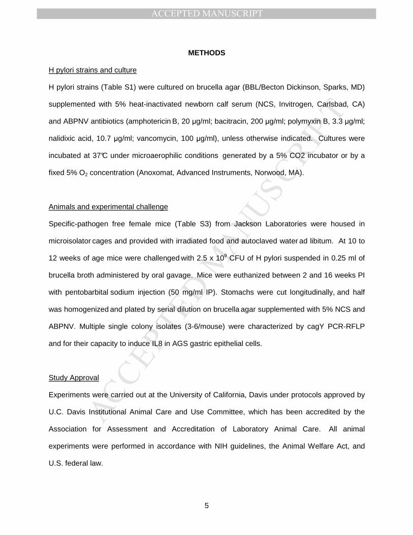
METHODS
H pylori strains and culture
H pylori strains (Table S1) were cultured on brucella agar (BBL/Becton Dickinson, Sparks, MD)
supplemented with 5% heat-inactivated newborn calf serum (NCS, Invitrogen, Carlsbad, CA)
and ABPNV antibiotics (amphotericin B, 20 µg/ml; bacitracin, 200 µg/ml; polymyxin B, 3.3 µg/ml;
nalidixic acid, 10.7 µg/ml; vancomycin, 100 µg/ml), unless otherwise indicated. Cultures were
incubated at 37°C under microaerophilic conditions generated by a 5% CO2 incubator or by a
fixed 5% O2 concentration (Anoxomat, Advanced Instruments, Norwood, MA).
Animals and experimental challenge
Specific-pathogen free female mice (Table S3) from Jackson Laboratories were housed in
microisolator cages and provided with irradiated food and autoclaved water ad libitum. At 10 to
12 weeks of age mice were challenged with 2.5 x 10⁹ CFU of H pylori suspended in 0.25 ml of
brucella broth administered by oral gavage. Mice were euthanized between 2 and 16 weeks PI
with pentobarbital sodium injection (50 mg/ml IP). Stomachs were cut longitudinally, and half
was homogenized and plated by serial dilution on brucella agar supplemented with 5% NCS and
ABPNV. Multiple single colony isolates (3-6/mouse) were characterized by cagY PCR-RFLP
and for their capacity to induce IL8 in AGS gastric epithelial cells.
Study Approval
Experiments were carried out at the University of California, Davis under protocols approved by
U.C. Davis Institutional Animal Care and Use Committee, which has been accredited by the
Association for Assessment and Accreditation of Laboratory Animal Care. All animal
experiments were performed in accordance with NIH guidelines, the Animal Welfare Act, and
U.S. federal law.

MANUSCRIP
T
ACCEPTED
ACCEPTED MANUSCRIPT
6
IL8 ELISA
H pylori induction of IL8 was measured as described previously 12. WT H pylori PMSS1, its
isogenic cagY deletion, and brucella broth were included on every plate as positive and
negative controls. IL8 values were normalized to WT H pylori, arbitrarily set to 1.0.
Adoptive Transfer
Rag1-/-mice were reconstituted with 1x10⁶ CD4+ T cells isolated from WT or Ifnγ-/- C57BL/6
mice, which had been infected for 8 weeks with PMSS1. Mice were euthanized with
pentobarbital sodium injection (50 mg/ml IP) and single cell suspensions were obtained by
passing spleens through a 40µm cell mesh into PBS. Cells were pelleted by centrifugation and
erythrocytes lysed for 2 min at room temperature with AKC buffer (0.15M NH4Cl, 10mM KHCO3,
0.1mM EDTA, pH7.35). Cells were washed with PBS, pooled, and CD4+ T cells were isolated
by using anti-CD4+ magnetic beads (Miltenyi, San Diego, CA) over a magnetic column,
resulting in > 90% purity of CD3+CD4+ cells demonstrated by flow cytometry. CD4+ T cells
were resuspended in PBS at 5x10⁶ cells/ml per 200µl and injected into uninfected Rag1-/- mice
via tail vein.
Flow Cytometry
Following RBC lysis, splenocytes were resuspended and washed once in FACS buffer (1XPBS,
0.5% BSA). Cells were stained with anti-CD3 FITC (clone 17A2, BD Biosciences) and anti-CD4
PE (clone RM4-5, BD Biosciences) in FACS stain buffer (FACS buffer, 20% mouse serum) for
15 minutes at room temperature. Cells were washed two times in FACS buffer and fixed with
2% PFA. Data were collected on a BD FACSCalibur and analyzed using FlowJo software 8.8.7
(Treestar).

MANUSCRIP
T
ACCEPTED
ACCEPTED MANUSCRIPT
7
Immunoblots and CagA translocation
Immunoblots of CagA and CagA translocation were performed as described previously using an
MOI of 100:1 and 22 hours of culture at 37°C in 5% CO2 8. CagY expression was detected by
electrophoresis of sonicated bacterial proteins on a 7.5% polyacrylamide gel, incubating with
rabbit antiserum (1:10,000) to CagY 7 as primary antibody and HRP-conjugated anti-rabbit IgG
(1:20,000) as secondary antibody.
cagY PCR-RFLP
cagY genotyping was performed by polymerase chain reaction-restriction fragment length
polymorphism (PCR-RFLP) using primers in Table S4 as previously described 8. Changes in
PCR-RFLP patterns are referred to as recombination based on DNA sequence analysis from
prior experiments 8, although this was not formally demonstrated.
Contraselection for genetic exchange of cagY
Alleles of cagY were exchanged between H pylori strains using contraselectable streptomycin
susceptibility as described previously 8 using primers in Table S4 and plasmid constructs in
Table S2.
Competition experiment
cagY was deleted from PMSS1 and replaced with either wild type PMSS1 cagY
(PMSS1∆cagY[PMSS1]) or SS1 cagY (PMSS1∆cagY[SS1]) using contraselection. Strains
expressing cagY from PMSS1 or SS1 were also marked by replacing bases 343-360 of the rdxA
locus with an antibiotic resistance gene encoding either kanamycin or chloramphenicol
resistance, respectively. Briefly, plasmid pJ318 (Table S2) was constructed in pBluescript SK-
by amplifying fragments 1194 bp upstream and 904 bp downstream of the rdxA deletion site in
PMSS1, and ligating to a kanamycin resistance cassette 13. pJ318 was then used to naturally
MANUSCRIP
T
ACCEPTED
ACCEPTED MANUSCRIPT
8
transform PMSS1∆cagY[PMSS1] with selection on 25 µg/ml kanamycin. pJ319 (Table S2),
which was created in the same fashion but with a chloramphenicol resistance gene, was
similarly used to naturally transform PMSS1∆cagY[SS1], with selection on 5 µg/ml
chloramphenicol. Gastric tissue from mice challenged with a 1:1 mixture of the marked
PMSS1∆cagY[PMSS1] and PMSS1∆cagY[SS1] strains was plated separately on brucella agar
with ABPNV plus either kanamycin or chloramphenicol to enumerate the relative abundance of
each strain. This was expressed as a log10 competition index calculated as CFU of
PMSS1∆cagY [SS1]/[PMSS1], normalized to the abundance of each strain in the input inoculum.
Histology Scoring
Sections of the glandular stomach were formalin fixed and stained with hematoxylin and eosin.
Each microscopic field was scored separately for the presence or absence of neutrophilic
infiltration (polymorphonuclear leukocytes), gastritis, and epithelial metaplasia using a system
previously validated in mice 14. The results were reported as the mean percentage of fields
affected for each mouse averaged over the three histologic criteria (minimum10 fields/sample).
Statistics
H pylori colonization (CFU/g) was analyzed using a 2-tailed Student’s t test (Prism 6.0) after
log10 transformation. Normalized IL8 levels were compared between groups using Wilcoxon
rank-sum tests. Analysis of gastritis and proportions of samples with changed cagY were
compared between groups using chi-square tests. In experiments with more than two groups,
logistic regression was used to evaluate pairwise differences in proportion of output strains with
recombination in cagY. A P value ≤ 0.05 was considered statistically significant.

MANUSCRIP
T
ACCEPTED
ACCEPTED MANUSCRIPT
9
RESULTS
CD4+ T cells are required for in vivo selection of cagY recombination and loss of T4SS
function
H pylori SS1 is a mouse-passaged strain that was proposed as the standard for H pylori
studies in mice 15, and was later found to have a defective T4SS that we showed was a result of
cagY recombination 8. PMSS1, which has a functional T4SS and readily colonizes mice, is the
original H pylori human isolate that gave rise to the SS1 strain after serial passage mice 16.
Unlike in wild type (WT) mice, PMSS1 does not undergo cagY recombination or lose T4SS
function when recovered from Rag1-/- mice 8, which do not have functional B or T cells. To
identify which arm of the adaptive immune response is responsible for loss of T4SS function,
knockout (KO) mice lacking functional B cells (µMT) or T cells (TCR β/δ-/-) were challenged with
PMSS1 and sacrificed 8 weeks post infection (PI). WT and Rag1-/- mice were challenged
simultaneously as controls. The H pylori bacterial burden in Rag1-/- and T cell KO mice was
approximately 10-fold higher than in WT mice and in B cell KO mice (Figure 1A). Loss of T4SS
function (reduced IL8 induction) and recombination in cagY (defined as a change in PCR-RFLP)
occurred commonly during infection of WT and B cell KO mice, but never in RAG-/- mice and
only occasionally in T cell KO mice (Figure 1B, C).
Since H pylori infection in mice primarily triggers a CD4+ Th1 immune response 10, 11, we
also asked if CD4+ T cells alone could select for H pylori with cagY alleles that encode a non-
functional T4SS that is no longer capable of inducing IL8. To examine this possibility, we
performed adoptive transfer experiments in which CD4+ T cells were isolated from WT mice that
were infected with H pylori for 8 weeks, and then transferred into Rag1-/- mice (Rag1-/-WT CD4+)
24 hours before H pylori challenge. Flow cytometry on splenocytes from adoptively transferred
mice demonstrated engraftment, with a mean of 8.9% (± 1.1% SEM) CD3+CD4+ cells in the
lymphocyte gate. The bacterial burden 8 weeks PI was significantly lower in Rag1-/-WT CD4+ mice
than in Rag1-/- mice (Figure 1A). It was also lower than in WT mice, which has been observed

MANUSCRIP
T
ACCEPTED
ACCEPTED MANUSCRIPT
10
previously and likely reflects a relative failure of Treg engraftment 17. Loss of T4SS function
(Figure 1B) and recombination in cagY (Figure 1C) were also more common in Rag1-/-WT CD4+
mice than in RAG-/- mice, though the difference in IL8 induction did not reach statistical
significance. Together, these data suggest that CD4+ T cells are essential for control of
bacterial burden and for selection of H pylori with CagY variants that form a non-functional
T4SS.
Selection of cagY variants and loss of T4SS function requires IFNγ signaling
The development of gastritis and control of H pylori bacterial burden are mediated by
CD4+ T cells 18, which also drive CagY-mediated loss of T4SS function (Figure 1). Since CD4+
T cells are a major source of IFNγ, we next asked if loss of T4SS function is mediated
downstream of IFNγ. WT mice and mice lacking the IFNγ receptor (IfnγR-/-) were infected with
H pylori PMSS1 and sacrificed 4 or 8 weeks PI. Similar to the bacterial burden in Rag1-/- mice,
IfnγR-/- mice were colonized at approximately 10-fold higher levels than WT mice (Figure 2A).
H pylori isolated from WT mice 4 and 8 weeks PI showed gradual loss of T4SS function
associated with recombination in cagY. In contrast, H pylori from IfnγR-/- mice retained T4SS
function (Figure 2B) and showed no cagY recombination (Figure 2C). To determine if IFNγ from
CD4+ T cells alone is sufficient for selection of cagY variants and loss of T4SS function, we
performed adoptive transfer. CD4+ T cells from Ifnγ-/- mice infected with PMSS1 for 8 weeks
were adoptively transferred into Rag1-/-mice (Rag1-/- Ifnγ-/- CD4+), which were then infected with H
pylori PMSS1 and sacrificed 8 weeks PI. Flow cytometry on splenocytes from adoptively
transferred mice demonstrated engraftment, with a mean of 9.3% (± 0.7% SEM) CD3+CD4+
cells in the lymphocyte gate. Adoptive transfer of Ifnγ-/- CD4+ T cells was sufficient to control
bacterial load (Figure 2D), but did not select H pylori variants with loss of T4SS function (Figure
2E) or recombination in cagY (Figure 2F). These results indicate that signaling downstream of

MANUSCRIP
T
ACCEPTED
ACCEPTED MANUSCRIPT
11
IFNγ derived from CD4+ T cells is essential for CagY-mediated loss of T4SS function, but is not
strictly required for control of H pylori bacterial load.
Variation in cagY functions as a molecular rheostat to alter the inflammatory capacity of
H pylori
Most H pylori strains that recombined cagY showed a markedly reduced capacity to
induce IL8, though we occasionally identified strains with changes in cagY but intermediate
levels of IL8 induction (e.g., WT infected mice 8 weeks PI, Figure 2B). This suggests that
recombination in cagY may function to modulate T4SS function rather than eliminate it. In other
words, cagY may function more like a rheostat than a switch. To test this hypothesis, we first
identified mouse output strains with unique cagY RFLP patterns (Figure 3A) that reproducibly
showed high (Out1, cagY PCR-RFLP equivalent to WT PMSS1), intermediate (Out2), or low
(Out3) induction of IL8 (Figure 3B, grey bars). We next used contraselection 8 to replace the
cagY gene in PMSS1 with that from each output, which was confirmed by PCR-RFLP (Figure
3A). Transformants complemented with cagY from output strains (∆Y[Out1], ∆Y[Out2],
∆Y[Out3]) restored the capacity to induce IL8 (Figure 3B, white bars) and translocate CagA
(Figure 3C) to levels similar to that of the respective output strain. These results suggest that
different cagY alleles vary in the extent to which they enable the bacterial cell to induce IL8 and
translocate CagA, and that recombination in cagY functions as a molecular rheostat to modulate
H pylori T4SS function.
cagY recombination and loss of T4SS function are strain-dependent and associated with
the capacity of H. pylori to induce inflammation
H pylori strains encoding a T4SS can differ markedly in their capacity to induce IL8,
despite having an intact cagPAI 19. Since loss of T4SS function and cagY recombination are
immune driven, we hypothesized that the capacity of H pylori to induce IL8 would be inversely
MANUSCRIP
T
ACCEPTED
ACCEPTED MANUSCRIPT
12
related to cagY recombination and loss of T4SS function during in vivo infection. To test this
hypothesis, we first compared the in vitro response of AGS gastric epithelial cells to H pylori
strains J166 and PMSS1, which show relatively low and high induction of IL8, respectively
(Figure 4A). Mice infected with PMSS1 had significantly more inflammation in the gastric
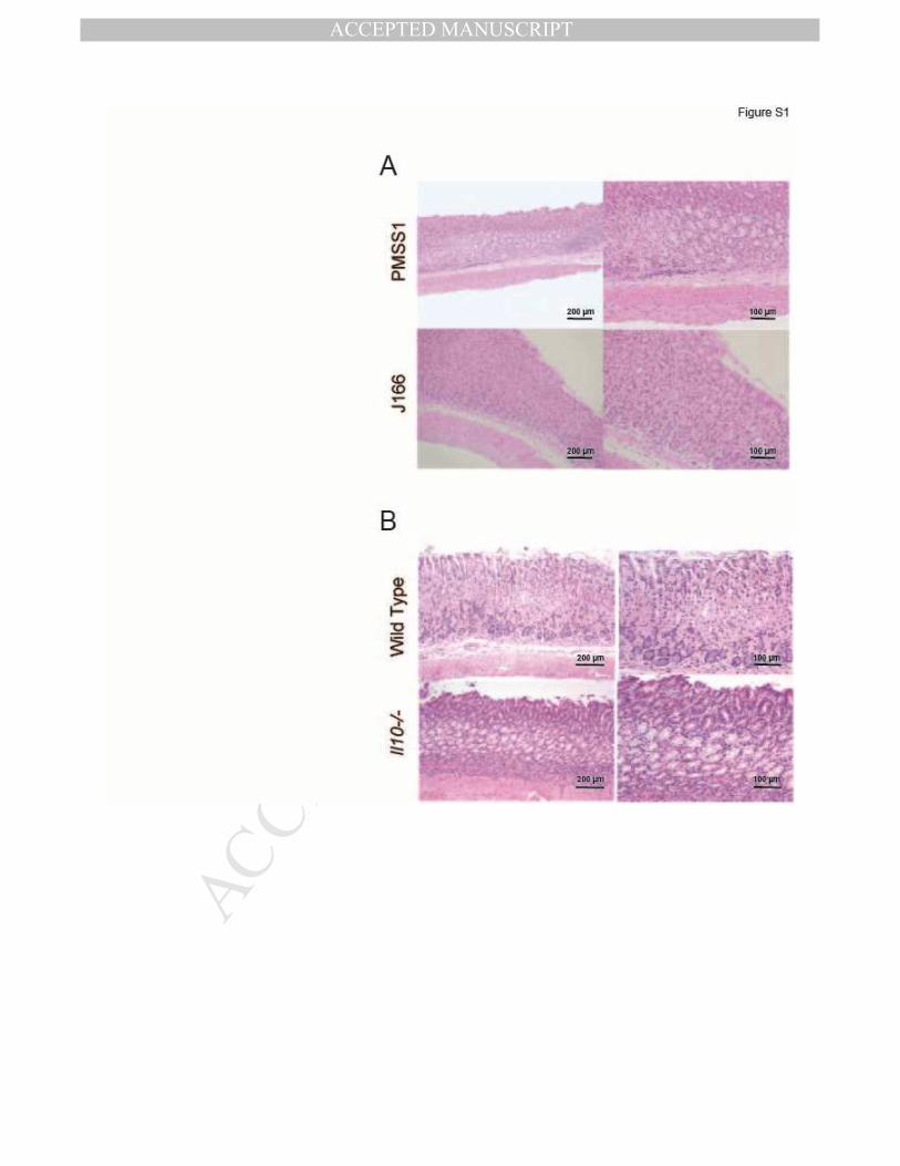
mucosa compared to J166 (Figure 4B, Figure S1), though bacterial loads were similar (data not
shown). Consistent with the greater capacity of H pylori PMSS1 to induce IL8 in vitro, and
induce inflammation in vivo, PMSS1 infected mice also showed more rapid and more complete
loss of T4SS function that was associated with cagY recombination (Figure 4C,D).
To examine this more systematically using isogenic strains, we next infected WT
C57BL/6 mice with PMSS1 bearing the cagY from Out1 or Out3, which have high and low T4SS
function (Figure 3), respectively, and sacrificed them 8 weeks PI. Colonization density was
significantly greater in mice challenged with ∆Y[Out3], which has poor T4SS function, compared
to ∆Y[Out1] (Figure 4E). Recombination in cagY occurred only in output colonies from mice
infected with ∆Y[Out1] (Figure 4F), though the frequency in this experiment was lower than
observed previously. Similarly, complete elimination of T4SS function by deletion of cagE 16,
which encodes an ATPase that is essential for T4SS function, increased bacterial load and
eliminated recombination in cagY 8 weeks PI (Figure S2). These data suggest that control of
bacterial load and selection of H pylori with a nonfunctional T4SS are enhanced in H pylori
strains that induce a more robust host immune response.
Competitive advantage of CagY-mediated loss of T4SS function increases progressively
during H pylori infection
Recombination in cagY and loss of T4SS function increase over time during infection of
WT mice, beginning around 4 weeks PI. Since loss of T4SS function is immune-mediated, this
may simply reflect the time required for development of adaptive immunity. On the other hand,
we previously reported that early during infection of rhesus macaques we could detect cagY-

MANUSCRIP
T
ACCEPTED
ACCEPTED MANUSCRIPT
13
mediated gain of T4SS function 8, suggesting the possibility that there may be selection for a
functional T4SS very early during infection. To address this question, we used contraselection
to construct isogenic strains of PMSS1 bearing either the WT cagY (PMSS1∆cagY[PMSS1]) or
the non-functional cagY from SS1 (PMSS1∆cagY[SS1]), which were marked respectively in the
neutral rdxA locus with antibiotic resistance to kanamycin or chloramphenicol. We then
performed a competition experiment in which WT C57BL/6 mice were inoculated with a 1:1
mixture of both strains and sacrificed between 1 and 8 weeks PI. Gastric contents were plated
on kanamycin and chloramphenicol to permit calculation of a competition index. The results
demonstrated progressive selection for loss of T4SS function beginning 4 weeks PI, with > 300-
fold competitive advantage by 8 weeks PI (Figure 5A). As expected, competition index showed
a strong inverse correlation (R2=0.64, P≤0.0001) with IL8 induction performed on colony sweeps
from each mouse (Figure 5B), which confirms that loss of T4SS function was due to selection
for PMSS1∆cagY[SS1], and not to a mutation in PMSS1∆cagY[PMSS1]. These results are
consistent with progressive loss of T4SS function that results from development of adaptive
immunity, with no fitness advantage to a functional T4SS early during infection in mice.
CagY-mediated loss of T4SS function promotes bacterial persistence in the setting of
increased inflammation
CagY-mediated loss of T4SS function occurs less commonly in mice with impaired
immunity (Figures 1, 2). This suggests that mice with an enhanced immune response might
have a greater selection for H pylori strains with loss of T4SS function, which might be a
bacterial strategy to persist in the face of inflammation. To test this hypothesis, we infected
Il10-/- mice with H pylori, which triggers a robust inflammatory response with severe gastritis
20,.21 and increased levels of IFNγ and other Th1 cytokines 22 compared to WT mice. Because
we anticipated that the aggressive inflammatory response would clear the infection at later time
points, mice were sacrificed at 2 and 4 weeks PI, rather than 8 weeks PI as usual. Il10-/- mice
MANUSCRIP
T
ACCEPTED
ACCEPTED MANUSCRIPT
14
infected with H pylori PMSS1 showed more inflammation (Figure 6A) and a lower bacterial
burden (Figure 6B) compared to WT mice. Colonization in Il10-/- mice was significantly lower
than in WT—often near the limit of detection (~100 CFU/g) 4 weeks PI (Figure 6B)—and was
undetectable in 3 mice. At 4 weeks PI, recombination in cagY occurred in 22 of 82 colonies
recovered from Il10-/- mice but only 10 of 75 colonies from WT mice (chi-square=4.23, P≤0.05,
Figure 6C). All colonies recovered 4 weeks PI from the 3 mice that showed the highest
colonization levels (similar to WT mice), also induced a low level of IL8 and showed
recombination in cagY (Figure 6B,C; bracketed data points). Moreover, the average IL8
induction of H pylori isolates from Il10-/- mice showed a highly significant inverse correlation
with the bacterial burden (Figure 6D). These data suggest that CagY-mediated loss of T4SS
function allows for increased H pylori colonization in the face of a robust immune response.
Recombination in cagY modulates T4SS function during chronic infection in humans
T4SS function (IL8 induction) is highly variable among H pylori clinical isolates, even when the
cagPAI is fully intact 19. The explanation for this is unknown, but it is intriguing that cagY is
under strong diversifying selective pressure—second only to cagA among genes on the cagPAI
19. To examine the possible role of cagY recombination in modulating T4SS function during
chronic infection of humans, we examined paired isolates that were previously collected from 14
patients over intervals ranging from 3.0 to 10.2 yrs (mean=6.1 yrs). Multilocus sequencing
typing analysis demonstrated that each pair was clonal, but showed microevolution during
prolonged infection 23. Of the 14 pairs, one showed changes in cagY PCR-RFLP (Figure 7A)
together with a significant decrease in the capacity to induce IL8 between the A and B isolates,
which were collected 7.4 years apart (Figure 7B). Deletion of cagY completely eliminated IL8
induction in both the A and B isolates (Figure 7B), which demonstrated that the T4SS was intact
in both. To determine if recombination in cagY was responsible for the change in T4SS function,
we used contraselection to exchange cagY genes between the A and B isolates, and confirmed

MANUSCRIP
T
ACCEPTED
ACCEPTED MANUSCRIPT
15
it by PCR RFLP. Exchange of cagY genes demonstrated that change in cagY was sufficient to
explain the differences in IL8 induction of the A and B isolates (Figure 7B). Control experiments
in which cagY was deleted from the A and B strains, and then reinserted by contraselection,
recovered the IL8 induction of the parent strain (data not shown). These results demonstrate
that recombination in cagY during chronic human infection can modulate T4SS function.
MANUSCRIP
T
ACCEPTED
ACCEPTED MANUSCRIPT
16
DISCUSSION
The T4SS system encoded on the cagPAI is the key bacterial virulence factor
associated with progression to peptic ulcer disease or gastric cancer, rather than asymptomatic
gastritis. Analysis of the PAI in vivo has been hampered by the observation that T4SS function
is lost during experimental infection of mice 9, which was initially viewed as an artifact of
infecting mice with a bacterium that is naturally found only in humans and some non-human
primates. The mechanism was unknown. We recently demonstrated that loss of T4SS in mice
is typically due to in-frame recombination in the middle repeat region of the cagY gene, which
encodes an essential component of the H pylori T4SS 4. Loss of T4SS does not occur in RAG-/-
mice, which lack functional B or T cells 8. While indels or SNPs in any of the essential genes
can result in loss of T4SS function, cagY seems specifically designed for recombinatorial
variation, suggesting that it is a bacterial contingency locus 24 that modulates or “tunes” the host
inflammatory response.
Here we have further investigated the immunologic basis and functional significance for
loss of T4SS function during H pylori infection of mice and humans. Previous investigators
speculated that recombination in cagY was a form of antigenic variation to avoid antibody
responses directed against a surface-exposed component of the H pylori T4SS pilus 7.
However, this was difficult to reconcile with the general lack of human antibody response to
CagY 7 and the evidence that humoral immunity is generally not thought to play an important
role in control of H pylori infection 25. Our results suggest that loss of T4SS function and
recombination in cagY are largely independent of B cells, but instead require CD4+ T cells
expressing IFNγ (Figures 1, 2). The modest loss of T4SS function we observed in B cell KO
mice (Figure 1B) may actually reflect a decrease in B cell-mediated immunoregulation 26, rather
than B cell control of infection. Together, these observations are consistent with seminal vaccine
studies demonstrating that MHC class II-restricted, Th1-polarized T cells are essential to control
H pylori infection 27, 28.

MANUSCRIP
T
ACCEPTED
ACCEPTED MANUSCRIPT
17
Several lines of evidence suggest that the variation in cagY and T4SS function that we
have observed in animal models is relevant to human H pylori infection. First, it is not simply an
artifact of the mouse model, because cagY mediated loss of T4SS function also occurs in
rhesus macaques 8, which most closely mimic human infection. Second, cagY recombination
can both up- and down-modulate T4SS function 8, in a graded fashion (Figure 3), suggesting
that this observation is a window into the biology of H pylori, which actually has the capacity to
“tune” or optimize the host inflammatory response to achieve a homeostatic balance. Third,
there is marked variability in the capacity of different PAI positive H pylori strains to induce IL8,
varying by up to 20-fold 19. Since cagY is second only to cagA in the percentage of codons
under positive selection 19, CagY diversity may be important for adaptation to chronic human
infection. Most importantly, here we demonstrate that cagY recombination within an individual
patient can modulate T4SS function (Figure 7). The cagY variants in the sequential isolates
might represent the dominant population present at each time point, which underwent
recombination and functional change under pressure from changes in, for example, host
physiology. Alternatively, we cannot exclude the possibility that the cagY variants we observed
represent diversity that was present at each time point, because only a single A and B isolate
were examined. Regardless of which scenario is correct, these results provide proof-of-
principle that cagY recombination can modulate T4SS function during H pylori infection in
humans. H pylori adaptation during acute and chronic human infection has also been
demonstrated at other virulence loci, such as the babA adhesion 29, 30, and more broadly using
whole genome sequencing 31.
When might cagY recombination occur during human infection, and why? Clearly, a
functional T4SS enhances bacterial fitness, probably by increasing its capacity to acquire iron
and other nutrients 32-34. But rearrangement in cagY that confers loss of T4SS function may be
more advantageous under conditions that are unfavorable for H pylori growth, because loss of
T4SS function decreases the host inflammatory response, increases bacterial load 16, 35, and

MANUSCRIP
T
ACCEPTED
ACCEPTED MANUSCRIPT
18
thus increases the likelihood of transmission to a new host. It may occur soon after acquisition,
as in mice and monkeys, or perhaps during some environmental event, which from the bacterial
perspective tips the balance for or against inflammation. If so, it may be difficult to “catch it in
the act” because acute H pylori infection is rarely detected in humans and the hypothetical
environmental events are unknown. One possibility is that cagY-mediated down regulation of
T4SS function is a bacterial strategy to persist in the setting of an intercurrent infectious disease
such as malaria, tuberculosis or typhoid—which, along with H pylori, have evolved with humans
since antiquity and might cause sufficient systemic inflammation that would otherwise reduce H
pylori colonization and perhaps even clear the infection. A similar phenomenon has been
observed experimentally in the mouse model of herpes viruses, where viral infection can non-
specifically protect against bacterial challenge with Listeria or Yersinia pestis by inducing IFNγ
and systemic activation of macrophages 36. This hypothesis is also supported by the results in
Il10-/- mice, which have an exaggerated inflammatory response to H pylori and demonstrate a
strong inverse correlation between bacterial load and cagY-mediated loss of T4SS function
(Figure 6). Alternatively, perhaps cagY-mediated loss of T4SS enhances bacterial persistence
in the setting of atrophic gastritis, in which elevated gastric pH reduces H pylori burden and
sometimes leads to bacterial clearance 37. Both are testable hypotheses that we are currently
examining.

MANUSCRIP
T
ACCEPTED
ACCEPTED MANUSCRIPT
19
FIGURE LEGENDS
Figure 1. CD4+ T cells are required to control H pylori colonization density and select
strains with loss of T4SS function and recombination in cagY.
(A) H pylori colonization density was significantly greater in Rag1-/- and T cell KO mice than in
wild type mice. Adoptive transfer of WT CD4+ T cells into Rag1-/- mice markedly reduced H
pylori colonization compared to Rag1-/-. Each data point represents CFU/g for an individual
mouse 8 weeks PI (N=7-8 mice/group). Horizontal lines indicate mean ± standard error of the
mean (SEM). (B) Single colonies recovered from WT and B cell KO mice, and mice adoptively
transferred with WT CD4+ T cells, showed marked loss in the capacity to induce IL8 that was
accompanied by recombination in cagY (open circles). In contrast, all colonies from Rag1-/-
mice and most from T cell KO mice induced IL8 and had the same cagY RFLP (closed circles)
as WT H pylori PMSS1. Each data point represents the result from a single colony (N=3-6
colonies/mouse). (C) Percent of colonies that underwent cagY recombination (open circles
divided by total colonies for each group in panel B). *P≤0.05, **P≤0.01, ***P≤0.001.
Figure 2. Selection of CagY variants is mediated downstream of IFN-γ signaling.
(A) H pylori colonization density was significantly higher in IfnγR-/- mice compared to WT at
both 4 and 8 weeks PI. Each data point represents CFU/g from an individual mouse
(N=6/group). (B) Single colonies (N=3-6/mouse) recovered from WT mice showed loss in the
capacity to induce IL8 that was associated with recombination in cagY (open circles), but
colonies from IfnγR-/- mice induced IL8 similarly to WT PMSS1 and had no changes in cagY
(closed circles). (C) Percent of colonies that underwent cagY recombination (open circles
divided by total colonies for each group in panel B). Adoptive transfer of Ifnγ-/- CD4+ T cells
into Rag1-/- mice was sufficient to control bacterial load 8 weeks PI (D), but did not select H

MANUSCRIP
T
ACCEPTED
ACCEPTED MANUSCRIPT
20
pylori variants with loss of IL8 induction or change in cagY PCR-RFLP (E,F). Horizontal lines
indicate mean ± SEM. *P≤0.05, **P≤0.01, ***P≤0.001, ****P≤0.0001
Figure 3. CagY is a molecular rheostat that alters the inflammatory capacity of H pylori.
Three single colonies recovered from WT mice infected with PMSS1 (Out1, with cagY PCR-
RFLP equivalent to wild type cagY from PMSS1; Out2; Out3) had unique cagY PCR-RFPL
patterns (A), and induced high, intermediate, or low IL8, respectively (B) (gray bars) compared
to PMSS1 and its cagY deletion mutant (black bars). Complementation of ∆cagY with Out1 (∆Y
[Out1]), Out2 (∆Y [Out2]), or Out3 (∆Y [Out3]) phenocopied the IL8 induction of the respective
output strain (white bars). Data represent mean ± SEM of four replicates. *P≤0.05. (C) Out1,
Out2 and Out3 also demonstrated decreasing translocation of phosphorylated CagA (α-PY99),
which was phenocopied when PMSS1∆cagY was complemented with the respective cagY gene.
Differences in CagY (α-CagY) were also apparent by immunoblot. Arrowheads in panel A
indicate unique bands.
Figure 4. Kinetics of cagY recombination and loss of T4SS function are associated with
the capacity of H. pylori to induce inflammation.
(A) Replicate IL8 assays (N=10) for WT H pylori strains PMSS1 and J166. Data for both strains
are normalized to PMSS1. (B) Mice infected with H pylori PMSS1 showed increased
inflammation in gastric tissue compared to J166, which was statistically significant at 8 and 16
weeks PI (B). (C) Colonies recovered from PMSS1-infected WT mice lost the capacity to induce
IL8 and changed cagY (open symbols) more rapidly and more completely than colonies
recovered from J166-infected mice. Data for each strain are normalized to their respective WT.
(D) Percent of colonies that underwent cagY recombination (open circles divided by total
colonies for each group in panel C. H pylori recovered 8 weeks after challenge of WT mice
with ∆Y[Out1], which induces high IL8, were at a lower bacterial density (E) and underwent

MANUSCRIP
T
ACCEPTED
ACCEPTED MANUSCRIPT
21
cagY recombination more frequently (F) than H pylori from mice colonized with ∆Y[Out3]. Bars
represent mean ± SEM. **P≤0.01, ***P≤0.001, ****P≤0.0001.
Figure 5. Competitive advantage of CagY-mediated loss of T4SS function increases
progressively over time.
(A) Output colonies from mice infected with an equal mixture of isogenic H pylori PMSS1
strains bearing either the functional (PMSS1) or non-functional (SS1) cagY allele were
enumerated by selective plating, and used to calculate the log10 competition index. Each data
point represents a single mouse; horizontal lines=geometric mean. At early time points there
was no selective advantage, but by 8 weeks PI the PMSS1 strain bearing the SS1 cagY was
present at > 300-fold greater abundance. *P≤0.05, **P≤0.01. (B) Normalized IL8 induction of a
sweep culture from each mouse showed a strong inverse correlation with log10 competition
index.
Figure 6. CagY-mediated loss of T4SS function promotes bacterial persistence during an
intense inflammatory response.
(A) Il10-/- mice inoculated with H pylori PMSS1 showed significantly increased gastritis
compared to WT mice 4 weeks PI. (B) H pylori bacterial burden was significantly higher in WT
compared to Il10-/- mice. By 4 weeks PI, bacterial burden in Il10-/- mice was frequently near
the level of detection and 3 mice were uninfected (not shown). Mice whose CFU are shown in
brackets yielded the colonies whose IL8 induction is shown in brackets in panel C. (C) Loss of
the capacity to induce IL8 associated with changes in cagY PCR RFLP (open circles) was more
apparent in H pylori colonies recovered from Il10-/- compared to WT mice, particularly in
colonies from mice that showed colonization density that resembled that in WT mice. All
colonies whose IL8 induction is shown in brackets in panel C were recovered from the mice
whose CFU are bracketed in panel B. (D) Average normalized IL8 induction of all colonies from

MANUSCRIP
T
ACCEPTED
ACCEPTED MANUSCRIPT
22
each mouse showed a strong inverse correlation with bacterial burden (R2=0.78, P≤0.0001).
**P≤0.01,***P≤0.001.
Figure 7. Recombination in cagY modulates T4SS function during chronic infection in
humans.
(A) cagY PCR-RFLP analysis of sequential A and B H pylori isolates. Arrowheads denote
bands that changed in isolate A and B, which were collected from the same patient 7.4 years
apart. (B) IL8 induction normalized to strain PMSS1 for sequential H pylori isolates A and B,
their cagY knockouts (∆), and strains in which their cagY genes have been exchanged.
***P≤0.001,****P≤0.0001.

MANUSCRIP
T
ACCEPTED
ACCEPTED MANUSCRIPT
23
REFERENCES
1. Wroblewski LE, Peek RM, Jr., Wilson KT. Helicobacter pylori and gastric cancer: factors
that modulate disease risk. Clin Microbiol Rev 2010;23:713-39.
2. Odenbreit S, Püls J, Sedlmaier B, et al. Translocation of Helicobacter pylori CagA into
gastric epithelial cells by type IV secretion. Science 2000;287:1497-500.
3. Segal ED, Lange C, Covacci A, et al. Induction of host signal transduction pathways by
Helicobacter pylori. Proc Natl Acad Sci U S A 1997;94:7595-9.
4. Fischer W, Püls J, Buhrdorf R, et al. Systematic mutagenesis of the Helicobacter pylori
cag pathogenicity island: essential genes for CagA translocation in host cells and
induction of interleukin-8. Mol Microbiol 2001;42:1337-1348.
5. Kutter S, Buhrdorf R, Haas J, et al. Protein subassemblies of the Helicobacter pylori Cag
type IV secretion system revealed by localization and interaction studies. J Bacteriol
2008;190:2161-71.
6. Frick-Cheng AE, Pyburn TM, Voss BJ, et al. Molecular and Structural Analysis of the
Helicobacter pylori cag Type IV Secretion System Core Complex. MBio 2016;7.
7. Aras RA, Fischer W, Perez-Perez GI, et al. Plasticity of repetitive DNA sequences within
a bacterial (Type IV) secretion system component. J Exp Med 2003;198:1349-60.
8. Barrozo RM, Cooke CL, Hansen LM, et al. Functional plasticity in the type IV secretion
system of Helicobacter pylori. PLoS Pathog 2013;9:e1003189.
9. Philpott DJ, Belaid D, Troubadour P, et al. Reduced activation of inflammatory
responses in host cells by mouse-adapted Helicobacter pylori isolates. Cell Microbiol
2002;4:285-96.
10. Bamford KB, Fan X, Crowe SE, et al. Lymphocytes in the human gastric mucosa during
Helicobacter pylori have a T helper cell 1 phenotype. Gastroenterol 1998;114:482-92.

MANUSCRIP
T
ACCEPTED
ACCEPTED MANUSCRIPT
24
11. Lundgren A, Trollmo C, Edebo A, et al. Helicobacter pylori-specific CD4+ T cells home to
and accumulate in the human Helicobacter pylori-infected gastric mucosa. Infect Immun
2005;73:5612-9.
12. Israel DA, Salama N, Arnold CN, et al. Helicobacter pylori strain-specific differences in
genetic content, identified by microarray, influence host inflammatory responses. J Clin
Invest 2001;107:611-20.
13. Menard R, Sansonetti PJ, Parsot C. Nonpolar mutagenesis of the ipa genes defines
IpaB, IpaC, and IpaD as effectors of Shigella flexneri entry into epithelial cells. J
Bacteriol 1993;175:5899-906.
14. Eaton KA, Danon SJ, Krakowka S, et al. A reproducible scoring system for quantification
of histologic lesions of inflammatory disease in mouse gastric epithelium. Comp Med
2007;57:57-65.
15. Lee A, O'Rourke J, De Ungria MC, et al. A standardized mouse model of Helicobacter
pylori infection: introducing the Sydney strain. Gastroenterol 1997;112:1386-97.
16. Arnold IC, Lee JY, Amieva MR, et al. Tolerance rather than immunity protects from
Helicobacter pylori-induced gastric preneoplasia. Gastroenterol 2010;140:199-209.
17. Gray BM, Fontaine CA, Poe SA, et al. Complex T cell interactions contribute to
Helicobacter pylori gastritis in mice. Infect Immun 2013;81:740-52.
18. Sayi A, Kohler E, Hitzler I, et al. The CD4+ T cell-mediated IFN-gamma response to
Helicobacter infection is essential for clearance and determines gastric cancer risk. J
Immunol 2009;182:7085-101.
19. Olbermann P, Josenhans C, Moodley Y, et al. A global overview of the genetic and
functional diversity in the Helicobacter pylori cag pathogenicity island. PLoS Genet
2010;6:e1001069.

MANUSCRIP
T
ACCEPTED
ACCEPTED MANUSCRIPT
25
20. Chen W, Shu D, Chadwick VS. Helicobacter pylori infection: mechanism of colonization
and functional dyspepsia Reduced colonization of gastric mucosa by Helicobacter pylori
in mice deficient in interleukin-10. J Gastroenterol Hepatol 2001;16:377-83.
21. Ismail HF, Fick P, Zhang J, et al. Depletion of neutrophils in IL10(-/-) mice delays
clearance of gastric Helicobacter infection and decreases the Th1 immune response to
Helicobacter. J Immunol 2003;170:3782-9.
22. Lee CW, Rao VP, Rogers AB, et al. Wild-type and interleukin-10-deficient regulatory T
cells reduce effector T-cell-mediated gastroduodenitis in RAG2-/- mice, but only wild-
type regulatory T cells suppress Helicobacter pylori gastritis. Infect Immun
2007;75:2699-707.
23. Morelli G, Didelot X, Kusecek B, et al. Microevolution of Helicobacter pylori during
prolonged infection of single hosts and within families. PLoS Genet 2010;6:e1001036.
24. Moxon R, Bayliss C, Hood D. Bacterial contingency loci: the role of simple sequence
DNA repeats in bacterial adaptation. Annu Rev Genet 2006;40:307-33.
25. Ermak TH, Giannasca PJ, Nichols R, et al. Immunization of mice with urease vaccine
affords protection against Helicobacter pylori infection in the absence of antibodies and
is mediated by MHC class II-restricted responses. J Exp Med 1998;188:2277-88.
26. Sayi A, Kohler E, Toller IM, et al. TLR-2-activated B cells suppress Helicobacter-induced
preneoplastic gastric immunopathology by inducing T regulatory-1 cells. J Immunol
2011;186:878-890.
27. Akhiani AA, Pappo J, Kabok Z, et al. Protection against Helicobacter pylori infection
following immunization is IL-12-dependent and mediated by Th1 cells. J Immunol
2002;169:6977-6984.
28. Myers GA, Ermak TH, Georgakopoulos K, et al. Oral immunization with recombinant
Helicobacter pylori urease confers long-lasting immunity against Helicobacter felis
infection. Vaccine 1999;17:1394-403.

MANUSCRIP
T
ACCEPTED
ACCEPTED MANUSCRIPT
26
29. Moonens K, Gideonsson P, Subedi S, et al. Structural Insights into Polymorphic ABO
Glycan Binding by Helicobacter pylori. Cell Host Microbe 2016;19:55-66.
30. Nell S, Kennemann L, Schwarz S, et al. Dynamics of Lewis b binding and sequence
variation of the babA adhesin gene during chronic Helicobacter pylori infection in
humans. MBio 2014;5.
31. Linz B, Windsor HM, McGraw JJ, et al. A mutation burst during the acute phase of
Helicobacter pylori infection in humans and rhesus macaques. Nat Commun
2014;5:4165-4172.
32. Noto JM, Lee JY, Gaddy JA, et al. Regulation of Helicobacter pylori virulence within the
context of iron deficiency. J Infect Dis 2014;211:1790-94.
33. Tan S, Noto JM, Romero-Gallo J, et al. Helicobacter pylori perturbs iron trafficking in the
epithelium to grow on the cell surface. PLoS Pathog 2011;7:e1002050.
34. Tan S, Tompkins LS, Amieva MR. Helicobacter pylori usurps cell polarity to turn the cell
surface into a replicative niche. PLoS Pathog 2009;5:e1000407.
35. Rieder G, Merchant JL, Haas R. Helicobacter pylori cag-type IV secretion system
facilitates corpus colonization to induce precancerous conditions in Mongolian gerbils.
Gastroenterol 2005;128:1229-42.
36. Barton ES, White DW, Cathelyn JS, et al. Herpesvirus latency confers symbiotic
protection from bacterial infection. Nature 2007;447:326-9.
37. Karnes WE, Jr., Samloff IM, Siurala M, et al. Positive serum antibody and negative
tissue staining for Helicobacter pylori in subjects with atrophic body gastritis.
Gastroenterol 1991;101:167-74.

MANUSCRIP
T
ACCEPTED
ACCEPTED MANUSCRIPT

MANUSCRIP
T
ACCEPTED
ACCEPTED MANUSCRIPT

MANUSCRIP
T
ACCEPTED
ACCEPTED MANUSCRIPT

MANUSCRIP
T
ACCEPTED
ACCEPTED MANUSCRIPT

MANUSCRIP
T
ACCEPTED
ACCEPTED MANUSCRIPT

MANUSCRIP
T
ACCEPTED
ACCEPTED MANUSCRIPT

MANUSCRIP
T
ACCEPTED
ACCEPTED MANUSCRIPT

MANUSCRIP
T
ACCEPTED
ACCEPTED MANUSCRIPT
SUPPLEMENTARY FIGURE LEGENDS Figure S1. H. pylori strain PMSS1 causes a greater inflammatory response than
J166.
(A) Representative photomicrographs of mouse gastric tissue stained with hematoxylin
and eosin 16 weeks after challenge with H. pylori PMSS1 (top) or J166 (bottom). (B)
Representative photomicrographs of gastric tissue stained with hematoxylin and eosin 4
weeks after challenge of WT (top) or Il10-/- (bottom) mice with H. pylori PMSS1.
Figure S2.
WT H. pylori PMSS1 recovered 8 weeks PI showed lower bacterial load (A) and frequent
cagY recombination (B), compared to PMSS1∆cagE. Horizontal lines indicate mean ±
SEM. *P≤0.05, ***P≤0.001, ****P≤0.0001.

MANUSCRIP
T
ACCEPTED
ACCEPTED MANUSCRIPT
Table S3. Mouse Strains Name Stock Number1
C57BL/6J (WT) 000664
IfnγR-/- 003288
Ifnγ-/- 002287
Rag1-/- 002216
T-Cell-/- (TCR beta/delta -/-) 002122
B-Cell-/- (IgHmuMT) 002288
Il10-/- 002251 1Jackson Labs
MANUSCRIP
T
ACCEPTED
ACCEPTED MANUSCRIPT

MANUSCRIP
T
ACCEPTED
ACCEPTED MANUSCRIPT

MANUSCRIP
T
ACCEPTED
ACCEPTED MANUSCRIPT
Table S1. Bacterial Strains
Strain Description Antibiotic Resistancea
Source (Reference)
PMSS1 Wild Type (1)
PMSS1∆cagE PMSS1 with cagE replaced by cat Cm (1)
PMSS1∆cagY PMSS1 with cagY replaced by cat:rpsL Cm This study
PMSS1∆cagY[PMSS1] PMSS1∆cagY replaced with cagY from PMSS1 Str, Km This study
PMSS1∆cagY[SS1] PMSS1∆cagY replaced with cagY from SS1 Str, Cm This study
PMSS1 Out 1 PMSS1 mouse output 8 weeks PI This study
PMSS1 Out 2 PMSS1 mouse output 8 weeks PI This study
PMSS1 Out 3 PMSS1 mouse output 8 weeks PI This study
PMSS1∆cagY [Out 1] PMSS1∆cagY replaced with cagY from PMSS1 Out1 Str This study
PMSS1∆cagY [Out 2] PMSS1∆cagY replaced with cagY from PMSS1 Out2 Str This study
PMSS1∆cagY [Out 3] PMSS1∆cagY replaced with cagY from PMSS1 Out3 Str This study
J166 Wild type (2)
KUS13A Clinical isolate from patient KUS13 (3)
KUS13B Isolate from patient KUS13 7.4 yrs after isolate A (3)
KUS13A∆cagY KUS13A with cagY replaced by cat::rpsL Cm This study
KUS13B∆cagY KUS13B with cagY replaced by cat::rpsL Cm This study
KUS13A∆cagY[KUS13B] KUS13A∆cagY replaced with cagY from KUS13B Str This study
KUS13A∆cagY[KUS13A] KUS13A∆cagY replaced with cagY from KUS13A Str This study
KUS13B∆cagY[KUS13A] KUS13B∆cagY replaced with cagY from KUS13A Str This study
KUS13B∆cagY[KUS13B] KUS13B∆cagY replaced with cagY from KUS13B Str This study
E. coli Top10 Cloning strain Invitrogen
aCm, chloramphenicol; Str, streptomycin; Ap, ampicillin; Km, kanamycin 1. Arnold, I.C., Lee, J.Y., Amieva, M.R., Roers, A., Flavell, R.A., Sparwasser, T., and Muller, A.
2010. Tolerance rather than immunity protects from Helicobacter pylori-induced gastric preneoplasia. Gastroenterol 140:199-209.
2. Dubois, A., Berg, D.E., Incecik, E.T., Fiala, N., Heman-Ackah, L.M., Perez-Perez, G.I., and Blaser, M.J. 1996. Transient and persistent experimental infection of nonhuman primates with Helicobacter pylori: Implications for human disease. Infect Immun 64:2885-2891.
3. Morelli, G., Didelot, X., Kusecek, B., Schwarz, S., Bahlawane, C., Falush, D., Suerbaum, S., and Achtman, M. 2010. Microevolution of Helicobacter pylori during prolonged infection of single hosts and within families. PLoS Genet 6:e1001036.

MANUSCRIP
T
ACCEPTED
ACCEPTED MANUSCRIPT
Table S2. Bacterial Plasmids
Plasmid Description Antibiotic Resistancea Source (Reference)
pBluescript SK- Cloning vector Ap Stratagene
pJ261 pBluescript SK- with CAT_rpsL replacing J166 cagY (bp 13-6,135), and flanked by upstream (1,348 bp) and downstream (1,122 bp) DNA
Ap, Cm (1)
pJ318
pBluescript SK- with kanamycin resistance gene replacing PMSS1 rdxA (bp 343-360), and flanked by upstream (1,194 bp) and downstream (904 bp) DNA
Ap, Km
This study
pJ319 pBluescript SK- with chloramphenicol resistance gene replacing PMSS1 rdxA (bp 343-360), and flanked by upstream (1,194 bp) and downstream (904 bp) DNA
Ap, Cm This study
aCm, chloramphenicol; Str, streptomycin; Ap, ampicillin; Km, kanamycin
1. Barrozo RM, Cooke CL, Hansen LM, Lam AM, Gaddy JA, Johnson EM, Cariaga TA, Suarez G, Peek RM, Jr., Cover TL, et al. Functional plasticity in the type IV secretion system of Helicobacter pylori. PLoS pathogens. 2013;9(2):e1003189.

MANUSCRIP
T
ACCEPTED
ACCEPTED MANUSCRIPT
Table S4. Primers used for PCR and cloning Name Sequence (5' to 3')
Contraselection for genetic exchange of cagY RpsLF AAC GAG CTC GAT GCT TTA TAA CTA TGG ATT AAA CAC C2CamR AAC GGA TCC TTA TCA GTG CGA CAA ACT GGG AT cagXF AAC CTC GAG TAA AGG TTG GAG TAT TGT GCC TA cagYR AAC GAG CTC TTC TTC ATT CAT GTC TTA ACG C cagYF AAC GGA TCC CAT GAA GAA ATC ACC ACA AGC C virB11R AAC GCG GCC GCC ATT CGC TAA ATT GCT GCT CA
Cloning to introduce kanamycin or chloramphenicol resistance cassette into rdxA HP0955:1U22 AAC GCG GCC GCA TGA ACG CTT GGA ATA CGA TTT
HP0954:318L25 AAC CTG CAG AAA ATC GAT GAT CAC TCT AAC TTT ATA AGA CTC C
HP0954:361U21 AAC GTC GAC CTT GGC GTG AGA TTC AAC CAC
HP0953:547L21 AAC CTC GAG CTA CCT TAA CGC ACA AAC GCT
Kan-F AAT CTG CAG GGT ACC CGG GTG AC
Kan-R AAC GTC GAC TCT AGA GGA TCC CC
CAT-F AAC CTG CAG GCG GAC AAC GAG TAA AAG AG
CAT-R AAC GTC GAC GCA GGA CGC ACT ACT CTC G
cagY amplification for PCR-RFLP cagY:5157L24 CCG TTC ATG TTC CAT ACA TCT TTG cagX:1515U22 CTA TGG TGA ATT GGA GCG TGT G