Embed Size (px)
Citation preview

Consideration in the local management of breast cancer
during pregnancy
Omar Zakaria Youssef M.DA.Professor of surgical oncology
NCI- Cairo University

Definition
• Pregnancy associated breast cancer (PABC)
Breast cancer is diagnosed during pregnancy or up to
1 year post partum (or at any time during lactation)

PABC
Subdivided into:
1.Breast cancer during pregnancy (BCDP)
2.Breast cancer during lactation
3.Fertility and pregnancy after breast cancer
treatment

Epidemiology
• Frequency ranges from 1 in 3000 to 1 in 10000
deliveries
• At least 10% of patients with breast cancer who are
younger than 40y will be pregnant at diagnosis
• It is expected to be increasingly common as women
delay childbearing until later in life.
Woo et al. Arch Surg. 2003;138:91-98


Breast
• Anatomy and development

Breast changes during pregnancy
• Pregnancy
Distal ducts grow and branch; breasts enlarge to twice their
normal weight; increase in mammary blood flow leads to
vascular engorgement and areolar pigmentation; sometimes
bloody nipple discharge occurs due to hypervascularity.
• Lactation
Acini are dilated and engorged with colostrum and then milk.

Clinical picture
Mean breast weight normally doubles in pregnancy from
200 g to 400 g, and the resulting breast firmness and
density make the clinical examination and mammogram
more difficult to interpret.
70 to 80% of breast lumps during pregnancy are benign
(Scott-Connor C, Schorr S. Am J Surg. 1995;170:401-405.)

Clinical picture
• Breast cancer appears as painless lump, firm
• Skin thickness, induration and edema
• Nipple discharge
• Nipple retraction
• Axillary mass
• Milk rejection sign

• A 1-month delay in primary tumor treatment
increases the risk of axillary metastases by 0.9%,
given a tumor-doubling time of 130 days. A 6-
month delay increases the risk by 5.1%.
(Nettleton J, Long J, Kuban D, et al. Obstet Gynecol. 1996; 87:414-418.)
differential diagnosis
• Lactating adenoma• Fibroadenoma• Breast Infarcts• Galactocele• Infection• Although 80% of breast masses are benign, any
mass persisting for 2 to 4 weeks deserves further workup

Radiological work-up
• 1st trimester: • Chest X-ray seems safe with appropriate
radioprotection (lead apron). • Pelvi-abdominal U/S• MRI if needed to search for metastasis,
should be done with no contrast

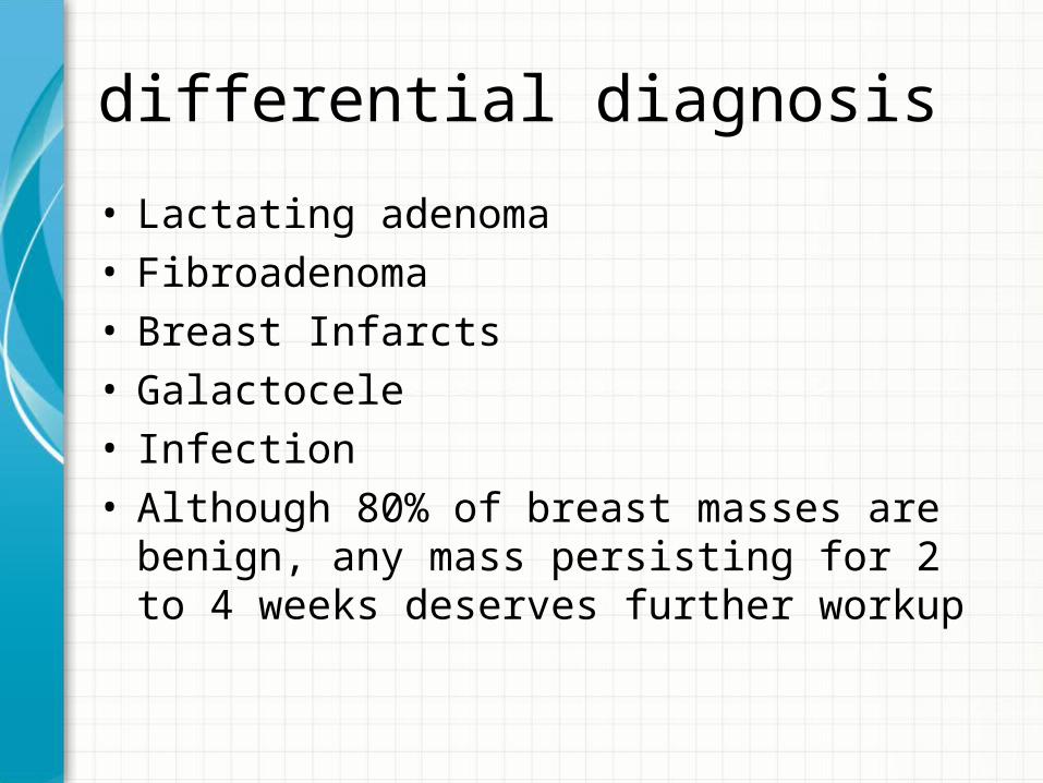
Diagnostic work-up
• ionizing radiation might cause pregnancy loss,
malformations, growth retardation, and
neurobehavioral defects.
• These anomalies appear at fetal doses in excess of
200 mGy, although avoidance of exposure to doses
higher than 100 mGy is advised
International Commission on Radiological Protection 2003; Kal and Struikmans 2005).

No single diagnostic procedure results in a radiation dose that
threatens the well being of the developing embryo and fetusAmerican college of radiology

Tissue diagnosis
• FNAC• Core needle biopsy• Vacuum assisted Breast biopsy• ?? Incisional/Excisional biopsy• F.S

Pathology
• Type : Carcinoma ( invasive/noninvasive) other
pathology ( e.g. Phyllodes T, others)
• grade
• hormonal status

Surgical Management
• The decision to proceed to mastectomy or breast
conservative surgery (BCS) should follow the
standard practice as in the non-pregnant setting.
• Both can be safely performed throughout the
course of gestation.
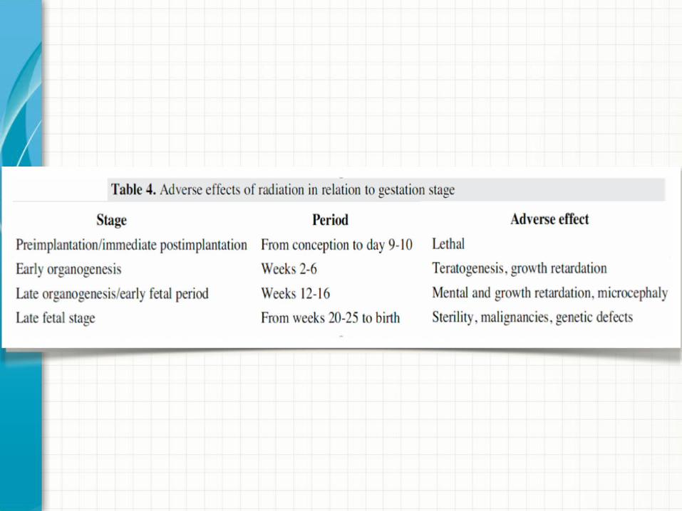
Surgical management
• 1st Trimester:Termination of pregnancy (non-therapeutic)Surgery: Mastectomy and axillary stagingNo role for BCS because RT will not be
delivered until end of pregnancy ( almost 6 months)

Surgical management
• 2nd trimester and early 3rd trimesterSurgery: Mastectomy vs BCS
Axillary stagingFollowed by adjuvant treatment
ORNeoadjuvant CT followed by surgery

Surgical management
• Late 3rd trimester:
Surgery: either mastectomy or BCS and axillary staging
Followed by adjuvant treatment postpartum
Surgical management of the Axilla
• Routine ALND• Role of SLNB:
• Only one clinical series involving 12 pregnant breast
cancer patients has been reported to date, No fetal
defects secondary to SLNB were observed and no
evidence of axillary relapse was encountered at a
median follow-up of 32 months.
Gentilini et al. Eur J Nucl Med Mol Imaging (2010) 37:78–83

ESMO recommendations
• It is clear that more data on SLNB are needed in the
pregnancy setting; however, we would not
discourage SLNB in pregnant breast cancer patients
in centers in which SLNB is routine practice in the
non-pregnant setting We discourage the use of vital
blue dye in pregnant patients, which is associated
with 2% risk of allergic reactions that could be life-
threatening


Conclusion
• Breast cancer in pregnancy will increase as more
women postpone childbearing until middle age.
• Breast examination at the first prenatal visit and
maintain a high index of suspicion for cancer.
• Although pregnancy-associated cancers tend to occur
at a later stage and are more often ER- negative, they
carry a similar prognosis to other breast cancers
when matched for stage and age.

Conclusion
• Mastectomy and axillary dissection is the traditional
treatment of choice.
• Therapeutic radiation during pregnancy cannot be
recommended because of the risk to the fetus.
• Surgical management should be tailored as for non-
pregnant breast cancer patients