Embed Size (px)
Citation preview

Linköping University Medical Dissertation No. 963
Brain Stem Involvement in Immune and Aversive Challenge
Jakob Paues
Department of Biomedicine and Surgery, Faculty of Health Sciences, Linköping University, SE-582 85, Linköping, Sweden
Linköping 2006

Published articles and figures have been reprinted with the permission of the respective copyright holder.
Printed in Sweden by LiU-Tryck, Linköping 2006
ISBN: 91-85643-81-5 ISSN: 0345-0082

There are in fact two things, science and opinion;the former begets knowledge, the latter ignorance.
Hippocrates (460 BC - 377 BC)
This thesis is based on the following papers, which will be referred to in the text by their Roman numerals:
I. Engström L., Engblom D., Örtegren U., Mackerlova L., Paues J., Blomqvist A. (2001) Preproenkephalin mRNA expression in rat parabrachial neurons: relation to cells activated by systemic immune challenge. Neurosci Lett316:165-8
II. Paues J., Engblom D., Mackerlova L., Ericsson-Dahlstrand A., Blomqvist A. (2001) Feeding-related immune responsive brain stem neurons: association with CGRP. Neuroreport 12:2399-403. Erratum in: Neuroreport 2001;12(16):inside back cover. Neuroreport 2001;12(13):inside back cover.
III. Richard S., Engblom D., Paues J., Mackerlova L., Blomqvist A. (2005) Activation of the parabrachio-amygdaloid pathway by immune challenge or spinal nociceptive input: a quantitative study in the rat using Fos immunohistochemistry and retrograde tract tracing. J Comp Neurol481:210-9. Erratum in: J Comp Neurol. 2005; 483:489-90.
IV. Paues J., Mackerlova L., Blomqvist A. (2006) Expression of melanocortin-4 receptor by rat parabrachial neurons responsive to immune and aversive stimuli. Neuroscience 141:287-97
CONTENTS
ABSTRACT................................................................................................................ 9
ABBREVIATIONS.................................................................................................... 11
INTRODUCTION ...................................................................................................... 13
Bacterial products and cytokines as mediators of disease symptoms ............................... 14Endotoxins............................................................................................................................ 14 Cytokines.............................................................................................................................. 15
Tumour necrosis factor-α................................................................................................. 16 Interleukin-1 ..................................................................................................................... 17 Interleukin-6 ..................................................................................................................... 18
Cytokines in immune-to-brain signalling............................................................................. 19Interaction with circumventricular organs ........................................................................... 20 Interaction with free nerve endings...................................................................................... 21 Signalling across the blood brain barrier.............................................................................. 22
Sickness behaviour ................................................................................................................. 22HPA-axis activation ............................................................................................................. 23 Lethargy ............................................................................................................................... 23 Fever..................................................................................................................................... 24 Anorexia ............................................................................................................................... 24
Appetite in disease ........................................................................................................... 25Aversion ............................................................................................................................... 26
Aversion in disease........................................................................................................... 27LiCl as a model for aversion ............................................................................................ 28
Pain .......................................................................................................................................... 28
Central nervous structures studied in this thesis ................................................................ 29Amygdala ............................................................................................................................. 29 Parabrachial nucleus............................................................................................................. 30
The external lateral subnucleus (PBel)............................................................................. 31 The external medial subnucleus (PBem).......................................................................... 32 The dorsal lateral subnucleus (PBdl) ............................................................................... 32 The central lateral subnucleus (PBcl)............................................................................... 32 The superior lateral subnucleus (PBsl)............................................................................. 32 The internal lateral subnucleus (PBil) .............................................................................. 33 The ventral lateral subnucleus (PBvl) .............................................................................. 33 The medial subnucleus (PBm) ......................................................................................... 33
Neuropeptides ......................................................................................................................... 33Calcitonin gene related peptide ............................................................................................ 34 Enkephalins .......................................................................................................................... 35 Melanocortins....................................................................................................................... 36
AIMS ........................................................................................................................ 39
Specific aims............................................................................................................................ 39
METHODS................................................................................................................ 41
Animals used........................................................................................................................... 41
Intravenous injection of LPS (paper I-IV)........................................................................... 41
Intraperitoneal injection of LiCl (paper IV)........................................................................ 41
Subcutaneous injection of formalin (paper III)................................................................. 41
Time points.............................................................................................................................. 42
Retrograde tracing combined with nociceptive or inflammatory stimuli (paper III) .... 42
Histology.................................................................................................................................. 42
Double-labelling immunohistochemistry (paper III) .......................................................... 43
Dual-labelling in situ-hybridization (paper II and IV) ....................................................... 43
Immunohistochemistry combined with in situ-hybridization (paper I)............................ 44
RESULTS................................................................................................................. 45
Systemic immune challenge activates very few parabrachial enkephalin-expressing neurons (paper I) .................................................................................................................... 45
Systemic immune challenge activates parabrachial CGRP-expressing neurons (paper II).................................................................................................................................................. 45
Parabrachial amygdala-projecting neurons are activated by systemic immune challenge but not by nociceptive stimulation (paper III)..................................................................... 45
Parabrachial neurons expressing the melanocortin-4 receptor are activated by immune and aversive stimuli (paper IV)............................................................................................. 46
DISCUSSION ........................................................................................................... 47
Methodological considerations.............................................................................................. 47Models that simulate acute disease symptoms..................................................................... 47 Fos as a marker for neuronal activation ............................................................................... 48 Studying the parabrachial nucleus........................................................................................ 49 In situ hybridization ............................................................................................................. 49 Immunohistochemistry combined with in situ hybridization............................................... 50
Anorexia and aversion in disease.......................................................................................... 50

The importance of the parabrachio-amygdaloid pathway in noxious versus immune challenge.................................................................................................................................. 54
Opioids in immune and aversive signalling ......................................................................... 55
Gustatory versus aversive pathways in the parabrachial nucleus..................................... 55
CONCLUSION ......................................................................................................... 57
ACKNOWLEDGEMENTS ........................................................................................ 59
LITERATURE CITED ............................................................................................... 61

9
ABSTRACT
Activation of the immune system by e.g. bacteria induces the acute-phase-response and sickness behaviour. The latter encompasses among other things fever, lethargy, anorexia and hyperalgesia. An often used model to study sickness behaviour is the intravenous injection of the gram negative bacterial endotoxin lipopolysaccharide (LPS). LPS induces the production of inflammatory mediators, such as cytokines and prostaglandins, which in turn can interact with the central nervous system (CNS) to affect behaviour. The CNS also memorises substances that have made us sick in the past to avoid future harm, a phenomenon called conditioned taste aversion (CTA). An often used model to study CTA is the intraperitoneal injection of LiCl.
The pontine parabrachial nucleus (PB) is an autonomic relay nucleus situated in the rostral brain stem that integrates afferent somatosensory and interoceptive information and forwards this information to the hypothalamus and limbic structures. PB is crucial for the acquisition of CTA and PB neurons are activated by many anorexigenic substances. Further, PB neurons express neuropeptides, among those calcitonin gene related peptide (CGRP) and enkephalin, both of which have been implicated in immune signalling, nociception, food intake, and aversion.
By using a dual-labelling immunohistochemical/in situ hybridization technique we investigated if enkephalinergic neurons in PB are activated by systemic immune challenge. While there were many neurons in the external lateral parabrachial subnucleus (PBel) that expressed the immediate early gene fos after intravenous injection of LPS and while a large proportion of the PBel neurons expressed preproenkephalin, there were very few double-labelled cells. The fos-expressing cells were predominantly located to the outer part of the PBel (PBelo), whereas the preproenkephalin-expressing PBel neurons were located closest to the peduncle. Thus we conclude that although enkephalin has been implicated in autonomic and immune signalling, enkephalinergic neurons in PB do not seem to be activated by immune stimulation (paper I). To further characterise the PBelo neurons activated by immune challenge we investigated if these neurons expressed CGRP. Dual-labelling in situ hybridisation showed that PBelo neurons that expressed fos after intravenous injection of LPS to a large extent co-expressed CGRP mRNA, indicating that CGRP may be involved in the regulation of the sickness response in immune challenge (paper II). Using dual-labelling immunohistochemistry we examined if PBel neurons activated by an immune stimulus projected to the amygdala, a limbic structure implicated in the affective response to homeostatic challenge. Animals were injected with the retrograde tracer substance cholera toxin b (CTb) into the amygdala and subsequently subjected to immune challenge. We found that approximately a third of the neurons that expressed fos after the intravenous injection of LPS also were labelled with CTb. Thus PBel neurons activated by immune challenge project to the amygdala. The PBel-
10
amygdala pathway has earlier been suggested to be important in nociceptive signalling. To investigate if amygdala-projecting PBel neurons are activated by nociceptive stimuli we again injected animals with CTb into the amygdala. After recovery the animals were injected with formalin into a hindpaw. Dual-labelling immunohistochemistry against fos and CTb showed that very few noxiously activated PB neurons projected to the amygdala. Thus, the PBel-amygdala projection seems to be important in immune challenge but not in nociceptive signalling (paper III). Many PBel neurons express fos after intraperitoneal injection of LiCl. Melanocortins are neuropeptides that recently have been implicated in metabolism, food intake and aversive mechanisms. The PB is known to express melanocortin receptor-4 (MC4-R) mRNA. Using dual-labelling in situ hybridization we investigated if PB neurons activated by intravenous injection of LPS or intraperitoneal injection of LiCl expressed MC4-R mRNA. We found that many PBelo neurons were activated by either LPS or LiCl and that a large proportion of such activated neurons expressed MC4-R mRNA. Further, using dual-labelling in situ hybridization against MC4-R mRNA and CGRP mRNA, we found that a large proportion of the CGRP positive PBelo neurons also expressed MC4-R mRNA.
In summary, this thesis shows that CGRP-expressing neurons in the PBel are activated by peripheral immune challenge, that lipopolysaccharide-activated PBel neurons project to the amygdala, that the amygdala-projecting neurons in the PBel are CGRP-positive, and that PBel neurons activated by immune or aversive challenge express MC4-R. Taken together, these data suggest the presence of a melanocortin-regulated CGRP-positive pathway from the PBel to the amygdala that relays information of importance to certain aspects of sickness behaviour.
11
ABBREVIATIONS
ACTH adrenocorticotropic hormone AgRP agouti related peptide AP-1 activated protein-1 BBB blood-brain barrier BSA bovine serum albumin CCK cholecystokinin CFA conditioned food aversion CGRP calcitonin gene related peptide CNS central nervous system COX-2 cyclooxygenase-2 CRLR calcitonin receptor-like receptor CRE cAMP-responsive element CREB cAMP response element-binding protein CT calcitonin CTA conditioned taste aversion CTb cholera toxin-b CVO circumventricular organs DCV dense core vesicle HPA-axis hypothalamic-pituitary-adrenal-axisIEG immediate early gene IL-1 interleukin-1 IL-1R interleukin-1 receptor IL-6 interleukin-6 IL-6R interleukin-6 receptor ISH in situ hybridisation LBP lipopolysaccharide binding protein LiCl lithium chloride LPS lipopolysaccharide MC-R melanocortin receptor MC-4R melanocortin receptor-4 α-, β-, γ-MSH α-, β-, γ-melanocyte stimulating hormone NTS nucleus of the solitary tract PB parabrachial nucleus PBcl -central lateral subnucleus PBdl -dorsal lateral subnucleus PBel -external lateral subnucleus PBelo -external lateral subnucleus, outer partPBeli -external lateral subnucleus, inner part PBem -external medial subnucleus PBil -internal lateral subnucleus
12
PBm -medial subnucleus PBsl -superior lateral subnucleus PBvl -ventral lateral subnucleus PBS phosphate buffered saline PGE2 prostaglandin E2POMC proopiomelanocortin ppENK preproenkephalin RAMP1 receptor activity-modifying protein 1 RCP receptor component protein SCP superior cerebellar peduncle SSC standard saline citrate TLR Toll-like receptor TNF-α tumour necrosis factor-αVPpc parvicellular part of the ventral posterior
thalamic nucleus
13
INTRODUCTION
The cells of our bodies all work together to maintain a stable and optimal inner environment. This process is called homeostasis and has proved to be a very efficient mean to survive in a hostile external environment with shifting temperature, humidity and access to nutrients. An important aspect of the maintenance of homeostasis is the ability to recognise “friend from foe”. Thus, all our cells express identification-markers to show that they belong to our body and those that cannot identify them-selves are readily destroyed by sentinel cells of the immune system. One threat to homeostasis is invading pathogens such as viruses or bacteria. Another threat is cells that start to divide without restraint, causing malignant cancer. Whatever the nature of the threat is, the immune system responds in similar ways. The strategy is to swiftly destroy the aggressor and if this fails, try to limit the extent of the infection. Thus humans can have dormant tuberculosis infections that are locally controlled until the individual become weakened by disease or old age. Likewise, premalignant cells are either destroyed or forced into senescence.
In order to maximize the efficiency of the immune response, it is coordinated by the central nervous system (CNS), which orchestrates well known sickness symptoms as fever, anorexia and fatigue. The CNS also memorises ingested substances that made us sick in the past and makes us avoid these substances in the future by a phenomenon called conditioned taste aversion. The immune system signals the CNS through molecules called cytokines, released by activated immune cells. Cytokines interact with free nerve endings, the blood-brain barrier, or areas in the CNS that lack a blood-brain barrier to induce fever, anorexia and fatigue. These symptoms are all effective defences in a short term illness, helping us combat the pathogens by concentrating our energy reserves to the immune system and creating an inner environment less suitable for the invaders. However, in the long run, increased metabolism and reduced food intake can start to work against the individual and lead to the anorexia-cachexia syndrome seen in severe chronic diseases, such as organ failure, protracted infection, or cancer. Cachexia is characterized by increased metabolism, weight loss, anaemia and fatigue and it is responsible for shortened life-span, increased co-morbidity, and suffering of afflicted patients.
Another important aspect of homeostasis is the maintenance of tissue integrity. Whenever the skin barrier is broken or tissues in the body are damaged, free nerve endings, nociceptors, signal to the brain in order to adapt behaviour to minimize the damage. Nociception is a potent modulator of motivated behaviour, and nociceptive stimuli activate the sympathetic branch of the autonomic nervous system. Also, the immune system and the nociceptive system work in conjunction with each other. For example, in an inflamed tissue nociceptors are sensitised by inflammatory mediators, a phenomenon known as hyperalgesia.

14
Bacterial products and cytokines as mediators of disease symptoms
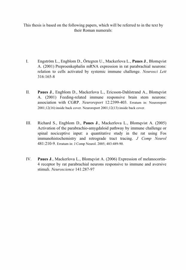
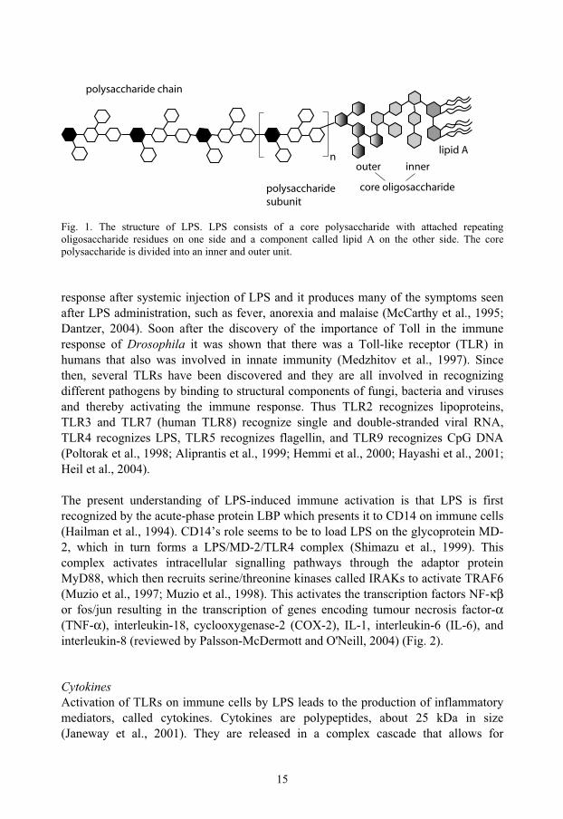
EndotoxinsThe release of toxins as a cause of disease has been suspected since the days of Hippocrates (ca 460-370 BC), but the toxin theory could not explain how a single sick person could afflict several thousands of others (Beutler and Rietschel, 2003). It was not until the beginning of the 20th century that there was a solution to this conundrum when first Henle suggested “multiplication of the toxic matter” (Henle, 1910) and then Pasteur proved that microbes were necessary and sufficient for the induction of infectious disease (reviewed by Beutler and Rietschel, 2003). Endotoxins were first described in 1892 by Pfeiffer as a mean to distinguish toxins appearing after bacterial lysis from those secreted by bacteria (exotoxins) (Pfeiffer, 1892). In 1933 Boivin and collaborators found that endotoxins in gram-negative bacteria are made of lipopolysaccharides (LPS) (Boivin et al., 1933). LPS consists of a core polysaccharide to which repeating oligosaccharide residues are attached as well as a component called lipid A (Fig. 1) (Brooks et al., 2004). Whereas the polysaccharide seems important in solubilizing the complex, lipid A is the portion that confers toxicity and it consists of a glucosamine disaccharide with attached fatty-acids and phosphate. Endotoxins are heat-stable, anchored to the outer bacterial membrane, and they are produced in different variants by several gram negative strains such as Escherichia coli,Salmonella and Neisseriae (Brooks et al., 2004). When injected into the bloodstream, LPS produces a dose-dependent response, with fever, anorexia, loose faeces and lethargy at low doses, and hypotension and death at high doses (Elmquist et al., 1996; Tkacs et al., 1997; Huang et al., 1999; Cuzzocrea et al., 2006).
Although the discovery of LPS revealed how bacteria could cause severe illness and immune system activation in animals, it was still unknown how this effect was produced. Some clues to the underlying mechanism were unravelled when a LPS binding, liver-produced, plasma protein was discovered (LBP), and it was shown that this protein could activate immune cells by forming a complex with CD14, a plasma membrane-bound receptor protein (Wright et al., 1990). However this complex did not have a cytoplasmic part and therefore there were no immediate clues as to how an intracellular signal was induced (Wright et al., 1990). It was not until recently that the mechanism underlying LPS interaction with the immune system was unravelled. In 1996 it was reported that mutations in Toll, a protein known to be involved in the dorso-ventral axis formation in Drosophila, caused impaired response to fungal infection in this species (Lemaitre et al., 1996). This awoke interest since it was prev-iously known that Toll activates the transcription factor NF-κβ, just like interleukin-1 (IL-1) and LPS (Shakhov et al., 1990; Wasserman, 1993), and that the cytoplasmic domain of Toll is homologous to the cytoplasmic domain of the IL-1 receptor (Gay and Keith, 1991). IL-1 is a cytokine that is involved in the immune
15
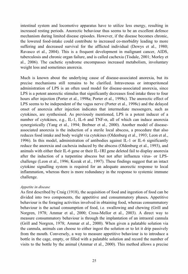
Fig. 1. The structure of LPS. LPS consists of a core polysaccharide with attached repeating oligosaccharide residues on one side and a component called lipid A on the other side. The core polysaccharide is divided into an inner and outer unit.
response after systemic injection of LPS and it produces many of the symptoms seen after LPS administration, such as fever, anorexia and malaise (McCarthy et al., 1995; Dantzer, 2004). Soon after the discovery of the importance of Toll in the immune response of Drosophila it was shown that there was a Toll-like receptor (TLR) in humans that also was involved in innate immunity (Medzhitov et al., 1997). Since then, several TLRs have been discovered and they are all involved in recognizing different pathogens by binding to structural components of fungi, bacteria and viruses and thereby activating the immune response. Thus TLR2 recognizes lipoproteins, TLR3 and TLR7 (human TLR8) recognize single and double-stranded viral RNA, TLR4 recognizes LPS, TLR5 recognizes flagellin, and TLR9 recognizes CpG DNA (Poltorak et al., 1998; Aliprantis et al., 1999; Hemmi et al., 2000; Hayashi et al., 2001; Heil et al., 2004).
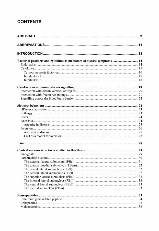
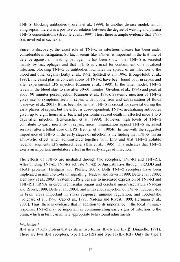
The present understanding of LPS-induced immune activation is that LPS is first recognized by the acute-phase protein LBP which presents it to CD14 on immune cells (Hailman et al., 1994). CD14’s role seems to be to load LPS on the glycoprotein MD-2, which in turn forms a LPS/MD-2/TLR4 complex (Shimazu et al., 1999). This complex activates intracellular signalling pathways through the adaptor protein MyD88, which then recruits serine/threonine kinases called IRAKs to activate TRAF6 (Muzio et al., 1997; Muzio et al., 1998). This activates the transcription factors NF-κβor fos/jun resulting in the transcription of genes encoding tumour necrosis factor-α(TNF-α), interleukin-18, cyclooxygenase-2 (COX-2), IL-1, interleukin-6 (IL-6), and interleukin-8 (reviewed by Palsson-McDermott and O'Neill, 2004) (Fig. 2).
CytokinesActivation of TLRs on immune cells by LPS leads to the production of inflammatory mediators, called cytokines. Cytokines are polypeptides, about 25 kDa in size (Janeway et al., 2001). They are released in a complex cascade that allows for
polysaccharide chain
polysaccharidesubunit
core oligosaccharide
outer inner
lipid An

16
maintenance and fine tuning of the immune response. The cytokines can augment local defences by inducing differentiation of local immune cells or they can travel in the bloodstream to distant target organs, such as the liver or brain to activate the acute-phase response and the hypothalamic-pituitary-adrenal-axis (HPA-axis) (Ericsson et al., 1994; Fattori et al., 1994; Billingsley et al., 1996; Dunn, 2000; Ek et al., 2001; Dantzer, 2004; Hessle et al., 2005). When discussing the role of different cytokines in disease-models one has to bear in mind that different stimuli produce different cytokine patterns. Injection of Staphylococcal enterotoxin A induces the production of TNF-α, interleukin-2 and interferon-γ (Brebner et al., 2000), whereas LPS results in the release of TNF-α, IL-1 and IL-6 (Cannon et al., 1990; Givalois et al., 1994). This could be explained by the above-mentioned fact that the activation of the innate immune-system involves Toll receptors that are specific to different pathogens. The most studied cytokines in immune-to-brain signalling are TNF-α, IL-1 and IL-6. Therefore these cytokines will be briefly described below.
Fig. 2. Current understanding of LPS induction of cytokine gene transcription. LPS is first recognized by the acute-phase protein LBP which presents it to CD14 on immune cells. CD14 loads LPS on the glycoprotein MD-2, which in turn forms a LPS/MD-2/TLR4 complex. This complex activates intracellular signalling pathways through the adaptor protein MyD88. Activation of transcription factors NF-κβ or fos/jun results in the transcription of genes encoding tumour necrosis factor-α (TNF-α), interleukin-1 (IL-1), interleukin-6 (IL-6) and cyclooxygenase-2 (COX-2).
Tumour necrosis factor-αTNF-α was first named cachectin when discovered because of its ability to induce anorexia and wasting when injected systemically (Beutler et al., 1985a; Tracey et al., 1988). Similarly, a TNF-α producing tumour induced cachexia in rodents (Tracey et al., 1990). In rats, tumour-induced cachexia can be reduced by the administration of
LBPTLR4
LPS
CD14MD2
MyD88
IRAKs
TRAF6
NF-fos/jun
Activation of transcription factors
IL-1, IL-6TNF- , COX-2
Nucleus
Cell membrane
17
TNF-α- blocking antibodies (Torelli et al., 1999). In another disease-model, simul-ating sepsis, there was a positive correlation between the degree of wasting and plasma TNF-α concentrations (Breuille et al., 1999). Thus, there is ample evidence that TNF-α is involved in cachexia.
Since its discovery, the exact role of TNF-α in infectious disease has been under considerable investigation. So far, it seems like TNF-α is important in the first line of defence against an invading pathogen. It has been shown that TNF-α is secreted mainly by macrophages and that TNF-α is crucial for containment of a localized infection; blocking TNF-α by antibodies facilitates the spread of an infection to the blood and other organs (Leiby et al., 1992; Sjöstedt et al., 1996; Broug-Holub et al., 1997). Increased plasma concentrations of TNF-α have been found both in sepsis and after experimental LPS injection (Cannon et al., 1990). In the latter model, TNF-αlevels in the blood start to rise after 30-60 minutes (Givalois et al., 1994) and peak at about 90 minutes post-injection (Cannon et al., 1990). Systemic injection of TNF-αgives rise to symptoms seen in sepsis with hypotension and extravasation of fluids (Janeway et al., 2001). It has been shown that TNF-α is crucial for survival during the early phases of sepsis, but the effect is dose-dependent. TNF-α neutralizing antibodies given up to eight hours after bacterial peritonitis caused death in affected mice 1 to 3 days after infection (Echtenacher et al., 1990). However, high levels of TNF-αcontribute to early mortality in sepsis, since immunization against TNF-α increased survival after a lethal dose of LPS (Beutler et al., 1985b). In line with the suggested importance of TNF-α in the early stages of infection is the finding that TNF-α has an antipyretic effect when administered together with LPS and that TNF-α soluble receptor augments LPS-induced fever (Klir et al., 1995). This indicates that TNF-αexerts an important modulatory effect in the early stages of infection.
The effects of TNF-α are mediated through two receptors, TNF-RI and TNF-RII. After binding TNF-α, TNF-Rs activate NF-κβ or Jun pathways through TRADD and TRAF proteins (Hehlgans and Pfeffer, 2005). Both TNF-α receptors have been implicated in immune-to-brain signalling (Nadeau and Rivest, 1999; Bette et al., 2003; Dempsey et al., 2003). Systemic LPS gives rise to increased expression of TNF-RI and TNF-RII mRNA in circumventricular organs and cerebral microvasculature (Nadeau and Rivest, 1999; Bette et al., 2003), and intravenous injection of TNF-α induces c-fos in brain areas important in stress response, immune regulation, and food-intake (Tolchard et al., 1996; Cao et al., 1998; Nadeau and Rivest, 1999; Hermann et al., 2003). Thus, there is evidence that in addition to its importance in the local immune-response, TNF-α may be important in communicating early signs of infection to the brain, which in turn can initiate appropriate behavioural adjustments.
Interleukin-1IL-1 is a 17 kDa protein that exists in two forms, IL-1α and IL-1β (Dinarello, 1991). There are two IL-1 receptors, type I (IL-1RI) and type II (IL-1RII). Only the type I

18
receptor seems to be able to activate intracellular signalling pathways. The type II receptor has therefore been suggested to be a decoy receptor involved in the negative regulation of IL-1 signalling (Dinarello, 1991; Bluthé et al., 2000). Upon ligand binding, the IL-1RI activates similar intracellular pathways as the TLRs as outlined above (Shakhov et al., 1990; Wasserman, 1993). IL-1 receptors are e.g. expressed on cells of the circumventricular organs and on vascular cells lining the capillaries of the brain (Ericsson et al., 1995; Konsman et al., 2004). IL-1R mRNA has also been found in dorsal root ganglion cells, suggesting that IL-1 receptors may be expressed on peripheral nerves (Ek et al., 1998).
There are diverging data concerning the role of IL-1 in the immune response. While, IL-1 has been proven to mediate many of the effects of LPS, it remains to be clarified if the actions of IL-1 are mediated through local or systemic mechanisms. There is evidence that the main effect of IL-1 is achieved through interaction with local nerve-endings and immune cells with subsequent neural activation and/or release of other, systemically acting cytokines like IL-6 (Leon et al., 1996). Accordingly, animals lacking the IL-1RI do not develop fever or anorexia after a local turpentine abscess (Leon et al., 1996). However, these animals were also found to have an intact response after a systemic immune-challenge by intraperitoneal injection of LPS, suggesting that IL-1 is more important at the local than the systemic level in infection.
However, IL-1 also seems to play a role for the systemic immune response, because circulating IL-1 has been found in sepsis and after LPS injection in human subjects (Cannon et al., 1990) Likewise, intravenous injection of IL-1 induces IL-6 production, fever and anorexia (Engblom et al., 2003; Turnbull et al., 2003; Elander et al., 2006). Blockage of IL-1 actions through receptor antagonists do not abolish, but significantly decrease LPS-induced fever, anorexia and IL-6 plasma concentrations (Luheshi et al., 1996; Swiergiel and Dunn, 1999). However, if TNF-α and IL-6 are blocked alongside IL-1 after intraperitoneal injection of LPS, the anorectic response is abolished (Swiergiel and Dunn, 1999). Thus IL-1 plays an important local role, whereas there seems to be a certain degree of redundancy in the cytokine response at the systemic level.
Interleukin-6IL-6 is a pleiotropic cytokine synthesised by both immune and non-immune cells (Papanicolaou et al., 1998). It binds to the IL-6 receptor but the ligand-receptor complex cannot in itself transduce a signal intracellularly. Instead, the receptor heterodimerizes with another transmembrane receptor protein, gp130 (Naka et al., 2002). The heterodimerization activates the gp130-associated JAK/STAT pathway. Jak1 and 2 are tyrosine kinases that phosphorylate the gp130 receptor and allow binding of STAT proteins that in turn become phosphorylated and dimerize. After dimerization STAT proteins can translocate to the nucleus and act as transcription factors (Heinrich et al., 1998; Naka et al., 2002; Carbia-Nagashima and Arzt, 2004).

19
IL-6 is an acute-phase protein upregulated during inflammation (Luheshi et al., 1997). It stimulates the differentiation of macrophages (Nicola et al., 1983) and the production of other acute-phase proteins like C-reactive protein, fibrinogen, and α1-antitrypsin (Castell et al., 1988). IL-6 has been implicated in fever, anorexia and HPA-axis activation in disease (Perlstein et al., 1991; Spath et al., 1994; Oka et al., 1996; Kozak et al., 1997; Leon et al., 1998) and it has been related to disease parameters in human cancer (Oka et al., 1996). IL-6 has consistently been found in the blood during disease, real or modelled, as well as after psychological stress (LeMay et al., 1990; Turnbull and Rivier, 1999; Alesci et al., 2005; Eijsbouts et al., 2005). Thus there is ample evidence that IL-6 plays an important regulatory role in response to disease.
Which role IL-6 plays compared to other cytokines in the signalling cascade during the acute-phase response has been under considerable investigation. The available literature indicates that IL-6 production is induced locally at an inflammatory site by other cytokines, such as IL-1. IL-6 then exerts its effects at the systemic level. Thus, the cachexia and activation of the acute-phase response after the induction of a sterile abscess is partly mediated by IL-1-induced IL-6 signalling. (Oldenburg et al., 1993) Further, mice lacking IL-6 did not develop fever, anorexia or cachexia after a sterile abscess (Kozak et al., 1997). The cytokine production in the micro-environment of a pathological process has also been studied and it has been found that local macrophages synthesise IL-1 and TNF-α, which in turn stimulate tumour cells to produce IL-6 (Billingsley et al., 1996). However, the systemic response to disease is not completely dependent on IL-6; there seems to be a degree of redundancy in the signalling cascade since LPS treated IL-6 deficient mice did not display any difference in anorexia and loss of body weight compared to wild-type controls (Fattori et al., 1994). Interestingly, in the latter study, the knock-out mice had elevated TNF-α levels compared to the wild-type animals, suggesting that TNF-α replaces IL-6 in this model.
Exactly how IL-6 mediates its effects on metabolism, food intake and fever remains to be shown, but there is evidence pointing at an important role for IL-6-signalling in the cerebral vasculature and in the brain parenchyma. After systemic injection of LPS, IL-6 receptor (IL-6R) mRNA was found to be increased in the circumventricular organs, the cerebral vasculature, the paraventricular nucleus of the hypothalamus, the amygdala, and the bed nucleus of the stria terminalis (Vallières and Rivest, 1997). Similar to the systemic administration of LPS, intravenous injection of IL-6 induces c-fos mRNA in the circumventricular organs (Elmquist et al., 1996; Vallières and Rivest, 1997). Thus IL-6 seems to be able to interact with central nervous structures important in homeostatic regulation.
Cytokines in immune-to-brain signalling The CNS is protected from substances circulating in the blood by the blood-brain barrier (BBB). The BBB, which was first described by Paul Erlich (1885), is made up
20
of tight junctions in the capillaries lining the brain, effectively protecting the sensitive neurons from potentially harmful substances in the blood stream (Hawkins and Davis, 2005). This means that the communication between immune cells, cytokines and neurons in most circumstances must rely on other pathways than direct interaction. Several such pathways for immune-to-brain signalling have been suggested:
1) Direct interaction of LPS or cytokines with neurons in the circumventricular organs, which lack a complete BBB.
2) LPS or cytokine activation of free nerve endings. 3) Signalling across the blood-brain barrier via induced prostaglandin synthesis.
All three pathways are probably important. The contribution of the different pathways are likely to be dependent on the location, duration and intensity of the infection. Hence, a local abscess may signal through peripheral nerve endings at the site of infection even though cytokines also are bound to leak into the circulation constantly or intermittently. Sepsis is a full front assault on the organism and threatens to over-throw the defences in a short time. Therefore, in this situation, circumventricular organ and blood-brain barrier signalling is probably important as well as signalling via nerve afferents from the liver. In the beginning of a disease the activation pattern may be different from that present during chronic conditions. In the latter case compensatory mechanisms have probably developed. A very aggressive infection probably activates more pathways than a small localised tumour. With this in mind the different pathways will be briefly outlined below.
Interaction with circumventricular organs The circumventricular organs (CVO) are areas in the brain that lack a proper BBB due to fenestrated capillaries with high permeability. The CVOs are situated adjacent to the third and fourth ventricles of the brain. In mammals there are four CVOs: the area postrema, the median eminence, the organum vasculosum of the lamina terminalis, and the subfornical organ (Ganong, 2000). The fenestrated capillaries allow CVO neurons to sample concentration of hormones, cytokines and ions in the blood (Ganong, 2000). This information can then be forwarded to other brain areas.
The area postrema is a chemoreceptor zone in the floor of the fourth ventricle sensing ion and satiety hormone levels in the circulation and it has been implicated in satiety, malaise and IL-1β induced HPA-axis activation (Yamamoto et al., 1992; Lee et al., 1998; Rinaman et al., 1998). The median eminence is important in energy homeo-stasis, reproduction and pituitary signalling (Elias et al., 2000; Ganong, 2000; Daftary and Gore, 2005). The organum vasculosum of the lamina terminalis has been implicated in fever (Blatteis, 2000), and the subfornical organ in salt balance (Noda, 2006). Toll and cytokine receptors have been found to be expressed in CVOs (Ericsson et al., 1995; Vallières and Rivest, 1997; Nadeau and Rivest, 1999; Laflamme and Rivest, 2001), and CVOs are activated by immune stimuli (Ericsson et al., 1994; Elmquist et al., 1996). However, as indicated above, there seems to be a specialisation
21
in the role of the different CVOs. For example, the IL-1RI is predominantly found in the area postrema (Ericsson et al., 1995), and disruption of the ascending pathways to the diencephalon attenuates IL-1 induced neuronal activation of paraventricular hypothalamic neurons (Ericsson et al., 1994). Further, ablation of the area postrema abolishes c-fos expression in the paraventricular hypothalamic nucleus and the HPA-axis activation seen after intravenous IL-1β (Lee et al., 1998). These findings suggest the presence of an important area postrema-forebrain pathway playing a pivotal role in the central nervous system response to immune stimulation.
Interaction with free nerve endings Free nerve endings expressing a wide variety of receptors are ubiquitous in most tissues, and they are positioned to sense the composition of the extracellular fluid, including molecules secreted by immune cells or supportive tissue. Accordingly, it has been suggested that peripheral nerves are important in modulating tumour development or the liver response to sepsis (Borovikova et al., 2000; Gidron et al., 2005). There are several potential mechanisms for immune-to-brain communication through peripheral nerves. One possibility is that infectious agents directly interact with sensory nerve endings through Toll receptor binding, which is supported by the fact that TLR-4 protein and mRNA and TLR-9 mRNA have been found in rodent nodose ganglion neurons (Hosoi et al., 2005; Sako et al., 2005). Another possible mechanism is cytokines interacting with peripheral nerves. TNF-receptor mRNA is expressed in dorsal root and trigeminal ganglion cells and is upregulated by systemic injection of LPS (Cunningham et al., 1997; Li et al., 2004b), and IL-1R mRNA is present in nodose ganglion cells (Ek et al., 1998). IL-6R mRNA is found in dorsal root ganglia (Gadient and Otten, 1996) and IL-1 and LPS activation of trigeminal neurons seems to be partly dependent on IL-6 (Kobierski et al., 2000). The most studied nerve in this context is the vagus nerve which innervates every internal organ of the body except the pelvic viscera. Liver vagal afferents can sample blood concentrations of cytokines or become stimulated by Kupffer cells, i.e. resident liver macrophages. Kupffer cells are thought to be important sentinels in the response to systemic infection (Gregory et al., 1998). Many attempts have been made to elucidate the importance of the vagal nerve in immune-to-brain signalling, but so far there is no conclusive answer (Porter et al., 1998c). Stimulus location and dose seem to be important. An intraperitoneal injection of a low dose of LPS seems to be more dependent on vagal afferent signalling than a high dose or systemic injection of the substance (Porter et al., 1998c). Possibly, the lower dose only interacts with local, vagal, nerve endings whereas a high intraperitoneal dose results in endotoxin reaching the systemic circulation allowing for parallel activation of additional pathways. A much used technique in studies addressing the importance of the vagus nerve in immune-to-brain-communication is vagotomy. At first glance vagotomy seems like a straight-forward method for studying the role of the vagus in the sickness response. However, vagotomy induces changes in intestinal peristaltics, with e.g. slowed gastric emptying as a consequence (Mistiaen et al., 2001). Further, vagotomy can induce

22
changes in the barrier function of the intestinal system affecting the immune-response and the ability to contain bacteria in the intestines (Doganay et al., 1997; van Westerloo et al., 2005). This in turn will result in already primed animals when a subsequent cytokine or LPS injection is given, and one prior dose of endotoxin suffices to significantly reduce the anorexia elicited by subsequent doses (Porter et al., 1998b). Further, in addition to dose-frequency, the magnitude of a given dose also seems to be important: Area postrema activation after IL-1 requires a dose of one magnitude higher than the dose required to activate the adjacent vagus nerve termination site, the nucleus of the solitary tract (NTS) (Ericsson et al., 1994). The cell bodies of the vagus nerve are located in the nodose ganglion and IL-1RI mRNA expressing nodose ganglion neurons are activated by the lower dose of IL-1 (Ek et al., 1998; Copray et al., 2001). Supposedly, in situations with low circulating levels of cytokines or with local confinement of cytokines, peripheral nerve signalling is more important than CVO and BBB signalling. This would be in line with the previously mentioned variability of the sickness response according to the severity of the infection.
Signalling across the blood brain barrier As stated above, the BBB is impermeable to cytokines. The only way for a cytokine to pass the BBB is via a specific transporter. Such transporters have been found (Banks et al., 1995), but it remains to be proven that they can transport sufficient amounts of cytokines to be of any importance. Another possibility is the binding of circulating cytokines to their receptors on endothelial or perivascular cells in the cerebral vasculature. After receptor activation another mediator is released to convey the signal into the brain parenchyma. Such a mechanism is the synthesis of prostaglandin E2
(PGE2) in vascular cells of the brain after systemic injection of IL-1 (Ek et al., 2001). Lack of the PGE2 synthesising enzyme, mPGES, inhibits LPS induced fever (Engblom et al., 2003). PGE2 receptors have been found on neurons situated in areas involved in homeostatic regulation and that are activated by LPS and IL-1 (Ek et al., 2000; Engblom et al., 2001). This points to an important role for prostaglandins in mediating immune-to-brain signalling. Other evidence for the importance of prostaglandin in disease is the extensive use of inhibitors of prostaglandin synthesis as antipyretics and analgesics (Houry et al., 1999; Cepeda et al., 2005). In addition to the effects on fever and pain, these drugs also have an effect on anorexia in some models of cancer and infectious disease (Strelkov et al., 1989; Lundholm et al., 1994; McCarthy, 2000; Wang et al., 2005).
Sickness behaviour The sickness response denotes the different symptoms displayed in many diseases. It encompasses fever, anorexia, lethargy, HPA-axis activation and hyperalgesia. The sickness response is thought to be beneficial to the organism and to help defending the body by depriving the invading pathogens of a friendly environment and at the same

23
time focusing all resources to the optimisation of the immune response (Konsman et al., 2002; Vollmer-Conna et al., 2004). The most frequently used and studied example is infectious diseases, but similar symptoms can be found in autoimmune diseases and malignant cancer. In the short run the sickness response is adaptive and speeds recovery. However in a chronic condition like AIDS, tuberculosis or small-cell lung cancer, the loss of body weight and lethargy contributes to co-morbidity and shortens survival. Considering this in an evolutionary perspective we seem to be more adapted to overcoming acute disease than to sustaining chronic conditions.
HPA-axis activation Stress activates the HPA-axis (Beishuizen and Thijs, 2003). This activation results in the secretion of glucocorticoids from the adrenal glands into the circulation: cortisol in humans or corticosterone in rats. Cortisol is a hormone involved in balancing the immune response and it is essential for surviving stress. IL-1, IL-6 and TNF-αsynergistically stimulate the release of cortisol either through HPA-axis activation or through a direct action on the adrenal gland (Naitoh et al., 1988; Perlstein et al., 1991; Horai et al., 1998; Brebner et al., 2000; Dunn, 2000; Silverman et al., 2004). The cytokine-induced HPA-axis activation seems to be at least partly mediated by prostaglandins (Murakami and Watanabe, 1989; Rivier and Vale, 1991). Accordingly, the PGE2 receptors EP1 and EP3 are expressed on hypothalamic corticotropin releasing hormone neurons and seem to be involved in adrenocorticotropic hormone (ACTH) release after systemic injection of LPS (Matsuoka et al., 2003). As mentioned above, HPA-axis activation is important in orchestrating an appropriate response in disease. Further, in addition to its role in maintaining homeostasis, the HPA-axis can be linked to disease states (Jacobson, 2005) such as the HPA-axis dysfunction associated with cognitive impairments in multiple sclerosis (Heesen et al., 2006).
Lethargy Infections induce fatigue, tiredness and sometimes anhedonia and depression. Disease-associated fatigue has been correlated to the serum levels of IL-1, IL-6 and TNF-α(Vollmer-Conna et al., 2004; Heesen et al., 2006). Animals display decreased social exploratory behaviour after immune challenge (Dantzer, 2004). In cancer, fatigue is a dominating symptom. Up to 78 % of cancer patients report that they suffer from fatigue, severely affecting their quality of life and possibility to sustain intensive treatment (Morrow et al., 2005). The most common cause of cancer fatigue seems to be anaemia, but there is also evidence that cytokines may be involved (Lee et al., 2004; Morrow et al., 2005). Thus, there seems to be a link between circulating cytokines and fatigue in disease. Like in anorexia there could be a survival advantage in resting compared to foraging. This is supported by the finding that sleep promotes survival during bacterial infections (Toth et al., 1993).

24
FeverNormal body temperature is 36.8° C +/- 0.4° C when measured orally. It fluctuates during the day; the upper normal limit in the morning has been suggested to be 37.2 and in the evening 37.7° C (Mackowiak et al., 1992). Hyperthermia is a term describing an elevation of body temperature above the normal range. Its opposite is hypothermia. Hyperthermia can be caused by excess external heat or increased internal heat production. Examples of factors increasing internal heat production are exercise, drugs and fever. Fever is an adaptive response to infection and represents the change of the temperature set point to a new, higher level. Recently the mechanisms underlying fever have received much attention and many of its components have been elucidated (Engblom et al., 2003; Lazarus, 2006).
The brain region believed to be crucial in changing the temperature set point is the preoptic area of the anterior hypothalamus (Scammell et al., 1996; Scammell et al., 1998). The pathway that leads to the activation of this area could be any of the above-mentioned, but it is understood that PGE2 plays an decisive role, since animals lacking microsomal prostaglandin E synthase-1, one of the enzymes involved in synthesising PGE2, do not develop fever after LPS or cytokine challenge (Engblom et al., 2003; Saha et al., 2005). After the set point has been elevated the body tries to increase its temperature. This can be accomplished in a variety of ways: behavioural adaptations like seeking a warmer environment, internal heat production such as increased brown fat utilization, muscle shivering, and vasoconstriction.
AnorexiaLoss of appetite is a hallmark of disease displayed in many conditions, such as infection, cancer and organ failure. Anorectic behaviour is an active defence strategy and lowered energy intake during an infection seems to have a positive survival value (Exton, 1997). Animals starved 72 hours before inoculation with Listeria monocytogenes had a 5 % mortality rate compared to the 95 % mortality observed in the ad libitum fed mice (Wing and Young, 1980; Exton, 1997). Further, it has been found that macrophage and general immune function is enhanced by starvation (Wing et al., 1983a; Wing et al., 1983b). Even after the debut of symptoms, lowered food intake seems to have a protective effect, since infected force-fed animals have a higher mortality rate than their anorectic counterparts (Murray and Murray, 1979). The reason for the increased survival value of anorexia in disease seems to be that the pathogens are deprived of nutritious elements, while at the same time they are challenged by the immune system. In line with this idea, endotoxin has been shown to decrease plasma iron levels about five-fold (Emody et al., 1974; Blatteis et al., 1981; Tegowska and Wasilewska, 1992) and bacterial growth rate at febrile temperatures was found to be significantly lowered in an iron-deficient environment (Kluger and Rothenburg, 1979). Further, when iron was administered to infected animals, mortality increased (Grieger and Kluger, 1978). Another proposed advantage of anorexia concerns channelling energy to the immune system. This means that other organs such as the gastro-
25
intestinal system and locomotive apparatus have to utilize less energy, resulting in increased resting periods. Anorectic behaviour thus seems to be an excellent defence mechanism during limited disease episodes. However, if the disease becomes chronic, the lowered food-intake could contribute to increased co-morbidity leading to more suffering and decreased survival for the afflicted individual (Dewys et al., 1980; Ravasco et al., 2004). This is a frequent development in malignant cancer, AIDS, tuberculosis and chronic organ failure, and is called cachexia (Tisdale, 2001; Morley et al., 2006). The cachetic syndrome encompasses increased metabolism, involuntary weight loss and sometimes anorexia.
Much is known about the underlying cause of disease-associated anorexia, but its precise mechanisms still remains to be clarified. Intravenous or intraperitoneal administration of LPS is an often used model for disease-associated anorexia, since LPS is a potent anorectic stimulus that significantly decreases food intake three to four hours after injection (Porter et al., 1998a; Porter et al., 1998c). The anorectic effect of LPS seems to be independent of the vagus nerve (Porter et al., 1998c) and the delayed onset of anorexia after injection indicates that intermediate messengers, such as cytokines, are synthesised. As previously mentioned, LPS is a potent inducer of a number of cytokines, e.g., IL-1, IL-6 and TNF-α, all of which can induce anorexia synergistically (Yang et al., 1994; Brebner et al., 2000). Another model of disease-associated anorexia is the induction of a sterile local abscess, a procedure that also reduces food intake and body weight via cytokines (Oldenburg et al., 1993; Leon et al., 1996). In this model, administration of antibodies against IL-1 or IL-6 significantly reduce the anorexia and cachexia induced by the abscess (Oldenburg et al., 1993), and animals with either their IL-6 gene or their IL-1RI gene deleted fail to display anorexia after the induction of a turpentine abscess but not after influenza virus- or LPS-challenge (Leon et al., 1996; Kozak et al., 1997). These findings suggest that an intact cytokine signalling system is required for an adequate anorectic response to local inflammation, whereas there is more redundancy in the response to systemic immune challenge.
Appetite in disease As first described by Craig (1918), the acquisition of food and ingestion of food can be divided into two components, the appetitive and consummatory phases. Appetitive behaviour is the foraging activities involved in obtaining food, whereas consummatory behaviour is the actual consumption of food, i.e. swallowing and chewing (Grill and Norgren, 1978; Ammar et al., 2000; Cross-Mellor et al., 2003). A direct way to measure consummatory behaviour is through the implantation of an intraoral cannula (Grill and Norgren, 1978; Ammar et al., 2000). When given a palatable solution via the cannula, animals can choose to either ingest the solution or to let it drip passively from the mouth. Conversely, a way to measure appetitive behaviour is to introduce a bottle in the cage, empty, or filled with a palatable solution and record the number of visits to the bottle by the animal (Ammar et al., 2000). This method allows a precise
26
evaluation of the effects of substances that affect food intake such as neuropeptide Y, leptin and cholecystokinin (CCK) (Ammar et al., 2000; Ammar et al., 2005). Similarly this method can be used to study the effects of immune activating or aversive substances. Animals with an implanted oral cannula decrease their consumption after an intraperitoneal injection of LiCl whereas their consumption of orally presented liquid is unaffected by intraperitoneal injection of LPS (Cross-Mellor et al., 2003). However, in the same study it was shown that both intraperitoneal LiCl and LPS decreased the voluntary consumption of a palatable liquid available in a bottle in their home cage. This indicates that an aversive substance like LiCl affects both consummatory and appetitive behaviour whereas an immune stimulating substance like LPS only affects appetitive behaviour. On a general level, the effect of LiCl could be interpreted as a defence mechanism against consuming substances associated with visceral malaise whereas the effect of LPS could be interpreted as a mechanism to conserve energy when ill. Food intake can also be described in terms of meal frequency and meal size forming a pattern of food consumption (Davies, 1977; Geary and Schwartz, 2005). In a laboratory environment, meal frequency describes how often the animal goes to the food dispenser to acquire food, whereas meal size defines how much the animal eats while at the food-dispenser. An anorectic stimulus can affect either meal size, meal frequency or both. Analyses of the anorectic effect of LPS have suggested that it affects meal frequency and not meal size (Porter et al., 1998a; Geary et al., 2004), which is in line with the above mentioned studies of appetitive and consummatory behaviour. This is in contrast to most satiating hormones which mainly affect meal size and not meal frequency (Asarian and Langhans, 2005). The precise mechanisms behind the LPS-induced reduction in appetitive behaviour and meal frequency remain to be elucidated. Recently, the peptide hormone ghrelin has been implicated in regulating meal frequency. Ghrelin is released from endocrine cells in the stomach, it potently stimulates feeding, and has been suggested to be a meal initiation signal (Wren et al., 2000; Tolle et al., 2002; Basa et al., 2003). After LPS administration, plasma levels of ghrelin are decreased (Basa et al., 2003). Thus, the LPS effect on ghrelin could be a mechanism for affecting meal frequency.
AversionAll organisms need to feed in order to survive. However, when feeding, we expose ourselves to the risk of ingesting something toxic and potentially lethal. In order to minimise the risks of eating, we have developed several protective measures. The first is smell. Food with a repulsive odour is avoided. The second is taste. We are unwilling to swallow something that tastes bad. Unfortunately there are toxic substances with a neutral, or even a pleasant smell and taste. As a consequence, it is unavoidable that an organism sometimes happen to ingest something harmful. Therefore, many organisms have developed a mechanism to couple the taste and/or smell of a substance with experienced negative consequences after ingestion of that substance. This associative learning is called conditioned taste aversion (CTA) or conditioned food aversion (CFA) and it helps to avoid future harm from a substance that previously has proven to
27
be harmful. CFA and CTA share many characteristics and CTA is often considered to be a form of CFA (Scalera, 2002). The memory produced by CTA or CFA is so strong that in many cases the mere thought of the provoking substance can induce nausea and discomfort. The CTA memory does not have to be conscious, i.e., we may not be aware of the event that created the CTA (Bermudez-Rattoni et al., 1988). In experimental settings CTA can be elicited through pairing a palatable substance, e.g. saccharin or sweetened milk, with a toxic or discomforting substance, e.g. LiCl or apomorphine. The palatable substance is called the conditioned stimulus and the aversive substance the unconditioned stimulus. In aversive conditioning, the retention of the inducing event is very strong and CTA can be elicited for a very long time after the initial exposure (Balcom et al., 1981). The mechanism of learning in CTA is potent. Often one trial is enough, even when there is a delay between conditioned stimulus and unconditioned stimulus of minutes to hours (Bernstein, 1999). CTA acquisition is rapid if the food is novel but slower if an animal has had a prior safe exposure (Revusky and Bedarf, 1967). It can be assumed that the capacity of CTA is of survival value since it has been retained throughout phylogeny (Wright et al., 1992; Scalera, 2002; Zhang et al., 2005). Hence, there are similarities between humans and rodents in CTA learning. In both species it suffices with a single trial and there can be a long delay between the conditioned stimulus and the unconditioned stimulus (Bernstein, 1999). An interesting aspect of the study of aversion is that it brings knowledge about how the internal environment of the organism can affect behaviour and vice versa (Garcia et al., 1974).
Aversion in disease Many cancer patients undergoing radio- or chemotherapy report aversion and loss of appetite (Mattes et al., 1992; Jacobsen et al., 1993). Food aversions during chemotherapy are more often directed towards proteins than carbohydrates (Midkiff and Bernstein, 1985). A possible explanation to this difference could perhaps be found on a pathophysiological level; a protein-rich diet is coupled to increased bacterial translocation and decreased survival in sepsis (Nelson et al., 1996). Thus there is a survival value coupled to avoiding proteins during sepsis and perhaps this is relevant in other diseases as well.
Studies in rats have shown that the tumour itself may induce CTA to the chow administered to rats during tumour development. When given a different diet, the rats ate more (Bernstein and Sigmundi, 1980). In contrast, anorexia after LPS challenge does not seem to involve aversive mechanisms. LPS-stimulated rats decreased their operant responding for food and consumption of palatable liquid presented in bottles. However, if liquid was presented in their mouths through an intraoral cannula, there was no difference in the amount ingested between LPS-injected and saline treated control animals. In contrast, animals given LiCl significantly reduced their intake of intraorally administered liquid (Bret-Dibat and Dantzer, 2000; Cross-Mellor et al., 2003).
28
LiCl as a model for aversion LiCl is a toxic salt commonly used as the unconditioned stimulus in many CTA paradigms. Even though its taste is similar to NaCl and considered palatable, rats will avoid lithium if they have consumed it before (Nachman, 1963). Intraperitoneal injection of LiCl causes profound visceral illness and provokes clay-eating in non-emetic species like rats (Mitchell et al., 1977; Seeley et al., 2000; Andrews and Horn, 2006). So far, it is not known exactly how LiCl induces CTA. However there are circumstantial clues; LiCl provokes elevated plasma levels of ACTH and corticosterone (Smotherman, 1985). The HPA-axis seems to be important for CTA learning, since blockade of HPA-activation through adrenalectomy impairs acquisition of LiCl-induced CTA (Peeters and Broekkamp, 1994). The acquisition and retention of CTA involve memory processes. A transcription factor implicated in neuronal plasticity and memory formation is cAMP response element-binding protein (CREB). Earlier studies have shown that LiCl can induce phosphorylation of CREB in the amygdala and insular cortex (Swank, 2000) and it has been demonstrated that LiCl-induced CTA is dependent on CREB activity in the amygdala since local inhibition of CREB activity in the amygdala impaired long-term CTA memory (Lamprecht et al., 1997).
Pain Pain is an unpleasant feeling associated with imminent or de facto tissue damage. It is a multi facetted entity encompassing the registration of noxious stimuli by peripheral nociceptors and the cognitive emotion of pain (Basbaum and Jessell, 2000). Lately it has been suggested that pain afferent pathways constitute an important part of the autonomic system’s ability to maintain homeostasis (Craig, 2003b). Tissue damage threatens homeostasis and thus our health, therefore painful stimuli results in behavioural and autonomic adaptations such as removing our hand from the object that caused pain and adjusting heart and respiratory rate to ensure adequate supply of nutrients to the damaged region (Craig, 2003a; Craig, 2003b). The sensory nerves that are responsible for the registration of ongoing or imminent tissue damage are small diameter myelinated Aδ-fibers or unmyelinated C-fibers with their cell bodies located in the dorsal root ganglia. Their afferent axons terminate in lamina I of the spinal dorsal horn where they synapse on ascending projection neurons. Lamina I is the major recipient of afferent sympathetic information about peripheral tissue status, where tissue integrity is but one component. The corresponding afferent parasympathetic information terminates in the nucleus of the solitary tract (Craig, 2003b). The ascending lamina I afferents project to brain stem and mesencephalic areas important in homeostatic regulation, such as the catecholaminergic cell groups, parabrachial nucleus and periaqueductal gray (the mesencephalic autonomic motor nucleus) (Wiberg and Blomqvist, 1984; Wiberg et al., 1987; Blomqvist et al., 1989; Craig, 1995; Andrew et al., 2003). In most mammals the parabrachial nucleus is the main termination site for spinal lamina I afferents (Wiberg and Blomqvist, 1984;
29
Cechetto et al., 1985; Craig, 1995). The PB integrates autonomic information and forwards it to the PAG, limbic structures, the thalamus and hypothalamus and the insular cortex (Saper and Loewy, 1980; Fulwiler and Saper, 1984; Cechetto and Saper, 1987). In primates there are also direct thalamo-cortical projections from the brain stem and the spinal cord, allowing cortical re-representation of visceral information. This re-representation is thought to underlie the ability to have emotions and an appreciation of one-self as a feeling organism (Craig, 2003a).
Central nervous structures studied in this thesis
Amygdala Since its first description by Burdach as an almond shaped structure deep in the temporal cortex (Burdach, 1819–1822), the amygdala has received much attention due to its role in modulating behaviour. Later, the amygdala was suggested to be divided in a phylogenetically older central and medial part and a more recent cortical, basal and lateral part (Johnston, 1923). Recently, Swanson and Petrovich (1998) have described the amygdala as a heterogenous region, consisting of an extension of the striatum on the one hand (central and medial nuclei, anterior area), and of the caudal olfactory cortex and the claustrum (lateral, basal and posterior nuclei) on the other hand. The amygdala receives input from all sensory modalities as well as visceral input. The olfactory, gustatory and visceral inputs are direct, whereas information from the other modalities are relayed via cortical or thalamic structures (Swanson and Petrovich, 1998; Price, 2003). The output from the amygdala stems either from its central or basolateral part. The central amygdala sends projections mainly to the hypothalamus, midbrain and the brain stem. The basolateral amygdala projects to insular and orbitofrontal cortical areas. The former projection is thus involved in affecting basal autonomic functions, whereas the latter projection is involved in visceral and autonomic modulation of mood, reward and behaviour (Swanson and Petrovich, 1998; Price, 2003). Early work by Brown and Schäfer (1888) pointed to an important role for the temporal lobe regarding behaviour. Monkeys with their temporal lobes removed displayed increased appetite and loss of fear. Similar findings were later described by Bucy and Klüver. They found that after temporal lobe lesions monkeys displayed visual agnosia, they compulsively put objects, even if inedible, in their mouth, they lacked fear or anger and they displayed indiscriminate sexual behaviour (Klüver and Bucy, 1937; Klüver and Bucy, 1939). These phenomena were later called the Klüver-Bucy syndrome (Gross, 2005). In some cases the monkeys put potentially dangerous objects in their mouths like snakes and pieces of glass (Bucy and Klüver, 1955). Subsequent studies have shown that the amygdala is the temporal lobe structure crucial for the association of a pleasant conditioned stimulus with an unpleasant unconditioned stimulus (Bernstein, 1999; Everitt et al., 2003). Different parts of the amygdala seem occupied with different aspects of stimulus-response association. Lesions of the basolateral amygdala but not CeA impairs CTA and neophobia (Morris
30
et al., 1999; Rollins et al., 2001). CTA studies using different ways of administrating the conditioned stimulus suggest that the CeA may be more involved in classical pavlovian stimulus response conditioning, whereas the basolateral amygdala encodes the conditioned stimulus with an affective value (Everitt et al., 2003; Wilkins and Bernstein, 2006). Intraamygdalar circuits provide a mean to interpret different information about the environment and forward this information to circuits involved in motivational behaviour (Everitt et al., 2003).
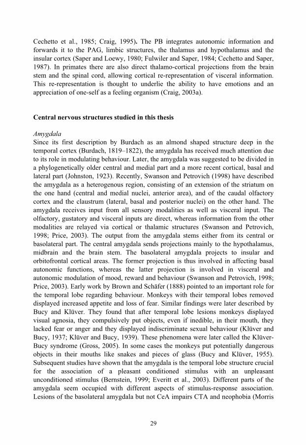
Fig. 3. A transverse section of the right parabrachial nucleus showing a schematic representation of the parabrachial subnuclei.. Up is dorsal and right is lateral. 4v is the fourth ventricle, meV is the mesencephalic trigeminal nucleus, mtV is the mesencephalic trigeminal tract, and scp is the superior cerebellar peduncle.
Parabrachial nucleus The parabrachial nucleus (PB) was named after its location around the brachium conjunctivum (superior cerebellar peduncle, SCP) in the pons (Olszewski and Baxter, 1954; Taber, 1961). Its rostro-caudal extent in humans is about 2.2 mm and it consists of several subnuclei. The different subnuclei can be identified by their location, neuronal morphology, neuropeptide expression and projection patterns (Saper and Loewy, 1980; Fulwiler and Saper, 1984; Mantyh and Hunt, 1984; Block et al., 1989).
scp
eloeli
em
PBm
dl
slcl
il
meVvl
4v
mtV
hypothalamus
intralaminar thalamus
cortex
amygdala,bed nucleusof the striaterminalis,basal forebrain
thalamicvisceralrelay
31
In rats, ten different subnuclei have been identified (Fulwiler and Saper, 1984) (Fig. 3). The medial part of the PB is involved in the processing of gustatory information (Norgren and Leonard, 1973), whereas the lateral part is involved in processing somatosensory and visceral information (Nagai et al., 1987; Chamberlin and Saper, 1992; Yamamoto et al., 1992; Li and Rowland, 1993; Chamberlin and Saper, 1994; Yamamoto et al., 1994; Saper, 1995; Hermanson and Blomqvist, 1996; Hermanson and Blomqvist, 1997c; Sakai and Yamamoto, 1997; Singewald and Sharp, 2000; Ballesteros et al., 2002). The PB receives afferent fibers from spinal and trigeminal lamina I neurons and the NTS (Wiberg et al., 1987; Ma and Peschanski, 1988; Blomqvist et al., 1989; Herbert et al., 1990; Jia et al., 1994; Feil and Herbert, 1995; Karimnamazi et al., 2002; Dallel et al., 2004) and it projects to the PAG, amygdala, thalamus, hypothalamus, and insular cortex (Saper and Loewy, 1980; Block and Schwartzbaum, 1983; Fulwiler and Saper, 1984; Bernard et al., 1991; Halsell, 1992; Bernard et al., 1993; Alden et al., 1994; Bester et al., 1999; Tkacs and Li, 1999; Krout and Loewy, 2000; Richard et al., 2005). The PB is reciprocally connected to the areas which it sends axons to (Hopkins and Holstege, 1978; Veening et al., 1984; Simerly and Swanson, 1988; Zardetto-Smith et al., 1988; Moga et al., 1989; Moga et al., 1990a; Moga et al., 1990b; Sim and Joseph, 1991; Petrovich and Swanson, 1997; Krout et al., 1998). The projection patterns of PB are largely conserved throughout phylogeny, but there are some differences. In primates, there are no PB-cortical projections and the hypothalamic projections are more limited than in the rat. Further, the PB does not seem to be as important as gustatory relay in primates as in rodents. This is in contrast to the PB-amygdala projection that is as prominent in primates as it is in rodents (Pritchard et al., 2000).
The external lateral subnucleus (PBel) The PBel cells are multipolar and somewhat larger and more densely packed than the cells of the neighbouring central lateral subnucleus. Dorsally PBel borders on the central lateral subnucleus (PBcl), medially it is juxtaposed to the SCP and at the dorsolateral tip of the peduncle it borders on the PBem. Laterally, PBel neurons borders on the lateral lemniscus, except in the middle part of its rostrocaudal extent where a distinct cell group, called the extreme lateral subnucleus can be found between the lemniscus and PBel. The PBel extends throughout most of the rostrocaudal extension of PB and it can be divided, functionally and anatomically, into an inner and outer part relative to the SCP. The part closest to the peduncle is called PBeli and the outer part is called PBelo (Fig. 3). Tracing studies have shown that PBeli receives input from area postrema and medial NTS and projects to zona incerta and substantia innominata (Fulwiler and Saper, 1984; Herbert et al., 1990). The PBelo receives input from the dorsomedial NTS, the outer rim of the area postrema and lamina I of the trigeminal spinal nucleus, and its efferent fibers project to the amygdala, and the preoptic, lateral and paraventricular hypothalamus. The projection to the central amygdala is the dominant output of the PBelo (Fulwiler and Saper, 1984; Herbert et al., 1990; Feil and Herbert, 1995).
32
The external medial subnucleus (PBem) The PBem borders dorsolaterally on the PBel and dorsomedially on the medial PB giving it a position at the lateral tip of the SCP and extending medially (Fig. 3). PBem receives input from the medial NTS, the area postrema and the paratrigeminal nucleus (Herbert et al., 1990; Feil and Herbert, 1995) and it projects to the parvicellular part of the ventral posterior thalamic nucleus (VPpc) (the “gustatory thalamus”) (Krout and Loewy, 2000) and insular cortex (Cechetto and Saper, 1987).
The dorsal lateral subnucleus (PBdl) The PBdl is shorter in its rostrocaudal extension than the PBel, beginning rostral to the separation of the inferior colliculus from the pons and extending about 400 μmcaudally. Medially it borders on the internal lateral subnucleus and ventrally on the central lateral subnucleus (Fig. 3). Dorsally the PBdl adjoins the ventral spino-cerebellar tract. PBdl neurons project mainly to hypothalamic nuclei with the main terminal field being the median preoptic nucleus. There are also less prominent projections to the paraventricular, ventromedial, dorsomedial and lateral hypothalamus as well as to the bed nucleus of the stria terminalis (Fulwiler and Saper, 1984; Hermanson, 1997). The bulk of the afferent fibers to the PBdl comes from lamina I in the spinal cord, and the PBdl is the main PB termination site for lamina I PB projection neurons (Blomqvist et al., 1989; Slugg and Light, 1994; Bernard et al., 1995). There are some trigeminal fibers terminating in the PBdl as well as some input from the NTS and the ventrolateral medulla (Fulwiler and Saper, 1984; Herbert et al., 1990).
The central lateral subnucleus (PBcl) The PBcl is a loosely organised subnucleus with less dense concentration of neurons compared to the surrounding subnuclei. Its neurons are ovoid and fusiform in shape, as seen in Nissl-stained sections. In the horizontal plane PBcl extends from the PBel to the PBvl and is bordered by the PBdl dorsally and the SCP ventromedially (Fig. 3) (Fulwiler and Saper, 1984; Hermanson, 1997). The afferent input to the PBcl comes from the medial NTS, paratrigeminal nucleus, and the spinal cord (Herbert et al., 1990; Feil and Herbert, 1995). Its projection pattern is similar to that of PBdl, with the median preoptic nucleus and the bed nucleus of the stria terminalis being the principal targets (Fulwiler and Saper, 1984; Hermanson, 1997).
The superior lateral subnucleus (PBsl) The PBsl is confined to the rostral third of the PB and partly extends rostrally to other subnuclei. It consists of pyramidal or multipolar neurons with a prominent nucleus. It is located dorsally to the PBdl (Fig. 3). PBsl receives input from superficial and deeper layers of the trigeminal dorsal horn and spinal cord, the PAG, and some hypothalamic structures (Moga et al., 1990a; Bernard et al., 1995; Feil and Herbert, 1995; Krout et al., 1998). It projects primarily to the ventromedial hypothalamic nucleus (Fulwiler and Saper, 1984; Bester et al., 1997; Hermanson et al., 1998).

33
The internal lateral subnucleus (PBil) The PBil is in Nissl-stained sections an easily recognisable structure situated in the middle third of the rostrocaudal extent of PB. It constitutes a round group of rounded neurons situated dorsal and lateral to the medial tip of the SCP (Fig. 3) (Fulwiler and Saper, 1984). The deep laminae of the dorsal horn of the spinal cord project to the PBil, and PBil neurons send their axons to the intralaminar thalamic nuclei (Fulwiler and Saper, 1984; Bernard et al., 1995; Hermanson and Blomqvist, 1997b; Bester et al., 1999; Krout and Loewy, 2000).
The ventral lateral subnucleus (PBvl) The PBvl, located along the dorsal medial side of the SCP (Fig. 3), is discernible from the PBcl due to its higher packing density of neurons. The PBvl is found in the caudal to lower middle part of the PB. PBvl receives afferent fibers from the NTS and projects to the lateral hypothalamic area and the zona incerta (Fulwiler and Saper, 1984; Herbert et al., 1990).
The medial subnucleus (PBm) On the medial side of the SCP, a heterogenous collection of neurons called the PBm is found (Fig. 3). The neurons of the PBm differ in size and morphology and they project mainly to the thalamus but there are minor projections to the frontal, insular and infralimbic cortices, amygdala, substantia innominata and zona incerta as well (Fulwiler and Saper, 1984). The principal input to the PBm stems from the NTS with some fibers also coming from the trigeminal dorsal horn. In cats and primates the PBm receives additional input from the dorsal horn of the spinal cord (Wiberg and Blomqvist, 1984; Herbert et al., 1990; Craig, 1995).
Neuropeptides Neuropeptides are polypeptide molecules that are used as neurotransmittors in the CNS. They are capable of long or short term modulation of the postsynaptic neuron. After synthesis, neuropeptides are packed in dense core vesicles (DCV) and transported from the soma to the presynaptic boutons (Burbach et al., 2001). Thus the levels of neuropeptides are usually low in the soma and when one wants to study the site of origin of a neuropeptide it is easier to localise its mRNA than the peptide itself. Since there are no re-uptake mechanisms, neuropeptides must be re-synthesised once released into the synaptic cleft (MacArthur and Eiden, 1996). Accordingly, it has been shown that a stimulus causing neuropeptide release also leads to synthesis of the same peptide, “stimulus-secretion-synthesis coupling” (Eiden et al., 1984). Conceivably, the amount of neuropeptide mRNA in a cell is dependent on at least three variables: the rate of synthesis, the presence of mRNA stabilisers and the rate of mRNA degradation. It has been shown that Ca2+-signalling can induce mRNA stability in neurons (Fukuchi et al., 2004). This means that the absence of increased mRNA transcription after a stimulus does not mean the absence of increased mRNA translation. Further, many neuronal somatas are located at considerable distance from
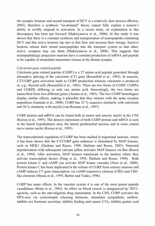
34
the synaptic boutons and axonal transport of DCV is a relatively slow process (Brown, 2003), therefore a synthesis “on-demand” theory cannot fully explain a neuron’s ability to swiftly respond to activation. In a recent study, an explanation to this discrepancy has been put forward (Shakiryanova et al., 2006). In this study it was shown that there is a constant synthesis and transportation of neuropeptide-containing DCV and that active boutons tap into to this flow and increase their storage. Inactive boutons release their stored neuropeptides into the transport system so that other, active, synapses may use them (Shakiryanova et al., 2006). This suggests that neuropeptidergic projection neurons have a constant production of mRNA and peptide to be capable of immediate transmitter release at the distant synapse.
Calcitonin gene related peptide Calcitonin gene related peptide (CGRP) is a 37 amino acid peptide generated through alternative splicing of the calcitonin (CT) gene (Rosenfeld et al., 1983). In neurons, CT/CGRP gene activation leads to CGRP production whereas calcitonin is produced in, e.g., thyroid cells (Rosenfeld et al., 1983). There are two forms of CGRP, CGRPand CGRP , differing in only one amino acid. Interestingly, the two forms are transcribed from two different genes (Amara et al., 1985). The two CGRP homologues display similar effects, making it plausible that they interact with the same receptor population (Juaneda et al., 2000). CGRP has 25 % sequence similarity with calcitonin and 50 % similarity with amylin (van Rossum et al., 1997).
CGRP protein and mRNA can be found both in motor and sensory nuclei in the CNS (Kresse et al., 1995). The densest expression of both CGRP protein and mRNA is seen in the lateral hypothalamic area, the lateral parabrachial nucleus and in some cranial nerve motor nuclei (Kresse et al., 1995).
The transcriptional regulation of CGRP has been studied in trigeminal neurons, where it has been shown that the CT/CGRP gene enhancer is stimulated by MAP kinases, such as MEK1 (Durham and Russo, 1998; Durham and Russo, 2003). Neuronal depolarization with subsequent calcium influx activates MAP kinases via Ras (Rosen et al., 1994). After activation, MAP kinases translocate to the nucleus where they activate transcription factors (Pang et al., 1995; Durham and Russo, 1998). Both protein kinase C and cAMP can activate MAP kinase cascades (Naor et al., 2000). Protein kinase C has been implicated in the release of CGRP from sensory neurons and cAMP induces CT gene transcription via cAMP-responsive element (CRE) and CRE-like elements (Monia et al., 1995; Barber and Vasko, 1996).
CGRP has many effects. In the vascular system it is one of the most potent peptide vasodilators (Brain et al., 1985). Its effect on blood vessels is antagonized by 5HT1-agonists, such as the anti-migraine drug sumatriptan. In the CNS, CGRP activates the HPA-axis via corticotropin releasing hormone, stimulates sympathetic outflow, inhibits sex hormone secretion, inhibits feeding and causes CTA, inhibits gastric acid
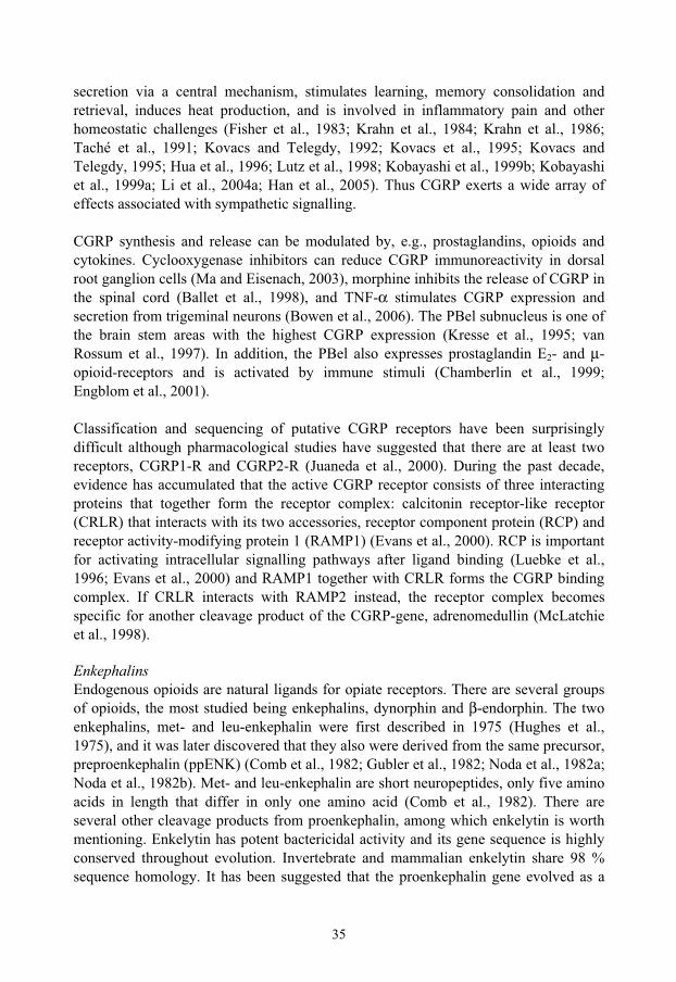
35
secretion via a central mechanism, stimulates learning, memory consolidation and retrieval, induces heat production, and is involved in inflammatory pain and other homeostatic challenges (Fisher et al., 1983; Krahn et al., 1984; Krahn et al., 1986; Taché et al., 1991; Kovacs and Telegdy, 1992; Kovacs et al., 1995; Kovacs and Telegdy, 1995; Hua et al., 1996; Lutz et al., 1998; Kobayashi et al., 1999b; Kobayashi et al., 1999a; Li et al., 2004a; Han et al., 2005). Thus CGRP exerts a wide array of effects associated with sympathetic signalling.
CGRP synthesis and release can be modulated by, e.g., prostaglandins, opioids and cytokines. Cyclooxygenase inhibitors can reduce CGRP immunoreactivity in dorsal root ganglion cells (Ma and Eisenach, 2003), morphine inhibits the release of CGRP in the spinal cord (Ballet et al., 1998), and TNF-α stimulates CGRP expression and secretion from trigeminal neurons (Bowen et al., 2006). The PBel subnucleus is one of the brain stem areas with the highest CGRP expression (Kresse et al., 1995; van Rossum et al., 1997). In addition, the PBel also expresses prostaglandin E2- and μ-opioid-receptors and is activated by immune stimuli (Chamberlin et al., 1999; Engblom et al., 2001).
Classification and sequencing of putative CGRP receptors have been surprisingly difficult although pharmacological studies have suggested that there are at least two receptors, CGRP1-R and CGRP2-R (Juaneda et al., 2000). During the past decade, evidence has accumulated that the active CGRP receptor consists of three interacting proteins that together form the receptor complex: calcitonin receptor-like receptor (CRLR) that interacts with its two accessories, receptor component protein (RCP) and receptor activity-modifying protein 1 (RAMP1) (Evans et al., 2000). RCP is important for activating intracellular signalling pathways after ligand binding (Luebke et al., 1996; Evans et al., 2000) and RAMP1 together with CRLR forms the CGRP binding complex. If CRLR interacts with RAMP2 instead, the receptor complex becomes specific for another cleavage product of the CGRP-gene, adrenomedullin (McLatchie et al., 1998).
Enkephalins Endogenous opioids are natural ligands for opiate receptors. There are several groups of opioids, the most studied being enkephalins, dynorphin and β-endorphin. The two enkephalins, met- and leu-enkephalin were first described in 1975 (Hughes et al., 1975), and it was later discovered that they also were derived from the same precursor, preproenkephalin (ppENK) (Comb et al., 1982; Gubler et al., 1982; Noda et al., 1982a; Noda et al., 1982b). Met- and leu-enkephalin are short neuropeptides, only five amino acids in length that differ in only one amino acid (Comb et al., 1982). There are several other cleavage products from proenkephalin, among which enkelytin is worth mentioning. Enkelytin has potent bactericidal activity and its gene sequence is highly conserved throughout evolution. Invertebrate and mammalian enkelytin share 98 % sequence homology. It has been suggested that the proenkephalin gene evolved as a
36
defence against microbes and that the noxious sensation of invading pathogens was the seed of the emotion pain. Along this line of reasoning, the analgesic peptide enkephalin may have evolved in parallel with enkelytin to stop us from being crippled of pain and unable to respond when threatened (Stefano et al., 2005). Indeed, opioids seem to be able to modulate the immune-response (Buller et al., 2005).
Transcription of ppENK can be initiated via the ENKCRE2 promoter which is sufficient for basal and inducible ppENK transcription (Kobierski et al., 1991). The ENKCRE2 promoter contains binding sites for CRE and AP-1 transcription factors (Comb et al., 1988) and ppENK transcription is preceded by c-fos, c-jun and junB gene upregulation (Bacher et al., 1996). The regulation of the ppENK gene by either CREB or AP-1 seems to be stimulus and tissue specific (Bacher et al., 1996; Kobierski et al., 1999; Hahm et al., 2003). In the CNS, morphological studies have implicated both CREB and c-fos in ppENK transcription (Hermanson and Blomqvist, 1997a; Engström et al., 2003).
The densest expression of ppENK mRNA in the brain stem is in the PB (Harlan et al., 1987), where it can be found in the PBil, PBvl and PBel subnuclei (Hermanson and Blomqvist, 1997b). Enkephalin binds to δ- and μ- opioid receptors (Waldhoer et al., 2004). PBel neurons also heavily express μ-opioid receptors and many of the μ-opioid receptor expressing neurons project to the amygdala (Chamberlin et al., 1999). Activation of parabrachial μ- opioid receptor results in increased feeding and chronic food restriction results in decreased μ-opioid receptor binding in the PBel and PBem (Wolinsky et al., 1996; Wilson et al., 2003).
Melanocortins Melanocortins are a family of peptides that bind to the melanocortin receptors (MC-R). There are five MC-Rs discovered so far. MC1-R is expressed on melanocytes and is involved in regulating the production of melanin, the pigment that determines skin colour. MC2-R is expressed in the adrenal cortex and controls the secretion of glucocorticoids. MC3 and 4-R are expressed in the CNS and are involved in appetite and metabolic control. MC5-R is implicated in the regulation of secretion from exocrine glands (Tatro, 1996; Balthasar et al., 2005). The melanocortin receptors are G-protein-coupled receptors that increase cAMP and intracellular calcium upon activation (Gantz et al., 1993; Mountjoy et al., 2001). Further, MC4-R activation also leads to MAP kinase activation through inositol triphosphate signalling (Daniels et al., 2003; Vongs et al., 2004). Activated MAP kinases can then lead to altered gene regulation (Pearson et al., 2001). The melanocortins derive from a precursor molecule, proopiomelanocortin (POMC). Cleavage of POMC yields melanocortins or β-endorphins. The melanocortins are ACTH and α-, β- or γ-melanocyte stimulating hormone (α-, β-, γ-MSH) (Cone, 2005). All melanocortins are agonists to the MC-R. There is also a natural antagonist, agouti related peptide (AgRP) (Wilson et al., 1999).
37
Recently there has been focus on the cerebral MC3 and 4 receptors due to their impact on appetite and metabolic signalling (Hagan et al., 1999; Marks et al., 2003; Balthasar et al., 2004; Balthasar et al., 2005). Activation of CNS melanocortin receptors decreases appetite and MC4-R knockouts display obesity, hyperinsulinemia and hyperphagia (Huszar et al., 1997). The hormone leptin, which is produced by the adipose tissue and signals the size of fat depots to the CNS, activates arcuate POMC neurons and MC4-R expressing cells in the hypothalamus (Balthasar et al., 2004). MC4-R are widely expressed in the brain and they can be found in many regions involved in metabolic and appetite control like the paraventricular and ventromedial hypothalamus (Kishi et al., 2003). POMC, the ligand precursor, is only expressed in the arcuate nucleus and the NTS, whereas AgRP is only expressed in the arcuate nucleus. However, even if their cell bodies are confined to a few nuclei, α-MSH and AgRP positive fibers can be found in many areas of the CNS (Palkovits et al., 1987; Joseph and Michael, 1988; Broberger et al., 1998; Bagnol et al., 1999; Fekete et al., 2000). Mostly AgRP and POMC fibers follow suit (Bagnol et al., 1999). There are few studies on the origin of brain stem α-MSH fibers but the available evidence suggests that half of them derive from the hypothalamus and the other half from the NTS (Joseph and Michael, 1988; Cone, 2005). So far, the hypothalamic melanocortin pathways have been most studied, and it is clear that melanocortins play an important role in the hypothalamus as regulators of metabolism and food intake (Cone, 2005). There is much less data on the role of melanocortins in the brain stem. The brain stem is important in the short term regulation of food intake and satiety but does seem to lack the capability to respond adequately to long term food-deprivation (Grill and Kaplan, 2002). The forebrain on the other hand is capable of responding to the availability of energy resources over time, but in order to accomplish this, it seems to be dependent on brain stem input (Grill and Kaplan, 2002). Thus, the activation of brain stem melanocortin receptors is sufficient to decrease food intake (Grill et al., 1998) and MC4-R are expressed in important brain stem autonomic nuclei such as the NTS and PB (Kishi et al., 2003). Further, NTS POMC neurons are involved in mediating CCK-induced satiety, and brain stem MC4-Rs are important in mediating the deceased food intake seen after the intraperitoneal injection of CCK (Fan et al., 2004). However, there has been no anatomical studies on the influence of the melanocortin system on brain stem areas activated after immune and aversive stimulation.

38
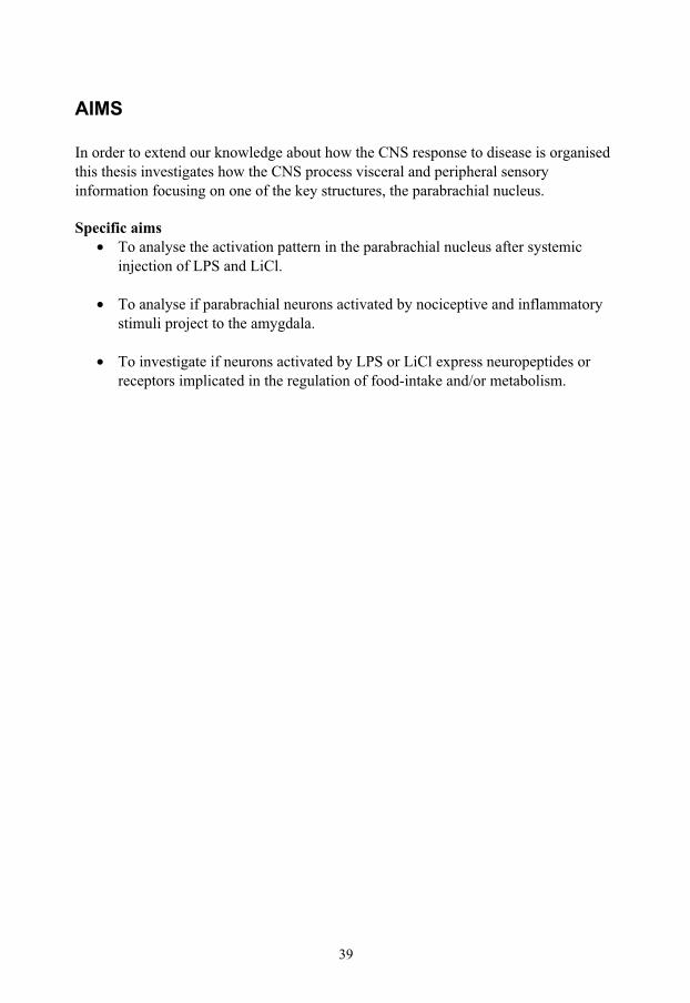
39
AIMS
In order to extend our knowledge about how the CNS response to disease is organised this thesis investigates how the CNS process visceral and peripheral sensory information focusing on one of the key structures, the parabrachial nucleus.
Specific aims • To analyse the activation pattern in the parabrachial nucleus after systemic
injection of LPS and LiCl.
• To analyse if parabrachial neurons activated by nociceptive and inflammatory stimuli project to the amygdala.
• To investigate if neurons activated by LPS or LiCl express neuropeptides or receptors implicated in the regulation of food-intake and/or metabolism.

40
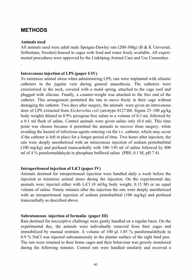
41
METHODS
Animals used All animals used were adult male Sprague-Dawley rats (200-300g) (B & K Universal, Sollentuna, Sweden) housed in cages with food and water freely available. All experi-mental procedures were approved by the Linköping Animal Care and Use Committee. .
Intravenous injection of LPS (paper I-IV) To minimize animal stress when administering LPS, rats were implanted with siliastic catheters in the jugular vein during general anaesthesia. The catheters were exteriorized in the neck, covered with a metal spring, attached to the cage roof and plugged with silicone. Finally, a counter-weight was attached to the free end of the catheter. This arrangement permitted the rats to move freely in their cage without damaging the catheter. Two days after surgery, the animals were given an intravenous dose of LPS extracted from Escherichia coli (serotype 0127:B8, Sigma 25–100 μg/kg body weight) diluted in 0.9% pyrogene-free saline to a volume of 0.3 ml, followed by a 0.1 ml flush of saline. Control animals were given saline only (0.4 ml). This time point was chosen because it permitted the animals to recover from surgery, while avoiding the hazard of infectious agents entering via the i.v. catheter, which may occur if the catheter is left in place for a longer period of time. Two hours after injection, the rats were deeply anesthetized with an intravenous injection of sodium pentobarbital (100 mg/kg) and perfused transcardially with 100–150 ml of saline followed by 800 ml of 4 % paraformaldehyde in phosphate buffered saline (PBS, 0.1 M, pH 7.4).
Intraperitoneal injection of LiCl (paper IV) Animals destined for intraperitoneal injection were handled daily a week before the injection to minimize animal stress during the injection. On the experimental day animals were injected either with LiCl (8 ml/kg body weight; 0.15 M) or an equal volume of saline. Ninety minutes after the injection the rats were deeply anesthetized with an intraperitoneal injection of sodium pentobarbital (100 mg/kg) and perfused transcardially as described above.
Subcutaneous injection of formalin (paper III) Rats destined for nociceptive challenge were gently handled on a regular basis. On the experimental day, the animals were individually removed from their cages and immobilized by manual restraint. A volume of 100 μl 1.85 % paraformaldehyde in 0.9 % NaCl was injected subcutaneously in the plantar surface of the right hind paw. The rats were returned to their home cages and their behaviour was grossly monitored during the following minutes. Control rats were handled similarly and received a
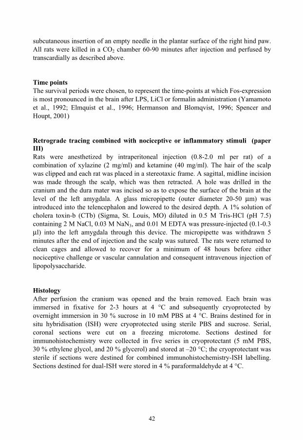
42
subcutaneous insertion of an empty needle in the plantar surface of the right hind paw. All rats were killed in a CO2 chamber 60-90 minutes after injection and perfused by transcardially as described above.
Time points The survival periods were chosen, to represent the time-points at which Fos-expression is most pronounced in the brain after LPS, LiCl or formalin administration (Yamamoto et al., 1992; Elmquist et al., 1996; Hermanson and Blomqvist, 1996; Spencer and Houpt, 2001)
Retrograde tracing combined with nociceptive or inflammatory stimuli (paper III)Rats were anesthetized by intraperitoneal injection (0.8-2.0 ml per rat) of a combination of xylazine (2 mg/ml) and ketamine (40 mg/ml). The hair of the scalp was clipped and each rat was placed in a stereotaxic frame. A sagittal, midline incision was made through the scalp, which was then retracted. A hole was drilled in the cranium and the dura mater was incised so as to expose the surface of the brain at the level of the left amygdala. A glass micropipette (outer diameter 20-50 μm) was introduced into the telencephalon and lowered to the desired depth. A 1% solution of cholera toxin-b (CTb) (Sigma, St. Louis, MO) diluted in 0.5 M Tris-HCl (pH 7.5) containing 2 M NaCl, 0.03 M NaN3, and 0.01 M EDTA was pressure-injected (0.1-0.3 μl) into the left amygdala through this device. The micropipette was withdrawn 5 minutes after the end of injection and the scalp was sutured. The rats were returned to clean cages and allowed to recover for a minimum of 48 hours before either nociceptive challenge or vascular cannulation and consequent intravenous injection of lipopolysaccharide.
HistologyAfter perfusion the cranium was opened and the brain removed. Each brain was immersed in fixative for 2-3 hours at 4 °C and subsequently cryoprotected by overnight immersion in 30 % sucrose in 10 mM PBS at 4 °C. Brains destined for in situ hybridisation (ISH) were cryoprotected using sterile PBS and sucrose. Serial, coronal sections were cut on a freezing microtome. Sections destined for immunohistochemistry were collected in five series in cryoprotectant (5 mM PBS, 30 % ethylene glycol, and 20 % glycerol) and stored at –20 °C; the cryoprotectant was sterile if sections were destined for combined immunohistochemistry-ISH labelling. Sections destined for dual-ISH were stored in 4 % paraformaldehyde at 4 °C.
43
Double-labelling immunohistochemistry (paper III) Immunohistochemistry was performed at room temperature on free-floating sections. To permit comparisons across the treatment groups, sections from different animals were processed simultaneously. In addition, a standardized protocol was strictly adhered to. Unless otherwise specified, 10 mM PBS was used to dilute reagents and to wash sections between steps. Endogenous peroxidases were blocked with 0.3 % H2O2.Subsequently, sections were incubated overnight with primary antibodies directed against Fos (rabbit anti-Fos antibody; Santa Cruz Biotechnology, Santa Cruz, CA; diluted 1:6,000) and CTb (goat anti-CTb antibody; List Biological Laboratories, Campbell, CA; diluted 1:10,000) with 1 % bovine serum albumin (BSA; Sigma), followed by secondary antibody incubation. For the visualization of anti-Fos antibodies sections were incubated with rabbit peroxidase antiperoxidase complex (Dako, 1:150) with 1 % BSA, followed by 0.02 % 3,3´diaminobenzidine (Sigma), 2.5 % ammonium nickel sulfate, and 0.01 % H2O2 in NaAc. Subsequently, anti-CTb antibodies were visualized by incubation with avidin-biotin complex (Vectastain ABC kit; Vector Laboratories, Burlingame, CA; 1:2,000), followed by 0.02 % 3,3´diamino-benzidine and 0.01 % H2O2 in tris buffer. Sections were mounted onto glass slides, air-dried overnight, dehydrated in 100 % ethanol, defatted in xylene, mounted in DPX (VWR International, Stockholm, Sweden), and coverslipped. Adjacent sections were stained with thionin.
Dual-labelling in situ-hybridization (paper II and IV) Dual labelling in situ hybridization was performed as previously described (Engblom et al., 2001; Engström et al., 2003). Sections were mounted on sterile slides and dried overnight. The slides were post-fixed in 4 % paraformaldehyde, rinsed and incubated for 20 min at 37 °C in 0.001 % proteinase K.. The slides were then rinsed, dehydrated and defatted in 100 % chloroform, rehydrated in ethanol and air dried. Hybridization was performed under coverslips in a hybridization chamber at 60 °C for 20 hours. Probe concentrations used were 1:1000 or 1:2000 for the digoxigenin-labelled probe and 107 cpm/ml for the radio-labelled probe. The MC4-R probe was labelled with [α-35S]-UTP (New England Nuclear, Boston, MA) whereas the CGRP-probe was labelled either with [α-35S]-UTP (paper II) or digoxigenin (paper IV). The c-fos probes were digoxigenin-labelled. Specificity of the c-fos, MC4-R and CGRP probes was shown by comparing their labelling pattern with earlier data (Rosenfeld et al., 1983; Amara et al., 1985; Mountjoy et al., 1994; Paues et al., 2001; Kishi et al., 2003). The specificity of the c-fos expression after LPS and LiCl was determined by comparing the obtained results with previous data on the distribution of c-fos mRNA and FOS protein, respectively, after immune and aversive stimuli and by the absence of expression in PB in naïve animals (Yamamoto et al., 1992; Elmquist et al., 1996; Swank, 1999; Paues et al., 2001).

44
Following hybridization the slides were soaked in standard saline citrate buffer (SSC; pH 7.0), after which the coverslips were removed. Subsequently the slides were rinsed, incubated in 0.002 % RNase A, rinsed in decreasing concentrations of SSC, subjected to a stringency bath heated to 73 °C in 0.1X SSC, and finally rinsed and air dried. For immunohistochemical detection of the digoxigenin-labelled probe, slides were first immersed in blocking buffer for 2 h at room temperature. They were then rinsed and incubated in anti-digoxigenin Fab fragments coupled to alkaline phosphatase (Roche, Mannheim, Germany) diluted 1:5000. After rinsing the sections were developed in 4-nitro blue tetrazolium chloride (100 mg/ml; Roche), 5-bromo-4-chloro-3-indolyl-phosphate (50 mg/ml; Roche), and levimasole (0.25 mg/ml; Sigma). Following new rinsing, the slides were dipped quickly in 70 % ethanol and air dried. In order to avoid darkening of the sections induced by the emulsion or the developer, slides were dipped twice in 2 % collodion/isoamyl acetate solution (Electron Microscopy Sciences, Fort Washington, PA), after which they were allowed to dry for 48 h before being dipped in photographic emulsion (Kodak NTB-2; Kodak, Rochester, NY). After exposure for 2-3 weeks the slides were developed in D-19 (Kodak), fixed, rinsed and coverslipped.
Immunohistochemistry combined with in situ-hybridization (paper I) In short, sections were incubated overnight with a primary antibody directed against Fos (rabbit anti-Fos antibody; Santa Cruz Biotechnology, Santa Cruz, CA; diluted 1:6,000), followed by incubation in a secondary antibody. Visualisation was made by rabbit peroxidase antiperoxidase complex (Dako, 1:150), followed after intervening washes by 0.02 % 3,3´diaminobenzidine (Sigma), 2.5 % ammonium nickel sulfate, and 0.01 % H2O2 in NaAc. All liquids and reagents used were sterile to allow for subsequent mRNA detection by ISH. ISH was performed using a preproenkephalin riboprobe, synthesised with 33P- or 35S-labeled UTP (NEN, Boston, MA). The sections were hybridized for 48 h at 58 °C with a solution containing a final probe concentration of 1×10⁷ cpm/ml or 1×10⁶ cpm/ml. After posthybridization, dehydration and defatting, the slides were dipped in photographic emulsion (Kodak NTB2), stored at 4 °C for 10–14 days, developed in Kodak D-19 and coverslipped.
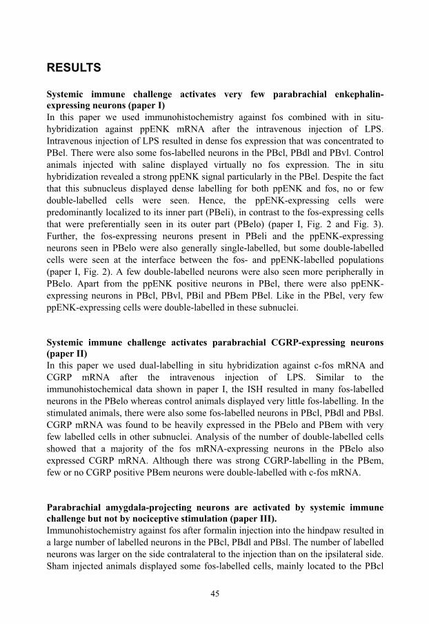
45
RESULTS
Systemic immune challenge activates very few parabrachial enkephalin-expressing neurons (paper I) In this paper we used immunohistochemistry against fos combined with in situ-hybridization against ppENK mRNA after the intravenous injection of LPS. Intravenous injection of LPS resulted in dense fos expression that was concentrated to PBel. There were also some fos-labelled neurons in the PBcl, PBdl and PBvl. Control animals injected with saline displayed virtually no fos expression. The in situ hybridization revealed a strong ppENK signal particularly in the PBel. Despite the fact that this subnucleus displayed dense labelling for both ppENK and fos, no or few double-labelled cells were seen. Hence, the ppENK-expressing cells were predominantly localized to its inner part (PBeli), in contrast to the fos-expressing cells that were preferentially seen in its outer part (PBelo) (paper I, Fig. 2 and Fig. 3). Further, the fos-expressing neurons present in PBeli and the ppENK-expressing neurons seen in PBelo were also generally single-labelled, but some double-labelled cells were seen at the interface between the fos- and ppENK-labelled populations (paper I, Fig. 2). A few double-labelled neurons were also seen more peripherally in PBelo. Apart from the ppENK positive neurons in PBel, there were also ppENK-expressing neurons in PBcl, PBvl, PBil and PBem PBel. Like in the PBel, very few ppENK-expressing cells were double-labelled in these subnuclei.
Systemic immune challenge activates parabrachial CGRP-expressing neurons (paper II) In this paper we used dual-labelling in situ hybridization against c-fos mRNA and CGRP mRNA after the intravenous injection of LPS. Similar to the immunohistochemical data shown in paper I, the ISH resulted in many fos-labelled neurons in the PBelo whereas control animals displayed very little fos-labelling. In the stimulated animals, there were also some fos-labelled neurons in PBcl, PBdl and PBsl. CGRP mRNA was found to be heavily expressed in the PBelo and PBem with very few labelled cells in other subnuclei. Analysis of the number of double-labelled cells showed that a majority of the fos mRNA-expressing neurons in the PBelo also expressed CGRP mRNA. Although there was strong CGRP-labelling in the PBem, few or no CGRP positive PBem neurons were double-labelled with c-fos mRNA.
Parabrachial amygdala-projecting neurons are activated by systemic immune challenge but not by nociceptive stimulation (paper III). Immunohistochemistry against fos after formalin injection into the hindpaw resulted in a large number of labelled neurons in the PBcl, PBdl and PBsl. The number of labelled neurons was larger on the side contralateral to the injection than on the ipsilateral side. Sham injected animals displayed some fos-labelled cells, mainly located to the PBcl
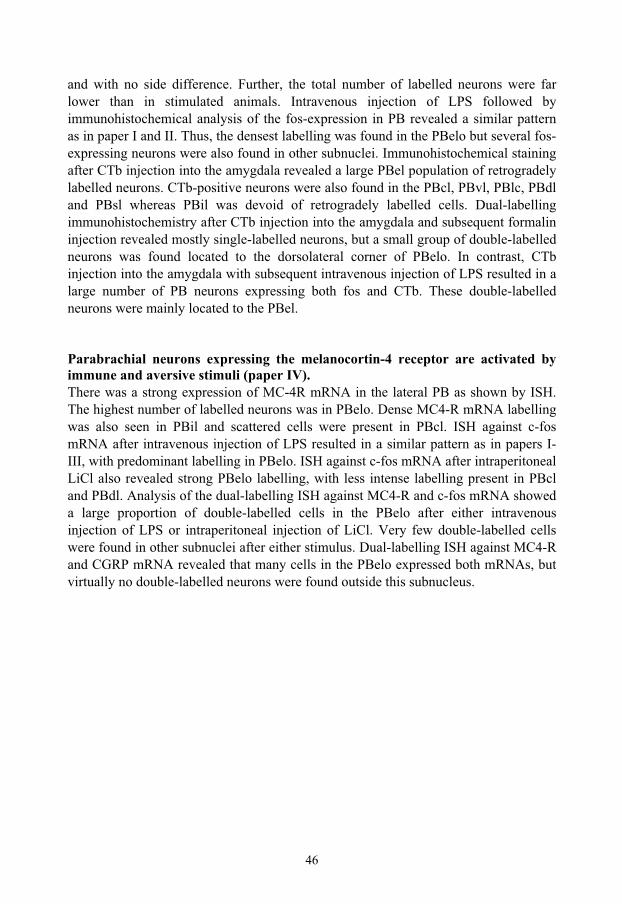
46
and with no side difference. Further, the total number of labelled neurons were far lower than in stimulated animals. Intravenous injection of LPS followed by immunohistochemical analysis of the fos-expression in PB revealed a similar pattern as in paper I and II. Thus, the densest labelling was found in the PBelo but several fos-expressing neurons were also found in other subnuclei. Immunohistochemical staining after CTb injection into the amygdala revealed a large PBel population of retrogradely labelled neurons. CTb-positive neurons were also found in the PBcl, PBvl, PBlc, PBdl and PBsl whereas PBil was devoid of retrogradely labelled cells. Dual-labelling immunohistochemistry after CTb injection into the amygdala and subsequent formalin injection revealed mostly single-labelled neurons, but a small group of double-labelled neurons was found located to the dorsolateral corner of PBelo. In contrast, CTb injection into the amygdala with subsequent intravenous injection of LPS resulted in a large number of PB neurons expressing both fos and CTb. These double-labelled neurons were mainly located to the PBel.
Parabrachial neurons expressing the melanocortin-4 receptor are activated by immune and aversive stimuli (paper IV). There was a strong expression of MC-4R mRNA in the lateral PB as shown by ISH. The highest number of labelled neurons was in PBelo. Dense MC4-R mRNA labelling was also seen in PBil and scattered cells were present in PBcl. ISH against c-fos mRNA after intravenous injection of LPS resulted in a similar pattern as in papers I-III, with predominant labelling in PBelo. ISH against c-fos mRNA after intraperitoneal LiCl also revealed strong PBelo labelling, with less intense labelling present in PBcl and PBdl. Analysis of the dual-labelling ISH against MC4-R and c-fos mRNA showed a large proportion of double-labelled cells in the PBelo after either intravenous injection of LPS or intraperitoneal injection of LiCl. Very few double-labelled cells were found in other subnuclei after either stimulus. Dual-labelling ISH against MC4-R and CGRP mRNA revealed that many cells in the PBelo expressed both mRNAs, but virtually no double-labelled neurons were found outside this subnucleus.
47
DISCUSSION
Methodological considerations
Models that simulate acute disease symptoms In this thesis we have used two models to simulate general illness and one model to evoke tissue damage.
Both endotoxin and LiCl cause general disease symptoms. Intravenous injection of endotoxin gives rise to symptoms seen during an infectious disease, e.g. fever, anorexia and lethargy. Intraperitoneal injection of LiCl creates a sense of nausea and visceral discomfort. Animals injected with LiCl become restless at first, evidenced by increased activity and chewing on cage contents. The increased activity is gradually superseded by inactivity. In contrast, hindpaw injection of formalin causes local signs of inflammation like pain, swelling and decreased use of the affected limb, but no fever or other systemic symptoms. All three models activate CNS structures and have been used to study mechanisms behind immune-to-brain signalling (LPS), adaptive behaviour in disease (LPS and LiCl), or nociceptive signalling (formalin).
A common denominator for all three disease models is that they are acute; the animals are killed within two hours after the stimulus. This is an advantage when studying the causal relationship between stimulus and response. It is more likely that an observed change in the CNS stems from the stimulus than from a confounding factor when using an acute rather than a chronic model. However, in humans, the disease conditions that cause most suffering in terms of unwanted weight loss, anorexia, pain and nausea are chronic. Therefore the ultimate goal is to find the underlying mechanisms behind the suffering seen in chronic diseases, like malignant tumours, infections and organ failure.
Can acute models then contribute to the understanding of CNS mechanisms in chronic diseases? Even if they only give a snap-shot of activity evoked in the CNS, acute models provide a starting point for the analyses. Many of the acutely activated nuclei and pathways and many of the implicated neurotransmitters are likely to be involved also in a chronic situation. Hence acute models can be useful to map pathways involved in chronic disease. The typical short-term disease is caused by micro-organisms like viruses and bacteria resulting in the common cold, flu or tonsillitis. In humans, such brief disease-episodes lasting a few days are quite common. Afflicted patients describe lethargy, anorexia and fever in addition to specific disease symptoms like a soar throat, running nose or muscular pain (Vollmer-Conna et al., 2004). These general disease symptoms can be mimicked by endotoxin injected intravenously (Konsman et al., 2002; Asarian and Langhans, 2005). Systemic injection of LPS causes fever, anorexia and lethargy in a dose-dependent manner (Givalois et al., 1994;
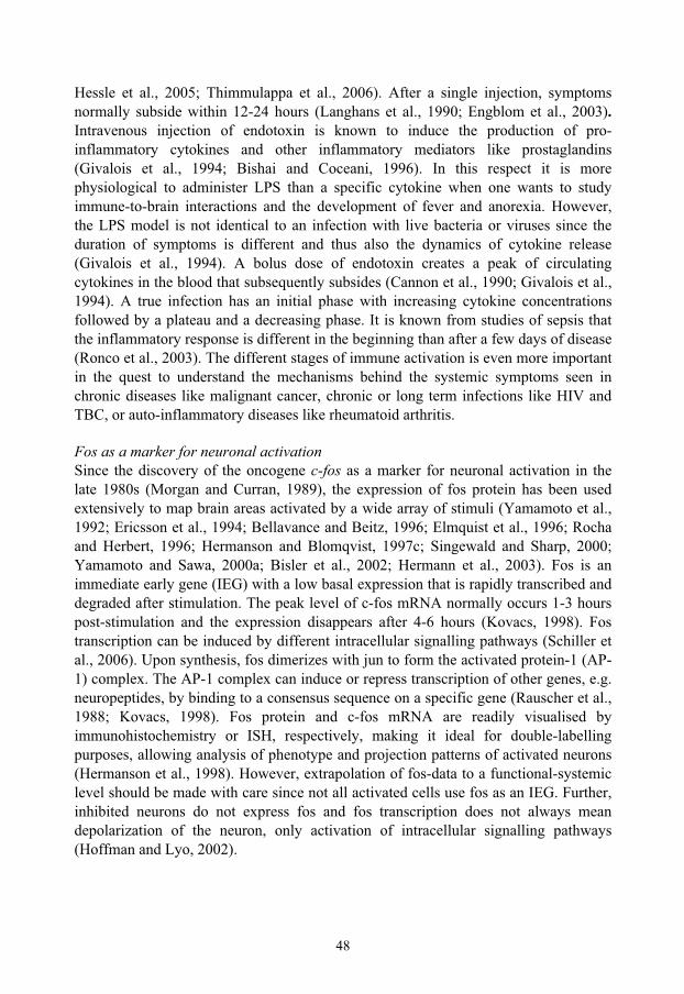
48
Hessle et al., 2005; Thimmulappa et al., 2006). After a single injection, symptoms normally subside within 12-24 hours (Langhans et al., 1990; Engblom et al., 2003).Intravenous injection of endotoxin is known to induce the production of pro-inflammatory cytokines and other inflammatory mediators like prostaglandins (Givalois et al., 1994; Bishai and Coceani, 1996). In this respect it is more physiological to administer LPS than a specific cytokine when one wants to study immune-to-brain interactions and the development of fever and anorexia. However, the LPS model is not identical to an infection with live bacteria or viruses since the duration of symptoms is different and thus also the dynamics of cytokine release (Givalois et al., 1994). A bolus dose of endotoxin creates a peak of circulating cytokines in the blood that subsequently subsides (Cannon et al., 1990; Givalois et al., 1994). A true infection has an initial phase with increasing cytokine concentrations followed by a plateau and a decreasing phase. It is known from studies of sepsis that the inflammatory response is different in the beginning than after a few days of disease (Ronco et al., 2003). The different stages of immune activation is even more important in the quest to understand the mechanisms behind the systemic symptoms seen in chronic diseases like malignant cancer, chronic or long term infections like HIV and TBC, or auto-inflammatory diseases like rheumatoid arthritis.
Fos as a marker for neuronal activation Since the discovery of the oncogene c-fos as a marker for neuronal activation in the late 1980s (Morgan and Curran, 1989), the expression of fos protein has been used extensively to map brain areas activated by a wide array of stimuli (Yamamoto et al., 1992; Ericsson et al., 1994; Bellavance and Beitz, 1996; Elmquist et al., 1996; Rocha and Herbert, 1996; Hermanson and Blomqvist, 1997c; Singewald and Sharp, 2000; Yamamoto and Sawa, 2000a; Bisler et al., 2002; Hermann et al., 2003). Fos is an immediate early gene (IEG) with a low basal expression that is rapidly transcribed and degraded after stimulation. The peak level of c-fos mRNA normally occurs 1-3 hours post-stimulation and the expression disappears after 4-6 hours (Kovacs, 1998). Fos transcription can be induced by different intracellular signalling pathways (Schiller et al., 2006). Upon synthesis, fos dimerizes with jun to form the activated protein-1 (AP-1) complex. The AP-1 complex can induce or repress transcription of other genes, e.g. neuropeptides, by binding to a consensus sequence on a specific gene (Rauscher et al., 1988; Kovacs, 1998). Fos protein and c-fos mRNA are readily visualised by immunohistochemistry or ISH, respectively, making it ideal for double-labelling purposes, allowing analysis of phenotype and projection patterns of activated neurons (Hermanson et al., 1998). However, extrapolation of fos-data to a functional-systemic level should be made with care since not all activated cells use fos as an IEG. Further, inhibited neurons do not express fos and fos transcription does not always mean depolarization of the neuron, only activation of intracellular signalling pathways (Hoffman and Lyo, 2002).
49
Studying the parabrachial nucleus The parabrachial nucleus is an autonomic relay nucleus where afferent visceral and somatic sensory information is integrated and transmitted to other brain stem and forebrain nuclei. This means that PB neurons are easily activated by changes in, e.g., blood pressure and heart rate. Altered blood pressure is inherent to stressful situations. Any experiment involving general anaesthesia or other stress is likely to cause PB activation. This problem is of particular importance when studying the PBel since neurons in its outer part and the bordering lateral crescent area have been shown to react to changes in blood pressure (Chamberlin and Saper, 1992; Rocha and Herbert, 1996). Accordingly, as described in the Methods section, great care was taken to minimise animal-stress. The intravenous injection model allowed injections unnoticed by the animals. Before intraperitoneal or hindpaw injections, animals were handled daily. These procedures resulted in very low background fos-labelling in control animals, implying that the fos-labelling seen in stimulated animals was caused by the stimulus and not caused by unspecific stress.
In situ hybridization ISH is a technique used to analyse the presence of a specific mRNA in a specific cell. The technique relies on the binding of a labelled probe to a target mRNA. Excess probe is washed away through consecutive baths with decreasing salt concentrations with a final stringency bath in a heated (72-73 C), low-salt solution. The amount of background is dependent on the specificity of the probe, duration of the hybridization, salt concentrations used during post-hybridization, temperature of the stringency bath and length of exposure of the photographic emulsion to the tissue.
In this thesis we used probes ranging from 167 bp (CGRP) to 2.1 kbp (fos). Shorter probes carry the risk of being less sensitive than longer probes, but the use of long probes may also result in unspecific labelling of mRNA due to sequence similarity. Even if the size-span of probes used was large, we saw very little background labelling in our experiments. This may be a result of very specific probes resulting in low cross-labelling, the high temperature of the stringency bath, or both. However, the effort put in to minimise unspecific labelling may have resulted in fewer positive cells. When examining the number of fos-positive cells after intravenous injection of LPS, immunohistochemical processing with antibodies directed against fos resulted in considerably more cells than ISH using a digoxigenin-labelled fos-probe.
There are also differences in sensitivity between different ISH protocols. The use of radioactively labelled probes with consecutive autoradiographic processing is a more sensitive technique than digoxigenin-labelled probes with subsequent immunohistochemical processing. Therefore radioactive ISH is more suitable for the detection of low-concentration mRNAs, such as receptor mRNA and some neuropeptide mRNAs.
50
Immunohistochemistry combined with in situ hybridization This technique allows the simultaneous detection of protein and mRNA (Hermanson et al., 1994). A drawback is that the immunohistochemical processing precedes the hybridization which may cause unwanted mRNA degradation through contamination with RNases, enzymes that degrade mRNA. To minimise this risk, all liquids and reagents used are sterilized and RNase inhibitors are added during antibody incubation steps.
Anorexia and aversion in disease Immune and aversive stimulation induced strong fos-expression in parabrachial neurons (paper I-IV). The most heavily activated subnucleus was the PBelo which represents an area where humoral as well as orofacial, gustatory and vagal information can be integrated. Accordingly, the PBelo is activated by other immune-activating compounds like IL-1, but also by orofacial nociceptive stimulation, CCK, leptin and dexfenfluramine, and sour and bitter taste stimuli (Li and Rowland, 1993; Ericsson et al., 1994; Hermanson and Blomqvist, 1997c; Elias et al., 2000; Yamamoto and Sawa, 2000b) and paper I-IV.
A large part of the LPS-activated PBelo neurons were found to project to the amygdala (paper III) (see also Tkacs and Li, 1999). The PBelo-amygdala projection has previously been found to be CGRP-ergic (Schwaber et al., 1988). CGRP is a neuropeptide suggested to be involved in modulating ascending visceral information and it has been implicated in anorexia, aversion and nociception (Krahn et al., 1984; Krahn et al., 1986; Kruger et al., 1988; Yasui et al., 1989; Blomqvist et al., 2000; Han et al., 2005). Intracerebroventricular injection of CGRP produces both anorexia and CTA. Similarly, aversion has been shown to produce elevated levels of CGRP in the insular cortex (Yamamoto et al., 1990). We found that a majority of LPS-activated PBelo neurons express CGRP (paper II). This finding, and the fact that a large portion of CGRP expressing PBelo neurons project to the amygdala (Schwaber et al., 1988), that many LPS-activated PBelo neurons project to the amygdala (paper III), and that an aversive stimulus induces strong fos-expression in the PBelo (paper IV), suggest the presence of a CGRP-positive pathway from the PBelo to the amygdala that is activated by immune and aversive stimuli. Further, the strong expression of MC-4R mRNA in PBelo neurons activated by immune and aversive stimuli suggests that this pathway may be regulated by melanocortins (Fig. 4 and paper IV).
The pathway leading to activation of the immune-responsive CGRP-expressing PBelo neurons has yet to be determined. Plausible routes are afferent input from the NTS or prostaglandins produced at the blood brain barrier. The NTS receives input from the vagus nerve and the area postrema. The vagus nerve innervates most of the viscera including the liver (Berthoud, 2004) and is as such ideally positioned to forward interoceptive information. The role of the vagus nerve in immune-to-brain signalling
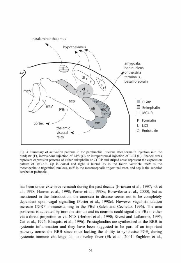
51
Fig. 4. Summary of activation patterns in the parabrachial nucleus after formalin injection into the hindpaw (F), intravenous injection of LPS (O) or intraperitoneal injection of LiCl (L). Shaded areas represent expression patterns of either enkephalin or CGRP and striped areas represent the expression pattern of MC-4R. Up is dorsal and right is lateral. 4v is the fourth ventricle, meV is the mesencephalic trigeminal nucleus, mtV is the mesencephalic trigeminal tract, and scp is the superior cerebellar peduncle.
has been under extensive research during the past decade (Ericsson et al., 1997; Ek et al., 1998; Hansen et al., 1998; Porter et al., 1998c; Borovikova et al., 2000), but as mentioned in the Introduction, the anorexia in disease seems not to be completely dependent upon vagal signalling (Porter et al., 1998c). However vagal stimulation increase CGRP immunostaining in the PBel (Saleh and Cechetto, 1996). The area postrema is activated by immune stimuli and its neurons could signal the PBelo either via a direct projection or via NTS (Herbert et al., 1990; Rivest and Laflamme, 1995; Cai et al., 1996; Elmquist et al., 1996). Prostaglandins are synthesised at the BBB in systemic inflammation and they have been suggested to be part of an important pathway across the BBB since mice lacking the ability to synthesise PGE2 during systemic immune challenge fail to develop fever (Ek et al., 2001; Engblom et al.,
FF
F
OO O
OOLL
LLL
L
F
FF
CGRP
Enkephalin
F Formalin
L LiCl
O Endotoxin
MC4-R
scp
eloeli
em
PBm
dl
slcl
il
meVvl
4v
mtV
hypothalamus
intralaminar thalamus
cortexthalamicvisceralrelay
amygdala,bed nucleusof the striaterminalis,basal forebrain
52
2003). PBelo neurons express the PGE2-receptor EP3 and these neurons are activated by intravenous LPS (Engblom et al., 2000; Engblom et al., 2001). A large proportion of EP3-expressing PBelo neurons also express CGRP (Engblom et al., 2004). PGE2-receptors have been shown to stimulate the release of CGRP from sensory neurons (Jenkins et al., 2001) and splice variants of the EP3-receptor have been implicated in signalling pathways known to induce CGRP transcription and sensitise sensory neurons (Coleman et al., 1994; Barber and Vasko, 1996; Southall and Vasko, 2001). Thus, there is evidence for a role of prostaglandins in activating PBelo-CGRP neurons in immune challenge (Fig. 5).
The presence of melanocortin receptors in the PBelo suggests additional mechanisms by which these neurons may be involved in the regulation of anorexic and aversive behaviours in disease (Fig. 4 and 5). The importance of melanocortin receptors and their ligands in energy homeostasis is well established (Cone, 2005). In paper IV we found that a large part of LPS-activated PBelo neurons also express MC4-R mRNA (paper IV). This fits well into what is known about the melanocortin system in immune-activation and the role of brain stem melanocortin receptors. Thus, the melanocortin agonist α-MSH potentiates and the non-specific MC3/4 receptor anta-gonist SHU9119 diminishes LPS-induced anorexia (Huang et al., 1999). Work by Grill and colleagues have shown that fourth ventricular application of melanocortin agonist and antagonists can have substantial impact on food intake and body weight (Grill et al., 1998) and in a recent study it was shown that 3rd ventricular application of SHU9119 is less potent in inhibiting CCK-induced anorexia than application of SHU9119 in the 4th ventricle, pointing to an important role for brain stem melanocortin receptors in the regulation of food intake (Fan et al., 2004). There is also evidence for an anti-inflammatory effect of melanocortins (Huang et al., 1999). The same study that implicated melanocortins in LPS-induced anorexia also found that α-MSH inhibits LPS-induced fever and it has been demonstrated that α-MSH can reduce hypothalamic COX-2 induction after LPS (Caruso et al., 2004). The presence of both α-MSH and AgRP-positive fibers in the PB presents a way for activation and inhibition of PBelo MC4-receptor expressing neurons (Yamazoe et al., 1984; Bagnol et al., 1999), although it remains to be shown if the α-MSH fibers originate in the arcuate nucleus or the NTS. Recently it has been shown that CCK activates commissural NTS POMC neurons (Appleyard et al., 2005). However, the commissural nucleus in the NTS projects to the PBeli and the lateral crescent area, not the PBelo (Herbert et al., 1990). This is enigmatic since MC4- and μ-opioid receptors are expressed in PBelo and not in the adjoining PBeli or lateral crescent area. One explanation is that the PBelo neurons receive melanocortin input from the arcuate nucleus and that the NTS POMC neurons project somewhere else. More studies are needed to clarify this issue.
Not only LPS activated PBelo neurons express MC4-R mRNA. There was also a high degree of co-localization between LiCl activated PBelo cells and MC4-R mRNA (Fig. 4 and paper IV). The melanocortin system has earlier been implicated in the regulation
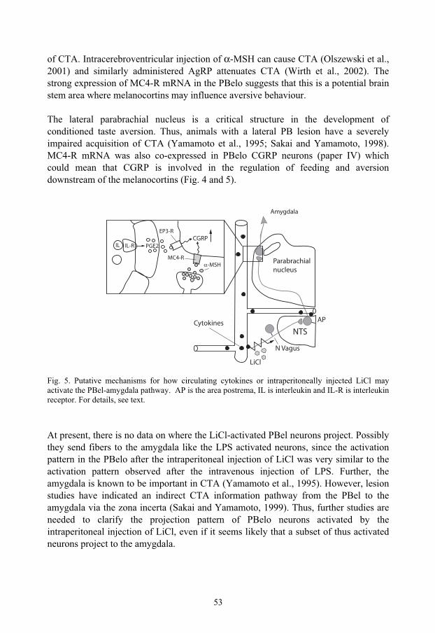
53
of CTA. Intracerebroventricular injection of α-MSH can cause CTA (Olszewski et al., 2001) and similarly administered AgRP attenuates CTA (Wirth et al., 2002). The strong expression of MC4-R mRNA in the PBelo suggests that this is a potential brain stem area where melanocortins may influence aversive behaviour.
The lateral parabrachial nucleus is a critical structure in the development of conditioned taste aversion. Thus, animals with a lateral PB lesion have a severely impaired acquisition of CTA (Yamamoto et al., 1995; Sakai and Yamamoto, 1998). MC4-R mRNA was also co-expressed in PBelo CGRP neurons (paper IV) which could mean that CGRP is involved in the regulation of feeding and aversion downstream of the melanocortins (Fig. 4 and 5).
Fig. 5. Putative mechanisms for how circulating cytokines or intraperitoneally injected LiCl may activate the PBel-amygdala pathway. AP is the area postrema, IL is interleukin and IL-R is interleukin receptor. For details, see text.
At present, there is no data on where the LiCl-activated PBel neurons project. Possibly they send fibers to the amygdala like the LPS activated neurons, since the activation pattern in the PBelo after the intraperitoneal injection of LiCl was very similar to the activation pattern observed after the intravenous injection of LPS. Further, the amygdala is known to be important in CTA (Yamamoto et al., 1995). However, lesion studies have indicated an indirect CTA information pathway from the PBel to the amygdala via the zona incerta (Sakai and Yamamoto, 1999). Thus, further studies are needed to clarify the projection pattern of PBelo neurons activated by the intraperitoneal injection of LiCl, even if it seems likely that a subset of thus activated neurons project to the amygdala.
CGRP
MC4-R
EP3-R
IL-RIL PGE2
-MSHParabrachialnucleus
Amygdala
NTSCytokines
LiCl
N Vagus
AP
54
The importance of the parabrachio-amygdaloid pathway in noxious versus immune challenge Recently, CGRP receptors in the amygdala have been implicated in the pain-affect reaction seen in an mono-arthritis model (Han et al., 2005). That study suggested an important role for CGRP in the amygdala after noxious stimulation, being in line with earlier findings that nociceptive stimulation activates the amygdala (Yamashiro et al., 1998). Further, multi-synaptic retrograde tracing studies have suggested that lamina I neurons contact amygdala-projecting parabrachial neurons (Jasmin et al., 1997). Since spinal lamina I fibers curve around the PBel to terminate in the PBdl (in contrast to trigeminal lamina I fibers which do terminate in the PBel (Feil and Herbert, 1995)), there is a discrepancy between the anatomical and the functional data. A possible explanation for the difference in the distribution of ascending afferent fibers and activation pattern evoked by these afferents as determined in electrophysiological studies is that dendrites of PBel neurons extend into the lamina I fiber-bundle that runs lateral to the PBel. The dendrites may then be contacted by collaterals from these fibers (Ma and Peschanski, 1988). However, in paper III we found very few noxiously-activated amygdala-projecting parabrachial neurons, being in contrast to previous data. One explanation for this discrepancy could be that these neurons simply do not use fos as a transcription factor after noxious stimuli. This in turn would imply that noxious stimulation of different body parts activates different transcription factors, since orofacial nociceptive stimulation evokes fos in the PBelo (Hermanson and Blomqvist, 1997c). Another explanation could be that in our study, we managed to reduce confounding, non-specific stress. The electrophysiological studies on the PBel-activation after noxious stimuli subjected the animals to general anaesthesia (Bernard et al., 1994), a stressful condition that may sensitise neurons, which in turn could lead to an altered PBel activity pattern (Krukoff et al., 1992). The model for noxious stimulation is also important. In paper III we used subcutaneous formalin injection into the hindpaw, with relatively short survival times (60-90 minutes). In the mono-arthritis study by Han et al. (2005) mentioned above, where kaolin and carrageenan was injected into a knee-joint, the survival period was six hours. It could be that the local and systemic inflammatory component was more developed in the arthritic model than in the formalin injection model. In comparison to local noxious and inflammatory stimuli, the systemic injection of LPS is a non-noxious stimulus that potently induces the acute-phase response (Gordon and Limaos, 1979; Fannin et al., 2005). Further, the intravenous injection of LPS activates many amygdala-projecting PBel neurons (paper III). Thus, the data supporting a role for the PBel-amygdala pathway in nociceptive signalling may very well have been confounded by the effects of general anaesthesia or systemic immune challenge. This would be in line with our finding in paper III that local noxious stimuli do not activate amygdala-projecting PBel neurons to any large extent.
55
Opioids in immune and aversive signallingPreproenkephalin mRNA was found to be expressed in the PBeli but not in the PBelo (paper I), and there seem to be very little overlap between the enkephalinergic neuronal population in the PBeli and the CGRP-ergic PBelo neurons. The PBeli forms a separate functional entity that is not to any significant extent activated by visceral stimuli, and that has unique afferent and efferent connections (Herbert et al., 1990; Krout and Loewy, 2000; Engblom et al., 2004). Enkephalin forms a part of the endogenous anti-nociceptive system (Akil et al., 1978; Inturrisi et al., 1980) and enkephalin can bind to μ-opioid receptors (Waldhoer et al., 2004), which have been suggested to be important in enkephalin induced analgesia (Gacel et al., 1981). Enkephalin and μ-opioid receptors have also been implicated in stimulating food intake and contributing to a basal positive hedonic state of the organism (Skoubis et al., 2001; Wilson et al., 2003; Skoubis et al., 2005) and the μ-opioid receptor is essential for the hedonic and analgesic effects of morphine (Matthes et al., 1996). Interestingly, many μ-opioid receptor-expressing neurons can be found in the PBelo. These neurons project to the amygdala and their dendrites extend into the PBeli (Chamberlin et al., 1999). The presence of enkephalin in the PBeli and the dense expression of the μ-opioid receptor in the PBelo open the possibility of a local PBeli-PBelo interaction influenced by enkephalin signalling. Earlier studies have suggested that many enkephalinergic neurons are interneurons (Hökfelt et al., 1977). Activation of opioid receptors produce post-synaptic inhibition (Childers, 1991). Taken together, enkephalinergic PBeli neurons could be interneurons that exert a tonic inhibitory influence on amygdala-projecting PBelo neurons. This influence could contribute to a basal positive hedonic state of the organism. Hedonically negative visceral information may override this basal tone, or perhaps inhibit the enkephalinergic neurons and thereby reinforce the negative hedonic value of information forwarded to the amygdala, which in turn forms the basis of the affective component of the response to homeostatic threat.
Gustatory versus aversive pathways in the parabrachial nucleus Together with the medial parabrachial nucleus, the PBem forms a parabrachial region implicated gustatory afferent signalling. The PBem receives input from the NTS, the spinal trigeminal nucleus oralis and paratrigeminal nucleus (Herbert et al., 1990; Feil and Herbert, 1995; Dallel et al., 2004) and it projects to the VPpc, representing the thalamocortical gustatory relay (Krout and Loewy, 2000). Neurons in the PBem are activated by orofacial nociceptive stimulation as well as sour and bitter taste (Yamamoto et al., 1994; Halsell and Travers, 1997; Hermanson and Blomqvist, 1997c). The data on activation patterns suggests that the PBem is involved in pro-cessing and integration of gustatory, visceral and sensory information from the gastro-intestinal tract. This idea is supported by the aforementioned input and output patterns of the PBem. It has been suggested that hedonic aspects of taste are represented in the PBem whereas the hedonics of visceral stimulation is represented in the PBelo
56
(Yamamoto and Sawa, 2000b; Sewards, 2004). The PBem-VPpc projection pattern suggests a pathway where hedonic gustatory information can be forwarded to the insular cortex in a specific manner allowing for a distinct adaptation of behaviour. In contrast, the PBelo-amygdala projection relays ascending homeostatic information to limbic structures and may provide a basis for affective responses to harmful stimuli. It is thus possible that PBelo neurons integrate information about taste stimuli with somatosensory information and relays this information to the amygdala. There are studies demonstrating a convergence of such information in the PBelo (Ogawa et al., 1982; Karimnamazi et al., 2002). This idea could explain why orofacial and not hindpaw noxious stimulation activates PBelo neurons and why systemic LPS or LiCl produce only a few activated cells in the PBem but a large number of activated cells in the PBelo (paper IV).
Like in PBelo, CGRP mRNA expression is dense in the PBem. As described above, CGRP has been implicated in processing gustatory and aversive information and it can be found throughout gustatory pathways from sensory neurons that innervate the tongue to the gustatory portion of the insular cortex (Mantyh and Hunt, 1984; Kruger et al., 1988; Silverman and Kruger, 1989; Dobolyi et al., 2005). Thus, CGRP seems to be associated with negative and aversive aspects of taste and gastrointestinal sensation, making it a putative marker of negative hedonic afferent signalling.
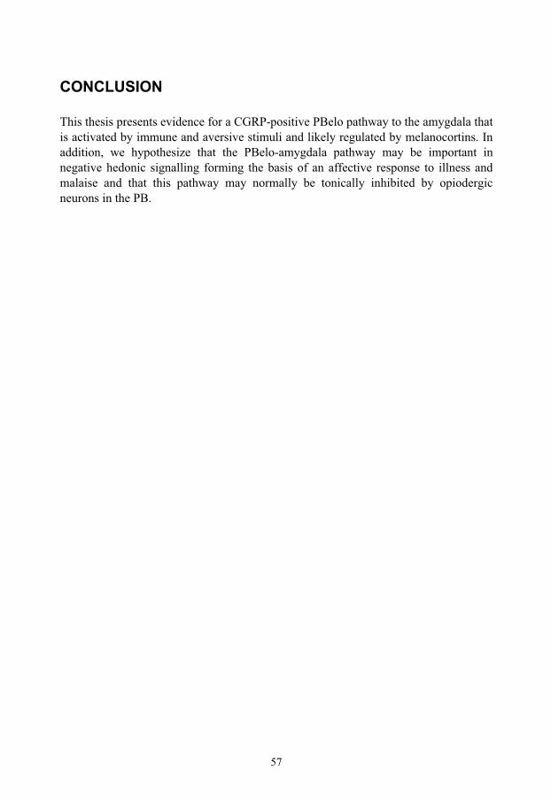
57
CONCLUSION
This thesis presents evidence for a CGRP-positive PBelo pathway to the amygdala that is activated by immune and aversive stimuli and likely regulated by melanocortins. In addition, we hypothesize that the PBelo-amygdala pathway may be important in negative hedonic signalling forming the basis of an affective response to illness and malaise and that this pathway may normally be tonically inhibited by opiodergic neurons in the PB.

58

59
ACKNOWLEDGEMENTS
I would like to express my sincere gratitude to everyone who has supported me and contributed to the work leading to this thesis. In particular, I would like to thank the following persons:
Professor Anders Blomqvist, my tutor and supervisor. I would like to thank you for introducing me into the field of neuroscience and sharing your deep knowledge. Further, I would like to thank you for your patience and your time, always ready to interrupt whatever you where doing to listen to my questions and ideas, no matter the subject.
Doctor David Engblom, for your friendship and for being a fantastic lab-partner, always ready to help with your skills and knowledge combined with a great sense of humour. Further, you always know when its Miller-time!
Doctor Martin Hallbeck, for your friendship and devotion to methodological development. You have taught me (or at least tried to teach me) the importance of patience in science. Also I would like to thank you for a great time when we were on parental leave.
Linda Engström, for being a very generous personality. A reliable friend in the lab, you are always ready to support and assist with your expertise. You have made in situ hybridization a more tolerable technique, something that was badly needed.
Ludmila Mackerlova, for your technical expertise in animal handling and lab administration, without your help, this thesis would not have been possible.
Ylva Kastrup, for you friendship and support during my time at level eleven, especially during our year in “forskarskolan”.
Johan Ruud and Louise Elander for your help, your patience with my daily scientific associations and suggestions, and for your contribution to making level eleven such a pleasant place.
Assistant professor Ola Hermanson, for always being supportive, optimistic and ingenious. You have always offered a helping hand when I needed it.
Åsa, Camilla, Björn, Karin, Emma, Helena, Anna, Christina, Reza, Amir, Nesar, David O,Khaddidja, Simon and Henric for contributing to a good scientific and social atmosphere.

60
Professor Claes Hildebrand for your inspiring love for science and interesting coffee-table discussions.
Professor Sivert Lindström for knowledge and interesting discussions about science and the mechanisms that controls it.
Professor Fredrik Elinder, for your enthusiasm and interest in science.
Eva Danielsson for making administrative problems disappear before they appear and Anette Wiklund for your patience with my ignorance of administration.
Doctor Sabine Richard for turning the defeat of being “scooped” to a victory.
My new and old friends and colleagues at the department for infectious medicine at Linköping University Hospital, for rapidly including me in your wonderful social and professional environment.
All my friends outside level 11, for your friendship and support. I especially would like to thank my brothers-in-arms in “98-oktan”, Stefan, Olov, Thomas, Johan, Mats and Osborn, your friendship through the years have been invaluable.
My parents for being loving, supportive and allowing me to pursue my interests. My siblings for being supportive and great personalities.
My mother-in-law, for always helping whenever needed.
Finally, I would like to thank Maria, Sofia and Oliver who are the joys of my life.
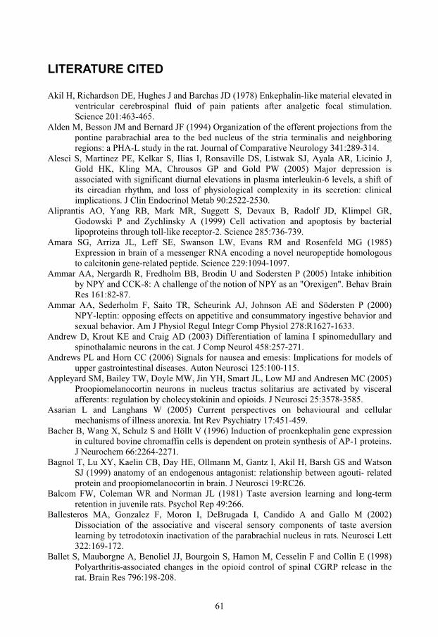
61
LITERATURE CITED
Akil H, Richardson DE, Hughes J and Barchas JD (1978) Enkephalin-like material elevated in ventricular cerebrospinal fluid of pain patients after analgetic focal stimulation. Science 201:463-465.
Alden M, Besson JM and Bernard JF (1994) Organization of the efferent projections from the pontine parabrachial area to the bed nucleus of the stria terminalis and neighboring regions: a PHA-L study in the rat. Journal of Comparative Neurology 341:289-314.
Alesci S, Martinez PE, Kelkar S, Ilias I, Ronsaville DS, Listwak SJ, Ayala AR, Licinio J, Gold HK, Kling MA, Chrousos GP and Gold PW (2005) Major depression is associated with significant diurnal elevations in plasma interleukin-6 levels, a shift of its circadian rhythm, and loss of physiological complexity in its secretion: clinical implications. J Clin Endocrinol Metab 90:2522-2530.
Aliprantis AO, Yang RB, Mark MR, Suggett S, Devaux B, Radolf JD, Klimpel GR, Godowski P and Zychlinsky A (1999) Cell activation and apoptosis by bacterial lipoproteins through toll-like receptor-2. Science 285:736-739.
Amara SG, Arriza JL, Leff SE, Swanson LW, Evans RM and Rosenfeld MG (1985) Expression in brain of a messenger RNA encoding a novel neuropeptide homologous to calcitonin gene-related peptide. Science 229:1094-1097.
Ammar AA, Nergardh R, Fredholm BB, Brodin U and Sodersten P (2005) Intake inhibition by NPY and CCK-8: A challenge of the notion of NPY as an "Orexigen". Behav Brain Res 161:82-87.
Ammar AA, Sederholm F, Saito TR, Scheurink AJ, Johnson AE and Södersten P (2000) NPY-leptin: opposing effects on appetitive and consummatory ingestive behavior and sexual behavior. Am J Physiol Regul Integr Comp Physiol 278:R1627-1633.
Andrew D, Krout KE and Craig AD (2003) Differentiation of lamina I spinomedullary and spinothalamic neurons in the cat. J Comp Neurol 458:257-271.
Andrews PL and Horn CC (2006) Signals for nausea and emesis: Implications for models of upper gastrointestinal diseases. Auton Neurosci 125:100-115.
Appleyard SM, Bailey TW, Doyle MW, Jin YH, Smart JL, Low MJ and Andresen MC (2005) Proopiomelanocortin neurons in nucleus tractus solitarius are activated by visceral afferents: regulation by cholecystokinin and opioids. J Neurosci 25:3578-3585.
Asarian L and Langhans W (2005) Current perspectives on behavioural and cellular mechanisms of illness anorexia. Int Rev Psychiatry 17:451-459.
Bacher B, Wang X, Schulz S and Höllt V (1996) Induction of proenkephalin gene expression in cultured bovine chromaffin cells is dependent on protein synthesis of AP-1 proteins. J Neurochem 66:2264-2271.
Bagnol T, Lu XY, Kaelin CB, Day HE, Ollmann M, Gantz I, Akil H, Barsh GS and Watson SJ (1999) anatomy of an endogenous antagonist: relationship between agouti- related protein and proopiomelanocortin in brain. J Neurosci 19:RC26.
Balcom FW, Coleman WR and Norman JL (1981) Taste aversion learning and long-term retention in juvenile rats. Psychol Rep 49:266.
Ballesteros MA, Gonzalez F, Moron I, DeBrugada I, Candido A and Gallo M (2002) Dissociation of the associative and visceral sensory components of taste aversion learning by tetrodotoxin inactivation of the parabrachial nucleus in rats. Neurosci Lett 322:169-172.
Ballet S, Mauborgne A, Benoliel JJ, Bourgoin S, Hamon M, Cesselin F and Collin E (1998) Polyarthritis-associated changes in the opioid control of spinal CGRP release in the rat. Brain Res 796:198-208.
62
Balthasar N, Coppari R, McMinn J, Liu SM, Lee CE, Tang V, Kenny CD, McGovern RA, Chua SC, Jr., Elmquist JK and Lowell BB (2004) Leptin receptor signaling in POMC neurons is required for normal body weight homeostasis. Neuron 42:983-991.
Balthasar N, Dalgaard LT, Lee CE, Yu J, Funahashi H, Williams T, Ferreira M, Tang V, McGovern RA, Kenny CD, Christiansen LM, Edelstein E, Choi B, Boss O, Aschkenasi C, Zhang CY, Mountjoy K, Kishi T, Elmquist JK and Lowell BB (2005) Divergence of melanocortin pathways in the control of food intake and energy expenditure. Cell 123:493-505.
Banks WA, Kastin AJ and Broadwell RD (1995) Passage of cytokines across the blood-brain barrier. Neuroimmunomodulation 2:241-248.
Barber LA and Vasko MR (1996) Activation of protein kinase C augments peptide release from rat sensory neurons. J Neurochem 67:72-80.
Basa NR, Wang L, Arteaga JR, Heber D, Livingston EH and Tache Y (2003) Bacterial lipopolysaccharide shifts fasted plasma ghrelin to postprandial levels in rats. Neurosci Lett 343:25-28.
Basbaum AI and Jessell TM, 2000. The Perception of Pain. In: Kandel, E. R. et al. (Eds.), Principles of Neural Science. McGraw-Hill Companies, International Edition, pp. 472-491.
Beishuizen A and Thijs LG (2003) Endotoxin and the hypothalamo-pituitary-adrenal (HPA) axis. J Endotoxin Res 9:3-24.
Bellavance LL and Beitz AJ (1996) Altered c-fos expression in the parabrachial nucleus in a rodent model of CFA-induced peripheral inflammation. Journal of Comparative Neurology 366:431-447.
Bermudez-Rattoni F, Forthman DL, Sanchez MA, Perez JL and Garcia J (1988) Odor and taste aversions conditioned in anesthetized rats. Behav Neurosci 102:726-732.
Bernard JF, Alden M and Besson JM (1993) The organization of the efferent projections from the pontine parabrachial area to the amygdaloid complex: a Phaseolus vulgaris leucoagglutinin (PHA-L) study in the rat. Journal of Comparative Neurology 329:201-229.
Bernard JF, Carroue J and Besson JM (1991) Efferent projections from the external parabrachial area to the forebrain: a Phaseolus vulgaris leucoagglutinin study in the rat. Neurosci Lett 122:257-260.
Bernard JF, Dallel R, Raboisson P, Villanueva L and Le Bars D (1995) Organization of the efferent projections from the spinal cervical enlargement to the parabrachial area and periaqueductal gray: a PHA-L study in the rat. J Comp Neurol 353:480-505.
Bernard JF, Huang GF and Besson JM (1994) The parabrachial area: electrophysiological evidence for an involvement in visceral nociceptive processes. J Neurophysiol 71:1646-1660.
Bernstein IL (1999) Taste aversion learning: a contemporary perspective. Nutrition 15:229-234.
Bernstein IL and Sigmundi RA (1980) Tumor anorexia: a learned food aversion? Science 209:416-418.
Berthoud HR (2004) Anatomy and function of sensory hepatic nerves. Anat Rec A Discov Mol Cell Evol Biol 280:827-835.
Bester H, Besson JM and Bernard JF (1997) Organization of efferent projections from the parabrachial area to the hypothalamus: a Phaseolus vulgaris-leucoagglutinin study in the rat. J Comp Neurol 383:245-281.
Bester H, Bourgeais L, Villanueva L, Besson JM and Bernard JF (1999) Differential projections to the intralaminar and gustatory thalamus from the parabrachial area: a PHA-L study in the rat. J Comp Neurol 405:421-449.
63
Bette M, Kaut O, Schafer MK and Weihe E (2003) Constitutive expression of p55TNFR mRNA and mitogen-specific up-regulation of TNF alpha and p75TNFR mRNA in mouse brain. J Comp Neurol 465:417-430.
Beutler B, Mahoney J, Le TN, Pekala P and Cerami A (1985a) Purification of cachectin, a lipoprotein lipase-suppressing hormone secreted by endotoxin-induced RAW 264.7 cells. Journal of Experimental Medicine 161:984-995.
Beutler B, Milsark IW and Cerami AC (1985b) Passive immunization against cachectin/tumor necrosis factor protects mice from lethal effect of endotoxin. Science 229:869-871.
Beutler B and Rietschel ET (2003) Innate immune sensing and its roots: the story of endotoxin. Nat Rev Immunol 3:169-176.
Billingsley KG, Fraker DL, Strassmann G, Loeser C, Fliot HM and Alexander HR (1996) Macrophage-derived tumor necrosis factor and tumor-derived of leukemia inhibitory factor and interleukin-6: possible cellular mechanisms of cancer cachexia. Annals of Surgical Oncology 3:29-35.
Bishai I and Coceani F (1996) Differential effects of endotoxin and cytokines on prostaglandin E2 formation in cerebral microvessels and brain parenchyma: implications for the pathogenesis of fever. Cytokine 8:371-376.
Bisler S, Schleicher A, Gass P, Stehle JH, Zilles K and Staiger JF (2002) Expression of c-Fos, ICER, Krox-24 and JunB in the whisker-to-barrel pathway of rats: time course of induction upon whisker stimulation by tactile exploration of an enriched environment. J Chem Neuroanat 23:187-198.
Blatteis CM (2000) The afferent signalling of fever. J Physiol 526 Pt 3:470. Blatteis CM, Mashburn TA, Jr. and Ahokas RA (1981) Fever and trace metal changes in
endotoxin-challenged neonates. Pflugers Arch 389:177-179. Block CH, Hoffman G and Kapp BS (1989) Peptide-containing pathways from the
parabrachial complex to the central nucleus of the amygdala. Peptides 10:465-471. Block CH and Schwartzbaum JS (1983) Ascending efferent projections of the gustatory
parabrachial nuclei in the rabbit. Brain Res 259:1-9. Blomqvist A, Ma W and Berkley KJ (1989) Spinal input to the parabrachial nucleus in the
cat. Brain Res 480:29-36. Blomqvist A, Zhang ET and Craig AD (2000) Cytoarchitectonic and immunohistochemical
characterization of a specific pain and temperature relay, the posterior portion of the ventral medial nucleus, in the human thalamus. Brain 123 Pt 3:601-619.
Bluthé RM, Layé S, Michaud B, Combe C, Dantzer R and Parnet P (2000) Role of interleukin-1beta and tumour necrosis factor-alpha in lipopolysaccharide-induced sickness behaviour: a study with interleukin-1 type I receptor-deficient mice. Eur J Neurosci 12:4447-4456.
Boivin A, Mesrobeanu L and Mesrobeanu I (1933) Technique pour la preparation des polysaccharides microbiens specifiques. Compt Rend 113:490-492.
Borovikova LV, Ivanova S, Zhang M, Yang H, Botchkina GI, Watkins LR, Wang H, Abumrad N, Eaton JW and Tracey KJ (2000) Vagus nerve stimulation attenuates the systemic inflammatory response to endotoxin. Nature 405:458-462.
Bowen EJ, Schmidt TW, Firm CS, Russo AF and Durham PL (2006) Tumor necrosis factor-alpha stimulation of calcitonin gene-related peptide expression and secretion from rat trigeminal ganglion neurons. J Neurochem 96:65-77.
Brain SD, Williams TJ, Tippins JR, Morris HR and MacIntyre I (1985) Calcitonin gene-related peptide is a potent vasodilator. Nature 313:54-56.
Brebner K, Hayley S, Zacharko R, Merali Z and Anisman H (2000) Synergistic effects of interleukin-1beta, interleukin-6, and tumor necrosis factor-alpha: central monoamine, corticosterone, and behavioral variations. Neuropsychopharmacology 22:566-580.
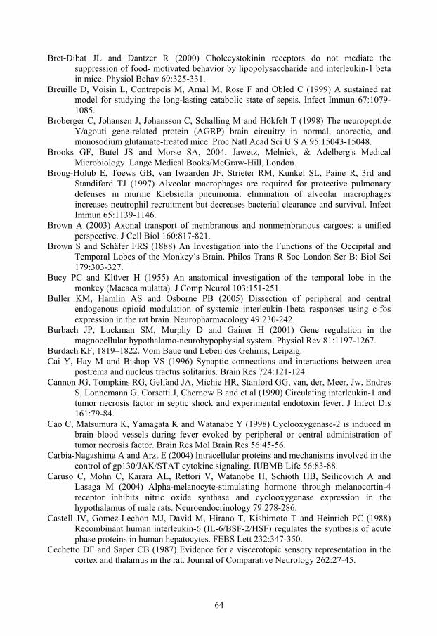
64
Bret-Dibat JL and Dantzer R (2000) Cholecystokinin receptors do not mediate the suppression of food- motivated behavior by lipopolysaccharide and interleukin-1 beta in mice. Physiol Behav 69:325-331.
Breuille D, Voisin L, Contrepois M, Arnal M, Rose F and Obled C (1999) A sustained rat model for studying the long-lasting catabolic state of sepsis. Infect Immun 67:1079-1085.
Broberger C, Johansen J, Johansson C, Schalling M and Hökfelt T (1998) The neuropeptide Y/agouti gene-related protein (AGRP) brain circuitry in normal, anorectic, and monosodium glutamate-treated mice. Proc Natl Acad Sci U S A 95:15043-15048.
Brooks GF, Butel JS and Morse SA, 2004. Jawetz, Melnick, & Adelberg's Medical Microbiology. Lange Medical Books/McGraw-Hill, London.
Broug-Holub E, Toews GB, van Iwaarden JF, Strieter RM, Kunkel SL, Paine R, 3rd and Standiford TJ (1997) Alveolar macrophages are required for protective pulmonary defenses in murine Klebsiella pneumonia: elimination of alveolar macrophages increases neutrophil recruitment but decreases bacterial clearance and survival. Infect Immun 65:1139-1146.
Brown A (2003) Axonal transport of membranous and nonmembranous cargoes: a unified perspective. J Cell Biol 160:817-821.
Brown S and Schäfer FRS (1888) An Investigation into the Functions of the Occipital and Temporal Lobes of the Monkey´s Brain. Philos Trans R Soc London Ser B: Biol Sci 179:303-327.
Bucy PC and Klüver H (1955) An anatomical investigation of the temporal lobe in the monkey (Macaca mulatta). J Comp Neurol 103:151-251.
Buller KM, Hamlin AS and Osborne PB (2005) Dissection of peripheral and central endogenous opioid modulation of systemic interleukin-1beta responses using c-fos expression in the rat brain. Neuropharmacology 49:230-242.
Burbach JP, Luckman SM, Murphy D and Gainer H (2001) Gene regulation in the magnocellular hypothalamo-neurohypophysial system. Physiol Rev 81:1197-1267.
Burdach KF, 1819–1822. Vom Baue und Leben des Gehirns, Leipzig. Cai Y, Hay M and Bishop VS (1996) Synaptic connections and interactions between area
postrema and nucleus tractus solitarius. Brain Res 724:121-124. Cannon JG, Tompkins RG, Gelfand JA, Michie HR, Stanford GG, van, der, Meer, Jw, Endres
S, Lonnemann G, Corsetti J, Chernow B and et al (1990) Circulating interleukin-1 and tumor necrosis factor in septic shock and experimental endotoxin fever. J Infect Dis 161:79-84.
Cao C, Matsumura K, Yamagata K and Watanabe Y (1998) Cyclooxygenase-2 is induced in brain blood vessels during fever evoked by peripheral or central administration of tumor necrosis factor. Brain Res Mol Brain Res 56:45-56.
Carbia-Nagashima A and Arzt E (2004) Intracellular proteins and mechanisms involved in the control of gp130/JAK/STAT cytokine signaling. IUBMB Life 56:83-88.
Caruso C, Mohn C, Karara AL, Rettori V, Watanobe H, Schioth HB, Seilicovich A and Lasaga M (2004) Alpha-melanocyte-stimulating hormone through melanocortin-4 receptor inhibits nitric oxide synthase and cyclooxygenase expression in the hypothalamus of male rats. Neuroendocrinology 79:278-286.
Castell JV, Gomez-Lechon MJ, David M, Hirano T, Kishimoto T and Heinrich PC (1988) Recombinant human interleukin-6 (IL-6/BSF-2/HSF) regulates the synthesis of acute phase proteins in human hepatocytes. FEBS Lett 232:347-350.
Cechetto DF and Saper CB (1987) Evidence for a viscerotopic sensory representation in the cortex and thalamus in the rat. Journal of Comparative Neurology 262:27-45.

65
Cechetto DF, Standaert DG and Saper CB (1985) Spinal and trigeminal dorsal horn projections to the parabrachial nucleus in the rat. J Comp Neurol 240:153-160.
Cepeda MS, Carr DB, Miranda N, Diaz A, Silva C and Morales O (2005) Comparison of morphine, ketorolac, and their combination for postoperative pain: results from a large, randomized, double-blind trial. Anesthesiology 103:1225-1232.
Chamberlin NL, Mansour A, Watson SJ and Saper CB (1999) Localization of mu-opioid receptors on amygdaloid projection neurons in the parabrachial nucleus of the rat. Brain Res 827:198-204.
Chamberlin NL and Saper CB (1992) Topographic organization of cardiovascular responses to electrical and glutamate microstimulation of the parabrachial nucleus in the rat. Journal of Comparative Neurology 326:245-262.
Chamberlin NL and Saper CB (1994) Topographic organization of respiratory responses to glutamate microstimulation of the parabrachial nucleus in the rat. J Neurosci 14:6500-6510.
Childers SR (1991) Opioid receptor-coupled second messenger systems. Life Sci 48:1991-2003.
Coleman RA, Smith WL and Narumiya S (1994) International Union of Pharmacology classification of prostanoid receptors: properties, distribution, and structure of the receptors and their subtypes. Pharmacol Rev 46:205-229.
Comb M, Mermod N, Hyman SE, Pearlberg J, Ross ME and Goodman HM (1988) Proteins bound at adjacent DNA elements act synergistically to regulate human proenkephalin cAMP inducible transcription. Embo J 7:3793-3805.
Comb M, Seeburg PH, Adelman J, Eiden L and Herbert E (1982) Primary structure of the human Met- and Leu-enkephalin precursor and its mRNA. Nature 295:663-666.
Cone RD (2005) Anatomy and regulation of the central melanocortin system. Nat Neurosci 8:571-578.
Copray JC, Mantingh I, Brouwer N, Biber K, Kust BM, Liem RS, Huitinga I, Tilders FJ, Van Dam AM and Boddeke HW (2001) Expression of interleukin-1 beta in rat dorsal root ganglia. J Neuroimmunol 118:203-211.
Craig AD (1995) Distribution of brainstem projections from spinal lamina I neurons in the cat and the monkey. J Comp Neurol 361:225-248.
Craig AD (2003a) Interoception: the sense of the physiological condition of the body. Curr Opin Neurobiol 13:500-505.
Craig AD (2003b) A new view of pain as a homeostatic emotion. Trends Neurosci 26:303-307.
Craig W (1918) Appetite and aversions as constituents of instincts. Biol Bull 34:91-107. Cross-Mellor SK, Roberts S, Kavaliers M and Ossenkopp KP (2003) Activation of the
immune system in rats with lipopolysaccharide reduces voluntary sucrose intake but not intraoral intake. Pharmacol Biochem Behav 76:153-159.
Cunningham ET, Jr., Stalder AK, Sanna PP, Liu SS, Bloom FE, Howes EL, Jr., Campbell IL and Margolis TP (1997) Distribution of tumor necrosis factor receptor messenger RNA in normal and herpes simplex virus infected trigeminal ganglia in the mouse. Brain Res 758:99-106.
Cuzzocrea S, Mazzon E, Di Paola R, Esposito E, Macarthur H, Matuschak GM and Salvemini D (2006) A role for nitric oxide-mediated peroxynitrite formation in a model of endotoxin induced shock. J Pharmacol Exp Ther.
Daftary SS and Gore AC (2005) IGF-1 in the brain as a regulator of reproductive neuroendocrine function. Exp Biol Med (Maywood) 230:292-306.

66
Dallel R, Ricard O and Raboisson P (2004) Organization of parabrachial projections from the spinal trigeminal nucleus oralis: an anterograde tracing study in the rat. J Comp Neurol 470:181-191.
Daniels D, Patten CS, Roth JD, Yee DK and Fluharty SJ (2003) Melanocortin receptor signaling through mitogen-activated protein kinase in vitro and in rat hypothalamus. Brain Res 986:1-11.
Dantzer R (2004) Cytokine-induced sickness behaviour: a neuroimmune response to activation of innate immunity. Eur J Pharmacol 500:399-411.
Davies RF (1977) Long-and short-term regulation of feeding patterns in the rat. J Comp Physiol Psychol 91:574-585.
Dempsey PW, Doyle SE, He JQ and Cheng G (2003) The signaling adaptors and pathways activated by TNF superfamily. Cytokine Growth Factor Rev 14:193-209.
Dewys WD, Begg C, Lavin PT, Band PR, Bennett JM, Bertino JR, Cohen MH, Douglass HO, Jr., Engstrom PF, Ezdinli EZ, Horton J, Johnson GJ, Moertel CG, Oken MM, Perlia C, Rosenbaum C, Silverstein MN, Skeel RT, Sponzo RW and Tormey DC (1980) Prognostic effect of weight loss prior to chemotherapy in cancer patients. Eastern Cooperative Oncology Group. Am J Med 69:491-497.
Dinarello CA (1991) Interleukin-1 and interleukin-1 antagonism. Blood 77:1627-1652. Dobolyi A, Irwin S, Makara G, Usdin TB and Palkovits M (2005) Calcitonin gene-related
peptide-containing pathways in the rat forebrain. J Comp Neurol 489:92-119. Doganay M, Kama NA, Yazgan A, Aksoy M, Ergul G and Tekeli A (1997) The effects of
vagotomy on bacterial translocation: an experimental study. J Surg Res 71:166-171. Dunn AJ (2000) Cytokine activation of the HPA axis. Ann N Y Acad Sci 917:608-617. Durham PL and Russo AF (1998) Serotonergic repression of mitogen-activated protein kinase
control of the calcitonin gene-related peptide enhancer. Mol Endocrinol 12:1002-1009. Durham PL and Russo AF (2003) Stimulation of the calcitonin gene-related peptide enhancer
by mitogen-activated protein kinases and repression by an antimigraine drug in trigeminal ganglia neurons. J Neurosci 23:807-815.
Echtenacher B, Falk W, Mannel DN and Krammer PH (1990) Requirement of endogenous tumor necrosis factor/cachectin for recovery from experimental peritonitis. J Immunol 145:3762-3766.
Eiden LE, Giraud P, Dave JR, Hotchkiss AJ and Affolter HU (1984) Nicotinic receptor stimulation activates enkephalin release and biosynthesis in adrenal chromaffin cells. Nature 312:661-663.
Eijsbouts AM, van den Hoogen FH, Laan RF, Hermus AR, Sweep CG and van de Putte LB (2005) Hypothalamic-pituitary-adrenal axis activity in patients with rheumatoid arthritis. Clin Exp Rheumatol 23:658-664.
Ek M, Arias C, Sawchenko P and Ericsson-Dahlstrand A (2000) Distribution of the EP3 prostaglandin E(2) receptor subtype in the rat brain: relationship to sites of interleukin-1-induced cellular responsiveness. J Comp Neurol 428:5-20.
Ek M, Engblom D, Saha S, Blomqvist A, Jakobsson PJ and Ericsson-Dahlstrand A (2001) Inflammatory response: Pathway across the blood-brain barrier. Nature 410:430-431.
Ek M, Kurosawa M, Lundeberg T and Ericsson A (1998) Activation of vagal afferents after intravenous injection of interleukin-1beta: role of endogenous prostaglandins. Journal of Neuroscience 18:9471-9479.
Elander L, Engström L, Hallbeck M and Blomqvist A (2006) IL-1{beta} and LPS induce anorexia by distinct mechanisms differentially dependent on microsomal prostaglandin E synthase-1. Am J Physiol Regul Integr Comp Physiol.

67
Elias CF, Kelly JF, Lee CE, Ahima RS, Drucker DJ, Saper CB and Elmquist JK (2000) Chemical characterization of leptin-activated neurons in the rat brain. J Comp Neurol 423:261-281.
Elmquist JK, Scammell TE, Jacobson CD and Saper CB (1996) Distribution of Fos-like immunoreactivity in the rat brain following intravenous lipopolysaccharide administration. J Comp Neurol 371:85-103.
Emody L, Ralovich B, Barna K, Brasch G and Ternak G (1974) Physiological effect of orally administered endotoxin to man. J Hyg Epidemiol Microbiol Immunol 18:454-458.
Engblom D, Ek M, Ericsson-Dahlstrand A and Blomqvist A (2001) Activation of prostanoid EP(3) and EP(4) receptor mRNA-expressing neurons in the rat parabrachial nucleus by intravenous injection of bacterial wall lipopolysaccharide. J Comp Neurol 440:378-386.
Engblom D, Ek M, Ericsson-Dahlstrand A and Blomqvist A (2004) EP3 and EP4 receptor mRNA expression in peptidergic cell groups of the rat parabrachial nucleus. Neuroscience 126:989-999.
Engblom D, Ek M, Hallbeck M, Ericsson-Dahlstrand A and Blomqvist A (2000) Distribution of prostaglandin EP(3) and EP(4) receptor mRNA in the rat parabrachial nucleus. Neurosci Lett 281:163-166.
Engblom D, Saha S, Engström L, Westman M, Audoly LP, Jakobsson PJ and Blomqvist A (2003) Microsomal prostaglandin E synthase-1 is the central switch during immune-induced pyresis. Nat Neurosci 6:1137-1138.
Engström L, Engblom D and Blomqvist A (2003) Systemic immune challenge induces preproenkephalin gene transcription in distinct autonomic structures of the rat brain. J Comp Neurol 462:450-461.
Ericsson A, Arias C and Sawchenko PE (1997) Evidence for an intramedullary prostaglandin-dependent mechanism in the activation of stress-related neuroendocrine circuitry by intravenous interleukin-1. Journal of Neuroscience 17:7166-7179.
Ericsson A, Kovacs KJ and Sawchenko PE (1994) A functional anatomical analysis of central pathways subserving the effects of interleukin-1 on stress-related neuroendocrine neurons. J Neurosci 14:897-913.
Ericsson A, Liu C, Hart RP and Sawchenko PE (1995) Type 1 interleukin-1 receptor in the rat brain: distribution, regulation, and relationship to sites of IL-1-induced cellular activation. J Comp Neurol 361:681-698.
Erlich P, 1885. Das sauerstufbudurfnis des organismus. Eine Farbenanalytische Studie, Hirschwald, Berlin.
Evans BN, Rosenblatt MI, Mnayer LO, Oliver KR and Dickerson IM (2000) CGRP-RCP, a novel protein required for signal transduction at calcitonin gene-related peptide and adrenomedullin receptors. J Biol Chem 275:31438-31443.
Everitt BJ, Cardinal RN, Parkinson JA and Robbins TW (2003) Appetitive behavior: impact of amygdala-dependent mechanisms of emotional learning. Ann N Y Acad Sci 985:233-250.
Exton MS (1997) Infection-induced anorexia: active host defence strategy. Appetite 29:369-383.
Fan W, Ellacott KL, Halatchev IG, Takahashi K, Yu P and Cone RD (2004) Cholecystokinin-mediated suppression of feeding involves the brainstem melanocortin system. Nat Neurosci 7:335-336.
Fannin RD, Auman JT, Bruno ME, Sieber SO, Ward SM, Tucker CJ, Merrick BA and Paules RS (2005) Differential gene expression profiling in whole blood during acute systemic inflammation in lipopolysaccharide-treated rats. Physiol Genomics 21:92-104.
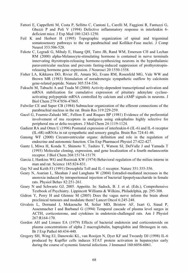
68
Fattori E, Cappelletti M, Costa P, Sellitto C, Cantoni L, Carelli M, Faggioni R, Fantuzzi G, Ghezzi P and Poli V (1994) Defective inflammatory response in interleukin 6-deficient mice. J Exp Med 180:1243-1250.
Feil K and Herbert H (1995) Topographic organization of spinal and trigeminal somatosensory pathways to the rat parabrachial and Kolliker-Fuse nuclei. J Comp Neurol 353:506-528.
Fekete C, Legradi G, Mihaly E, Huang QH, Tatro JB, Rand WM, Emerson CH and Lechan RM (2000) alpha-Melanocyte-stimulating hormone is contained in nerve terminals innervating thyrotropin-releasing hormone-synthesizing neurons in the hypothalamic paraventricular nucleus and prevents fasting-induced suppression of prothyrotropin-releasing hormone gene expression. J Neurosci 20:1550-1558.
Fisher LA, Kikkawa DO, Rivier JE, Amara SG, Evans RM, Rosenfeld MG, Vale WW and Brown MR (1983) Stimulation of noradrenergic sympathetic outflow by calcitonin gene-related peptide. Nature 305:534-536.
Fukuchi M, Tabuchi A and Tsuda M (2004) Activity-dependent transcriptional activation and mRNA stabilization for cumulative expression of pituitary adenylate cyclase-activating polypeptide mRNA controlled by calcium and cAMP signals in neurons. J Biol Chem 279:47856-47865.
Fulwiler CE and Saper CB (1984) Subnuclear organization of the efferent connections of the parabrachial nucleus in the rat. Brain Res 319:229-259.
Gacel G, Fournie-Zaluski MC, Fellion E and Roques BP (1981) Evidence of the preferential involvement of mu receptors in analgesia using enkephalins highly selective for peripheral mu or delta receptors. J Med Chem 24:1119-1124.
Gadient RA and Otten U (1996) Postnatal expression of interleukin-6 (IL-6) and IL-6 receptor (IL-6R) mRNAs in rat sympathetic and sensory ganglia. Brain Res 724:41-46.
Ganong WF (2000) Circumventricular organs: definition and role in the regulation of endocrine and autonomic function. Clin Exp Pharmacol Physiol 27:422-427.
Gantz I, Miwa H, Konda Y, Shimoto Y, Tashiro T, Watson SJ, DelValle J and Yamada T (1993) Molecular cloning, expression, and gene localization of a fourth melanocortin receptor. J Biol Chem 268:15174-15179.
Garcia J, Hankins WG and Rusiniak KW (1974) Behavioral regulation of the milieu interne in man and rat. Science 185:824-831.
Gay NJ and Keith FJ (1991) Drosophila Toll and IL-1 receptor. Nature 351:355-356. Geary N, Asarian L, Sheahan J and Langhans W (2004) Estradiol-mediated increases in the
anorexia induced by intraperitoneal injection of bacterial lipopolysaccharide in female rats. Physiol Behav 82:251-261.
Geary N and Schwartz GJ, 2005. Appetite. In: Sadock, B. J. et al. (Eds.), Comprehensive Textbook of Psychiatry. Lippincott Williams & Wilkins, Philadelphia, pp. 295-308.
Gidron Y, Perry H and Glennie M (2005) Does the vagus nerve inform the brain about preclinical tumours and modulate them? Lancet Oncol 6:245-248.
Givalois L, Dornand J, Mekaouche M, Solier MD, Bristow AF, Ixart G, Siaud P, Assenmacher I and Barbanel G (1994) Temporal cascade of plasma level surges in ACTH, corticosterone, and cytokines in endotoxin-challenged rats. Am J Physiol 267:R164-170.
Gordon AH and Limaos EA (1979) Effects of bacterial endotoxin and corticosteroids on plasma concentrations of alpha 2 macroglobulin, haptoglobin and fibrinogen in rats. Br J Exp Pathol 60:434-440.
Gregory SH, Wing EJ, Danowski KL, van Rooijen N, Dyer KF and Tweardy DJ (1998) IL-6 produced by Kupffer cells induces STAT protein activation in hepatocytes early during the course of systemic listerial infections. J Immunol 160:6056-6061.
69
Grieger TA and Kluger MJ (1978) Fever and survival: the role of serum iron. J Physiol 279:187-196.
Grill HJ, Ginsberg AB, Seeley RJ and Kaplan JM (1998) Brainstem application of melanocortin receptor ligands produces long- lasting effects on feeding and body weight. J Neurosci 18:10128-10135.
Grill HJ and Kaplan JM (2002) The neuroanatomical axis for control of energy balance. Front Neuroendocrinol 23:2-40.
Grill HJ and Norgren R (1978) The taste reactivity test. I. Mimetic responses to gustatory stimuli in neurologically normal rats. Brain Res 143:263-279.
Gross CG (2005) Processing the facial image: a brief history. Am Psychol 60:755-763. Gubler U, Seeburg P, Hoffman BJ, Gage LP and Udenfriend S (1982) Molecular cloning
establishes proenkephalin as precursor of enkephalin-containing peptides. Nature 295:206-208.
Hagan MM, Rushing PA, Schwartz MW, Yagaloff KA, Burn P, Woods SC and Seeley RJ (1999) Role of the CNS melanocortin system in the response to overfeeding. J Neurosci 19:2362-2367.
Hahm SH, Chen Y, Vinson C and Eiden LE (2003) A calcium-initiated signaling pathway propagated through calcineurin and cAMP response element-binding protein activates proenkephalin gene transcription after depolarization. Mol Pharmacol 64:1503-1511.
Hailman E, Lichenstein HS, Wurfel MM, Miller DS, Johnson DA, Kelley M, Busse LA, Zukowski MM and Wright SD (1994) Lipopolysaccharide (LPS)-binding protein accelerates the binding of LPS to CD14. J Exp Med 179:269-277.
Halsell CB (1992) Organization of parabrachial nucleus efferents to the thalamus and amygdala in the golden hamster. Journal of Comparative Neurology 317:57-78.
Halsell CB and Travers SP (1997) Anterior and posterior oral cavity responsive neurons are differentially distributed among parabrachial subnuclei in rat. J Neurophysiol 78:920-938.
Han JS, Li W and Neugebauer V (2005) Critical Role of Calcitonin Gene-Related Peptide 1 Receptors in the Amygdala in Synaptic Plasticity and Pain Behavior. J Neurosci 25:10717-10728.
Hansen MK, Taishi P, Chen Z and Krueger JM (1998) Vagotomy blocks the induction of interleukin-1beta (IL-1beta) mRNA in the brain of rats in response to systemic IL-1beta. J Neurosci 18:2247-2253.
Harlan RE, Shivers BD, Romano GJ, Howells RD and Pfaff DW (1987) Localization of preproenkephalin mRNA in the rat brain and spinal cord by in situ hybridization. J Comp Neurol 258:159-184.
Hawkins BT and Davis TP (2005) The blood-brain barrier/neurovascular unit in health and disease. Pharmacol Rev 57:173-185.
Hayashi F, Smith KD, Ozinsky A, Hawn TR, Yi EC, Goodlett DR, Eng JK, Akira S, Underhill DM and Aderem A (2001) The innate immune response to bacterial flagellin is mediated by Toll-like receptor 5. Nature 410:1099-1103.
Heesen C, Nawrath L, Reich C, Bauer N, Schulz KH and Gold SM (2006) Fatigue in multiple sclerosis: an example of cytokine mediated sickness behaviour? J Neurol Neurosurg Psychiatry 77:34-39.
Hehlgans T and Pfeffer K (2005) The intriguing biology of the tumour necrosis factor/tumour necrosis factor receptor superfamily: players, rules and the games. Immunology 115:1-20.
Heil F, Hemmi H, Hochrein H, Ampenberger F, Kirschning C, Akira S, Lipford G, Wagner H and Bauer S (2004) Species-specific recognition of single-stranded RNA via toll-like receptor 7 and 8. Science 303:1526-1529.
70
Heinrich PC, Behrmann I, Muller-Newen G, Schaper F and Graeve L (1998) Interleukin-6-type cytokine signalling through the gp130/Jak/STAT pathway. Biochem J 334 ( Pt 2):297-314.
Hemmi H, Takeuchi O, Kawai T, Kaisho T, Sato S, Sanjo H, Matsumoto M, Hoshino K, Wagner H, Takeda K and Akira S (2000) A Toll-like receptor recognizes bacterial DNA. Nature 408:740-745.
Henle J, 1910. vol.3. Joh. Ambrosius Barth, Leipzig, pp. 1-88. Herbert H, Moga MM and Saper CB (1990) Connections of the parabrachial nucleus with the
nucleus of the solitary tract and the medullary reticular formation in the rat. J Comp Neurol 293:540-580.
Hermann GE, Tovar CA and Rogers RC (2003) TNFalpha-stimulation of cFos-activation of neurons in the solitary nucleus is suppressed by TNFR:Fc adsorbant construct in the dorsal vagal complex. Brain Res 976:69-74.
Hermanson O, 1997. Molecular mechanisms of nociception in the rat brain. In: Department of Cell Biology), pp. 1-86. Faculty of Health Sciences, Linköping University, Linköping.
Hermanson O and Blomqvist A (1996) Subnuclear localization of FOS-like immunoreactivity in the rat parabrachial nucleus after nociceptive stimulation. Journal of Comparative Neurology 368:45-56.
Hermanson O and Blomqvist A (1997a) Differential expression of the AP-1/CRE-binding proteins FOS and CREB in preproenkephalin mRNA-expressing neurons of the rat parabrachial nucleus after nociceptive stimulation. Brain Res Mol Brain Res 51:188-196.
Hermanson O and Blomqvist A (1997b) Preproenkephalin messenger RNA-expressing neurons in the rat parabrachial nucleus: subnuclear organization and projections to the intralaminar thalamus. Neuroscience 81:803-812.
Hermanson O and Blomqvist A (1997c) Subnuclear localization of FOS-like immunoreactivity in the parabrachial nucleus after orofacial nociceptive stimulation of the awake rat. J Comp Neurol 387:114-123.
Hermanson O, Ericson H, Sanchez WG, Watts AG and Blomqvist A (1994) Autoradiographic visualization of 35S-labeled cRNA probes combined with immunoperoxidase detection of choleragenoid: a double-labeling light microscopic method for in situ hybridization and retrograde tract tracing. Journal of Histochemistry & Cytochemistry 42:827-831.
Hermanson O, Larhammar D and Blomqvist A (1998) Preprocholecystokinin mRNA-expressing neurons in the rat parabrachial nucleus: subnuclear localization, efferent projection, and expression of nociceptive-related intracellular signaling substances. J Comp Neurol 400:255-270.
Hessle CC, Andersson B and Wold AE (2005) Gram-positive and Gram-negative bacteria elicit different patterns of pro-inflammatory cytokines in human monocytes. Cytokine 30:311-318.
Hoffman GE and Lyo D (2002) Anatomical markers of activity in neuroendocrine systems: are we all 'fos-ed out'? J Neuroendocrinol 14:259-268.
Hopkins DA and Holstege G (1978) Amygdaloid projections to the mesencephalon, pons and medulla oblongata in the cat. Exp Brain Res 32:529-547.
Horai R, Asano M, Sudo K, Kanuka H, Suzuki M, Nishihara M, Takahashi M and Iwakura Y (1998) Production of mice deficient in genes for interleukin (IL)-1alpha, IL-1beta, IL-1alpha/beta, and IL-1 receptor antagonist shows that IL-1beta is crucial in turpentine-induced fever development and glucocorticoid secretion. J Exp Med 187:1463-1475.
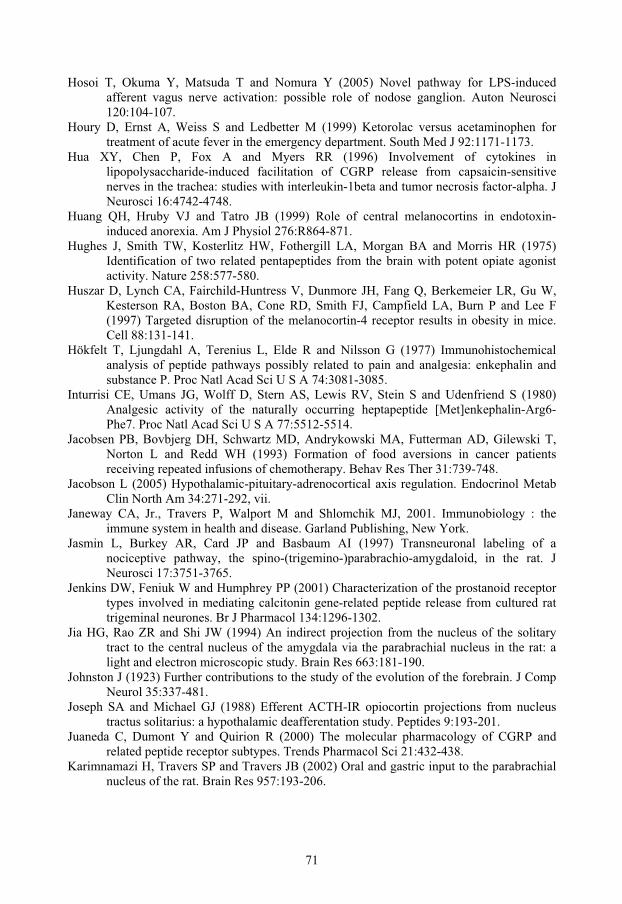
71
Hosoi T, Okuma Y, Matsuda T and Nomura Y (2005) Novel pathway for LPS-induced afferent vagus nerve activation: possible role of nodose ganglion. Auton Neurosci 120:104-107.
Houry D, Ernst A, Weiss S and Ledbetter M (1999) Ketorolac versus acetaminophen for treatment of acute fever in the emergency department. South Med J 92:1171-1173.
Hua XY, Chen P, Fox A and Myers RR (1996) Involvement of cytokines in lipopolysaccharide-induced facilitation of CGRP release from capsaicin-sensitive nerves in the trachea: studies with interleukin-1beta and tumor necrosis factor-alpha. J Neurosci 16:4742-4748.
Huang QH, Hruby VJ and Tatro JB (1999) Role of central melanocortins in endotoxin-induced anorexia. Am J Physiol 276:R864-871.
Hughes J, Smith TW, Kosterlitz HW, Fothergill LA, Morgan BA and Morris HR (1975) Identification of two related pentapeptides from the brain with potent opiate agonist activity. Nature 258:577-580.
Huszar D, Lynch CA, Fairchild-Huntress V, Dunmore JH, Fang Q, Berkemeier LR, Gu W, Kesterson RA, Boston BA, Cone RD, Smith FJ, Campfield LA, Burn P and Lee F (1997) Targeted disruption of the melanocortin-4 receptor results in obesity in mice. Cell 88:131-141.
Hökfelt T, Ljungdahl A, Terenius L, Elde R and Nilsson G (1977) Immunohistochemical analysis of peptide pathways possibly related to pain and analgesia: enkephalin and substance P. Proc Natl Acad Sci U S A 74:3081-3085.
Inturrisi CE, Umans JG, Wolff D, Stern AS, Lewis RV, Stein S and Udenfriend S (1980) Analgesic activity of the naturally occurring heptapeptide [Met]enkephalin-Arg6-Phe7. Proc Natl Acad Sci U S A 77:5512-5514.
Jacobsen PB, Bovbjerg DH, Schwartz MD, Andrykowski MA, Futterman AD, Gilewski T, Norton L and Redd WH (1993) Formation of food aversions in cancer patients receiving repeated infusions of chemotherapy. Behav Res Ther 31:739-748.
Jacobson L (2005) Hypothalamic-pituitary-adrenocortical axis regulation. Endocrinol Metab Clin North Am 34:271-292, vii.
Janeway CA, Jr., Travers P, Walport M and Shlomchik MJ, 2001. Immunobiology : the immune system in health and disease. Garland Publishing, New York.
Jasmin L, Burkey AR, Card JP and Basbaum AI (1997) Transneuronal labeling of a nociceptive pathway, the spino-(trigemino-)parabrachio-amygdaloid, in the rat. J Neurosci 17:3751-3765.
Jenkins DW, Feniuk W and Humphrey PP (2001) Characterization of the prostanoid receptor types involved in mediating calcitonin gene-related peptide release from cultured rat trigeminal neurones. Br J Pharmacol 134:1296-1302.
Jia HG, Rao ZR and Shi JW (1994) An indirect projection from the nucleus of the solitary tract to the central nucleus of the amygdala via the parabrachial nucleus in the rat: a light and electron microscopic study. Brain Res 663:181-190.
Johnston J (1923) Further contributions to the study of the evolution of the forebrain. J Comp Neurol 35:337-481.
Joseph SA and Michael GJ (1988) Efferent ACTH-IR opiocortin projections from nucleus tractus solitarius: a hypothalamic deafferentation study. Peptides 9:193-201.
Juaneda C, Dumont Y and Quirion R (2000) The molecular pharmacology of CGRP and related peptide receptor subtypes. Trends Pharmacol Sci 21:432-438.
Karimnamazi H, Travers SP and Travers JB (2002) Oral and gastric input to the parabrachial nucleus of the rat. Brain Res 957:193-206.
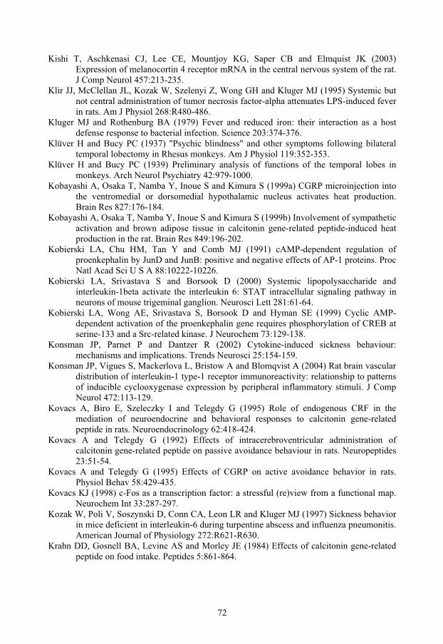
72
Kishi T, Aschkenasi CJ, Lee CE, Mountjoy KG, Saper CB and Elmquist JK (2003) Expression of melanocortin 4 receptor mRNA in the central nervous system of the rat. J Comp Neurol 457:213-235.
Klir JJ, McClellan JL, Kozak W, Szelenyi Z, Wong GH and Kluger MJ (1995) Systemic but not central administration of tumor necrosis factor-alpha attenuates LPS-induced fever in rats. Am J Physiol 268:R480-486.
Kluger MJ and Rothenburg BA (1979) Fever and reduced iron: their interaction as a host defense response to bacterial infection. Science 203:374-376.
Klüver H and Bucy PC (1937) "Psychic blindness" and other symptoms following bilateral temporal lobectomy in Rhesus monkeys. Am J Physiol 119:352-353.
Klüver H and Bucy PC (1939) Preliminary analysis of functions of the temporal lobes in monkeys. Arch Neurol Psychiatry 42:979-1000.
Kobayashi A, Osaka T, Namba Y, Inoue S and Kimura S (1999a) CGRP microinjection into the ventromedial or dorsomedial hypothalamic nucleus activates heat production. Brain Res 827:176-184.
Kobayashi A, Osaka T, Namba Y, Inoue S and Kimura S (1999b) Involvement of sympathetic activation and brown adipose tissue in calcitonin gene-related peptide-induced heat production in the rat. Brain Res 849:196-202.
Kobierski LA, Chu HM, Tan Y and Comb MJ (1991) cAMP-dependent regulation of proenkephalin by JunD and JunB: positive and negative effects of AP-1 proteins. Proc Natl Acad Sci U S A 88:10222-10226.
Kobierski LA, Srivastava S and Borsook D (2000) Systemic lipopolysaccharide and interleukin-1beta activate the interleukin 6: STAT intracellular signaling pathway in neurons of mouse trigeminal ganglion. Neurosci Lett 281:61-64.
Kobierski LA, Wong AE, Srivastava S, Borsook D and Hyman SE (1999) Cyclic AMP-dependent activation of the proenkephalin gene requires phosphorylation of CREB at serine-133 and a Src-related kinase. J Neurochem 73:129-138.
Konsman JP, Parnet P and Dantzer R (2002) Cytokine-induced sickness behaviour: mechanisms and implications. Trends Neurosci 25:154-159.
Konsman JP, Vigues S, Mackerlova L, Bristow A and Blomqvist A (2004) Rat brain vascular distribution of interleukin-1 type-1 receptor immunoreactivity: relationship to patterns of inducible cyclooxygenase expression by peripheral inflammatory stimuli. J Comp Neurol 472:113-129.
Kovacs A, Biro E, Szeleczky I and Telegdy G (1995) Role of endogenous CRF in the mediation of neuroendocrine and behavioral responses to calcitonin gene-related peptide in rats. Neuroendocrinology 62:418-424.
Kovacs A and Telegdy G (1992) Effects of intracerebroventricular administration of calcitonin gene-related peptide on passive avoidance behaviour in rats. Neuropeptides 23:51-54.
Kovacs A and Telegdy G (1995) Effects of CGRP on active avoidance behavior in rats. Physiol Behav 58:429-435.
Kovacs KJ (1998) c-Fos as a transcription factor: a stressful (re)view from a functional map. Neurochem Int 33:287-297.
Kozak W, Poli V, Soszynski D, Conn CA, Leon LR and Kluger MJ (1997) Sickness behavior in mice deficient in interleukin-6 during turpentine abscess and influenza pneumonitis. American Journal of Physiology 272:R621-R630.
Krahn DD, Gosnell BA, Levine AS and Morley JE (1984) Effects of calcitonin gene-related peptide on food intake. Peptides 5:861-864.
73
Krahn DD, Gosnell BA, Levine AS and Morley JE (1986) The effect of calcitonin gene-related peptide on food intake involves aversive mechanisms. Pharmacol Biochem Behav 24:5-7.
Kresse A, Jacobowitz DM and Skofitsch G (1995) Detailed mapping of CGRP mRNA expression in the rat central nervous system: comparison with previous immunocytochemical findings. Brain Res Bull 36:261-274.
Krout KE, Jansen AS and Loewy AD (1998) Periaqueductal gray matter projection to the parabrachial nucleus in rat. J Comp Neurol 401:437-454.
Krout KE and Loewy AD (2000) Parabrachial nucleus projections to midline and intralaminar thalamic nuclei of the rat. J Comp Neurol 428:475-494.
Kruger L, Sternini C, Brecha NC and Mantyh PW (1988) Distribution of calcitonin gene-related peptide immunoreactivity in relation to the rat central somatosensory projection. J Comp Neurol 273:149-162.
Krukoff TL, Morton TL, Harris KH and Jhamandas JH (1992) Expression of c-fos protein in rat brain elicited by electrical stimulation of the pontine parabrachial nucleus. J Neurosci 12:3582-3590.
Laflamme N and Rivest S (2001) Toll-like receptor 4: the missing link of the cerebral innate immune response triggered by circulating gram-negative bacterial cell wall components. Faseb Journal 15:155-163.
Lamprecht R, Hazvi S and Dudai Y (1997) cAMP response element-binding protein in the amygdala is required for long- but not short-term conditioned taste aversion memory. J Neurosci 17:8443-8450.
Langhans W, Harlacher R, Balkowski G and Scharrer E (1990) Comparison of the effects of bacterial lipopolysaccharide and muramyl dipeptide on food intake. Physiol Behav 47:805-813.
Lazarus M (2006) The differential role of prostaglandin E2 receptors EP3 and EP4 in regulation of fever. Mol Nutr Food Res 50:451-455.
Lee BN, Dantzer R, Langley KE, Bennett GJ, Dougherty PM, Dunn AJ, Meyers CA, Miller AH, Payne R, Reuben JM, Wang XS and Cleeland CS (2004) A cytokine-based neuroimmunologic mechanism of cancer-related symptoms. Neuroimmunomodulation 11:279-292.
Lee HY, Whiteside MB and Herkenham M (1998) Area postrema removal abolishes stimulatory effects of intravenous interleukin-1beta on hypothalamic-pituitary-adrenal axis activity and c-fos mRNA in the hypothalamic paraventricular nucleus. Brain Res Bull 46:495-503.
Leiby DA, Fortier AH, Crawford RM, Schreiber RD and Nacy CA (1992) In vivo modulation of the murine immune response to Francisella tularensis LVS by administration of anticytokine antibodies. Infect Immun 60:84-89.
Lemaitre B, Nicolas E, Michaut L, Reichhart JM and Hoffmann JA (1996) The dorsoventral regulatory gene cassette spatzle/Toll/cactus controls the potent antifungal response in Drosophila adults. Cell 86:973-983.
LeMay LG, Vander AJ and Kluger MJ (1990) The effects of psychological stress on plasma interleukin-6 activity in rats. Physiol Behav 47:957-961.
Leon LR, Conn CA, Glaccum M and Kluger MJ (1996) IL-1 type I receptor mediates acute phase response to turpentine, but not lipopolysaccharide, in mice. Am J Physiol 271:R1668-1675.
Leon LR, White AA and Kluger MJ (1998) Role of IL-6 and TNF in thermoregulation and survival during sepsis in mice. Am J Physiol 275:R269-277.
Li BH and Rowland NE (1993) Dexfenfluramine induces Fos-like immunoreactivity in discrete brain regions in rats. Brain Res Bull 31:43-48.
74
Li XF, Bowe JE, Mitchell JC, Brain SD, Lightman SL and O'Byrne KT (2004a) Stress-induced suppression of the gonadotropin-releasing hormone pulse generator in the female rat: a novel neural action for calcitonin gene-related peptide. Endocrinology 145:1556-1563.
Li Y, Ji A, Weihe E and Schafer MK (2004b) Cell-specific expression and lipopolysaccharide-induced regulation of tumor necrosis factor alpha (TNFalpha) and TNF receptors in rat dorsal root ganglion. J Neurosci 24:9623-9631.
Luebke AE, Dahl GP, Roos BA and Dickerson IM (1996) Identification of a protein that confers calcitonin gene-related peptide responsiveness to oocytes by using a cystic fibrosis transmembrane conductance regulator assay. Proc Natl Acad Sci U S A 93:3455-3460.
Luheshi G, Miller AJ, Brouwer S, Dascombe MJ, Rothwell NJ and Hopkins SJ (1996) Interleukin-1 receptor antagonist inhibits endotoxin fever and systemic interleukin-6 induction in the rat. Am J Physiol 270:E91-95.
Luheshi GN, Stefferl A, Turnbull AV, Dascombe MJ, Brouwer S, Hopkins SJ and Rothwell NJ (1997) Febrile response to tissue inflammation involves both peripheral and brain IL-1 and TNF-alpha in the rat. American Journal of Physiology.
Lundholm K, Gelin J, Hyltander A, Lönnroth C, Sandström R, Svaninger G, Korner U, Gulich M, Karrefors I, Norli B and et al (1994) Anti-inflammatory treatment may prolong survival in undernourished patients with metastatic solid tumors. Cancer Research 54:5602-5606.
Lutz TA, Rossi R, Althaus J, Del Prete E and Scharrer E (1998) Amylin reduces food intake more potently than calcitonin gene-related peptide (CGRP) when injected into the lateral brain ventricle in rats. Peptides 19:1533-1540.
Ma W and Eisenach JC (2003) Intraplantar injection of a cyclooxygenase inhibitor ketorolac reduces immunoreactivities of substance P, calcitonin gene-related peptide, and dynorphin in the dorsal horn of rats with nerve injury or inflammation. Neuroscience 121:681-690.
Ma W and Peschanski M (1988) Spinal and trigeminal projections to the parabrachial nucleus in the rat: electron-microscopic evidence of a spino-ponto-amygdalian somatosensory pathway. Somatosens Res 5:247-257.
MacArthur L and Eiden L (1996) Neuropeptide genes: targets of activity-dependent signal transduction. Peptides 17:721-728.
Mackowiak PA, Wasserman SS and Levine MM (1992) A critical appraisal of 98.6 degrees F, the upper limit of the normal body temperature, and other legacies of Carl Reinhold August Wunderlich. Jama 268:1578-1580.
Mantyh PW and Hunt SP (1984) Neuropeptides are present in projection neurones at all levels in visceral and taste pathways: from periphery to sensory cortex. Brain Res 299:297-312.
Marks DL, Butler AA, Turner R, Brookhart G and Cone RD (2003) Differential role of melanocortin receptor subtypes in cachexia. Endocrinology 144:1513-1523.
Matsuoka Y, Furuyashiki T, Bito H, Ushikubi F, Tanaka Y, Kobayashi T, Muro S, Satoh N, Kayahara T, Higashi M, Mizoguchi A, Shichi H, Fukuda Y, Nakao K and Narumiya S (2003) Impaired adrenocorticotropic hormone response to bacterial endotoxin in mice deficient in prostaglandin E receptor EP1 and EP3 subtypes. Proc Natl Acad Sci U S A 100:4132-4137.
Mattes RD, Curran WJ, Jr., Alavi J, Powlis W and Whittington R (1992) Clinical implications of learned food aversions in patients with cancer treated with chemotherapy or radiation therapy. Cancer 70:192-200.

75
Matthes HW, Maldonado R, Simonin F, Valverde O, Slowe S, Kitchen I, Befort K, Dierich A, Le Meur M, Dolle P, Tzavara E, Hanoune J, Roques BP and Kieffer BL (1996) Loss of morphine-induced analgesia, reward effect and withdrawal symptoms in mice lacking the mu-opioid-receptor gene. Nature 383:819-823.
McCarthy DO (2000) Tumor necrosis factor alpha and interleukin-6 have differential effects on food intake and gastric emptying in fasted rats. Res Nurs Health 23:222-228.
McCarthy HD, Dryden S and Williams G (1995) Interleukin-1 beta-induced anorexia and pyrexia in rat: relationship to hypothalamic neuropeptide Y. American Journal of Physiology 269:E852-E857.
McLatchie LM, Fraser NJ, Main MJ, Wise A, Brown J, Thompson N, Solari R, Lee MG and Foord SM (1998) RAMPs regulate the transport and ligand specificity of the calcitonin-receptor-like receptor. Nature 393:333-339.
Medzhitov R, Preston-Hurlburt P and Janeway CA, Jr. (1997) A human homologue of the Drosophila Toll protein signals activation of adaptive immunity. Nature 388:394-397.
Midkiff EE and Bernstein IL (1985) Targets of learned food aversions in humans. Physiol Behav 34:839-841.
Mistiaen W, Van Hee R, Blockx P, Bortier H and Harrisson F (2001) Gastric emptying rate for solid and for liquid test meals in patients with dyspeptic symptoms after partial gastrectomy and after vagotomy followed by partial gastrectomy. Hepatogastroenterology 48:299-302.
Mitchell D, Winter W and Morisaki CM (1977) Conditioned taste aversions accompanied by geophagia: evidence for the occurrence of "psychological" factors in the etiology of pica. Psychosom Med 39:401-412.
Moga MM, Herbert H, Hurley KM, Yasui Y, Gray TS and Saper CB (1990a) Organization of cortical, basal forebrain, and hypothalamic afferents to the parabrachial nucleus in the rat. Journal of Comparative Neurology 295:624-661.
Moga MM, Saper CB and Gray TS (1989) Bed nucleus of the stria terminalis: cytoarchitecture, immunohistochemistry, and projection to the parabrachial nucleus in the rat. Journal of Comparative Neurology 283:315-332.
Moga MM, Saper CB and Gray TS (1990b) Neuropeptide organization of the hypothalamic projection to the parabrachial nucleus in the rat. Journal of Comparative Neurology 295:662-682.
Monia YT, Peleg S and Gagel RF (1995) Cell type-specific regulation of transcription by cyclic adenosine 3,'5'-monophosphate-responsive elements within the calcitonin promoter. Mol Endocrinol 9:784-793.
Morgan JI and Curran T (1989) Stimulus-transcription coupling in neurons: role of cellular immediate- early genes. Trends Neurosci 12:459-462.
Morley JE, Thomas DR and Wilson MM (2006) Cachexia: pathophysiology and clinical relevance. Am J Clin Nutr 83:735-743.
Morris R, Frey S, Kasambira T and Petrides M (1999) Ibotenic acid lesions of the basolateral, but not the central, amygdala interfere with conditioned taste aversion: evidence from a combined behavioral and anatomical tract-tracing investigation. Behav Neurosci 113:291-302.
Morrow GR, Shelke AR, Roscoe JA, Hickok JT and Mustian K (2005) Management of cancer-related fatigue. Cancer Invest 23:229-239.
Mountjoy KG, Kong PL, Taylor JA, Willard DH and Wilkison WO (2001) Melanocortin receptor-mediated mobilization of intracellular free calcium in HEK293 cells. Physiol Genomics 5:11-19.
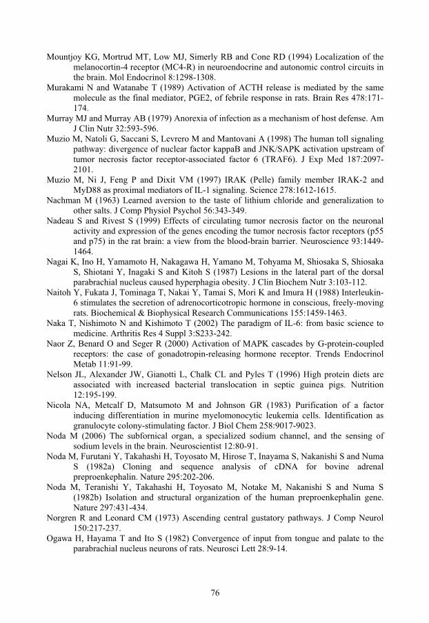
76
Mountjoy KG, Mortrud MT, Low MJ, Simerly RB and Cone RD (1994) Localization of the melanocortin-4 receptor (MC4-R) in neuroendocrine and autonomic control circuits in the brain. Mol Endocrinol 8:1298-1308.
Murakami N and Watanabe T (1989) Activation of ACTH release is mediated by the same molecule as the final mediator, PGE2, of febrile response in rats. Brain Res 478:171-174.
Murray MJ and Murray AB (1979) Anorexia of infection as a mechanism of host defense. Am J Clin Nutr 32:593-596.
Muzio M, Natoli G, Saccani S, Levrero M and Mantovani A (1998) The human toll signaling pathway: divergence of nuclear factor kappaB and JNK/SAPK activation upstream of tumor necrosis factor receptor-associated factor 6 (TRAF6). J Exp Med 187:2097-2101.
Muzio M, Ni J, Feng P and Dixit VM (1997) IRAK (Pelle) family member IRAK-2 and MyD88 as proximal mediators of IL-1 signaling. Science 278:1612-1615.
Nachman M (1963) Learned aversion to the taste of lithium chloride and generalization to other salts. J Comp Physiol Psychol 56:343-349.
Nadeau S and Rivest S (1999) Effects of circulating tumor necrosis factor on the neuronal activity and expression of the genes encoding the tumor necrosis factor receptors (p55 and p75) in the rat brain: a view from the blood-brain barrier. Neuroscience 93:1449-1464.
Nagai K, Ino H, Yamamoto H, Nakagawa H, Yamano M, Tohyama M, Shiosaka S, Shiosaka S, Shiotani Y, Inagaki S and Kitoh S (1987) Lesions in the lateral part of the dorsal parabrachial nucleus caused hyperphagia obesity. J Clin Biochem Nutr 3:103-112.
Naitoh Y, Fukata J, Tominaga T, Nakai Y, Tamai S, Mori K and Imura H (1988) Interleukin-6 stimulates the secretion of adrenocorticotropic hormone in conscious, freely-moving rats. Biochemical & Biophysical Research Communications 155:1459-1463.
Naka T, Nishimoto N and Kishimoto T (2002) The paradigm of IL-6: from basic science to medicine. Arthritis Res 4 Suppl 3:S233-242.
Naor Z, Benard O and Seger R (2000) Activation of MAPK cascades by G-protein-coupled receptors: the case of gonadotropin-releasing hormone receptor. Trends Endocrinol Metab 11:91-99.
Nelson JL, Alexander JW, Gianotti L, Chalk CL and Pyles T (1996) High protein diets are associated with increased bacterial translocation in septic guinea pigs. Nutrition 12:195-199.
Nicola NA, Metcalf D, Matsumoto M and Johnson GR (1983) Purification of a factor inducing differentiation in murine myelomonocytic leukemia cells. Identification as granulocyte colony-stimulating factor. J Biol Chem 258:9017-9023.
Noda M (2006) The subfornical organ, a specialized sodium channel, and the sensing of sodium levels in the brain. Neuroscientist 12:80-91.
Noda M, Furutani Y, Takahashi H, Toyosato M, Hirose T, Inayama S, Nakanishi S and Numa S (1982a) Cloning and sequence analysis of cDNA for bovine adrenal preproenkephalin. Nature 295:202-206.
Noda M, Teranishi Y, Takahashi H, Toyosato M, Notake M, Nakanishi S and Numa S (1982b) Isolation and structural organization of the human preproenkephalin gene. Nature 297:431-434.
Norgren R and Leonard CM (1973) Ascending central gustatory pathways. J Comp Neurol 150:217-237.
Ogawa H, Hayama T and Ito S (1982) Convergence of input from tongue and palate to the parabrachial nucleus neurons of rats. Neurosci Lett 28:9-14.
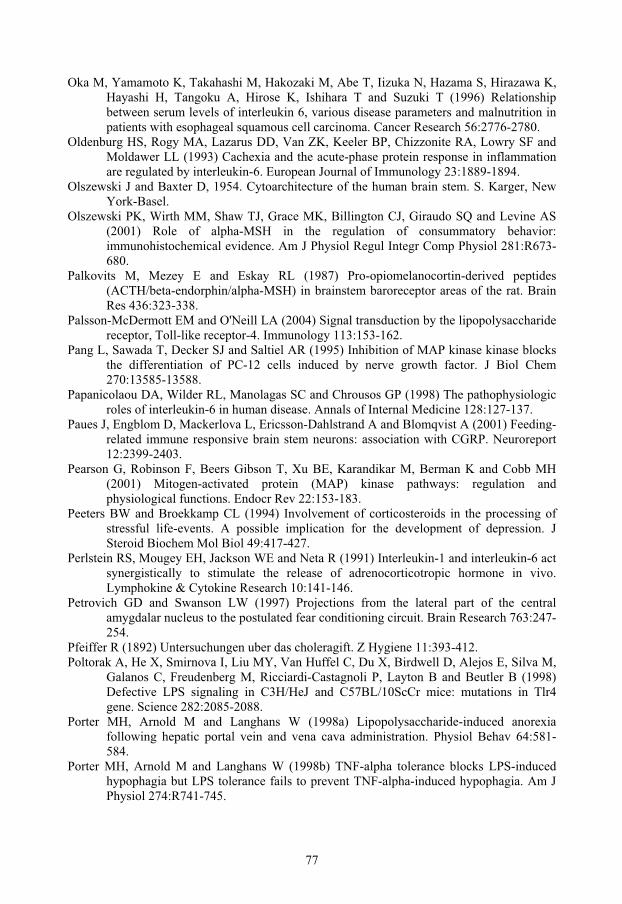
77
Oka M, Yamamoto K, Takahashi M, Hakozaki M, Abe T, Iizuka N, Hazama S, Hirazawa K, Hayashi H, Tangoku A, Hirose K, Ishihara T and Suzuki T (1996) Relationship between serum levels of interleukin 6, various disease parameters and malnutrition in patients with esophageal squamous cell carcinoma. Cancer Research 56:2776-2780.
Oldenburg HS, Rogy MA, Lazarus DD, Van ZK, Keeler BP, Chizzonite RA, Lowry SF and Moldawer LL (1993) Cachexia and the acute-phase protein response in inflammation are regulated by interleukin-6. European Journal of Immunology 23:1889-1894.
Olszewski J and Baxter D, 1954. Cytoarchitecture of the human brain stem. S. Karger, New York-Basel.
Olszewski PK, Wirth MM, Shaw TJ, Grace MK, Billington CJ, Giraudo SQ and Levine AS (2001) Role of alpha-MSH in the regulation of consummatory behavior: immunohistochemical evidence. Am J Physiol Regul Integr Comp Physiol 281:R673-680.
Palkovits M, Mezey E and Eskay RL (1987) Pro-opiomelanocortin-derived peptides (ACTH/beta-endorphin/alpha-MSH) in brainstem baroreceptor areas of the rat. Brain Res 436:323-338.
Palsson-McDermott EM and O'Neill LA (2004) Signal transduction by the lipopolysaccharide receptor, Toll-like receptor-4. Immunology 113:153-162.
Pang L, Sawada T, Decker SJ and Saltiel AR (1995) Inhibition of MAP kinase kinase blocks the differentiation of PC-12 cells induced by nerve growth factor. J Biol Chem 270:13585-13588.
Papanicolaou DA, Wilder RL, Manolagas SC and Chrousos GP (1998) The pathophysiologic roles of interleukin-6 in human disease. Annals of Internal Medicine 128:127-137.
Paues J, Engblom D, Mackerlova L, Ericsson-Dahlstrand A and Blomqvist A (2001) Feeding-related immune responsive brain stem neurons: association with CGRP. Neuroreport 12:2399-2403.
Pearson G, Robinson F, Beers Gibson T, Xu BE, Karandikar M, Berman K and Cobb MH (2001) Mitogen-activated protein (MAP) kinase pathways: regulation and physiological functions. Endocr Rev 22:153-183.
Peeters BW and Broekkamp CL (1994) Involvement of corticosteroids in the processing of stressful life-events. A possible implication for the development of depression. J Steroid Biochem Mol Biol 49:417-427.
Perlstein RS, Mougey EH, Jackson WE and Neta R (1991) Interleukin-1 and interleukin-6 act synergistically to stimulate the release of adrenocorticotropic hormone in vivo. Lymphokine & Cytokine Research 10:141-146.
Petrovich GD and Swanson LW (1997) Projections from the lateral part of the central amygdalar nucleus to the postulated fear conditioning circuit. Brain Research 763:247-254.
Pfeiffer R (1892) Untersuchungen uber das choleragift. Z Hygiene 11:393-412. Poltorak A, He X, Smirnova I, Liu MY, Van Huffel C, Du X, Birdwell D, Alejos E, Silva M,
Galanos C, Freudenberg M, Ricciardi-Castagnoli P, Layton B and Beutler B (1998) Defective LPS signaling in C3H/HeJ and C57BL/10ScCr mice: mutations in Tlr4 gene. Science 282:2085-2088.
Porter MH, Arnold M and Langhans W (1998a) Lipopolysaccharide-induced anorexia following hepatic portal vein and vena cava administration. Physiol Behav 64:581-584.
Porter MH, Arnold M and Langhans W (1998b) TNF-alpha tolerance blocks LPS-induced hypophagia but LPS tolerance fails to prevent TNF-alpha-induced hypophagia. Am J Physiol 274:R741-745.

78
Porter MH, Hrupka BJ, Langhans W and Schwartz GJ (1998c) Vagal and splanchnic afferents are not necessary for the anorexia produced by peripheral IL-1beta, LPS, and MDP. Am J Physiol 275:R384-389.
Price JL (2003) Comparative aspects of amygdala connectivity. Ann N Y Acad Sci 985:50-58.
Pritchard TC, Hamilton RB and Norgren R (2000) Projections of the parabrachial nucleus in the old world monkey. Exp Neurol 165:101-117.
Rauscher FJ, 3rd, Voulalas PJ, Franza BR, Jr. and Curran T (1988) Fos and Jun bind cooperatively to the AP-1 site: reconstitution in vitro. Genes Dev 2:1687-1699.
Ravasco P, Monteiro-Grillo I, Vidal PM and Camilo ME (2004) Cancer: disease and nutrition are key determinants of patients' quality of life. Support Care Cancer 12:246-252.
Revusky SH and Bedarf EW (1967) Association of illness with prior ingestion of novel foods. Science 155:219-220.
Richard S, Engblom D, Paues J, Mackerlova L and Blomqvist A (2005) Activation of the parabrachio-amygdaloid pathway by immune challenge or spinal nociceptive input: a quantitative study in the rat using Fos immunohistochemistry and retrograde tract tracing. J Comp Neurol 481:210-219.
Rinaman L, Baker EA, Hoffman GE, Stricker EM and Verbalis JG (1998) Medullary c-Fos activation in rats after ingestion of a satiating meal. Am J Physiol 275:R262-268.
Rivest S and Laflamme N (1995) Neuronal activity and neuropeptide gene transcription in the brains of immune-challenged rats. J Neuroendocrinol 7:501-525.
Rivier C and Vale W (1991) Stimulatory effect of interleukin-1 on adrenocorticotropin secretion in the rat: is it modulated by prostaglandins? Endocrinology 129:384-388.
Rocha MJ and Herbert H (1996) c-fos expression in the parabrachial nucleus following cardiovascular and blood volume changes. J Hirnforsch 37:389-397.
Rollins BL, Stines SG, McGuire HB and King BM (2001) Effects of amygdala lesions on body weight, conditioned taste aversion, and neophobia. Physiol Behav 72:735-742.
Ronco C, Tetta C, Mariano F, Wratten ML, Bonello M, Bordoni V, Cardona X, Inguaggiato P, Pilotto L, d'Intini V and Bellomo R (2003) Interpreting the mechanisms of continuous renal replacement therapy in sepsis: the peak concentration hypothesis. Artif Organs 27:792-801.
Rosen LB, Ginty DD, Weber MJ and Greenberg ME (1994) Membrane depolarization and calcium influx stimulate MEK and MAP kinase via activation of Ras. Neuron 12:1207-1221.
Rosenfeld MG, Mermod JJ, Amara SG, Swanson LW, Sawchenko PE, Rivier J, Vale WW and Evans RM (1983) Production of a novel neuropeptide encoded by the calcitonin gene via tissue-specific RNA processing. Nature 304:129-135.
Saha S, Engström L, Mackerlova L, Jakobsson PJ and Blomqvist A (2005) Impaired febrile responses to immune challenge in mice deficient in microsomal prostaglandin E synthase-1. Am J Physiol Regul Integr Comp Physiol 288:R1100-1107.
Sakai N and Yamamoto T (1997) Conditioned taste aversion and c-fos expression in the rat brainstem after administration of various USs. Neuroreport 8:2215-2220.
Sakai N and Yamamoto T (1998) Role of the medial and lateral parabrachial nucleus in acquisition and retention of conditioned taste aversion in rats. Behav Brain Res 93:63-70.
Sakai N and Yamamoto T (1999) Possible routes of visceral information in the rat brain in formation of conditioned taste aversion. Neurosci Res 35:53-61.
Sako K, Okuma Y, Hosoi T and Nomura Y (2005) STAT3 activation and c-FOS expression in the brain following peripheral administration of bacterial DNA. J Neuroimmunol 158:40-49.

79
Saleh TM and Cechetto DF (1996) Peptide changes in the parabrachial nucleus following cervical vagal stimulation. J Comp Neurol 366:390-405.
Saper CB (1995) The spinoparabrachial pathway: shedding new light on an old path. Journal of Comparative Neurology 353:477-479.
Saper CB and Loewy AD (1980) Efferent connections of the parabrachial nucleus in the rat. Brain Res 197:291-317.
Scalera G (2002) Effects of conditioned food aversions on nutritional behavior in humans. Nutr Neurosci 5:159-188.
Scammell TE, Elmquist JK, Griffin JD and Saper CB (1996) Ventromedial preoptic prostaglandin E2 activates fever-producing autonomic pathways. J Neurosci 16:6246-6254.
Scammell TE, Griffin JD, Elmquist JK and Saper CB (1998) Microinjection of a cyclooxygenase inhibitor into the anteroventral preoptic region attenuates LPS fever. Am J Physiol 274:R783-789.
Schiller M, Bohm M, Dennler S, Ehrchen JM and Mauviel A (2006) Mitogen- and stress-activated protein kinase 1 is critical for interleukin-1-induced, CREB-mediated, c-fos gene expression in keratinocytes. Oncogene.
Schwaber JS, Sternini C, Brecha NC, Rogers WT and Card JP (1988) Neurons containing calcitonin gene-related peptide in the parabrachial nucleus project to the central nucleus of the amygdala. J Comp Neurol 270:416-426, 398-419.
Seeley RJ, Blake K, Rushing PA, Benoit S, Eng J, Woods SC and D'Alessio D (2000) The role of CNS glucagon-like peptide-1 (7-36) amide receptors in mediating the visceral illness effects of lithium chloride. J Neurosci 20:1616-1621.
Sewards TV (2004) Dual separate pathways for sensory and hedonic aspects of taste. Brain Res Bull 62:271-283.
Shakhov AN, Collart MA, Vassalli P, Nedospasov SA and Jongeneel CV (1990) Kappa B-type enhancers are involved in lipopolysaccharide-mediated transcriptional activation of the tumor necrosis factor alpha gene in primary macrophages. J Exp Med 171:35-47.
Shakiryanova D, Tully A and Levitan ES (2006) Activity-dependent synaptic capture of transiting peptidergic vesicles. Nat Neurosci 9:896-900.
Shimazu R, Akashi S, Ogata H, Nagai Y, Fukudome K, Miyake K and Kimoto M (1999) MD-2, a molecule that confers lipopolysaccharide responsiveness on Toll-like receptor 4. J Exp Med 189:1777-1782.
Silverman JD and Kruger L (1989) Calcitonin-gene-related-peptide-immunoreactive innervation of the rat head with emphasis on specialized sensory structures. J Comp Neurol 280:303-330.
Silverman MN, Miller AH, Biron CA and Pearce BD (2004) Characterization of an interleukin-6- and adrenocorticotropin-dependent, immune-to-adrenal pathway during viral infection. Endocrinology 145:3580-3589.
Sim LJ and Joseph SA (1991) Arcuate nucleus projections to brainstem regions which modulate nociception. J Chem Neuroanat 4:97-109.
Simerly RB and Swanson LW (1988) Projections of the medial preoptic nucleus: a Phaseolus vulgaris leucoagglutinin anterograde tract-tracing study in the rat. J Comp Neurol 270:209-242.
Singewald N and Sharp T (2000) Neuroanatomical targets of anxiogenic drugs in the hindbrain as revealed by fos immunocytochemistry. Neuroscience 98:759-770.
Sjöstedt A, North RJ and Conlan JW (1996) The requirement of tumour necrosis factor-alpha and interferon-gamma for the expression of protective immunity to secondary murine
80
tularaemia depends on the size of the challenge inoculum. Microbiology 142 ( Pt 6):1369-1374.
Skoubis PD, Lam HA, Shoblock J, Narayanan S and Maidment NT (2005) Endogenous enkephalins, not endorphins, modulate basal hedonic state in mice. Eur J Neurosci 21:1379-1384.
Skoubis PD, Matthes HW, Walwyn WM, Kieffer BL and Maidment NT (2001) Naloxone fails to produce conditioned place aversion in mu-opioid receptor knock-out mice. Neuroscience 106:757-763.
Slugg RM and Light AR (1994) Spinal cord and trigeminal projections to the pontine parabrachial region in the rat as demonstrated with Phaseolus vulgaris leucoagglutinin. J Comp Neurol 339:49-61.
Smotherman WP (1985) Glucocorticoid and other hormonal substrates of conditioned taste aversion. Ann N Y Acad Sci 443:126-144.
Southall MD and Vasko MR (2001) Prostaglandin receptor subtypes, EP3C and EP4, mediate the prostaglandin E2-induced cAMP production and sensitization of sensory neurons. J Biol Chem 276:16083-16091.
Spath SE, Born J, Schrezenmeier H, Bornstein SR, Stromeyer P, Drechsler S, Fehm HL and Porzsolt F (1994) Interleukin-6 stimulates the hypothalamus-pituitary-adrenocortical axis in man. Journal of Clinical Endocrinology & Metabolism 79:1212-1214.
Spencer CM and Houpt TA (2001) Dynamics of c-fos and ICER mRNA expression in rat forebrain following lithium chloride injection. Brain Res Mol Brain Res 93:113-126.
Stefano GB, Fricchione G, Goumon Y and Esch T (2005) Pain, immunity, opiate and opioid compounds and health. Med Sci Monit 11:MS47-53.
Strelkov AB, Fields AL and Baracos VE (1989) Effects of systemic inhibition of prostaglandin production on protein metabolism in tumor-bearing rats. American Journal of Physiology.
Swank MW (1999) Coordinate regulation of Fos and Jun proteins in mouse brain by LiCl. Neuroreport 10:3685-3689.
Swank MW (2000) Phosphorylation of MAP kinase and CREB in mouse cortex and amygdala during taste aversion learning. Neuroreport 11:1625-1630.
Swanson LW and Petrovich GD (1998) What is the amygdala? Trends Neurosci 21:323-331. Swiergiel AH and Dunn AJ (1999) The roles of IL-1, IL-6, and TNFalpha in the feeding
responses to endotoxin and influenza virus infection in mice. Brain Behav Immun 13:252-265.
Taber E (1961) The cytoarchitecture of the brain stem of the cat. I. Brain stem nuclei of cat. J Comp Neurol 116:27-69.
Taché Y, Raybould H and Wei JY (1991) Central and peripheral actions of calcitonin gene-related peptide on gastric secretory and motor function. Adv Exp Med Biol 298:183-198.
Tatro JB (1996) Receptor biology of the melanocortins, a family of neuroimmunomodulatory peptides. Neuroimmunomodulation 3:259-284.
Tegowska E and Wasilewska E (1992) Effect of ambient temperature and E. coli endotoxin upon the plasma iron level in wild house mice in winter season. J Comp Physiol [B] 162:327-330.
Thimmulappa RK, Lee H, Rangasamy T, Reddy SP, Yamamoto M, Kensler TW and Biswal S (2006) Nrf2 is a critical regulator of the innate immune response and survival during experimental sepsis. J Clin Invest 116:984-995.
Tisdale MJ (2001) Cancer anorexia and cachexia. Nutrition 17:438-442. Tkacs NC and Li J (1999) Immune stimulation induces Fos expression in brainstem amygdala
afferents. Brain Res Bull 48:223-231.

81
Tkacs NC, Li J and Strack AM (1997) Central amygdala Fos expression during hypotensive or febrile, nonhypotensive endotoxemia in conscious rats. J Comp Neurol 379:592-602.
Tolchard S, Hare AS, Nutt DJ and Clarke G (1996) TNF alpha mimics the endocrine but not the thermoregulatory responses of bacterial lipopolysaccharide (LPS): correlation with FOS-expression in the brain. Neuropharmacology 35:243-248.
Tolle V, Bassant MH, Zizzari P, Poindessous-Jazat F, Tomasetto C, Epelbaum J and Bluet-Pajot MT (2002) Ultradian rhythmicity of ghrelin secretion in relation with GH, feeding behavior, and sleep-wake patterns in rats. Endocrinology 143:1353-1361.
Torelli GF, Meguid MM, Moldawer LL, Edwards CK, 3rd, Kim HJ, Carter JL, Laviano A and Fanelli FR (1999) Use of recombinant human soluble TNF receptor in anorectic tumor- bearing rats. Am J Physiol 277:R850-R855.
Toth LA, Tolley EA and Krueger JM (1993) Sleep as a prognostic indicator during infectious disease in rabbits. Proc Soc Exp Biol Med 203:179-192.
Tracey KJ, Morgello S, Koplin B, Fahey TJd, Fox J, Aledo A, Manogue KR and Cerami A (1990) Metabolic effects of cachectin/tumor necrosis factor are modified by site of production. Cachectin/tumor necrosis factor-secreting tumor in skeletal muscle induces chronic cachexia, while implantation in brain induces predominantly acute anorexia. J Clin Invest 86:2014-2024.
Tracey KJ, Wei H, Manogue KR, Fong Y, Hesse DG, Nguyen HT, Kuo GC, Beutler B, Cotran RS, Cerami A and et al (1988) Cachectin/tumor necrosis factor induces cachexia, anemia, and inflammation. Journal of Experimental Medicine 167:1211-1227.
Turnbull AV, Prehar S, Kennedy AR, Little RA and Hopkins SJ (2003) Interleukin-6 is an afferent signal to the hypothalamo-pituitary-adrenal axis during local inflammation in mice. Endocrinology 144:1894-1906.
Turnbull AV and Rivier CL (1999) Regulation of the hypothalamic-pituitary-adrenal axis by cytokines: actions and mechanisms of action. Physiol Rev 79:1-71.
Waldhoer M, Bartlett SE and Whistler JL (2004) Opioid receptors. Annu Rev Biochem 73:953-990.
Vallières L and Rivest S (1997) Regulation of the genes encoding interleukin-6, its receptor, and gp130 in the rat brain in response to the immune activator lipopolysaccharide and the proinflammatory cytokine interleukin-1beta. Journal of Neurochemistry 69:1668-1683.
van Rossum D, Hanisch UK and Quirion R (1997) Neuroanatomical localization, pharmacological characterization and functions of CGRP, related peptides and their receptors. Neurosci Biobehav Rev 21:649-678.
van Westerloo DJ, Giebelen IA, Florquin S, Daalhuisen J, Bruno MJ, de Vos AF, Tracey KJ and van der Poll T (2005) The cholinergic anti-inflammatory pathway regulates the host response during septic peritonitis. J Infect Dis 191:2138-2148.
Wang W, Andersson M, Lonnroth C, Svanberg E and Lundholm K (2005) Anorexia and cachexia in prostaglandin EP1 and EP3 subtype receptor knockout mice bearing a tumor with high intrinsic PGE2 production and prostaglandin related cachexia. J Exp Clin Cancer Res 24:99-107.
Wasserman SA (1993) A conserved signal transduction pathway regulating the activity of the rel-like proteins dorsal and NF-kappa B. Mol Biol Cell 4:767-771.
Veening JG, Swanson LW and Sawchenko PE (1984) The organization of projections from the central nucleus of the amygdala to brainstem sites involved in central autonomic regulation: a combined retrograde transport-immunohistochemical study. Brain Res 303:337-357.
82
Wiberg M and Blomqvist A (1984) The spinomesencephalic tract in the cat: its cells of origin and termination pattern as demonstrated by the intraaxonal transport method. Brain Res 291:1-18.
Wiberg M, Westman J and Blomqvist A (1987) Somatosensory projection to the mesencephalon: an anatomical study in the monkey. J Comp Neurol 264:92-117.
Wilkins EE and Bernstein IL (2006) Conditioning method determines patterns of c-fos expression following novel taste-illness pairing. Behav Brain Res 169:93-97.
Wilson BD, Bagnol D, Kaelin CB, Ollmann MM, Gantz I, Watson SJ and Barsh GS (1999) Physiological and anatomical circuitry between Agouti-related protein and leptin signaling. Endocrinology 140:2387-2397.
Wilson JD, Nicklous DM, Aloyo VJ and Simansky KJ (2003) An orexigenic role for mu-opioid receptors in the lateral parabrachial nucleus. Am J Physiol Regul Integr Comp Physiol 285:R1055-1065.
Wing EJ, Barczynski LK and Boehmer SM (1983a) Effect of acute nutritional deprivation on immune function in mice. I. Macrophages. Immunology 48:543-550.
Wing EJ, Stanko RT, Winkelstein A and Adibi SA (1983b) Fasting-enhanced immune effector mechanisms in obese subjects. Am J Med 75:91-96.
Wing EJ and Young JB (1980) Acute starvation protects mice against Listeria monocytogenes. Infect Immun 28:771-776.
Wirth MM, Olszewski PK, Levine AS and Giraudo SQ (2002) Effect of Agouti-related protein on development of conditioned taste aversion and oxytocin neuronal activation. Neuroreport 13:1355-1358.
Wolinsky TD, Carr KD, Hiller JM and Simon EJ (1996) Chronic food restriction alters mu and kappa opioid receptor binding in the parabrachial nucleus of the rat: a quantitative autoradiographic study. Brain Res 706:333-336.
Vollmer-Conna U, Fazou C, Cameron B, Li H, Brennan C, Luck L, Davenport T, Wakefield D, Hickie I and Lloyd A (2004) Production of pro-inflammatory cytokines correlates with the symptoms of acute sickness behaviour in humans. Psychol Med 34:1289-1297.
Vongs A, Lynn NM and Rosenblum CI (2004) Activation of MAP kinase by MC4-R through PI3 kinase. Regul Pept 120:113-118.
Wren AM, Small CJ, Ward HL, Murphy KG, Dakin CL, Taheri S, Kennedy AR, Roberts GH, Morgan DG, Ghatei MA and Bloom SR (2000) The novel hypothalamic peptide ghrelin stimulates food intake and growth hormone secretion. Endocrinology 141:4325-4328.
Wright MV, Frantz M and Van Houten JL (1992) Lithium fluxes in Paramecium and their relationship to chemoresponse. Biochim Biophys Acta 1107:223-230.
Wright SD, Ramos RA, Tobias PS, Ulevitch RJ and Mathison JC (1990) CD14, a receptor for complexes of lipopolysaccharide (LPS) and LPS binding protein. Science 249:1431-1433.
Yamamoto T, Fujimoto Y, Shimura T and Sakai N (1995) Conditioned taste aversion in rats with excitotoxic brain lesions. Neurosci Res 22:31-49.
Yamamoto T, Matsuo R, Ichikawa H, Wakisaka S, Akai M, Imai Y, Yonehara N and Inoki R (1990) Aversive taste stimuli increase CGRP levels in the gustatory insular cortex of the rat. Neurosci Lett 112:167-172.
Yamamoto T and Sawa K (2000a) c-Fos-like immunoreactivity in the brainstem following gastric loads of various chemical solutions in rats. Brain Res 866:135-143.
Yamamoto T and Sawa K (2000b) Comparison of c-fos-like immunoreactivity in the brainstem following intraoral and intragastric infusions of chemical solutions in rats. Brain Res 866:144-151.

83
Yamamoto T, Shimura T, Sakai N and Ozaki N (1994) Representation of hedonics and quality of taste stimuli in the parabrachial nucleus of the rat. Physiol Behav 56:1197-1202.
Yamamoto T, Shimura T, Sako N, Azuma S, Bai WZ and Wakisaka S (1992) C-fos expression in the rat brain after intraperitoneal injection of lithium chloride. Neuroreport 3:1049-1052.
Yamashiro T, Satoh K, Nakagawa K, Moriyama H, Yagi T and Takada K (1998) Expression of Fos in the rat forebrain following experimental tooth movement. J Dent Res 77:1920-1925.
Yamazoe M, Shiosaka S, Yagura A, Kawai Y, Shibasaki T, Ling N and Tohyama M (1984) The distribution of alpha-melanocyte stimulating hormone (alpha-MSH) in the central nervous system of the rat: an immunohistochemical study. II. Lower brain stem. Peptides 5:721-727.
Yang ZJ, Koseki M, Meguid MM, Gleason JR and Debonis D (1994) Synergistic effect of rhTNF-alpha and rhIL-1 alpha in inducing anorexia in rats. Am J Physiol 267:R1056-1064.
Yasui Y, Saper CB and Cechetto DF (1989) Calcitonin gene-related peptide immunoreactivity in the visceral sensory cortex, thalamus, and related pathways in the rat. J Comp Neurol 290:487-501.
Zardetto-Smith AM, Moga MM, Magnuson DJ and Gray TS (1988) Lateral hypothalamic dynorphinergic efferents to the amygdala and brainstem in the rat. Peptides 9:1121-1127.
Zhang Y, Lu H and Bargmann CI (2005) Pathogenic bacteria induce aversive olfactory learning in Caenorhabditis elegans. Nature 438:179-184.