Embed Size (px)
Citation preview

iologicalsychiatry
Archival Report BP
Brain Correlates of Suicide Attempt in 18,925Participants Across 18 International Cohorts
Adrian I. Campos, Paul M. Thompson, Dick J. Veltman, Elena Pozzi, Laura S. van Veltzen,Neda Jahanshad, Mark J. Adams, Bernhard T. Baune, Klaus Berger, Katharina Brosch,Robin Bülow, Colm G. Connolly, Udo Dannlowski, Christopher G. Davey, Greig I. de Zubicaray,Danai Dima, Tracy Erwin-Grabner, Jennifer W. Evans, Cynthia H.Y. Fu, Ian H. Gotlib,Roberto Goya-Maldonado, Hans J. Grabe, Dominik Grotegerd, Matthew A. Harris,Ben J. Harrison, Sean N. Hatton, Marco Hermesdorf, Ian B. Hickie, Tiffany C. Ho, Tilo Kircher,Axel Krug, Jim Lagopoulos, Hannah Lemke, Katie McMahon, Frank P. MacMaster,Nicholas G. Martin, Andrew M. McIntosh, Sarah E. Medland, Susanne Meinert, Tina Meller,Igor Nenadic, Nils Opel, Ronny Redlich, Liesbeth Reneman, Jonathan Repple,Matthew D. Sacchet, Simon Schmitt, Anouk Schrantee, Kang Sim, Aditya Singh,Frederike Stein, Lachlan T. Strike, Nic J.A. van der Wee, Steven J.A. van der Werff,Henry Völzke, Lena Waltemate, Heather C. Whalley, Katharina Wittfeld, Margaret J. Wright,Tony T. Yang, Carlos A. Zarate, Lianne Schmaal, and Miguel E. Rentería, for theENIGMA-MDD Working GroupISS
ABSTRACTBACKGROUND: Neuroimaging studies of suicidal behavior have so far been conducted in small samples, prone tobiases and false-positive associations, yielding inconsistent results. The ENIGMA-MDD Working Group aims toaddress the issues of poor replicability and comparability by coordinating harmonized analyses acrossneuroimaging studies of major depressive disorder and related phenotypes, including suicidal behavior.METHODS: Here, we pooled data from 18 international cohorts with neuroimaging and clinical measurements in18,925 participants (12,477 healthy control subjects and 6448 people with depression, of whom 694 had attemptedsuicide). We compared regional cortical thickness and surface area and measures of subcortical, lateral ventricular,and intracranial volumes between suicide attempters, clinical control subjects (nonattempters with depression), andhealthy control subjects.RESULTS: We identified 25 regions of interest with statistically significant (false discovery rate , .05) differencesbetween groups. Post hoc examinations identified neuroimaging markers associated with suicide attempt includingsmaller volumes of the left and right thalamus and the right pallidum and lower surface area of the left inferior parietallobe.CONCLUSIONS: This study addresses the lack of replicability and consistency in several previously publishedneuroimaging studies of suicide attempt and further demonstrates the need for well-powered samples andcollaborative efforts. Our results highlight the potential involvement of the thalamus, a structure viewed historicallyas a passive gateway in the brain, and the pallidum, a region linked to reward response and positive affect. Futurefunctional and connectivity studies of suicidal behaviors may focus on understanding how these regions relate tothe neurobiological mechanisms of suicide attempt risk.
https://doi.org/10.1016/j.biopsych.2021.03.015
Suicide is a leading cause of death worldwide and is a consid-erable health concern in both developed and developing coun-tries (1). While region- and country-specific estimates vary, theglobal average prevalence for suicide is estimated to be about10.6 deaths per 100,000 (2). Suicide attempts outnumber actualsuicides by 20- to 30-fold (3,4), which further increases theeconomic and social burden of suicidal behavior (5).
Suicidal behavior is more common in people living withmental illness (6–8). For a long time, suicidal behaviors were
ª 2021 Society of BioN: 0006-3223 Biolo
conceptualized as a symptom inherent to certain conditions,in particular, major depressive disorder (MDD). It is increas-ingly clear that suicidal behavior is complex (9). On the whole,a better understanding of suicidality, in terms of its underlyingmechanisms, could help identify individuals at increased riskof engaging in suicidal behaviors and inform better in-terventions (10).
Noninvasive neuroimaging technologies, such as magneticresonance imaging (MRI), allow brain structure and function to
logical Psychiatry. Published by Elsevier Inc. All rights reserved. 243gical Psychiatry August 15, 2021; 90:243–252 www.sobp.org/journal

Brain Correlates of Suicide AttemptBiologicalPsychiatry
be studied in vivo (11,12). The analysis of brain morphometryand neuroanatomical differences between individuals withmental illness and healthy control subjects has already provenuseful in conditions such as MDD (13), bipolar disorder (14),and schizophrenia (15). Similar approaches have been used tostudy suicidal behaviors, albeit in small samples. Briefly,several studies have reported lower gray matter volume andcortical thickness in the frontal, prefrontal, orbitofrontal,dorsolateral, and temporal lobes and white matter hyper-intensities associated with suicidal behaviors (16–29).
Nonetheless, small samples and heterogeneous analysismethods have led to a lack of replicability and inconsistentresults (11,30). The ENIGMA-MDD Working Group aims toaddress issues of poor replicability and comparability in neu-roimaging studies by coordinating harmonized analyses ofMDD and related phenotypes, including suicidal behavior. Inthe most recent meta-analysis of subcortical brain volumesconducted by our working group, we did not detect any sig-nificant morphological differences associated with suicidalbehavior independently of depression diagnosis (31). Identi-fying the neural substrates of suicide attempt is key to un-derstanding the etiology of suicide. This in turn might lead tothe development of novel therapeutic strategies based onbehavioral neuroscience and brain stimulation (32).
Previous studies have pinpointed some of these associa-tions, for example, alterations in the ventral and dorsal pre-frontal cortex, the insula, and regions involved in temporal,striatal, and posterior circuits (11,30). However, studies havebeen performed on samples of small size and with severalsources of potential bias, resulting in inconsistent findingsacross publications (11). Notably, subcortical associations didnot replicate in our previous meta-analyses (31). Thus, weconclude that prior literature might not be sufficiently robust tosupport the role of specific brain structures in suicide attempt.For that reason, we decided to conduct a comprehensiveexploratory investigation of neuroimaging correlates for suicideattempt in the largest participant sample to date. We per-formed a pooled mega-analysis of subcortical volumes andregional cortical surface area and thickness, using linearmixed-model regressions in a sample of 18,925 subjects from18 cohorts from around the world. We aimed to shed light onthe neural circuits that underlie suicidal behavior by comparingbrain morphometry between MDD cases with a history ofsuicide attempt versus those without, as well as versus healthycontrol subjects.
METHODS AND MATERIALS
Samples
We analyzed pooled data (mega-analysis) across 17 ENIGMA-MDD Working Group cohorts with clinical and neuroimagingdata available for participants fulfilling MDD criteria (33) (n =2533) and healthy control subjects (n = 4066) and participantsfrom the UK Biobank (n = 12,366). We defined three groups:suicide attempters; clinical control subjects, that is, partici-pants with depression and no history of suicide attempt; andhealthy control subjects. Descriptive statistics for each sampleare listed in Table 1 and Table S1. Each cohort assesseddepression status and history of a suicide attempt based onavailable clinical information. In the UK Biobank, lifetime
244 Biological Psychiatry August 15, 2021; 90:243–252 www.sobp.org
depression status (n = 3633) and lifetime suicide attempt (n =322) were ascertained using the Composite InternationalDiagnostic Interview. Participants with no history of depressionor suicide attempt (n = 8411) were defined as healthy controlsubjects. A psychiatric diagnostic interview was used to di-agnose participants across the ENIGMA-MDD groups. Infor-mation on the instruments used to determine suicide attemptand exclusion criteria per site are available in Tables S2 andS3, respectively. The combined sample comprised 12,477healthy control subjects and 6448 participants with a lifetimedepression diagnosis. Within the depression group, 694 par-ticipants reported at least one suicide attempt. All sites ob-tained approval from their local institutional ethics committeesand review boards to participate in this study, and all partici-pants provided informed consent at their local recruitmentinstitution.
Image Processing and Analysis
T1-weighted MRI structural brain scans were acquired andanalyzed locally at each site using the validated and automatedsegmentation software FreeSurfer (34) (http://surfer.nmr.mgh.harvard.edu/). Image acquisition parameters and softwareversions and descriptions are detailed in Table S2. The seg-mentation of cortical and subcortical phenotypes was visuallyinspected for accuracy following standardized protocolsdesigned to facilitate harmonized image analysis acrossmultiple sites (http://enigma.ini.usc.edu/protocols/imaging-protocols/). Within each cohort, measures were visually veri-fied for accuracy and excluded if they were not properlysegmented. Within-cohort outliers (defined as measurementgreater than 3 standard deviations away from the mean) wereexcluded from the analysis. We examined five global brainmeasures, including intracranial volume (ICV), total surfacearea of the left and right hemispheres and mean corticalthickness of the left and right hemispheres, 16 subcorticalbrain volume measures, and cortical surface area and thick-ness measures for 68 brain regions of interest (ROIs) asdefined by the Desikan-Killiany atlas (35).
Ascertainment of Suicide Attempt History
In this study, a suicide attempt was defined as any self-harmact with the intent to die. In this study, we focused on life-time suicide attempt, as opposed to other suicidal behaviors,to reduce potential heterogeneity arising from different suiciderisk assessment instruments used across cohorts. Attemptseverity was not assessed because of a lack of information inindividual studies. A description of instruments used to mea-sure suicide attempt in each site is available in Table S2. Co-horts also provided (where available) information on 1) whetherparticipants have used antidepressants; 2) depression severity,coded either as the Hamilton Depression Rating Scale scoreexcluding the suicide item, or as the number of DSM-IV MDDcriteria endorsed (ranging from 0 to 9); 3) age of depressiononset; and 4) whether depression was recurrent or a singleepisode.
Statistical Analyses
Linear Mixed-Effects Models. Statistical analyses wereperformed in R version 3.6.1 (R Foundation for Statistical
/journal

Table 1. Demographics and Clinical Measures Across Studied Groups
Characteristics Healthy Control Subjects Clinical Control Subjects Suicide Attempters
Total, n (%) 12,477 (66%) 5754 (30%) 694 (4%)
Sex, Female, n (%) 6076 (60%) 3736 (44%) 466 (4.5%)
Sex, Male, n (%) 6401 (74.0%) 2018 (23.3%) 228 (2.6%)
Age, Years, Mean (SD) 57.6 (14.8) 53.2 (15.4) 49.2 (16.3)
Beck Depression Inventory, Mean (SD)a,b 3.6 (4.0) 18 (10.9) 23 (11.8)
Hamilton Depression Rating Scale, Mean (SD)a,b 1.3 (2.0) 11.0 (6.9) 13.8 (6.9)
Depression Age of Onset, Years, Mean (SD)a NA 29.8 (14.3) 23.3 (14.1)
Antidepressant Use, %a 0.1% 36% 21%
Depression Recurrence, %a NA 21% 36%
NA, not applicable.aData available only for a subset of the sample.bSum score excluding the suicidal behaviors item.
Brain Correlates of Suicide AttemptBiologicalPsychiatry
Computing, Vienna, Austria) using the statistical packagenlme. Linear mixed-effects models were used to account forsite variation (with a random intercept for scan site) whilecorrecting for desired covariates as fixed effects. We modeledeach regional measure as an outcome while using an indicatorvariable per group of interest: healthy control subjects, patientswith MDD with no suicide attempt history (clinical controlsubjects), and patients with MDD with attempt history (suicideattempters). All models were adjusted for age and sex, whilesurface area and volumetric analyses were also adjusted forICV (except when ICV was the measure of interest). Main ef-fects of groups (i.e., differences between groups of healthycontrol subjects, clinical control subjects, and suicideattempters) were identified by performing a type II analysis ofvariance (F test) over the fitted linear mixed-effects modeldescribed above. We conducted follow-up (post hoc) analysesto assess whether the effects were driven by suicide attempt.ROIs were compared between suicide attempters and clinicalcontrol subjects, between suicide attempters and healthycontrol subjects, and between clinical control subjects andhealthy control subjects. Finally, we conducted several sensi-tivity analyses. First, to assess the effects of severity, recur-rence, and age of onset of depression and history ofantidepressant use on the observed associations, we repeatedthe post hoc analyses of the four regions showing evidence ofassociation with suicide attempt, including additional cova-riates one at a time. Then, to evaluate the contribution of thelargest cohort in the analysis to the observed results on thefour ROIs mentioned above, we conducted the analysesexcluding the UK Biobank cohort.
Statistical Significance Definition. We corrected formultiple comparisons using a false discovery rate procedure(36) for each set of morphometry measures separately. Thesignificance threshold to define ROIs for post hoc analyseswas set at false discovery rate p value , .05. For the post hoctests of the ROIs identified above, we used a matrix spectraldecomposition to identify the number of effective variables(37,38) coupled with Bonferroni correction to keep the type Ierror rate at 5%. In this review, a significant result survivedpost hoc multiple testing corrections (p , Bonferroni correctedthreshold), whereas a nominally significant result was onlysignificant before correction (p , .05).
Biological Ps
RESULTS
Suicide Attempt Prevalence and SampleDemographics
Details on the sample of each cohort included in this analysisare summarized in Table 1 and Table S1. Notably, not all co-horts had cases of suicide attempt, but still contributed data tothe healthy or clinical control groups. The pooled mean age(SD) was 56.22 (15.17) years. Differences in age and sexcomposition across cohorts were detected (Table S1) andused as covariates for all the analyses. The total sample sizecomprised 18,925 subjects, of which 3.67% (n = 694) had atleast one past suicide attempt. Furthermore, 30.40% (n =5754) of the total sample was diagnosed with depression butdid not report a previous suicide attempt. Methodologicaldifferences (e.g., scanner used or different parameters for thescan) between participating cohorts are listed in Table S2.
Subcortical Volumetric Measures
The thalamus (right and left), right pallidum, and total ICVexhibited a statistically significant group effect (i.e., any dif-ference between healthy control subjects, clinical controlsubjects, or suicide attempters) after correcting for multiplecomparisons. The left pallidum and the right nucleus accum-bens showed a nominally significant difference but did notsurvive correction for multiple comparisons (Table S4).Depressed attempters exhibited smaller volumes in the left andright thalamus and right pallidum compared with both clinical(Cohen’s d = 20.13, 20.14, and 20.12, respectively) andhealthy control subjects (Figure 1). Depressed attempters alsoexhibited smaller ICV compared with healthy control subjects(Cohen’s d = 20.13). This association did not reach signifi-cance, when comparing attempters and clinical control sub-jects, after accounting for multiple testing. None of the regionswith a significant group effect showed a significant differencewhen comparing clinical control subjects with healthy controlsubjects (Table S5).
Cortical Surface Area
Eight of the 68 cortical regions under analysis displayed asignificant group effect (Table S6). These regions included theleft and right pericalcarine, left and right cuneus, left inferior
ychiatry August 15, 2021; 90:243–252 www.sobp.org/journal 245
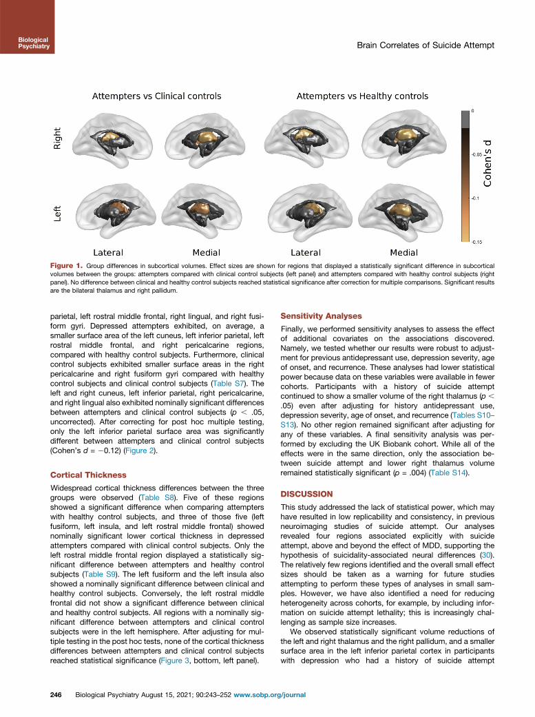
Figure 1. Group differences in subcortical volumes. Effect sizes are shown for regions that displayed a statistically significant difference in subcorticalvolumes between the groups: attempters compared with clinical control subjects (left panel) and attempters compared with healthy control subjects (rightpanel). No difference between clinical and healthy control subjects reached statistical significance after correction for multiple comparisons. Significant resultsare the bilateral thalamus and right pallidum.
Brain Correlates of Suicide AttemptBiologicalPsychiatry
parietal, left rostral middle frontal, right lingual, and right fusi-form gyri. Depressed attempters exhibited, on average, asmaller surface area of the left cuneus, left inferior parietal, leftrostral middle frontal, and right pericalcarine regions,compared with healthy control subjects. Furthermore, clinicalcontrol subjects exhibited smaller surface areas in the rightpericalcarine and right fusiform gyri compared with healthycontrol subjects and clinical control subjects (Table S7). Theleft and right cuneus, left inferior parietal, right pericalcarine,and right lingual also exhibited nominally significant differencesbetween attempters and clinical control subjects (p , .05,uncorrected). After correcting for post hoc multiple testing,only the left inferior parietal surface area was significantlydifferent between attempters and clinical control subjects(Cohen’s d = 20.12) (Figure 2).
Cortical Thickness
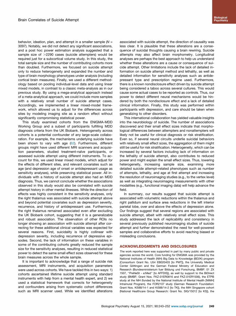
Widespread cortical thickness differences between the threegroups were observed (Table S8). Five of these regionsshowed a significant difference when comparing attempterswith healthy control subjects, and three of those five (leftfusiform, left insula, and left rostral middle frontal) showednominally significant lower cortical thickness in depressedattempters compared with clinical control subjects. Only theleft rostral middle frontal region displayed a statistically sig-nificant difference between attempters and healthy controlsubjects (Table S9). The left fusiform and the left insula alsoshowed a nominally significant difference between clinical andhealthy control subjects. Conversely, the left rostral middlefrontal did not show a significant difference between clinicaland healthy control subjects. All regions with a nominally sig-nificant difference between attempters and clinical controlsubjects were in the left hemisphere. After adjusting for mul-tiple testing in the post hoc tests, none of the cortical thicknessdifferences between attempters and clinical control subjectsreached statistical significance (Figure 3, bottom, left panel).
246 Biological Psychiatry August 15, 2021; 90:243–252 www.sobp.org
Sensitivity Analyses
Finally, we performed sensitivity analyses to assess the effectof additional covariates on the associations discovered.Namely, we tested whether our results were robust to adjust-ment for previous antidepressant use, depression severity, ageof onset, and recurrence. These analyses had lower statisticalpower because data on these variables were available in fewercohorts. Participants with a history of suicide attemptcontinued to show a smaller volume of the right thalamus (p ,
.05) even after adjusting for history antidepressant use,depression severity, age of onset, and recurrence (Tables S10–S13). No other region remained significant after adjusting forany of these variables. A final sensitivity analysis was per-formed by excluding the UK Biobank cohort. While all of theeffects were in the same direction, only the association be-tween suicide attempt and lower right thalamus volumeremained statistically significant (p = .004) (Table S14).
DISCUSSION
This study addressed the lack of statistical power, which mayhave resulted in low replicability and consistency, in previousneuroimaging studies of suicide attempt. Our analysesrevealed four regions associated explicitly with suicideattempt, above and beyond the effect of MDD, supporting thehypothesis of suicidality-associated neural differences (30).The relatively few regions identified and the overall small effectsizes should be taken as a warning for future studiesattempting to perform these types of analyses in small sam-ples. However, we have also identified a need for reducingheterogeneity across cohorts, for example, by including infor-mation on suicide attempt lethality; this is increasingly chal-lenging as sample size increases.
We observed statistically significant volume reductions ofthe left and right thalamus and the right pallidum, and a smallersurface area in the left inferior parietal cortex in participantswith depression who had a history of suicide attempt
/journal

Left
Rig
ht
Lateral Medial MedialLateral
Attempters vs Clinical controls Attempters vs Healthy controls
Cortical surface area
Clinical vs Healthy controls
-0.15 -0.12 -0.09 -0.06 -0.03 0Cohen's d
MedialLateral
Left
Rig
ht
p<
0.0
5(n
omin
ally
sig
nif
ican
t)p
<0
.00
33
3(s
tati
stic
ally
sig
nif
ican
t)
Figure 2. Group differences in cortical surface area. Effect sizes are shown for regions that displayed a (top) nominally significant (p , .05) or (bottom) astatistically significant (p , .00333 threshold after multiple test correction) post hoc difference between attempters and clinical control subjects (left panel),attempters and healthy control subjects (middle panel), and clinical control subjects compared with healthy control subjects (right panel). All of the coloredregions showed a statistically significant group effect (false discovery rate , .05).
Brain Correlates of Suicide AttemptBiologicalPsychiatry
compared with both clinical and healthy control subjects.Previous studies have suggested that these regions areassociated with suicidal behaviors (17,21,39–41); however, thelack of evidence for associations in better-powered studies(31) and the lack of consensus in the field (11,13) made itchallenging to reach definitive conclusions. A recent overviewof a brain model for suicidal behavior identified at least fourfunctional diathetic elements to suicide. These include sub-jective distress, impaired decision making, learning or memorydeficits, and social distortion as part of a cognitive impairment(42). The regions identified here are involved in decision-making behaviors such as impulsivity and planning, as wellas attention and concept of self (see below). All of these factorsare related to the four risk-increasing components discussedabove.
The pallidum has been linked to reward response, socialactivity mediation, and positive affect (43,44). Furthermore, arecent structural MRI study has linked the pallidum to suicidalideation severity and impulsivity in a small sample of Koreanpatients with MDD (45). Thus, alterations in the pallidum mayreflect changes in affect and impulsivity known to be associ-ated with suicidal behaviors (46,47). The thalamus, historicallyviewed as a passive gateway linking different brain regions,has been recently proposed to operate as an integrative hub of
Biological Ps
the brain (48), relaying signals between regions such as thebasal ganglia and the cortex, but also frontal-subcorticalconnections that have been linked to suicide attemptthrough fractional anisotropy (42). A growing body of evidencesuggests that the different nuclei within the thalamus areinvolved in high-order cognition (49). The thalamus serves as aregion where the integration of cortico-striatal-thalamic-cortical circuit takes place. These circuits modulate severalbehaviors including emotional drive and planning (50). Ourresults showing abnormalities in part of the basal ganglia(pallidum) and the thalamus would be consistent with impli-cating these circuits with suicide attempt. Furthermore,thalamic abnormalities and lesions have been linked to disor-ders such as addiction (51), bipolar disorder (52), and schizo-phrenia (53,54). Therefore, an impaired function in thethalamus and its related circuits with the basal ganglia andcortex might mediate suicide attempt through impaired affect,empathy, and processing of stimuli response relationships.
As previously reviewed (28), the parietal region is part of theexecutive control network, which exerts control over thought,emotion, and behavior. The inferior parietal lobe is further partof the posterior parietal cortex (55), known for its role inattention networks (56) and processing of visual information.The inferior parietal lobe has been linked to schizophrenia
ychiatry August 15, 2021; 90:243–252 www.sobp.org/journal 247

Figure 3. Group differences in cortical thickness. Effect sizes are shown for regions that displayed a (top) nominally significant (p , .05) or (bottom) astatistically significant (p , .001373 threshold after multiple test correction) post hoc difference between attempters and clinical control subjects (left panel),attempters and healthy control subjects (middle panel), and clinical control subjects compared with healthy control subjects (right panel). All of the coloredregions showed a statistically significant group effect (false discovery rate , .05).
Brain Correlates of Suicide AttemptBiologicalPsychiatry
through structural and functional differences including execu-tive function and concept-of-self functions (57). It has furtherbeen linked to suicide attempt within bipolar disorder (58).Connectivity and functional studies are necessary to comple-ment our results; however, studying suicidality using functionalMRI carries important challenges such as the immediate needto reduce suicide risk and establishing a valid (i.e., relevant)time frame before or after a suicide attempt.
Conversely, no cortical thickness differences survived posthoc multiple testing correction. Some regions differed betweenattempters and healthy control subjects, but the lack of dif-ferences between clinical control subjects and suicideattempters suggests that these associations may be driven bydepression status, rather than suicide attempt. The fact thatsubcortical volume and surface area associations had astronger association with suicide attempt than with corticalthickness could be of interest in light of genetic analyses thatidentified substantial differences in the genetic etiology ofcortical morphometry phenotypes (59–61). For example, sur-face area measurements were reported to have a higher heri-tability and to be more influenced by early developmentalgenetic influences than cortical thickness (60), suggesting thatbiomarkers associated with surface area are likely establishedearlier in life. In contrast, cortical thickness phenotypes may be
248 Biological Psychiatry August 15, 2021; 90:243–252 www.sobp.org
more variable and more susceptible to adverse environmentaleffects later in life, such as substance use or having a psy-chiatric condition.
The substantial overlap between depression severity andsuicide attempt (e.g., Table 1 shows that participants with apast suicide attempt also have higher Hamilton DepressionRating Scale and Beck Depression Inventory sum scores evenafter removing the suicide item) makes it difficult to differen-tiate whether an identified statistical effect is driven bydepression severity or suicide attempt status. This is alsosupported by the fact that several cortical associations alsodisplayed an effect when comparing clinical with healthycontrol subjects and that those results did not remain signifi-cant after sensitivity analyses that controlled for proxies ofdepression severity. Overall, effect sizes were smaller whencomparing clinical with healthy control subjects than thosewhen comparing suicide attempters with either control group.That might be due to the previously discussed collinearitybetween suicidal behaviors and depression severity. Many ofthe most severely depressed cases will be among thedepressed suicidal group, thus reducing the strength of anysignals associated with depression severity.
In our previous study, we investigated intracranial andsubcortical volumes for their association with suicidal
/journal
Brain Correlates of Suicide AttemptBiologicalPsychiatry
behavior, ideation, plan, and attempt in a smaller sample (N =3097). Notably, we did not detect any significant associations,and a post hoc power estimation analysis suggested that asample size of .2290 cases (suicide attempters) would berequired just for a subcortical volume study. In this study, thetotal sample size and the number of contributing cohorts morethan doubled. Furthermore, we focused on suicide attemptonly to reduce heterogeneity and expanded the number andtype of brain morphology phenotypes under analysis (includingcortical brain measures). Finally, we used a different method-ology based on pooling individual-level data and using linearmixed models, in contrast to a classic meta-analysis as in ourprevious study. By using a mega-analytical approach insteadof a meta-analytical approach, we could include more sampleswith a relatively small number of suicide attempt cases.Accordingly, we implemented a linear mixed-model frame-work, which allowed us to adjust for the differences acrosssites by modeling imaging site as a random effect withoutsignificantly compromising statistical power.
This study examined cohorts from the ENIGMA-MDDWorking Group and a subset of participants fulfilling MDDdiagnosis criteria from the UK Biobank. Heterogeneity acrosscohorts is a potential confounder of any large-scale collabo-ration. For example, the mechanisms underlying suicide havebeen shown to vary with age (62). Furthermore, differentgroups might have used different MRI scanners and acquisi-tion parameters, studied treatment-naïve participants, orassessed suicide attempt using different instruments. To ac-count for this, we used linear mixed models, which adjust forthe effects of different sites, and relevant covariates such asage (and depression age of onset or antidepressant usage assensitivity analyses), while preserving statistical power. All in-dividuals with a history of suicide attempt also had an MDDdiagnosis. Thus, we cannot conclude whether the associationsobserved in this study would also be correlated with suicideattempt history in other mental illnesses. While the direction ofeffects was highly consistent in the sensitivity analyses, onlythe right thalamus was associated with suicide attempt aboveand beyond potential covariates such as depression severity,recurrence, and history of antidepressant use. Furthermore,the right thalamus remained associated even after excludingthe UK Biobank cohort, suggesting that it is a generalizableand robust association. The observation of other ROIs nolonger showing an association with suicide attempt after cor-recting for these additional clinical variables was expected forseveral reasons. First, suicidality is highly collinear withdepression severity, including recurrence of depressive epi-sodes. Second, the lack of information on these variables insome of the contributing cohorts greatly reduced the samplesize for the sensitivity analyses, resulting in reduced statisticalpower to detect the same small effect sizes observed for thesebrain measures across the whole sample.
It is important to acknowledge that a range of suicide riskassessment, MRI instruments, and acquisition parameterswere used across cohorts. We have tackled this in two ways: 1)cohorts ascertained lifetime suicide attempt using standardinstruments with help from a mental health expert, and 2) weused a statistical framework that corrects for heterogeneityand confounders arising from systematic cohort differencessuch as MRI scanner used. While we identified alterations
Biological Ps
associated with suicide attempt, the direction of causality wasless clear. It is plausible that these alterations are a conse-quence of suicidal thoughts causing a brain rewiring. Suicideattempts may also affect brain morphometry. Longitudinalanalyses are perhaps the best approach to help us understandwhether these alterations are a cause or consequence of sui-cide attempt. Other limitations include the lack of detailed in-formation on suicide attempt method and lethality, as well asdetailed information for sensitivity analyses such as antide-pressant type and prescription regime used. Furthermore,there is a known nondisclosure effect driven by suicide attemptbeing considered a taboo across several cultures. This wouldcause some actual cases to be reported as controls. Thus, ourpower to detect different neural mechanisms would be hin-dered by both the nondisclosure effect and a lack of detailedclinical information. Finally, this study was performed withinparticipants with depression, and care should be taken whengeneralizing to other disorders.
This international collaboration has yielded valuable insightsinto the neurobiology of suicide. The number of associationsdiscovered and their small effect sizes indicate that morpho-logical differences between attempters and nonattempters willlikely not be useful for clinical diagnosis or risk stratification.Even so, if several neural circuits underlie suicidal behaviorswith relatively small effect sizes, the aggregation of them mightstill be useful for risk stratification. Heterogeneity, which can beincreased by several factors including lack of information onthe lethality of suicide attempt, also contributes to reducedpower and might explain the small effect sizes. Thus, loweringheterogeneity, increasing sample size, examining moredetailed suicide attempt–related phenotypes such as numberof attempts, lethality, and age at first attempt and increasingthe resolution of neuroimaging studies (e.g., to the vertex level)as well as integrating neuroimaging measurements with othermodalities (e.g., functional imaging data) will help advance thefield.
In summary, our results suggest that suicide attempt isassociated with volumetric reductions within the thalamus andright pallidum and surface area reductions in the left interiorparietal lobe, over and above the effects of depression alone.Our findings suggest that several regions are associated withsuicide attempt, albeit with relatively small effect sizes. Thisstudy addressed the lack of replicability and consistency inseveral previously published neuroimaging studies of suicideattempt and further demonstrated the need for well-poweredsamples and collaborative efforts to avoid reaching biased ormisleading conclusions.
ACKNOWLEDGMENTS AND DISCLOSURESThe work reported here was supported in part by many public and privateagencies across the world. Core funding for ENIGMA was provided by theNational Institutes of Health (NIH) Big Data to Knowledge (BD2K) program(Consortium Grant No. U54 EB020403 [to PMT]), the University MedicalCenter Göttingen and the German Federal Ministry of Education andResearch (Bundesministerium fuer Bildung und Forschung, BMBF: 01 ZX1507, ‘‘PreNeSt - e:Med’’ [to AFFDIS]), as well by support to the BiDirectstudy (BMBF; Grant Nos. FKZ-01ER0816 and FKZ-01ER1506), the ETPBstudy at the NIH (funded by the National Institute of Mental Health [NIMH]Intramural Program), the FOR2107 study (German Research Foundation:Grant Nos. Ki588/14-1 and Ki588/14-2 [to TK]), the IMH Singapore cohort(National Healthcare Group Research: Grant No. SIG/15012 [to KS]), the
ychiatry August 15, 2021; 90:243–252 www.sobp.org/journal 249
Brain Correlates of Suicide AttemptBiologicalPsychiatry
LOND study (supported in part by the Biomedical Research Centre, SouthLondon, and Maudsley NHS Foundation Trust, London, United Kingdom),the Stanford study (supported in part by the NIMH Grant R37-MH101495 [toIHG]), the Melbourne study (Australia’s National Health and MedicalResearch Council [NHMRC] Project Grants 1064643 [to principal investi-gator, BJH] and 1024570 [to principal investigator, CGD]), the QTIM study(National Institute of Child Health and Human Development Grant No. R01-HD050735 and NHMRC Australia Grant Nos. 486682 and 1009064 [toprincipal investigator, MJW]), the Pharmo study in Amsterdam (faculty re-sources of the Academic Medical Center, University of Amsterdam, and11.32050.26 ERA-NET PRIOMEDCHILD FP 6 [EU]), and the UCSF study(funded in part by the NIMH Grant Nos. R01MH085734 and K01MH117442[to TTY and TCH]; National Center for Complementary and IntegrativeHealth [NCCIH Grant No. 1R61AT009864]; and the American Foundation forSuicide Prevention [AFSP]). The Edinburgh group is funded by a WellcomeTrust Strategic Award “Stratifying Resilience and Depression Longitudinally”(STRADL) (Reference 104036/Z/14/Z [to AMM]). The SHIP cohort is part ofthe Community Medicine Research Network of the University MedicineGreifswald, which is supported by the German Federal State ofMecklenburg–West Pomerania. This work was also supported by a UQResearch Training Scholarship from The University of Queensland (UQ [toAIC]), the NHMRC and Australian Research Council (ARC) through aResearch Fellowship (Grant No. APP1102821 [to MER]), NHMRC CareerDevelopment Fellowships (Grant Nos. 1124472 and 1061757, respectively[to BJH and CGD]), NHMRC (Grant Nos. APP1103623, APP1158127, andAPP1172917 [to SEM]), NCCIH (Grant No. 1R61AT009864 [to TTY]), and theNIMH of the NIH (U.S.) under Award Number R01-MH117601 (to LS and NJ).
MER and LS conceived and jointly supervised the study. Authors from allsite cohorts were involved in data collection, processing, analysis, andfunding for their samples. AIC implemented the analysis pipeline, performedall statistical analyses, and generated the tables and figures. AIC, MER, andLS drafted the first draft of the manuscript with feedback and input from allcoauthors. All authors approved the content of this manuscript.
A previous version of this article was published as a preprint on medRxiv:https://www.medrxiv.org/content/10.1101/2020.05.06.20090191v1.
This project used data from the UK Biobank under applicationnumber 25331.
HJG has received travel grants and speaker’s honoraria from FreseniusMedical Care, Neuraxpharm, Servier, and Janssen Cilag as well as researchfunding from Fresenius Medical Care. CAZ is a full-time U.S. governmentemployee. He is listed as a coinventor on a patent for the use of ketamineand its metabolites in major depression and suicidal ideation. CAZ hasassigned his patent rights to the U.S. government but will share a per-centage of any royalties that may be received by the government. NJ andPMT are MPI of a research-related grant from Biogen, Inc., for work unre-lated to this manuscript. IBH is funded by NHMRC Fellowship (2013–2017and 2018–2022). He is the Chief Scientific Advisor to, and a 5% equityshareholder in, InnoWell Pty Ltd. InnoWell was formed by the University ofSydney (45% equity) and PwC (Australia; 45% equity) to deliver the $30 MAustralian Government–funded Project Synergy (2017–2020). All other au-thors report no biomedical financial interests or potential conflicts ofinterest.
ARTICLE INFORMATIONFrom the Genetic Epidemiology Lab (AIC, NGM, MER), QIMR BerghoferMedical Research Institute, Brisbane; Faculty of Medicine (AIC, SEM, MER),The University of Queensland, Brisbane; Institute of Health and BiomedicalInnovation (GidZ), Queensland University of Technology, Brisbane; Psychi-atric Genetics Lab (SEM), QIMR Berghofer Medical Research Institute,Brisbane; Queensland Brain Institute (LTS, MJW) and Centre for AdvancedImaging (MJW), The University of Queensland, Brisbane; School of Psy-chology (SEM), The University of Queensland, Brisbane; Herston ImagingResearch Facility & School of Clinical Sciences (KM), Queensland Universityof Technology, Brisbane; Thompson Institute (JL), University of the SunshineCoast, Sunshine Coast, Queensland; Orygen (EP, LSvV, LS), The NationalCentre of Excellence in Youth Mental Health, Parkville; Department ofPsychiatry (EP, LSvV, CGD, BJH), The University of Melbourne & MelbourneHealth, Melbourne; Department of Psychiatry (BTB) and The Florey Instituteof Neuroscience and Mental Health (BTB), The University of Melbourne,
250 Biological Psychiatry August 15, 2021; 90:243–252 www.sobp.org
Melbourne, Victoria; Brain and Mind Centre (SNH, IBH, JL), University ofSydney, Sydney, New South Wales, Australia; Amsterdam UMC (DJV), VrijeUniversiteit Amsterdam, Department of Psychiatry, Department of Anatomy& Neurosciences, Amsterdam Neuroscience, Amsterdam, Department ofRadiology and Nuclear Medicine (LR, AS), Amsterdam University MedicalCenters, location AMC, Amsterdam; Department of Psychiatry (NJAvdW,SJAvdW), Leiden University Medical Center, Leiden; Leiden Institute forBrain and Cognition (NJAvdW, SJAvdW), Leiden, the Netherlands; Divisionof Psychiatry (MJA, MAH, AMM, HCW), University of Edinburgh, Edinburgh;Department of Psychology (DD), School of Arts and Social Sciences, City,University of London, London; Department of Neuroimaging (DD), Instituteof Psychiatry, Psychology and Neuroscience, King’s College London, Lon-don; Centre for Affective Disorders (CHYF), Institute of Psychology, Psy-chiatry and Neuroscience, King’s College London, London; School ofPsychology (CHYF), University of East London, London, United Kingdom;Department of Psychiatry (BTB, UD, DG, HL, SM, NO, RR, JR, LW) andInstitute of Epidemiology and Social Medicine (KBe, MH), University ofMünster, Münster, North Rhine-Westphalia; Department of Psychiatry andPsychotherapy (AK), University of Bonn, Bonn, North Rhine-Westphalia;Department of Psychiatry (KBr, TK, AK, TM, IN, SS, FS), Philipps-UniversityMarburg, Marburg, Hesse; Institute of Diagnostic Radiology and Neurora-diology (RB), University Medicine Greifswald, Greifswald, Mecklenburg-Vorpommern; Laboratory of Systems Neuroscience and Imaging inPsychiatry (TE-G, RG-M, AS), Department of Psychiatry and Psychotherapy,University Medical Center, Göttingen, Lower Saxony; German Center forNeurodegenerative Disease (HJG, KW), Greifswald, Mecklenburg-Vorpom-mern; Institute for Community Medicine (HV), University Medicine Greifs-wald, Greifswald, Mecklenburg-Vorpommern; Germany; Department ofPediatrics and Psychiatry (FPM), Cumming School of Medicine, University ofCalgary, Calgary; Strategic Clinical Network for Addictions and MentalHealth (FPM), Alberta Health Services, Calgary, Alberta, Canada; Depart-ment of Biomedical Sciences (CGC), Florida State University, Tallahassee,Florida; Experimental Therapeutics and Pathophysiology Branch (JWE,CAZ), National Institute of Mental Health, National Institutes of Health,Bethesda, Maryland; Imaging Genetics Center (PMT, NJ), Mark & MaryStevens Neuroimaging & Informatics Institute, Keck School of Medicine,University of Southern California; Department of Psychology (IHG), StanfordUniversity, Stanford, California; Department of Psychiatry & Weill Institute forNeurosciences (TCH, TTY), University of California San Francisco, SanFrancisco, California; Center for Depression, Anxiety, and Stress Research(MDS), McLean Hospital, Harvard Medical School, Belmont, Massachusetts;West Region (KS), Institute of Mental Health, Buangkok View, Singapore;Yong Loo Lin School of Medicine (KS), National University of Singapore,Singapore.
Address correspondence to Miguel E. Rentería, Ph.D., at [email protected], or Lianne Schmaal, Ph.D., at [email protected].
Received Sep 26, 2020; revised Mar 12, 2021; accepted Mar 13, 2021.Supplementary material cited in this article is available online at https://
doi.org/10.1016/j.biopsych.2021.03.015.
REFERENCES1. World Health Organization: Suicide data Available at: https://www.
who.int/en/news-room/fact-sheets/detail/suicide. Accessed January27, 2021.
2. The Global Health Observatory, World Health Organization: Suicidemortality rate: World Health Statistics data visualizations dashboardAvailable at: http://apps.who.int/gho/data/node.sdg.3-4-viz-2?lan-g=en. Accessed January 27, 2021.
3. Lifeline Australia: Data and statistics Available at: https://www.lifeline.org.au/about-lifeline/lifeline-information/statistics-on-suicide-in-australia. Accessed January 27, 2021.
4. Zalsman G, Hawton K, Wasserman D, van Heeringen K, Arensman E,Sarchiapone M, et al. (2016): Suicide prevention strategies revisited:10-year systematic review. Lancet Psychiatry 3:646–659.
5. Shepard DS, Gurewich D, Lwin AK, Reed GA Jr, Silverman MM (2016):Suicide and suicidal attempts in the United States: Costs and policyimplications. Suicide Life Threat Behav 46:352–362.
/journal

Brain Correlates of Suicide AttemptBiologicalPsychiatry
6. Chesney E, Goodwin GM, Fazel S (2014): Risks of all-cause and sui-cide mortality in mental disorders: A meta-review. World Psychiatry13:153–160.
7. Bertolote JM, Fleischmann A (2002): Suicide and psychiatric diag-nosis: A worldwide perspective. World Psychiatry 1:181–185.
8. Rihmer Z (2007): Suicide risk in mood disorders. Curr Opin Psychiatry20:17–22.
9. Stone DM, Simon TR, Fowler KA, Kegler SR, Yuan K, Holland KM,et al. (2018): Vital signs: Trends in state suicide rates - United States,1999-2016 and circumstances contributing to suicide - 27 states,2015. MMWR Morb Mortal Wkly Rep 67:617–624.
10. Pompili M (2018): The increase of suicide rates: The need for a para-digm shift. Lancet 392:474–475.
11. Domínguez-Baleón C, Gutiérrez-Mondragón LF, Campos-González AI, Rentería ME (2018): Neuroimaging studies of suicidalbehavior and non-suicidal self-injury in psychiatric patients: A sys-tematic review. Front Psychiatry 9:500.
12. Desmyter S, van Heeringen C, Audenaert K (2011): Structural andfunctional neuroimaging studies of the suicidal brain. Prog Neuro-psychopharmacol Biol Psychiatry 35:796–808.
13. Schmaal L, Veltman DJ, van Erp TGM, Sämann PG, Frodl T,Jahanshad N, et al. (2016): Subcortical brain alterations in majordepressive disorder: Findings from the ENIGMA Major DepressiveDisorder working group. Mol Psychiatry 21:806–812.
14. Hibar DP, Westlye LT, Doan NT, Jahanshad N, Cheung JW,Ching CRK, et al. (2018): Cortical abnormalities in bipolar disorder: AnMRI analysis of 6503 individuals from the ENIGMA bipolar disorderWorking Group. Mol Psychiatry 23:932–942.
15. van Erp TGM, Walton E, Hibar DP, Schmaal L, Jiang W, Glahn DC,et al. (2018): Cortical brain abnormalities in 4474 individuals withschizophrenia and 5098 control subjects via the enhancing neuroimaging genetics through meta analysis (ENIGMA) consortium. BiolPsychiatry 84:644–654.
16. Wagner G, Koch K, Schachtzabel C, Schultz CC, Sauer H,Schlösser RG (2011): Structural brain alterations in patients with majordepressive disorder and high risk for suicide: Evidence for a distinctneurobiological entity? Neuroimage 54:1607–1614.
17. Taylor WD, Boyd B, McQuoid DR, Kudra K, Saleh A, MacFall JR(2015): Widespread white matter but focal gray matter alterations indepressed individuals with thoughts of death. Prog Neuro-psychopharmacol Biol Psychiatry 62:22–28.
18. Gosnell SN, Velasquez KM, Molfese DL, Molfese PJ, Madan A,Fowler JC, et al. (2016): Prefrontal cortex, temporal cortex, and hip-pocampus volume are affected in suicidal psychiatric patients. Psy-chiatry Res Neuroimaging 256:50–56.
19. Hwang JP, Lee TW, Tsai SJ, Chen TJ, Yang CH, Lirng JF,Tsai CF (2010): Cortical and subcortical abnormalities in late-onsetdepression with history of suicide attempts investigated with MRIand voxel-based morphometry. J Geriatr Psychiatry Neurol23:171–184.
20. Besteher B, Wagner G, Koch K, Schachtzabel C, Reichenbach JR,Schlösser R, et al. (2016): Pronounced prefronto-temporal corticalthinning in schizophrenia: Neuroanatomical correlate of suicidalbehavior? Schizophr Res 176:151–157.
21. Giakoumatos CI, Tandon N, Shah J, Mathew IT, Brady RO,Clementz BA, et al. (2013): Are structural brain abnormalities associ-ated with suicidal behavior in patients with psychotic disorders?J Psychiatr Res 47:1389–1395.
22. Wagner G, Schultz CC, Koch K, Schachtzabel C, Sauer H,Schlösser RG (2012): Prefrontal cortical thickness in depressed pa-tients with high-risk for suicidal behavior. J Psychiatr Res 46:1449–1455.
23. Lijffijt M, Rourke ED, Swann AC, Zunta-Soares GB, Soares JC (2014):Illness-course modulates suicidality-related prefrontal gray matterreduction in women with bipolar disorder. Acta Psychiatr Scand130:374–387.
24. Rüsch N, Spoletini I, Wilke M, Martinotti G, Bria P, Trequattrini A, et al.(2008): Inferior frontal white matter volume and suicidality in schizo-phrenia. Psychiatry Res 164:206–214.
Biological Ps
25. Aguilar EJ, García-Martí G, Martí-Bonmatí L, Lull JJ, Moratal D,Escartí MJ, et al. (2008): Left orbitofrontal and superior temporal gyrusstructural changes associated to suicidal behavior in patients withschizophrenia. Prog Neuropsychopharmacol Biol Psychiatry 32:1673–1676.
26. Soloff PH, Pruitt P, Sharma M, Radwan J, White R, Diwadkar VA(2012): Structural brain abnormalities and suicidal behavior inborderline personality disorder. J Psychiatr Res 46:516–525.
27. Soloff P, White R, Diwadkar VA (2014): Impulsivity, aggression andbrain structure in high and low lethality suicide attempters withborderline personality disorder. Psychiatry Res 222:131–139.
28. van Heeringen C, Bijttebier S, Godfrin K (2011): Suicidal brains: A re-view of functional and structural brain studies in association withsuicidal behaviour. Neurosci Biobehav Rev 35:688–698.
29. Pompili M, Innamorati M, Mann JJ, Oquendo MA, Lester D, DelCasale A, et al. (2008): Periventricular white matter hyperintensities aspredictors of suicide attempts in bipolar disorders and unipolardepression. Prog Neuropsychopharmacol Biol Psychiatry 32:1501–1507.
30. Schmaal L, van Harmelen AL, Chatzi V, Lippard ETC, Toenders YJ,Averill LA, et al. (2020): Imaging suicidal thoughts and behaviors: Acomprehensive review of 2 decades of neuroimaging studies. MolPsychiatry 25:408–427.
31. Rentería ME, Schmaal L, Hibar DP, Couvy-Duchesne B, Strike LT,Mills NT, et al. (2017): Subcortical brain structure and suicidalbehaviour in major depressive disorder: A meta-analysis from theENIGMA-MDD working group. Transl Psychiatry 7:e1116.
32. La Torre D, Della Torre A, Chirchiglia D, Volpentesta G, Guzzi G,Lavano A (2020): Deep brain stimulation for treatment-resistantdepression: A safe and effective option. Expert Rev Neurother20:449–457.
33. American Psychiatric Association (2013): Diagnostic and StatisticalManual of Mental Disorders (DSM-5®). Philadelphia: American Psy-chiatric Publishing.
34. Fischl B, Salat DH, Busa E, Albert M, Dieterich M, Haselgrove C, et al.(2002): Whole brain segmentation: Automated labeling of neuroana-tomical structures in the human brain. Neuron 33:341–355.
35. Desikan RS, Ségonne F, Fischl B, Quinn BT, Dickerson BC, Blacker D,et al. (2006): An automated labeling system for subdividing the humancerebral cortex on MRI scans into gyral based regions of interest.Neuroimage 31:968–980.
36. Benjamini Y, Hochberg Y (1995): Controlling the false discovery rate: Apractical and powerful approach to multiple testing. J R Stat Soc B(Methodol) 57:289–300.
37. Nyholt DR (2004): A simple correction for multiple testing for single-nucleotide polymorphisms in linkage disequilibrium with each other.Am J Hum Genet 74:765–769.
38. Li J, Ji L (2005): Adjusting multiple testing in multilocus analyses usingthe eigenvalues of a correlation matrix. Heredity (Edinb) 95:221–227.
39. Vang FJ, Ryding E, Träskman-Bendz L, van Westen D, Lindström MB(2010): Size of basal ganglia in suicide attempters, and its associationwith temperament and serotonin transporter density. Psychiatry Res183:177–179.
40. Jia Z, Wang Y, Huang X, Kuang W, Wu Q, Lui S, et al. (2014): Impairedfrontothalamic circuitry in suicidal patients with depression revealedby diffusion tensor imaging at 3.0 T. J Psychiatry Neurosci 39:170–177.
41. Chen Z, Zhang H, Jia Z, Zhong J, Huang X, Du M, et al. (2015):Magnetization transfer imaging of suicidal patients with majordepressive disorder. Sci Rep 5:9670.
42. Mann JJ, Rizk MM (2020): A brain-centric model of suicidal behavior.Am J Psychiatry 177:902–916.
43. Napier TC, Mickiewicz AL (2010): The role of the ventral pallidum inpsychiatric disorders. Neuropsychopharmacology 35:337.
44. Smith KS, Tindell AJ, Aldridge JW, Berridge KC (2009): Ventralpallidum roles in reward and motivation. Behav Brain Res 196:155–167.
45. Kim K, Shin JH, Myung W, Fava M, Mischoulon D, Papakostas GI,et al. (2019): Deformities of the globus pallidus are associated with
ychiatry August 15, 2021; 90:243–252 www.sobp.org/journal 251

Brain Correlates of Suicide AttemptBiologicalPsychiatry
severity of suicidal ideation and impulsivity in patients with majordepressive disorder. Sci Rep 9:7462.
46. Conner KR, Meldrum S, Wieczorek WF, Duberstein PR, Welte JW(2004): The association of irritability and impulsivity with suicidalideation among 15- to 20-year-old males. Suicide Life Threat Behav34:363–373.
47. Smith AR, Witte TK, Teale NE, King SL, Bender TW, Joiner TE (2008):Revisiting impulsivity in suicide: Implications for civil liability of thirdparties. Behav Sci Law 26:779–797.
48. Hwang K, Bertolero MA, Liu WB, D’Esposito M (2017): The humanthalamus is an integrative hub for functional brain networks.J Neurosci 37:5594–5607.
49. Wolff M, Vann SD (2019): The cognitive thalamus as a gateway tomental representations. J Neurosci 39:3–14.
50. Haber SN, Calzavara R (2009): The cortico-basal ganglia integrativenetwork: The role of the thalamus. Brain Res Bull 78:69–74.
51. Balleine BW, Morris RW, Leung BK (2015): Thalamocortical integrationof instrumental learning and performance and their disintegration inaddiction. Brain Res 1628:104–116.
52. Bielau H, Trübner K, Krell D, Agelink MW, Bernstein HG, Stauch R,et al. (2005): Volume deficits of subcortical nuclei in mood disordersA postmortem study. Eur Arch Psychiatry Clin Neurosci 255:401–412.
53. Pinault D (2011): Dysfunctional thalamus-related networks in schizo-phrenia. Schizophr Bull 37:238–243.
54. Anticevic A, Cole MW, Repovs G, Murray JD, Brumbaugh MS,Winkler AM, et al. (2014): Characterizing thalamo-cortical
252 Biological Psychiatry August 15, 2021; 90:243–252 www.sobp.org
disturbances in schizophrenia and bipolar illness. Cereb Cortex24:3116–3130.
55. Richter M, Amunts K, Mohlberg H, Bludau S, Eickhoff SB, Zilles K,Caspers S (2019): Cytoarchitectonic segregation of human posteriorintraparietal and adjacent parieto-occipital sulcus and its relation tovisuomotor and cognitive functions. Cereb Cortex 29:1305–1327.
56. Allan PG, Briggs RG, Conner AK, O’Neal CM, Bonney PA, Maxwell BD,et al. (2019): Parcellation-based tractographic modeling of the dorsalattention network. Brain Behav 9:e01365.
57. Torrey EF (2007): Schizophrenia and the inferior parietal lobule.Schizophr Res 97:215–225.
58. Benedetti F, Radaelli D, Poletti S, Locatelli C, Falini A, Colombo C,Smeraldi E (2011): Opposite effects of suicidality and lithium on graymatter volumes in bipolar depression. J Affect Disord 135:139–147.
59. Elliott LT, Sharp K, Alfaro-Almagro F, Shi S, Miller KL, Douaud G, et al.(2018): Genome-wide association studies of brain imaging phenotypesin UK Biobank. Nature 562:210–216.
60. Grasby KL, Jahanshad N, Painter JN, Colodro-Conde L, Bralten J,Hibar DP, et al. (2020): The genetic architecture of the human cerebralcortex. Science 367:eaay6690.
61. Hibar DP, Stein JL, Renteria ME, Arias-Vasquez A, Desrivières S,Jahanshad N, et al. (2015): Common genetic variants influence humansubcortical brain structures. Nature 520:224–229.
62. McGirr A, Renaud J, Bureau A, Seguin M, Lesage A, Turecki G (2008):Impulsive-aggressive behaviours and completed suicide across thelife cycle: A predisposition for younger age of suicide. Psychol Med38:407–417.
/journal