Embed Size (px)
Citation preview
Brachytherapy for Breast Cancer
SK Shrivastava, A. Budrukkar, R. Sarin, R. Jalali,
A. Munshi, KA Dinshaw, DD Deshpande
Department of Radiation Oncology & Medical Physics,
Tata Memorial Hospital, Mumbai
Brachytherapy for Breast Cancer
• Boost brachytherapy
• Radical brachytherapy
• Chest wall brachytherapy-recurrences
• Surface mould
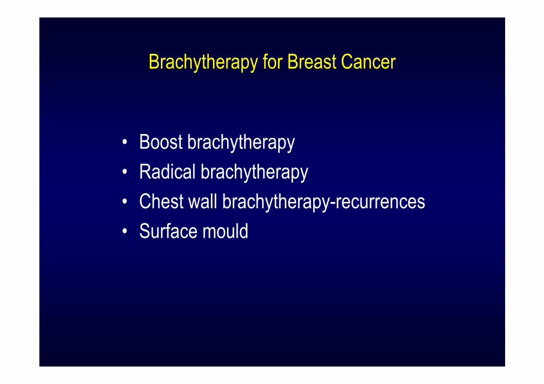
Breast conserving therapy: Standard treatment for early
breast cancer
Randomized trials comparing MRM vs BCT:
Comparable outcome
Better cosmetic outcome
Improved psychosocial impact
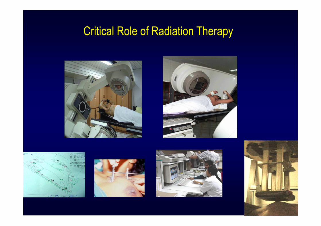
Critical Role of Radiation Therapy
• Current practice
• Rationale for boost
• To boost or not to boost.
• Dose and fractionations.
• Techniques of boost delivery.
• Comparison of above techniques.
• Delineation of boost volume.
• TMH Experience
• Identification of High Risk groups
Tumor Bed Boost
Rationale for boost..
Pathological basis ..
• N =441 pts (333 analysed) of Stage I & II Ca breast
• Aim – to define CTV for PBI
• All the pts underwent re-excision after lumpectomy.
• Results-
� 35.2% had no residual
� 20.1% had dis. 0-5 mm from tumour edge
� 24.9% extended from 5-10 mm
� 10.2% from 10-15 mm
� 9% extended > 15 mm
• Conclusion: In ~ 90% of pts margin of 10 mm is adequate.
Frank A. Vicini IJROBP. 60(3) :2004
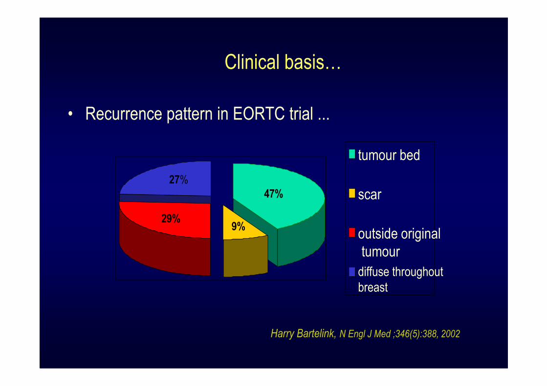
Clinical basis…
• Recurrence pattern in EORTC trial ...
tumour bed
scar
outside original
tumour
diffuse throughout
breast
47%
27%
29%9%
Harry Bartelink, N Engl J Med ;346(5):388, 2002
Boost Vs No Boost
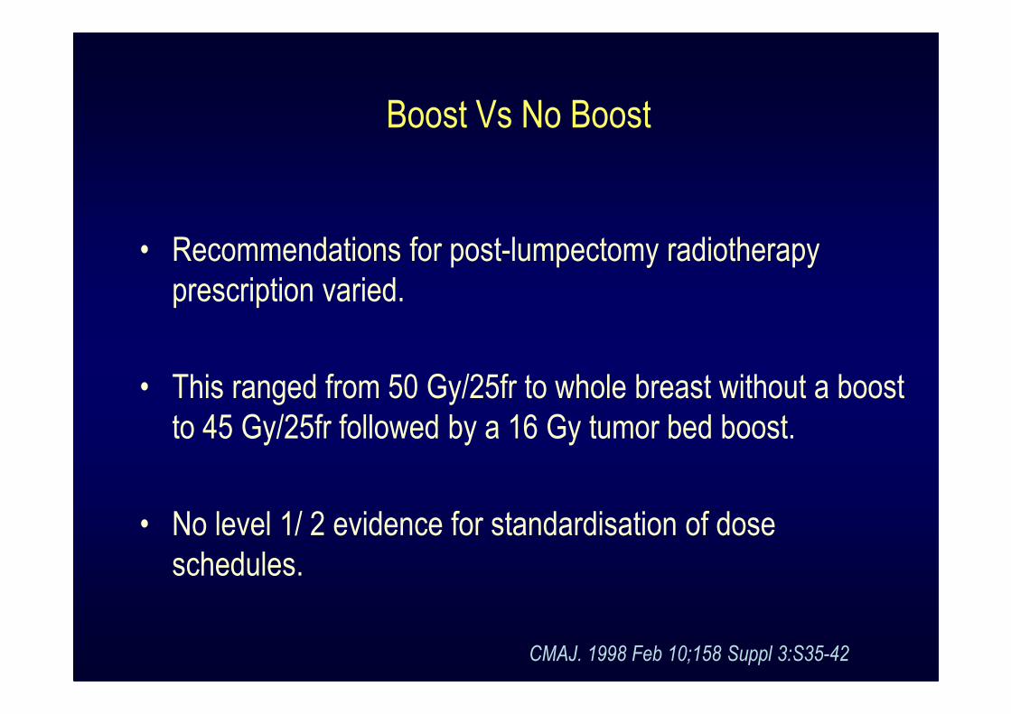
Boost Vs No Boost
• Recommendations for post-lumpectomy radiotherapy
prescription varied.
• This ranged from 50 Gy/25fr to whole breast without a boost
to 45 Gy/25fr followed by a 16 Gy tumor bed boost.
• No level 1/ 2 evidence for standardisation of dose
schedules.
CMAJ. 1998 Feb 10;158 Suppl 3:S35-42
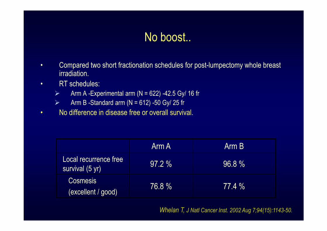
No boost..
• Compared two short fractionation schedules for post-lumpectomy whole breast irradiation.
• RT schedules:
� Arm A -Experimental arm (N = 622) -42.5 Gy/ 16 fr
� Arm B -Standard arm (N = 612) -50 Gy/ 25 fr
• No difference in disease free or overall survival.
Arm A Arm B
Local recurrence free
survival (5 yr)97.2 % 96.8 %
Cosmesis
(excellent / good)76.8 % 77.4 %
Whelan T, J Natl Cancer Inst. 2002 Aug 7;94(15):1143-50.
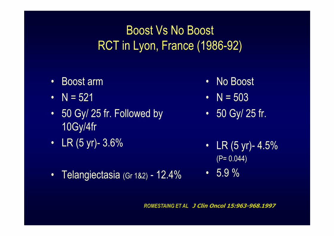
Boost Vs No Boost
RCT in Lyon, France (1986-92)
• Boost arm
• N = 521
• 50 Gy/ 25 fr. Followed by
10Gy/4fr
• LR (5 yr)- 3.6%
• Telangiectasia (Gr 1&2) - 12.4%
• No Boost
• N = 503
• 50 Gy/ 25 fr.
• LR (5 yr)- 4.5% (P= 0.044)
• 5.9 %
ROMESTAING ET AL , J Clin Oncol 15:963-968.1997
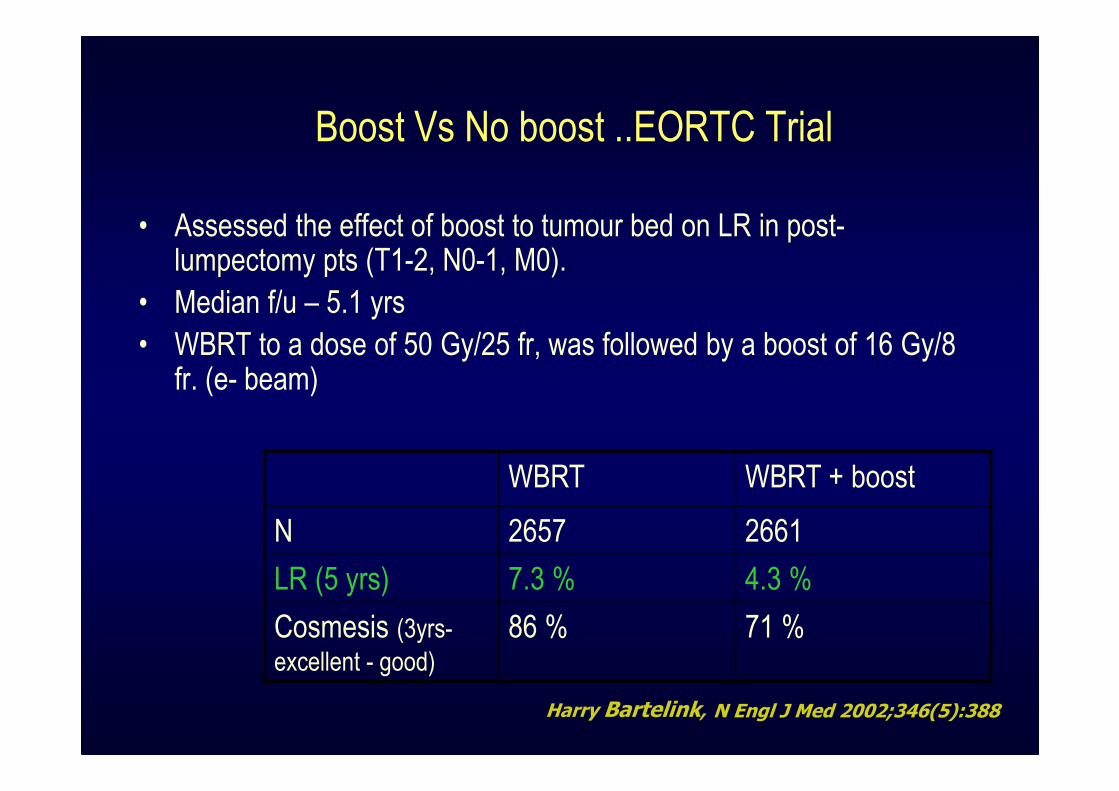
Boost Vs No boost ..EORTC Trial
• Assessed the effect of boost to tumour bed on LR in post-lumpectomy pts (T1-2, N0-1, M0).
• Median f/u – 5.1 yrs
• WBRT to a dose of 50 Gy/25 fr, was followed by a boost of 16 Gy/8 fr. (e- beam)
Harry Bartelink, N Engl J Med 2002;346(5):388
WBRT WBRT + boost
N 2657 2661
LR (5 yrs) 7.3 % 4.3 %
Cosmesis (3yrs-
excellent - good)
86 % 71 %
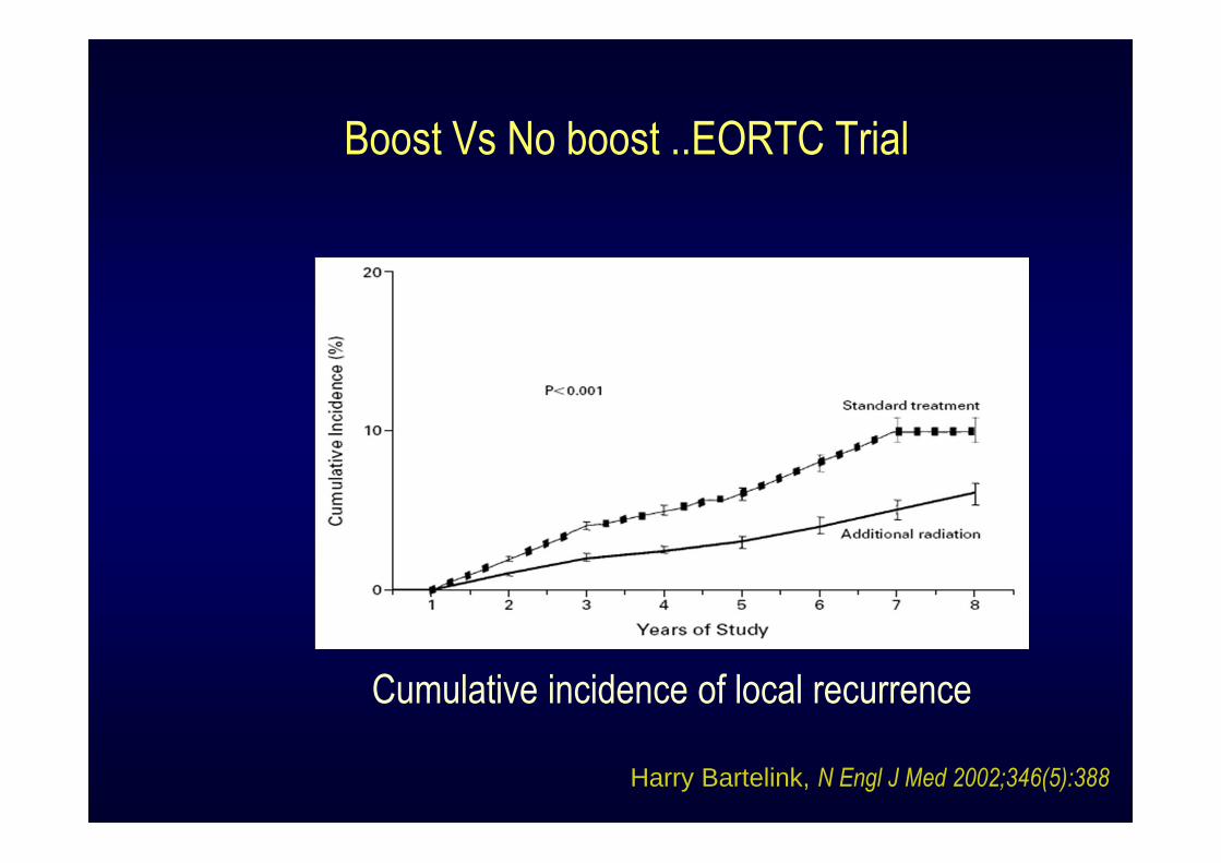
Boost Vs No boost ..EORTC Trial
Cumulative incidence of local recurrence
Harry Bartelink, N Engl J Med 2002;346(5):388
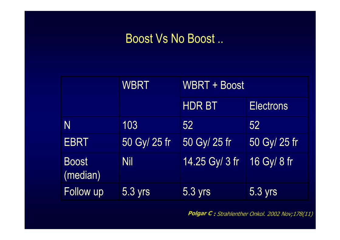
Boost Vs No Boost ..
WBRT WBRT + Boost
HDR BT Electrons
N 103 52 52
EBRT 50 Gy/ 25 fr 50 Gy/ 25 fr 50 Gy/ 25 fr
Boost
(median)
Nil 14.25 Gy/ 3 fr 16 Gy/ 8 fr
Follow up 5.3 yrs 5.3 yrs 5.3 yrs
Polgar C : Strahlenther Onkol. 2002 Nov;178(11)
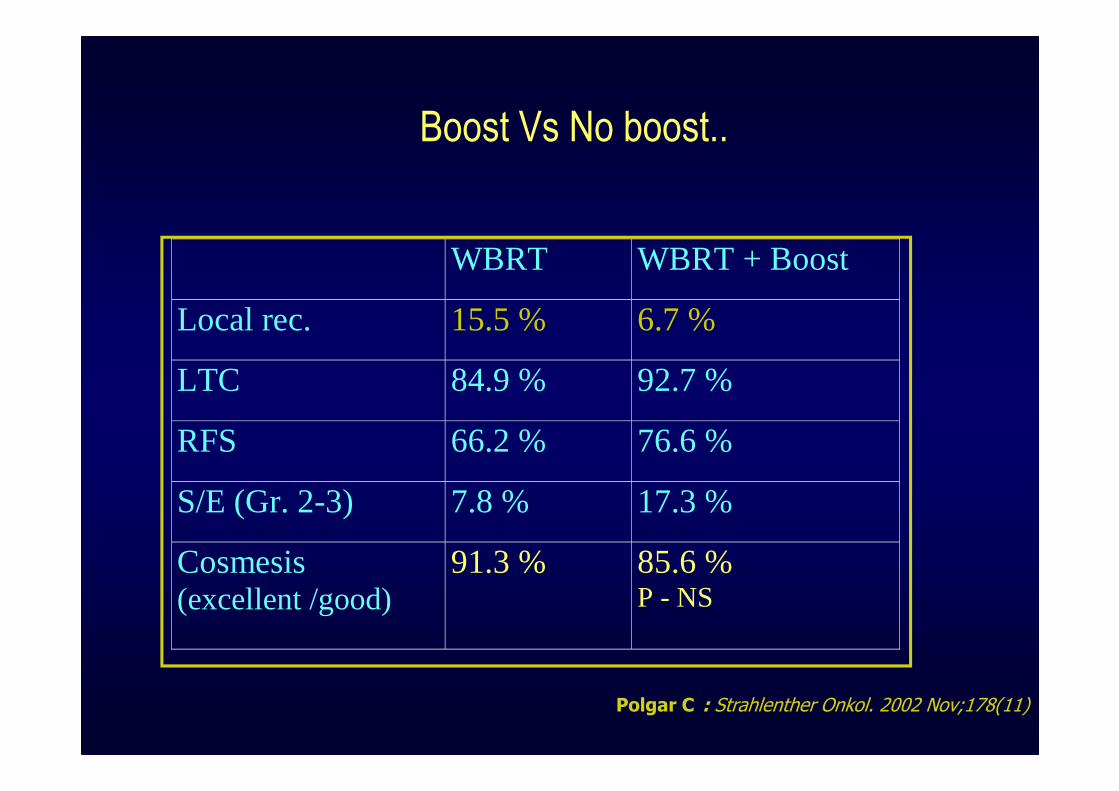
Boost Vs No boost..
WBRT WBRT + Boost
Local rec. 15.5 % 6.7 %
LTC 84.9 % 92.7 %
RFS 66.2 % 76.6 %
S/E (Gr. 2-3) 7.8 % 17.3 %
Cosmesis (excellent /good)
91.3 % 85.6 % P - NS
: Strahlenther Onkol. 2002 Nov;178(11)Polgar C
RT boost .. Dose & Fractionation
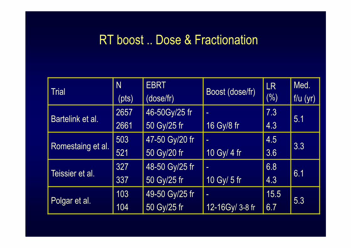
RT boost .. Dose & Fractionation
TrialN
(pts)
EBRT
(dose/fr)Boost (dose/fr)
LR
(%)
Med.
f/u (yr)
Bartelink et al.2657
2661
46-50Gy/25 fr
50 Gy/25 fr
-
16 Gy/8 fr
7.3
4.35.1
Romestaing et al.503
521
47-50 Gy/20 fr
50 Gy/20 fr
-
10 Gy/ 4 fr
4.5
3.63.3
Teissier et al.327
337
48-50 Gy/25 fr
50 Gy/25 fr
-
10 Gy/ 5 fr
6.8
4.36.1
Polgar et al.103
104
49-50 Gy/25 fr
50 Gy/25 fr
-
12-16Gy/ 3-8 fr
15.5
6.75.3
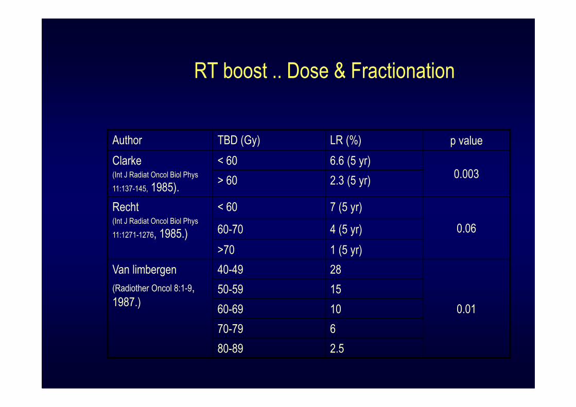
RT boost .. Dose & Fractionation
Author TBD (Gy) LR (%) p value
Clarke(Int J Radiat Oncol Biol Phys
11:137-145, 1985).
< 60 6.6 (5 yr)0.003
> 60 2.3 (5 yr)
Recht(Int J Radiat Oncol Biol Phys
11:1271-1276, 1985.)
< 60 7 (5 yr)
0.0660-70 4 (5 yr)
>70 1 (5 yr)
Van limbergen
(Radiother Oncol 8:1-9,
1987.)
40-49 28
0.01
50-59 15
60-69 10
70-79 6
80-89 2.5
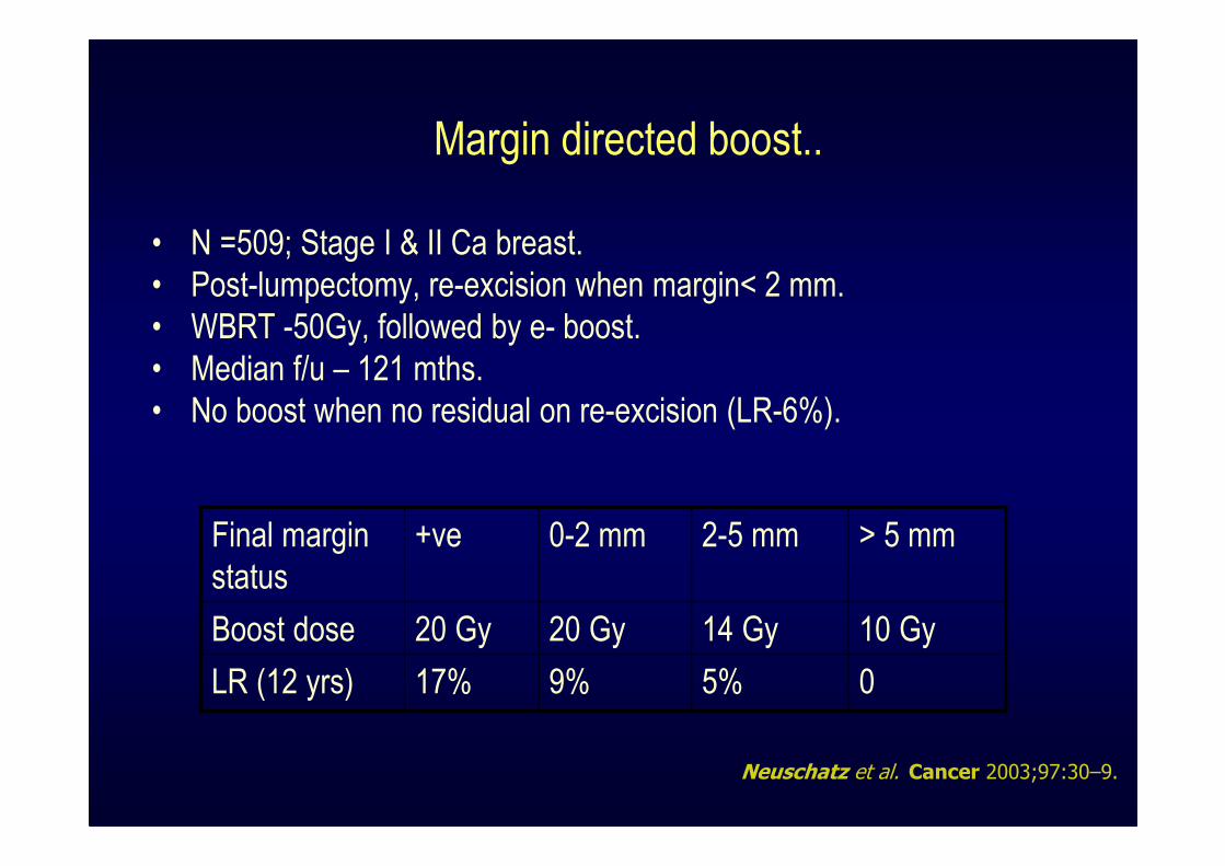
Margin directed boost..
• N =509; Stage I & II Ca breast.
• Post-lumpectomy, re-excision when margin< 2 mm.
• WBRT -50Gy, followed by e- boost.
• Median f/u – 121 mths.
• No boost when no residual on re-excision (LR-6%).
Final margin
status
+ve 0-2 mm 2-5 mm > 5 mm
Boost dose 20 Gy 20 Gy 14 Gy 10 Gy
LR (12 yrs) 17% 9% 5% 0
Cancer 2003;97:30–9.Neuschatz et al.

Boost delivery..
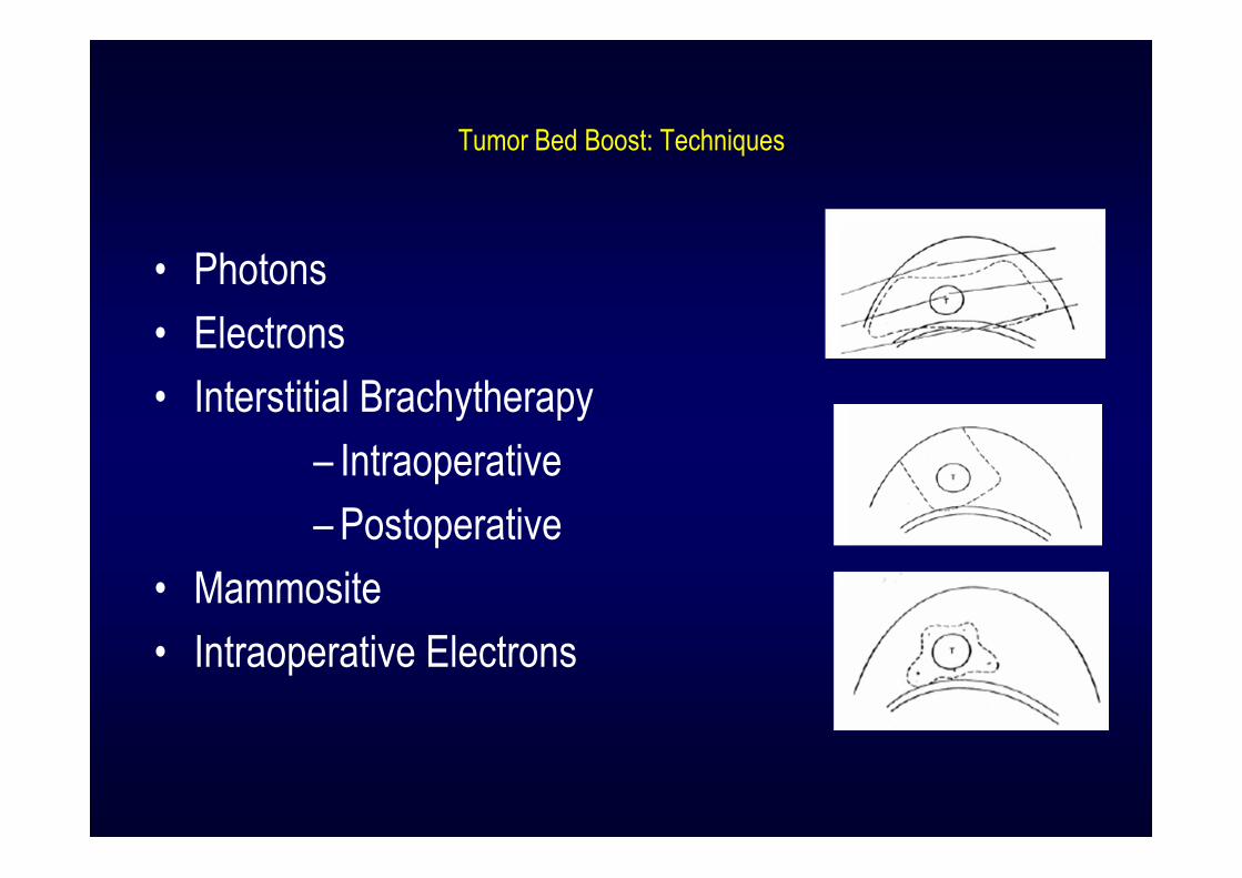
Tumor Bed Boost: Techniques
• Photons
• Electrons
• Interstitial Brachytherapy
– Intraoperative
– Postoperative
• Mammosite
• Intraoperative Electrons
Comparison of Boost Techniques
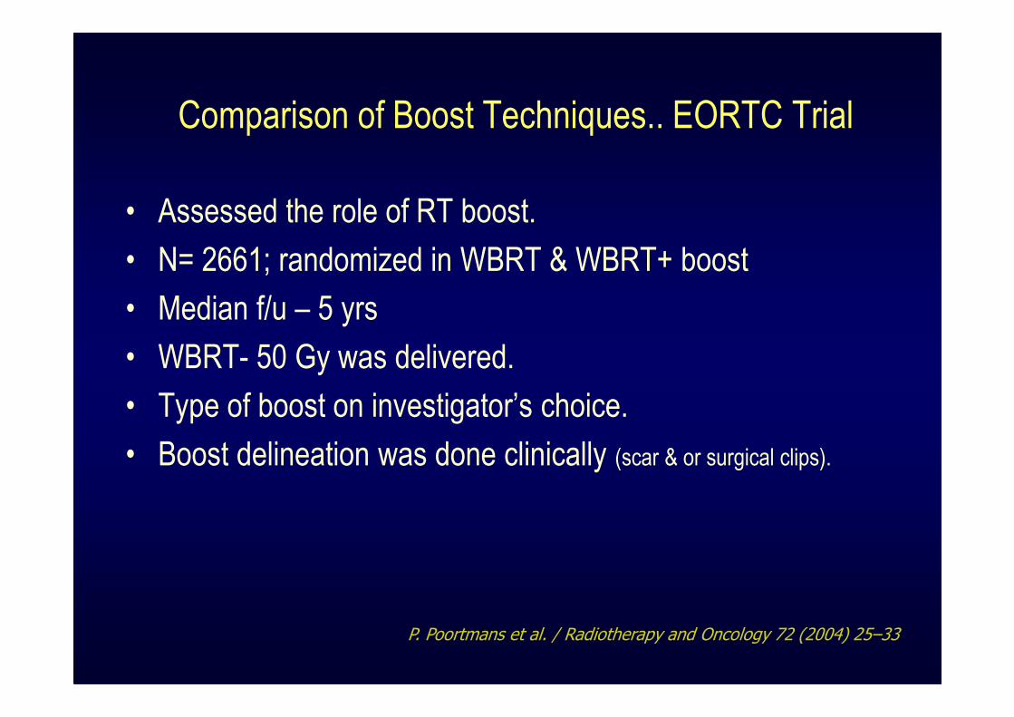
Comparison of Boost Techniques.. EORTC Trial
• Assessed the role of RT boost.
• N= 2661; randomized in WBRT & WBRT+ boost
• Median f/u – 5 yrs
• WBRT- 50 Gy was delivered.
• Type of boost on investigator’s choice.
• Boost delineation was done clinically (scar & or surgical clips).
P. Poortmans et al. / Radiotherapy and Oncology 72 (2004) 25–33
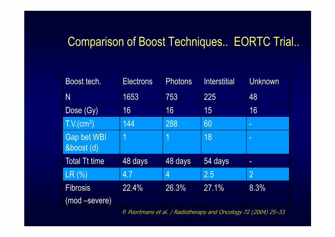
Comparison of Boost Techniques.. EORTC Trial..
Boost tech. Electrons Photons Interstitial Unknown
N 1653 753 225 48
Dose (Gy) 16 16 15 16
T.V.(cm3) 144 288 60 -
Gap bet WBI
&boost (d)
1 1 18 -
Total Tt time 48 days 48 days 54 days -
LR (%) 4.7 4 2.5 2
Fibrosis
(mod –severe)
22.4% 26.3% 27.1% 8.3%
P. Poortmans et al. / Radiotherapy and Oncology 72 (2004) 25–33
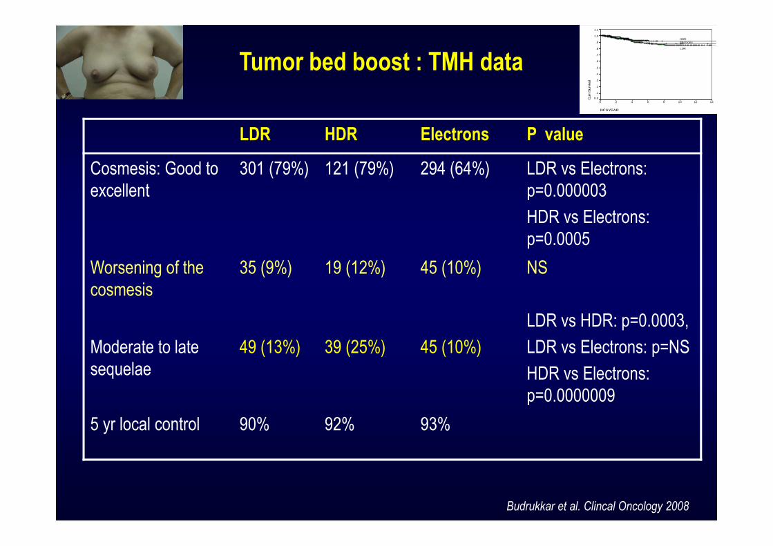
LDR HDR Electrons P value
Cosmesis: Good to
excellent
301 (79%) 121 (79%) 294 (64%) LDR vs Electrons:
p=0.000003
HDR vs Electrons:
p=0.0005
Worsening of the
cosmesis
35 (9%) 19 (12%) 45 (10%) NS
Moderate to late
sequelae
49 (13%) 39 (25%) 45 (10%)
LDR vs HDR: p=0.0003,
LDR vs Electrons: p=NS
HDR vs Electrons:
p=0.0000009
5 yr local control 90% 92% 93%
Tumor bed boost : TMH data
Budrukkar et al. Clincal Oncology 2008
DFSYEAR
14121086420
Cum
Sur
viva
l
1.1
1.0
.9
.8
.7
.6
.5
.4
.3
.2
.1
0.0
LDR
HDRElectrons
Delineation of lumpectomy
cavity..
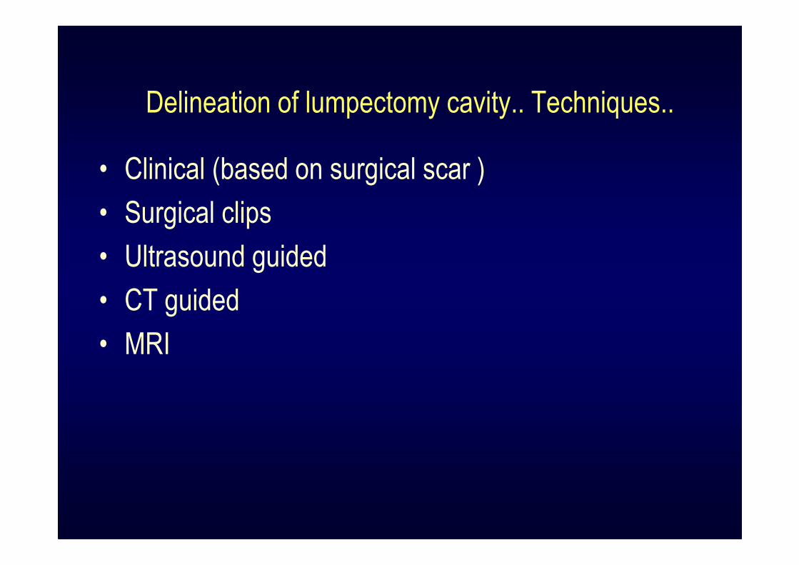
Delineation of lumpectomy cavity.. Techniques..
• Clinical (based on surgical scar )
• Surgical clips
• Ultrasound guided
• CT guided
• MRI
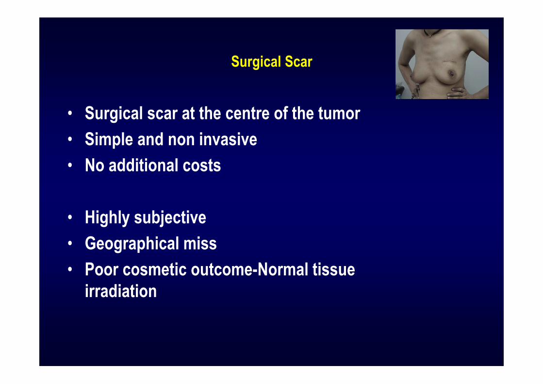
Surgical Scar
• Surgical scar at the centre of the tumor
• Simple and non invasive
• No additional costs
• Highly subjective
• Geographical miss
• Poor cosmetic outcome-Normal tissue
irradiation
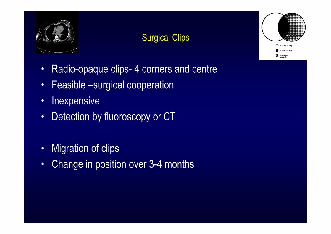
Surgical Clips
• Radio-opaque clips- 4 corners and centre
• Feasible –surgical cooperation
• Inexpensive
• Detection by fluoroscopy or CT
• Migration of clips
• Change in position over 3-4 months
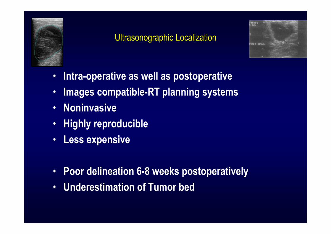
Ultrasonographic Localization
• Intra-operative as well as postoperative
• Images compatible-RT planning systems
• Noninvasive
• Highly reproducible
• Less expensive
• Poor delineation 6-8 weeks postoperatively
• Underestimation of Tumor bed
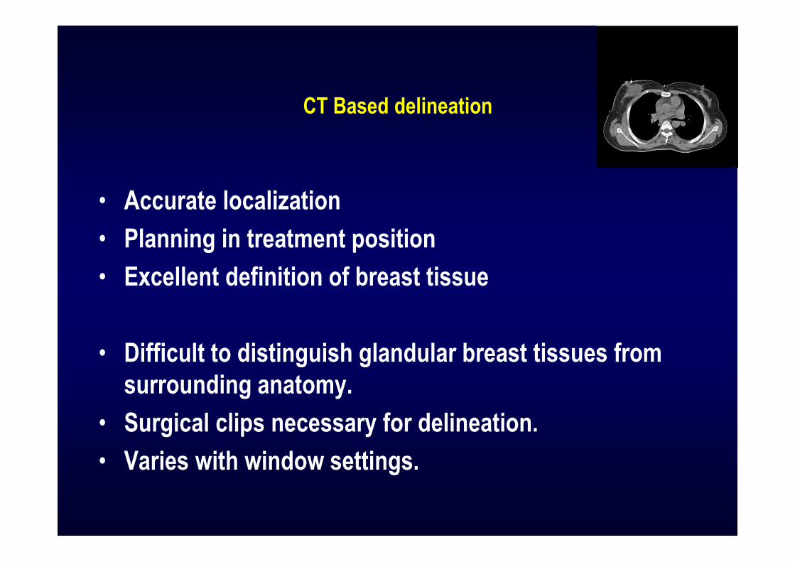
CT Based delineation
• Accurate localization
• Planning in treatment position
• Excellent definition of breast tissue
• Difficult to distinguish glandular breast tissues from
surrounding anatomy.
• Surgical clips necessary for delineation.
• Varies with window settings.
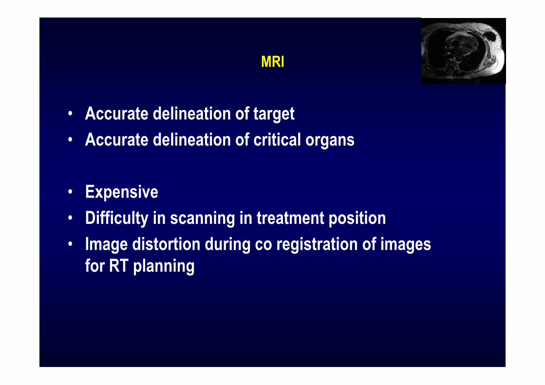
MRI
• Accurate delineation of target
• Accurate delineation of critical organs
• Expensive
• Difficulty in scanning in treatment position
• Image distortion during co registration of images
for RT planning
TMH Experience ..

Tata Memorial Hospital
Breast Conserving Therapy: 1980-2000
1022 patients
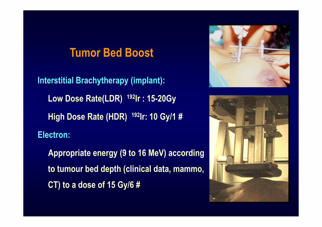
Interstitial Brachytherapy (implant):
Low Dose Rate(LDR) 192Ir : 15-20Gy
High Dose Rate (HDR) 192Ir: 10 Gy/1 #
Electron:
Appropriate energy (9 to 16 MeV) according
to tumour bed depth (clinical data, mammo,
CT) to a dose of 15 Gy/6 #
Tumor Bed Boost
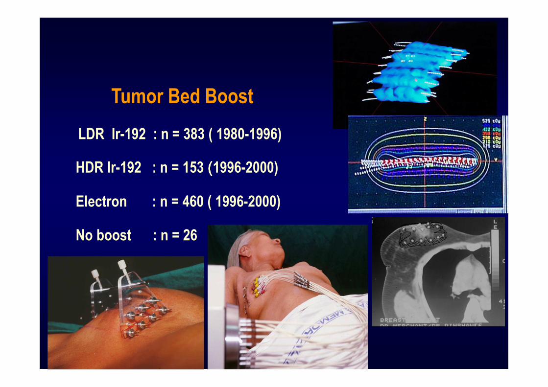
LDR Ir-192 : n = 383 ( 1980-1996)
HDR Ir-192 : n = 153 ((((1996-2000)
Electron : n = 460 ( 1996-2000)
No boost : n = 26
Tumor Bed Boost
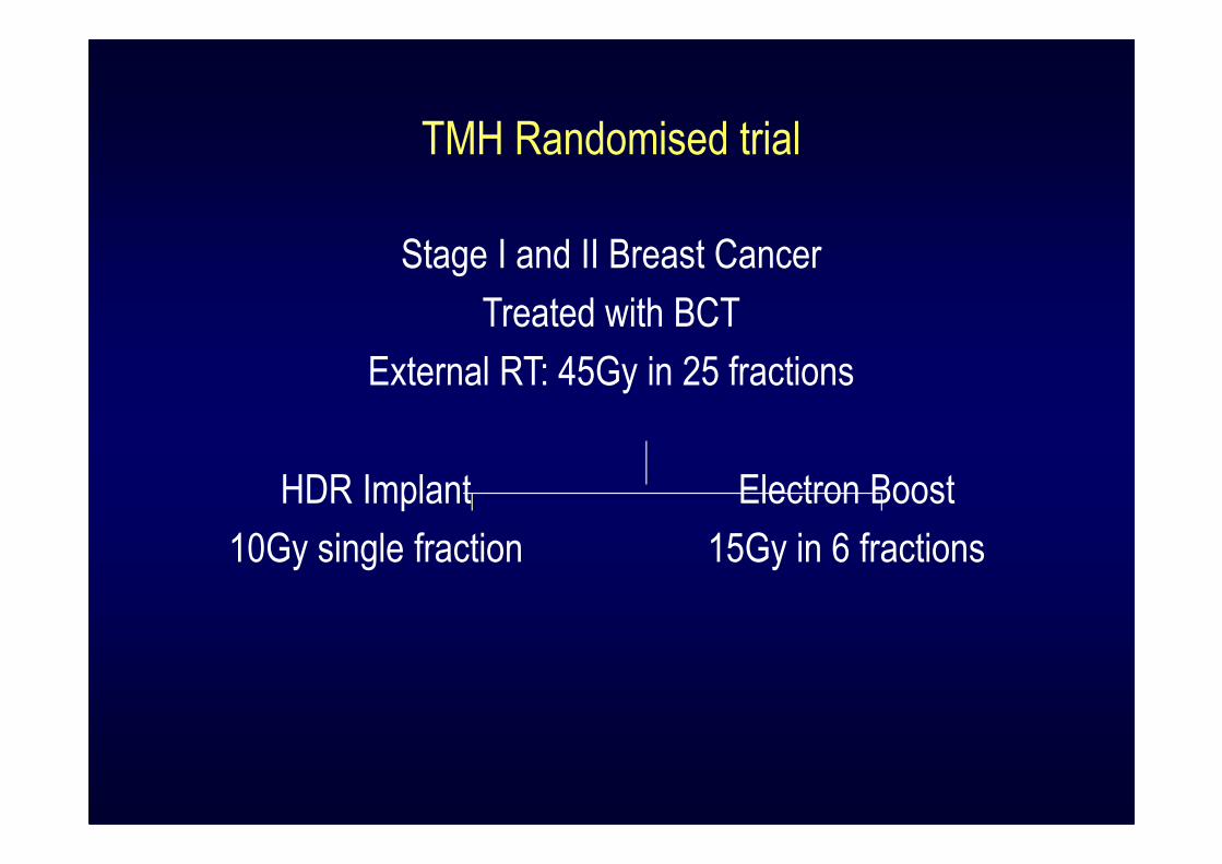
TMH Randomised trial
Stage I and II Breast Cancer
Treated with BCT
External RT: 45Gy in 25 fractions
HDR Implant
10Gy single fraction
Electron Boost
15Gy in 6 fractions
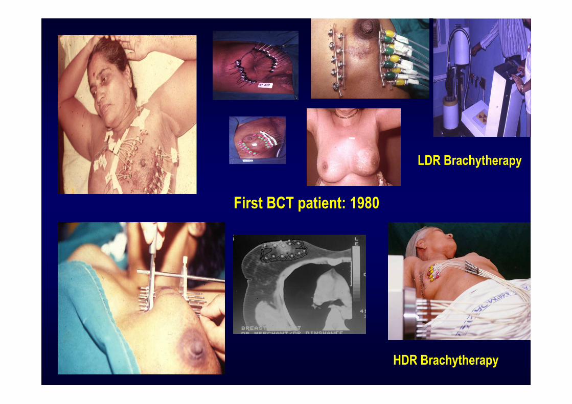
LDR Brachytherapy
First BCT patient: 1980
HDR Brachytherapy
Identification of High Risk patients
…
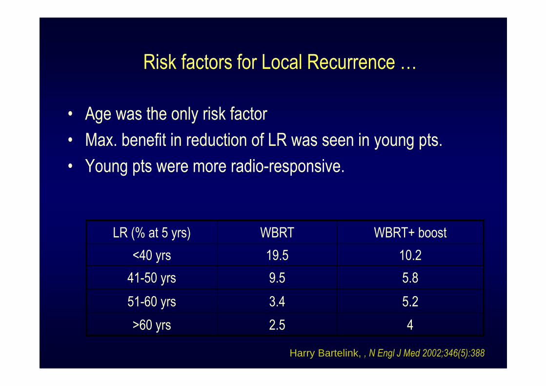
Risk factors for Local Recurrence …
• Age was the only risk factor
• Max. benefit in reduction of LR was seen in young pts.
• Young pts were more radio-responsive.
LR (% at 5 yrs) WBRT WBRT+ boost
<40 yrs 19.5 10.2
41-50 yrs 9.5 5.8
51-60 yrs 3.4 5.2
>60 yrs 2.5 4
Harry Bartelink, , N Engl J Med 2002;346(5):388
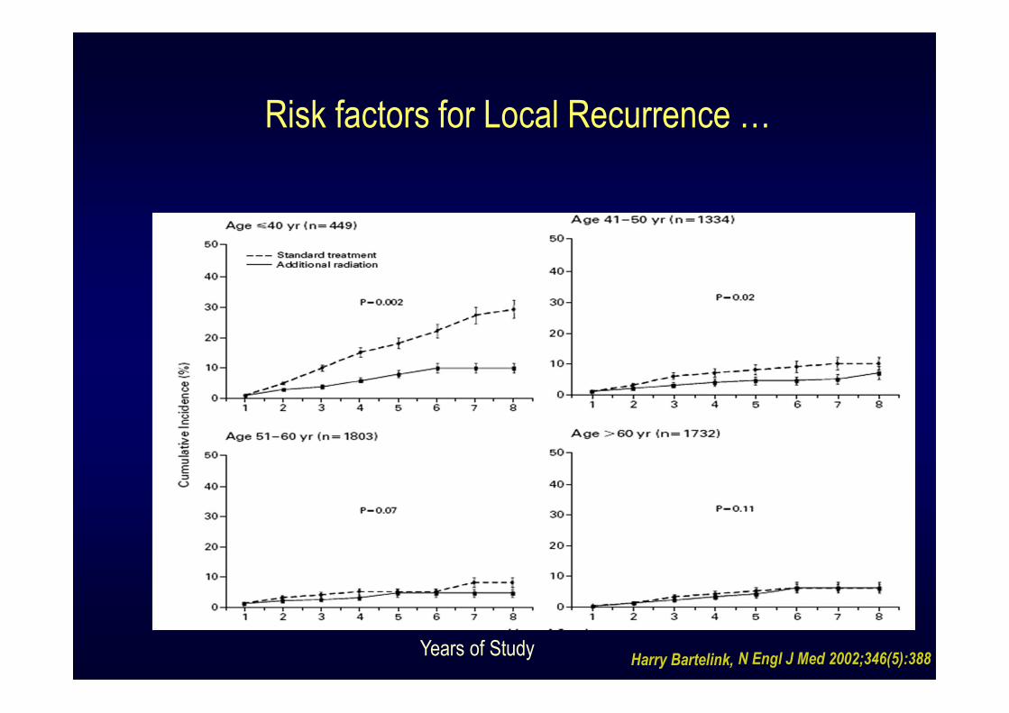
Risk factors for Local Recurrence …
Years of StudyHarry Bartelink, N Engl J Med 2002;346(5):388
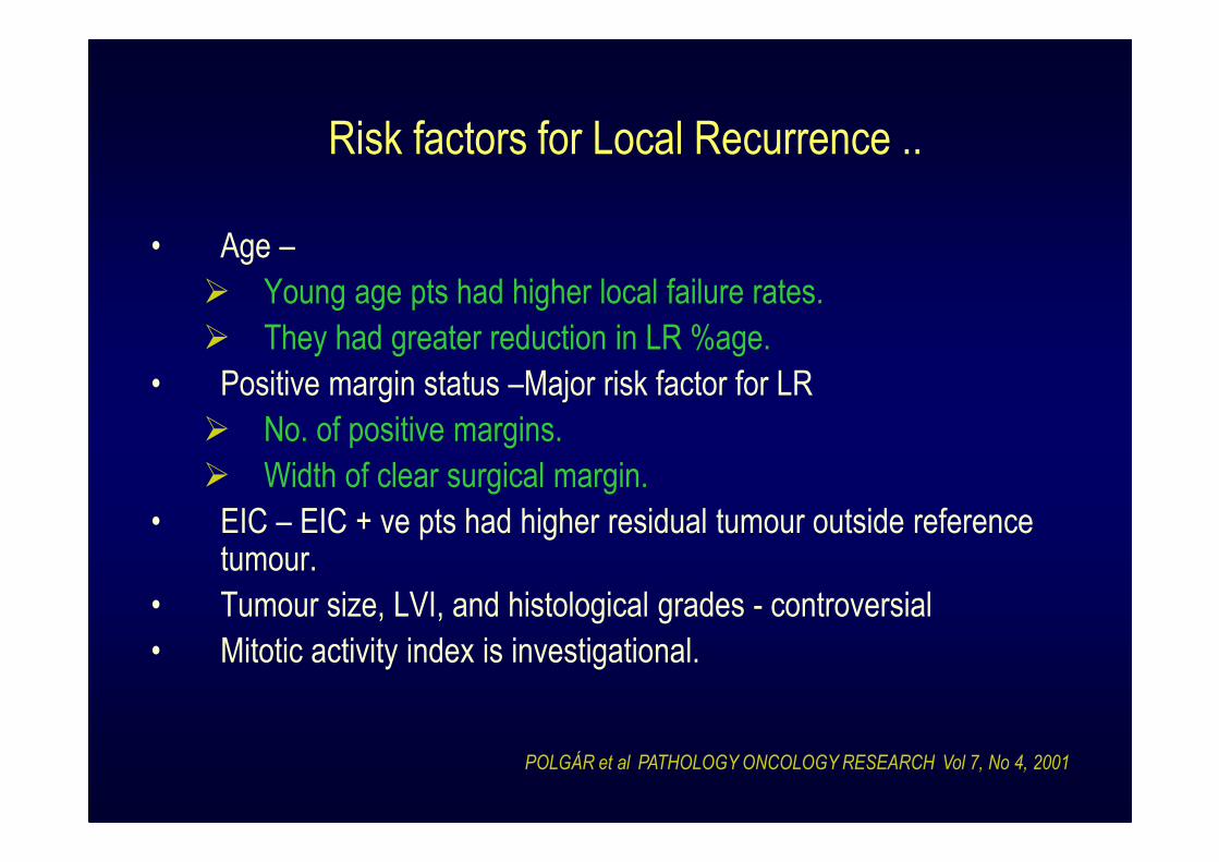
Risk factors for Local Recurrence ..
• Age –
� Young age pts had higher local failure rates.
� They had greater reduction in LR %age.
• Positive margin status –Major risk factor for LR
� No. of positive margins.
� Width of clear surgical margin.
• EIC – EIC + ve pts had higher residual tumour outside reference tumour.
• Tumour size, LVI, and histological grades - controversial
• Mitotic activity index is investigational.
POLGÁR et al PATHOLOGY ONCOLOGY RESEARCH Vol 7, No 4, 2001
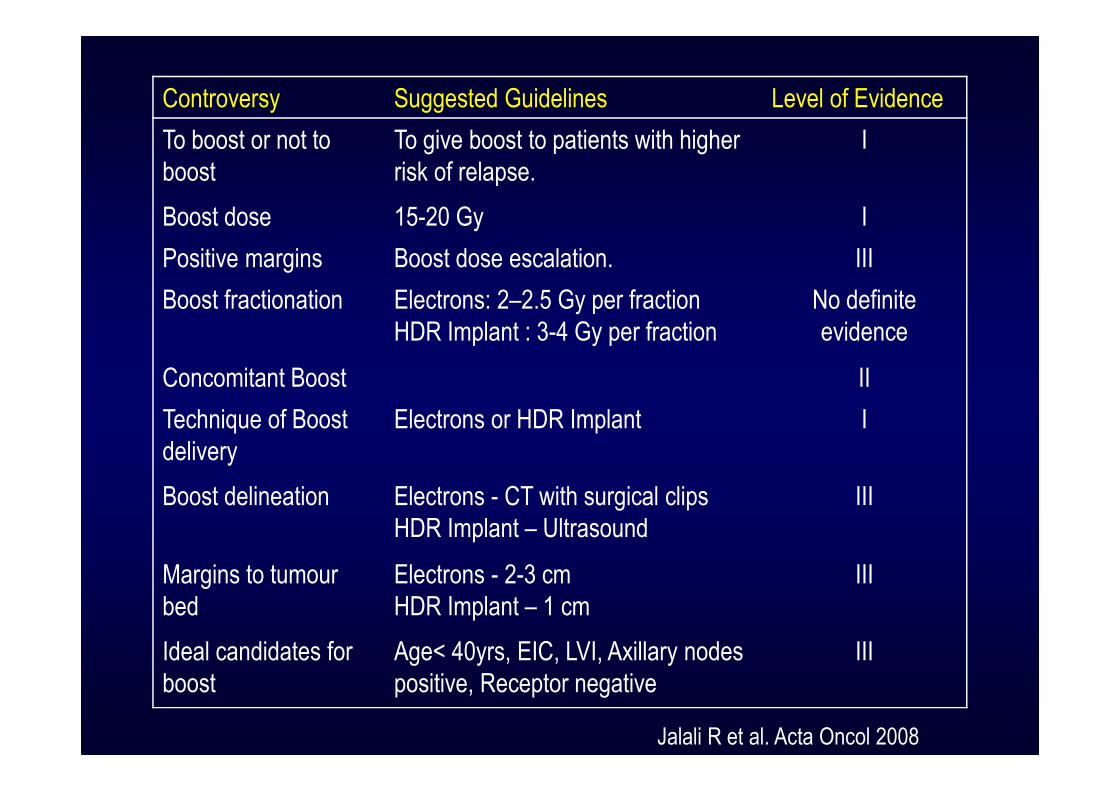
Controversy Suggested Guidelines Level of Evidence
To boost or not to
boost
To give boost to patients with higher
risk of relapse.
I
Boost dose 15-20 Gy I
Positive margins Boost dose escalation. III
Boost fractionation Electrons: 2–2.5 Gy per fraction
HDR Implant : 3-4 Gy per fraction
No definite
evidence
Concomitant Boost II
Technique of Boost
delivery
Electrons or HDR Implant I
Boost delineation Electrons - CT with surgical clips
HDR Implant – Ultrasound
III
Margins to tumour
bed
Electrons - 2-3 cm
HDR Implant – 1 cm
III
Ideal candidates for
boost
Age< 40yrs, EIC, LVI, Axillary nodes
positive, Receptor negative
III
Jalali R et al. Acta Oncol 2008
Accelerated Partial Breast Irradiation
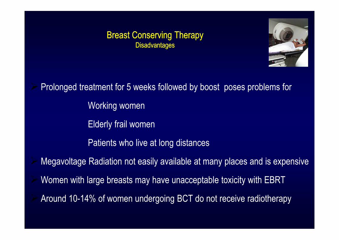
Breast Conserving TherapyDisadvantages
Breast Conserving TherapyDisadvantages
� Prolonged treatment for 5 weeks followed by boost poses problems for
Working women
Elderly frail women
Patients who live at long distances
� Megavoltage Radiation not easily available at many places and is expensive
� Women with large breasts may have unacceptable toxicity with EBRT
� Around 10-14% of women undergoing BCT do not receive radiotherapy
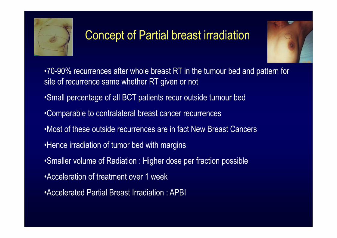
Concept of Partial breast irradiation
•70-90% recurrences after whole breast RT in the tumour bed and pattern for
site of recurrence same whether RT given or not
•Small percentage of all BCT patients recur outside tumour bed
•Comparable to contralateral breast cancer recurrences
•Most of these outside recurrences are in fact New Breast Cancers
•Hence irradiation of tumor bed with margins
•Smaller volume of Radiation : Higher dose per fraction possible
•Acceleration of treatment over 1 week
•Accelerated Partial Breast Irradiation : APBI

Selection Criteria for APBI
Criteria American Brachytherapy Society recommendation
TMH
Age 45 years or more 40 years
Tumour size Up to 3cm Up to 3 cm
Node Negative Negative
Histology Infiltrating duct carcinoma (IDC) IDC
Margins Microscopically negative Microscopically negative
EIC - Negative
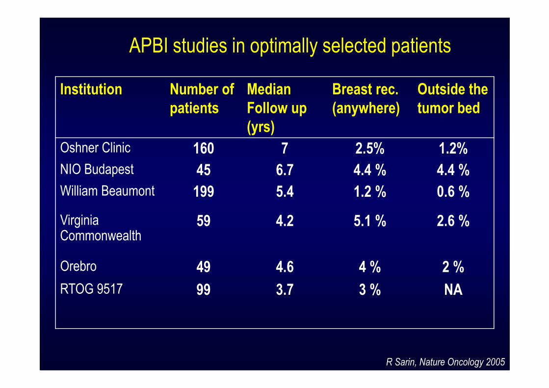
Institution Number of
patients
Median
Follow up
(yrs)
Breast rec.
(anywhere)
Outside the
tumor bed
Oshner Clinic 160 7 2.5% 1.2%
NIO Budapest 45 6.7 4.4 % 4.4 %
William Beaumont 199 5.4 1.2 % 0.6 %
Virginia Commonwealth
59 4.2 5.1 % 2.6 %
Orebro 49 4.6 4 % 2 %
RTOG 9517 99 3.7 3 % NA
APBI studies in optimally selected patients
R Sarin, Nature Oncology 2005
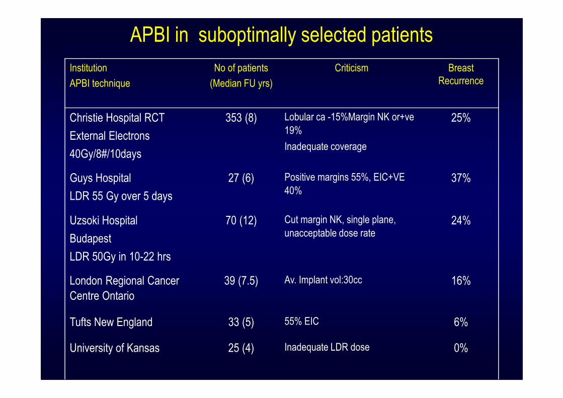
Institution
APBI technique
No of patients
(Median FU yrs)
Criticism Breast
Recurrence
Christie Hospital RCT
External Electrons
40Gy/8#/10days
353 (8) Lobular ca -15%Margin NK or+ve
19%
Inadequate coverage
25%
Guys Hospital
LDR 55 Gy over 5 days
27 (6) Positive margins 55%, EIC+VE
40%37%
Uzsoki Hospital
Budapest
LDR 50Gy in 10-22 hrs
70 (12) Cut margin NK, single plane,
unacceptable dose rate24%
London Regional Cancer
Centre Ontario
39 (7.5) Av. Implant vol:30cc 16%
Tufts New England 33 (5) 55% EIC 6%
University of Kansas 25 (4) Inadequate LDR dose 0%
APBI in suboptimally selected patients
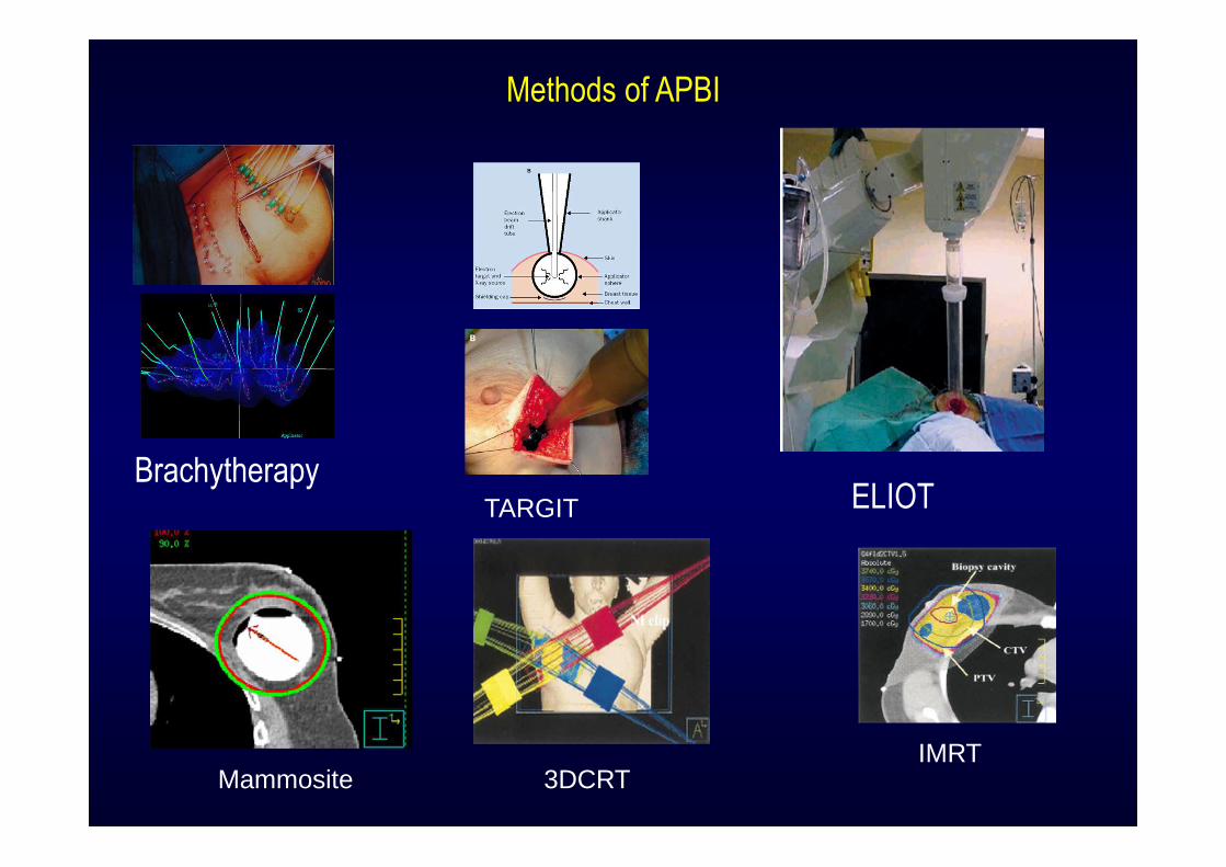
Methods of APBI
BrachytherapyTARGIT ELIOT
Mammosite 3DCRTIMRT
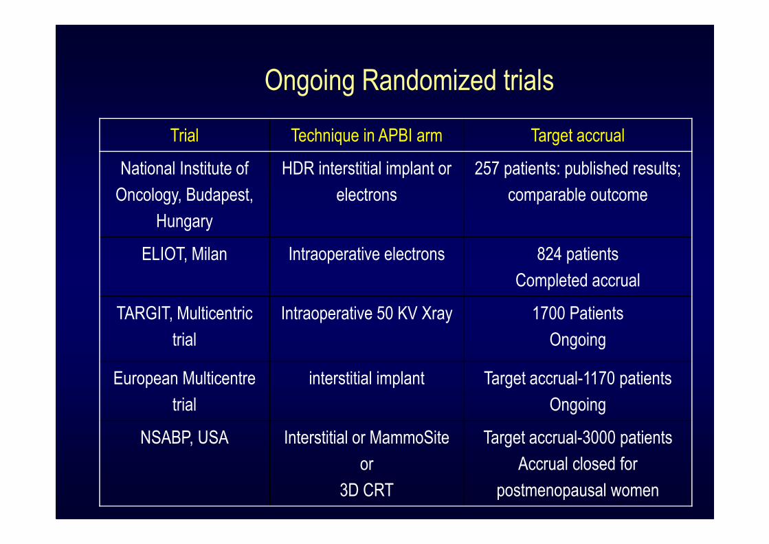
Trial Technique in APBI arm Target accrual
National Institute of
Oncology, Budapest,
Hungary
HDR interstitial implant or
electrons
257 patients: published results;
comparable outcome
ELIOT, Milan Intraoperative electrons 824 patients
Completed accrual
TARGIT, Multicentric
trial
Intraoperative 50 KV Xray 1700 Patients
Ongoing
European Multicentre
trial
interstitial implant Target accrual-1170 patients
Ongoing
NSABP, USA Interstitial or MammoSite
or
3D CRT
Target accrual-3000 patients
Accrual closed for
postmenopausal women
Ongoing Randomized trials
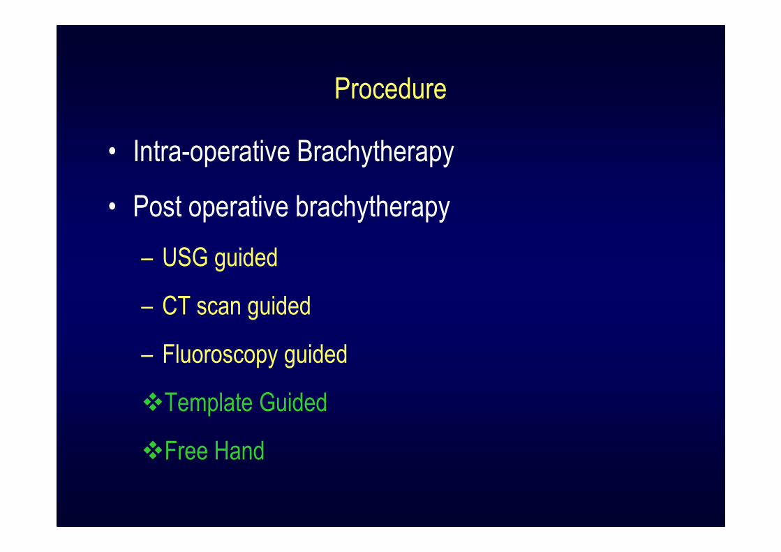
Procedure
• Intra-operative Brachytherapy
• Post operative brachytherapy
– USG guided
– CT scan guided
– Fluoroscopy guided
�Template Guided
�Free Hand
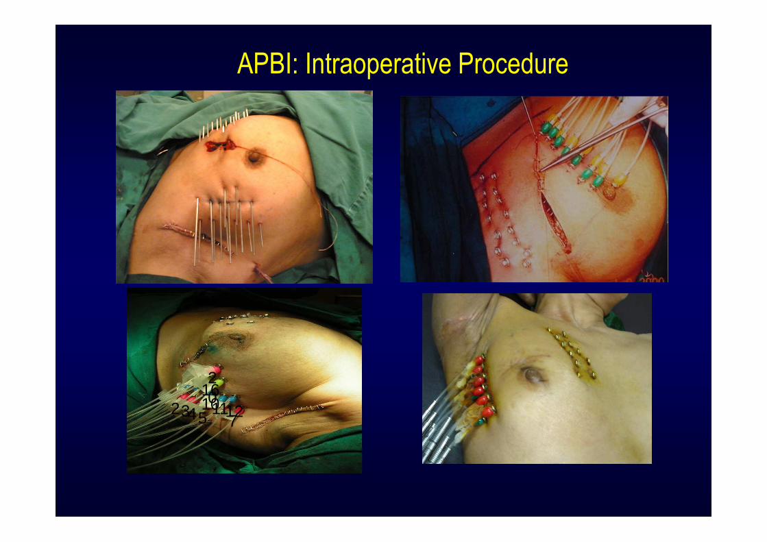
APBI: Intraoperative Procedure
75432 1112101620
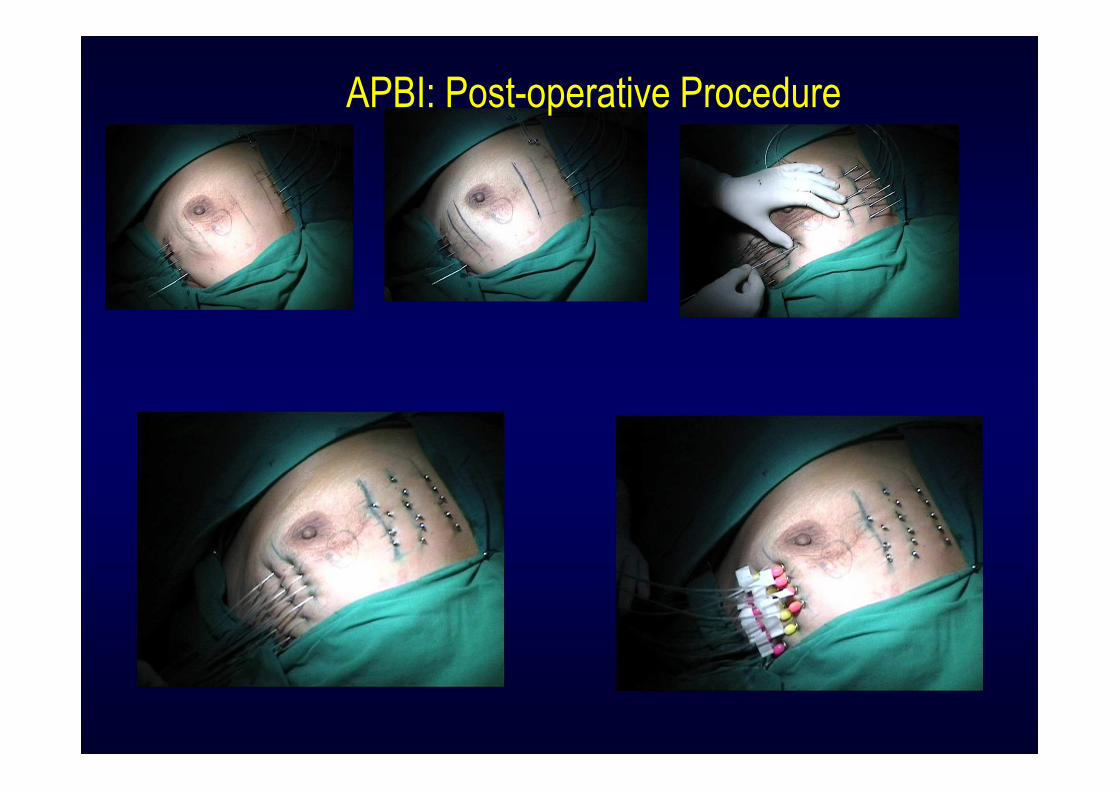
APBI: Post-operative Procedure
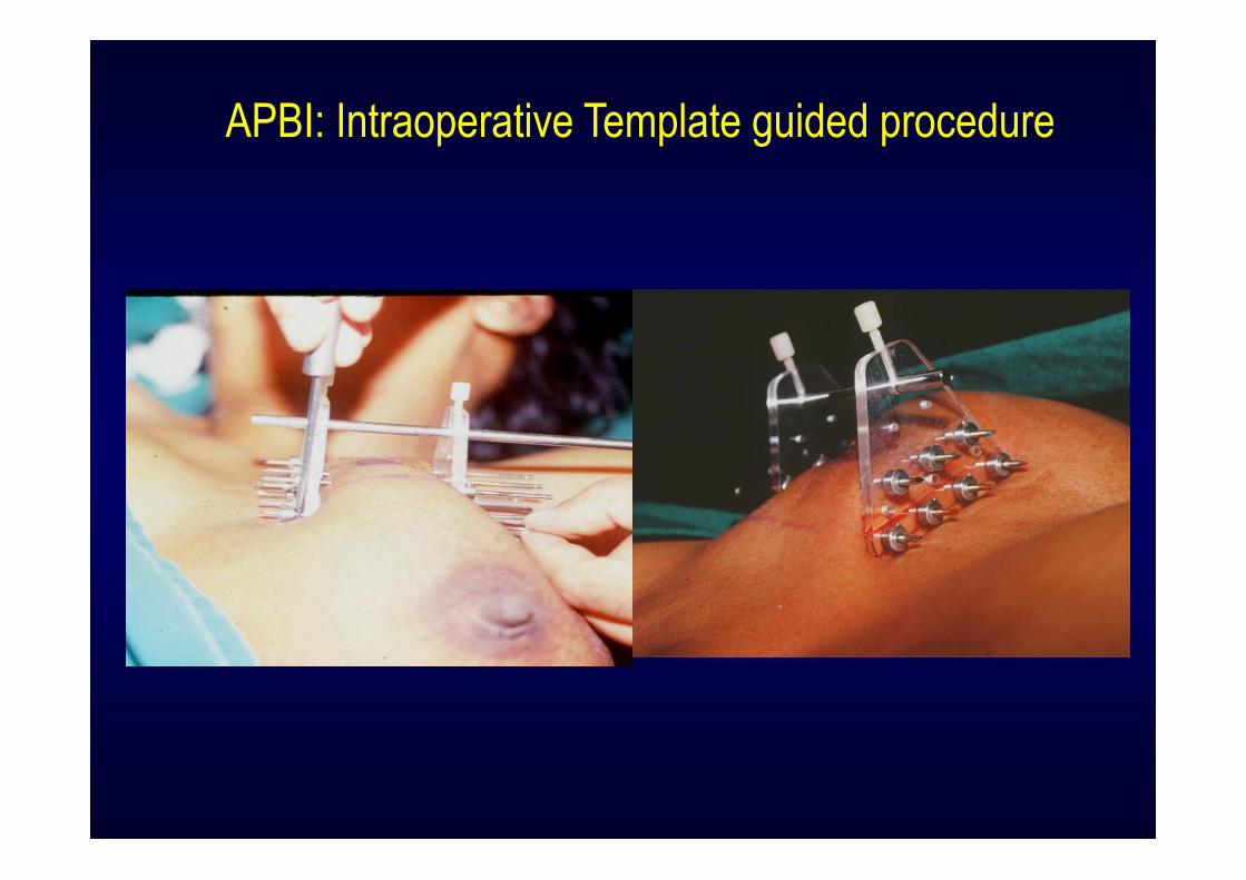
APBI: Intraoperative Template guided procedure
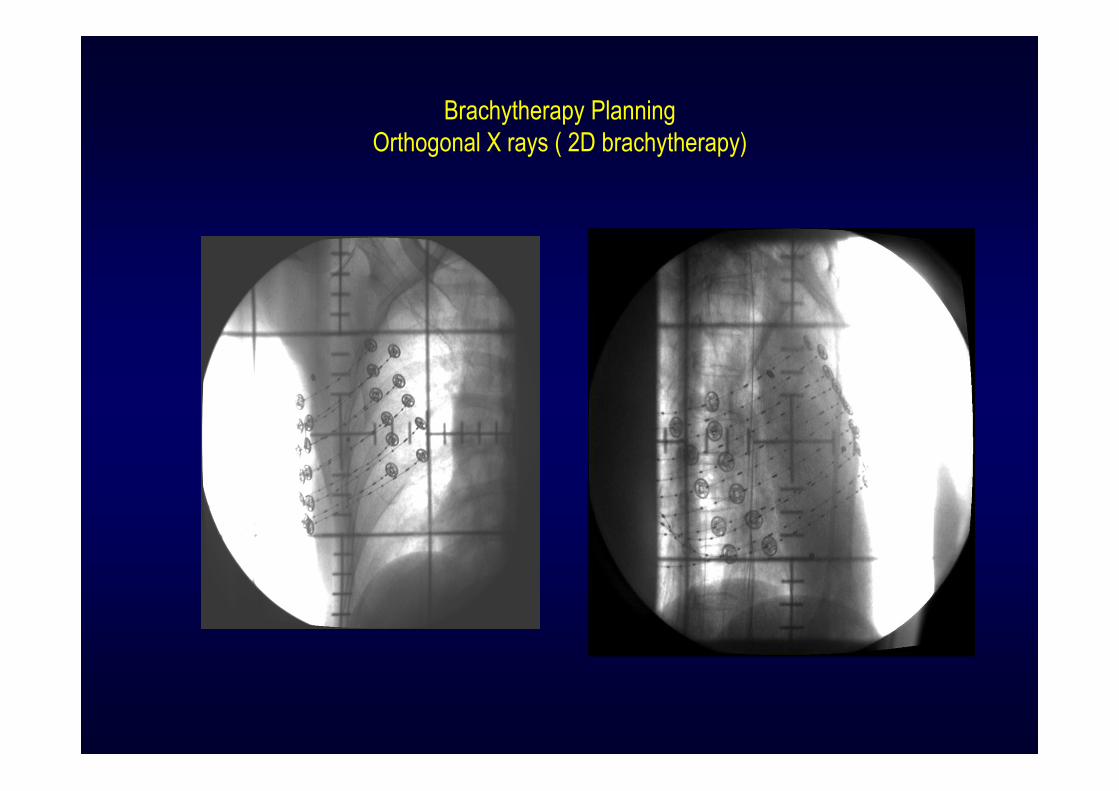
Brachytherapy Planning
Orthogonal X rays ( 2D brachytherapy)
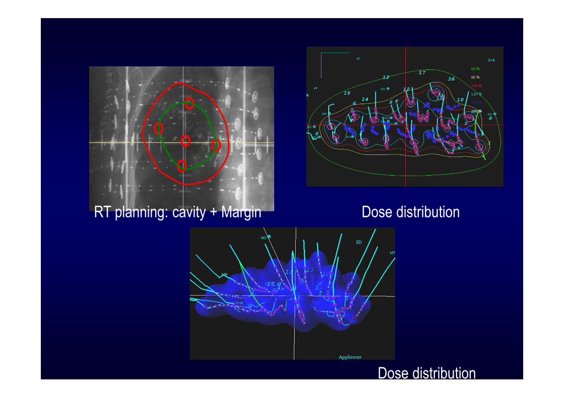
RT planning: cavity + Margin Dose distribution
Dose distribution

Dose prescription and Treatment delivery
• Dose: 34Gy in 10 fraction two fractions per day, 6 hrs apart
• Dose per fraction: 340cGy

Intraoperative Brachytherapy
W/E+ Axillary dissection
Confirmation of basic histopathological features on Frozen section
If suitable: Intraoperative placement of catheters in 2-4 planes
Radiotherapy planning X rays and CT scans on day 2/3
Treatment starts: day 3/4
Confirmation of final HPR before 5th fraction
Favorable: continue brachy Unfavorable: convert to boost
Ext RT to be followed
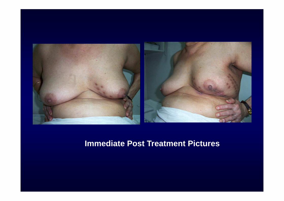
Immediate Post Treatment Pictures
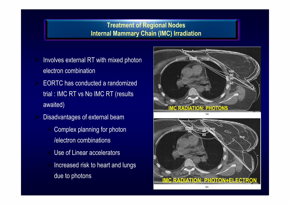
Treatment of Regional Nodes
Internal Mammary Chain (IMC) Irradiation
� Involves external RT with mixed photon
electron combination
� EORTC has conducted a randomized
trial : IMC RT vs No IMC RT (results
awaited)
� Disadvantages of external beam
� Complex planning for photon
/electron combinations
� Use of Linear accelerators
� Increased risk to heart and lungs
due to photons
IMC RADIATION: PHOTONS
IMC RADIATION: PHOTON+ELECTRON
IMC Brachytherapy: A Novel Approach
Potential advantages:
� Rapid fall off of the dose to the cardiac and other structures
� IMC nodes lie around the vessels, which are anyway dispensable
� Brachytherapy machine relatively more common and available (in
developing countries)
Potential advantages:
� Rapid fall off of the dose to the cardiac and other structures
� IMC nodes lie around the vessels, which are anyway dispensable
� Brachytherapy machine relatively more common and available (in
developing countries)
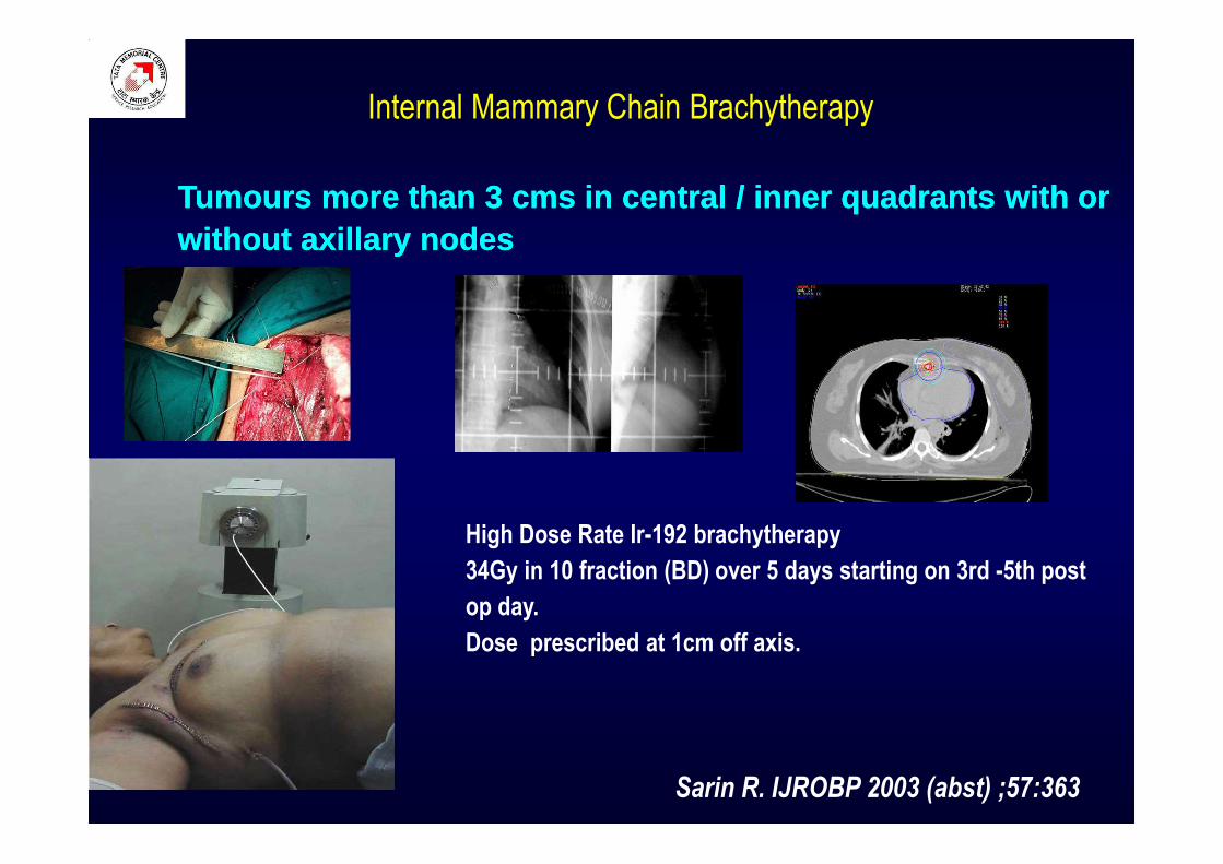
Tumours more than 3 cms in central / inner quadrants with or without axillary nodesTumours more than 3 cms in central / inner quadrants with or without axillary nodes
High Dose Rate Ir-192 brachytherapy
34Gy in 10 fraction (BD) over 5 days starting on 3rd -5th post
op day.
Dose prescribed at 1cm off axis.
Sarin R. IJROBP 2003 (abst) ;57:363
Internal Mammary Chain Brachytherapy
TATA MEMORIAL EXPERIENCE
IRIDIUM-192 HDR BRACHYTHERAPY FOR IMC
IN BREAST CANCER
TATA MEMORIAL EXPERIENCE
IRIDIUM-192 HDR BRACHYTHERAPY FOR IMC
IN BREAST CANCER
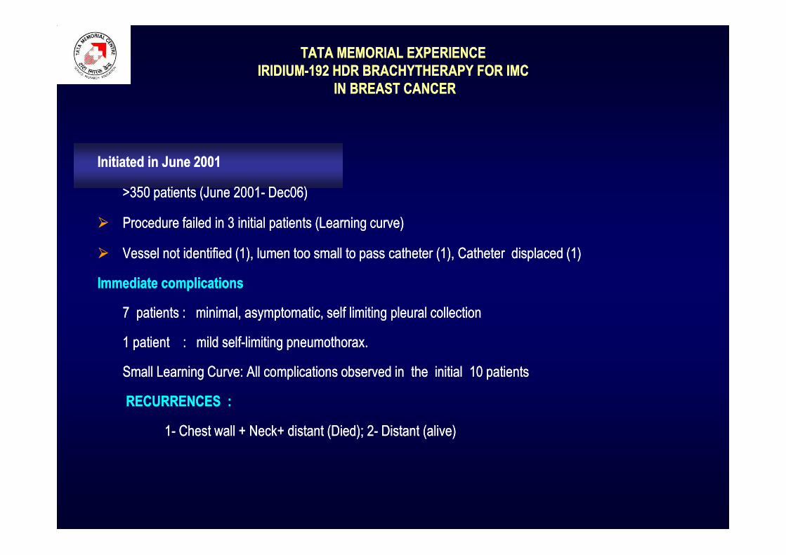
Initiated in June 2001
>350 patients (June 2001- Dec06)
� Procedure failed in 3 initial patients (Learning curve)
� Vessel not identified (1), lumen too small to pass catheter (1), Catheter displaced (1)
Immediate complications
7 patients : minimal, asymptomatic, self limiting pleural collection
1 patient : mild self-limiting pneumothorax.
Small Learning Curve: All complications observed in the initial 10 patients
RECURRENCES :
1- Chest wall + Neck+ distant (Died); 2- Distant (alive)
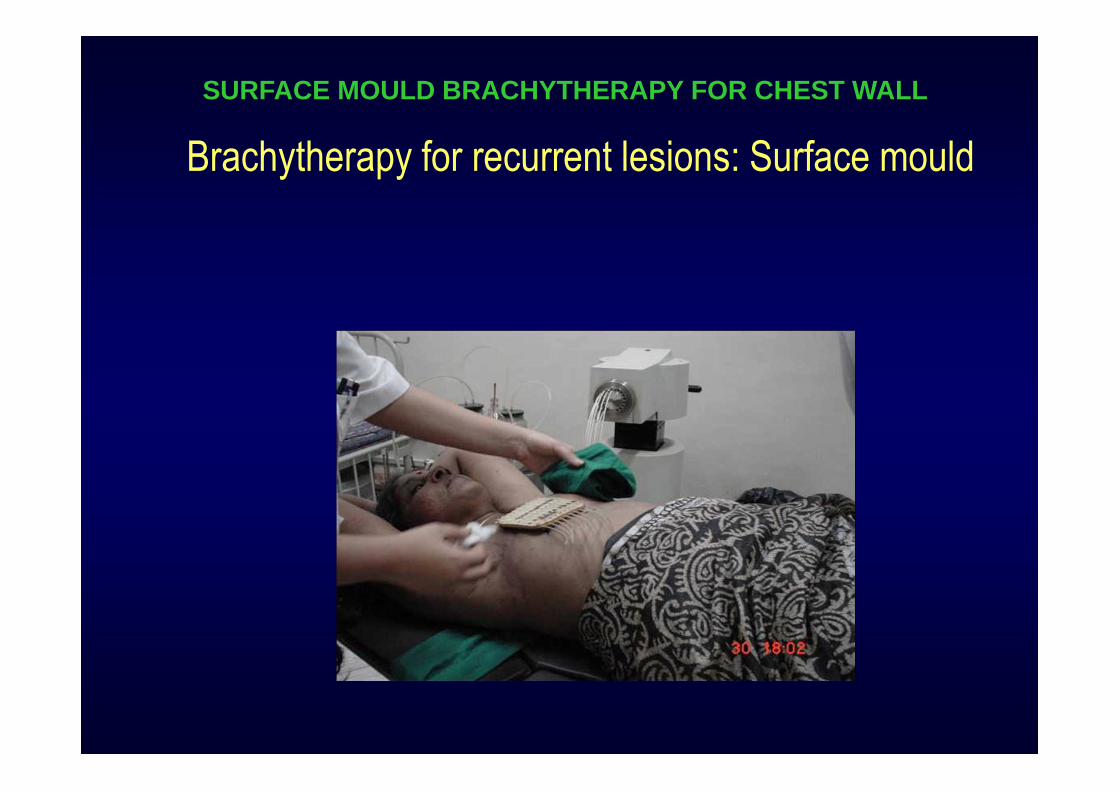
SURFACE MOULD BRACHYTHERAPY FOR CHEST WALL
Brachytherapy for recurrent lesions: Surface mould