Embed Size (px)
Citation preview

OVERVIEW OF RADIOTHERAPY IN BREAST CONSERVATION
Prof G K RathProfessor and Head of Radiotherapy &
Chief, Dr. BRA IRCH,
All India Institute of Medical Sciences, New Delhi

RT in Breast Cancer
�Combined Modality is the mainstayof treatment
�The adjuvant treatment aftersurgery should address
-Local recurrence (RT)
-Systemic disease (chemo, hormone)

3
Breast Conservation Therapy
Removal of only the tumor with a safe margin with axillary lymphadenectomy instead of mastectomy followed by radiotherapy to the breast
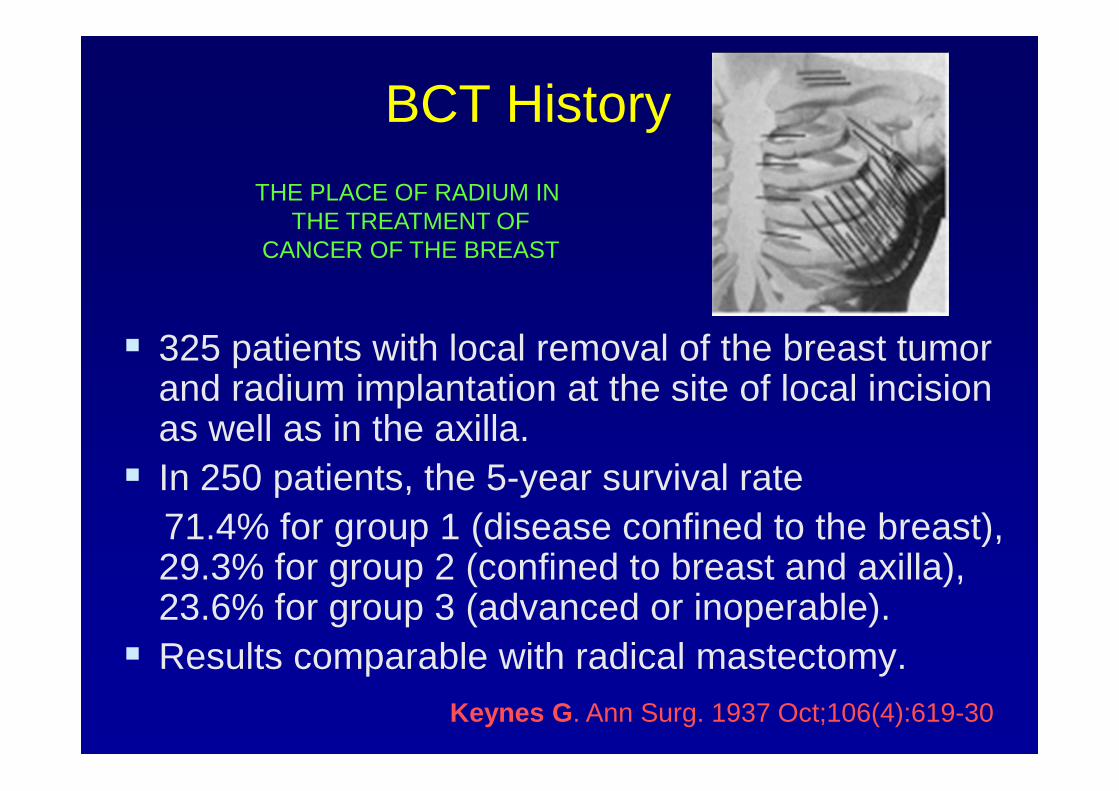
BCT History
� 325 patients with local removal of the breast tumor and radium implantation at the site of local incision as well as in the axilla.
� In 250 patients, the 5-year survival rate 71.4% for group 1 (disease confined to the breast), 29.3% for group 2 (confined to breast and axilla), 23.6% for group 3 (advanced or inoperable).
� Results comparable with radical mastectomy.
THE PLACE OF RADIUM IN THE TREATMENT OF
CANCER OF THE BREAST
Keynes G . Ann Surg. 1937 Oct;106(4):619-30
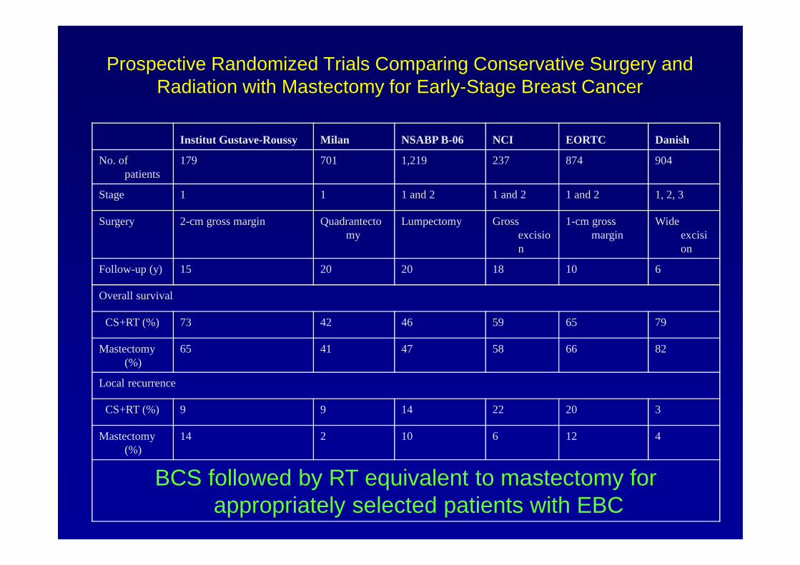
Prospective Randomized Trials Comparing Conservative Surgery and Radiation with Mastectomy for Early-Stage Breast Cancer
Institut Gustave-Roussy Milan NSABP B-06 NCI EORTC Danish
No. of patients
179 701 1,219 237 874 904
Stage 1 1 1 and 2 1 and 2 1 and 2 1, 2, 3
Surgery 2-cm gross margin Quadrantectomy
Lumpectomy Gross excision
1-cm gross margin
Wide excision
Follow-up (y) 15 20 20 18 10 6
Overall survival
CS+RT (%) 73 42 46 59 65 79
Mastectomy (%)
65 41 47 58 66 82
Local recurrence
CS+RT (%) 9 9 14 22 20 3
Mastectomy (%)
14 2 10 6 12 4
BCS followed by RT equivalent to mastectomy for appropriately selected patients with EBC

Early Stage Breast Cancer
NIH Consensus Development Conference Statement (1990)
“Breast conservation treatment is an appropriate method of primary therapy for the majority of women with Stage I and II breast cancer and is preferable because it provides survival equivalent to total mastectomy and axillary dissection while preserving the breast”
“The recommended technique for breast conservation includes: � local excision of primary tumor with clear margins� Level I-II axillary node dissection� breast irradiation to 4,500-5,000 cGy with or without a boost”
Rationale of BCT
� Breast cancer is a systemic disease with hematogenousspread early in the disease process
� Surgery and Radiation as a combined modality� Surgery alone- More failure at margins� Radiotherapy alone- More failure at the epicenter
� Using surgery to remove grossly visible tumor with a small margin and moderate-dose radiotherapy to treat the larger volume of tissue that may harbor residual disease
Criteria for BCT
Indications� Motivated Pts� R T facilities � Mammography� Tumor < 5 cms� Node N0/N1� Good tumor breast ratio
Contraindications:ABSOLUTE� High probability of
recurrence� Multicentric disease� Positive surgical margins
� High probability of complications from irradiation� CVD� Prior irradiation� Early pregnancy
Contraindications to BCT
RELATIVE:� High probability of subsequent breast cancers
� Poor cosmetic results� Unfavorable tumor-breast ratio� Oncologically necessary removal of nipple-areola
complex� Large medial lesions
� Personal preference of the patient

BCT: Technical aspects
� Pre-op evaluation of tumor by Radiation Oncologist
� Minimum margin 1 cm all around� Separate incisions preferred for primary and
axilla� Pectoralis minor may be divided or preserved � Surgical clips are left if brachytherapy not
planned

Standard approach in BCT� BCS
� WLE + ALND
� Whole breast RT � 45-50 Gy/25#/5 weeks� Tangential fields – medial and lateral (Co-60 or 6 MV
photons)� Newer techniques- IMRT, proton beam etc
� Regional RT- only when indicated by post-op HPR
� Boost to tumor bed� 10-16 Gy� Photon/ Electron/ Brachytherapy
Factors affecting cosmesis after BCS
� Removal of large volume of breast tissue� Removal of Nipple-areola complex� Location of tumor (Medial vs lateral)� Post radiation fibrosis

13
Risk Factors for Local Relapse � Young age increase risk� Positive margins increase risk� Systemic therapy lowers risk� Higher RT doses lowers risk� Extensive intra-ductal component increase risk� LCIS increase risk� Lobular histology - higher risk� BRCA1-2 - higher relapse� Larger tumors - higher local relapses � Node-positive - higher local relapse� ER/PR negative- higher local relapse� Her 2+ tumors

RT in BCT
� Volume of irradiationWhole breast ----� boost to the tumor siteAxilla and SCF if necessary
� 45-50 Gy to whole breast by Ext RT� 10-16 Gy boost by electron/photon/brachy


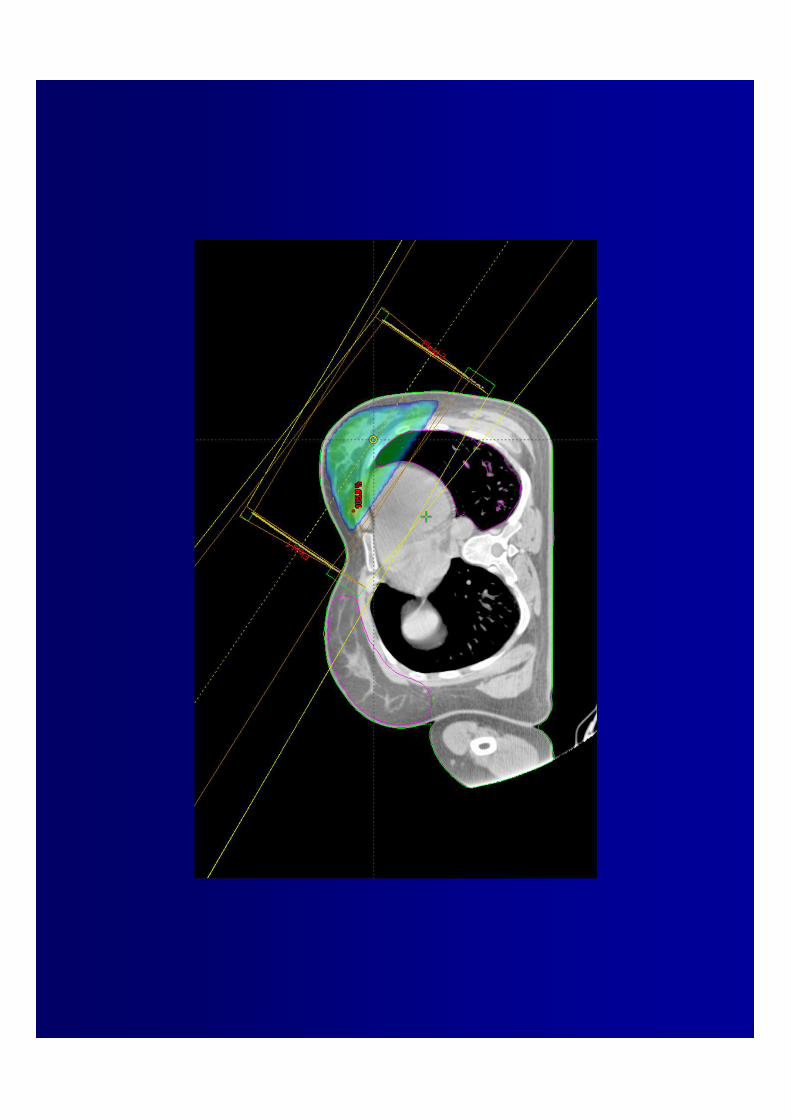

Limitations with conventional radiotherapy
� Dose inhomogenity- Due to continuous change of contour of breast .- 15-20 % dose inhomogenity may result in superior and
inferior plane of breast. - Medial and lateral aspect of breast may get higher dose
of radiation.
� Radiation accompaniments (lung, heart)aim of newer techniques is to further minimize the accompaniments.
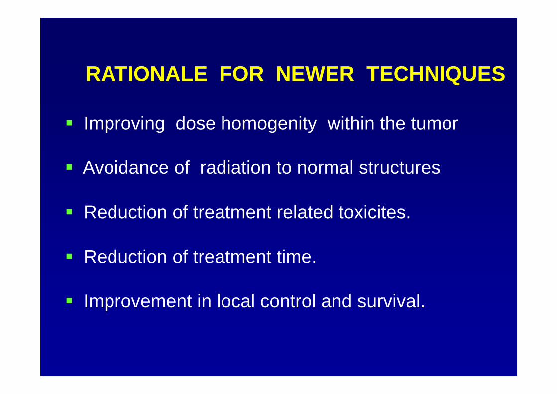
RATIONALE FOR NEWER TECHNIQUES
� Improving dose homogenity within the tumor
� Avoidance of radiation to normal structures
� Reduction of treatment related toxicites.
� Reduction of treatment time.
� Improvement in local control and survival.

NEWER EB-RADIOTHERAPY TECHNIQUES IN EBC
�3D CRT�IMRT �CT scan based planning� Use of Tissue Compensators�Gated Radiotherapy�Partial Breast Irradiation
IMRT
� IMRT is an approach to conformal therapy that notonly conforms high dose to tumor tissue but alsoconforms low dose to surrounding normal tissue.
� Dose intensity is varied in the tumor volumeA higher dose can be delivered to tumor tissueMinimal dose is delivered to surrounding normaltissues.
Higher tumor control probabilityMinimal side effects of radiotherapy

GATED RADIOTHERAPY
�Tumor motion taken into account while radiation treatment is being delivered.
� Techniques of Gating
A. Breath hold technique- Active- airway of patient is temporarily blocked by a valve- Passive- the patient temporarily holds the breath
B. Synchronized Gatingexternal devices are used to predict the phase ofrespiratory cycle while patient breathes freely
USE OF TISSUE COMPENSATORS
� Compensator- is a device which compensates formissing tissues.
� Use of tissue compensators improve dosimetryand reduce complications.
� Various types of tissue compensators are used
- Tissue equivalent materials
- MLC
Boost in BCT
METHODS
� Electron beam� Photon beam- 3DCRT/IMRT� Interstitial Brachytherapy� IORT� Mammosite

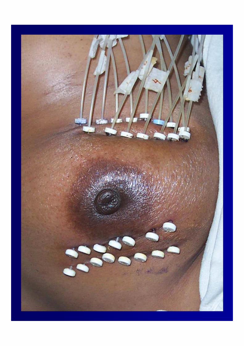
INTERSTITIAL BRACHYTHERAPY
� Main advantage lies in ability to tightly conformdose to a specified volume.
� Used as a boost following BCT along with EBRT
� Clinical situations where brachytherapy may be more useful than EBRT include – Large breasts
-Deep seated tumors-Extensive intra-ductal comp.-Uncertain margins.
� Shorter treatment times

INTRAOPERATIVE RADIOTHERAPY ( IORT)
� IORT is a radiotherapeutic technique which delivers a single dose of radiation to tumor bed or to expos ed tumor during surgery.
� It is used mainly as a boost to be followed by EBR T.
� Rationale : 85 % of relapses in BCT after RT occur in the operated area.
Techniques: IOHDRIOERT
MAMMOSITE
� Can be used for primary radiation or as a boost
� HDR with Iridium –192 source is used.
� Places the radiation source inside the lumpectomy cavity.
� Cosmetic results are good to excellent in 88% of cases.
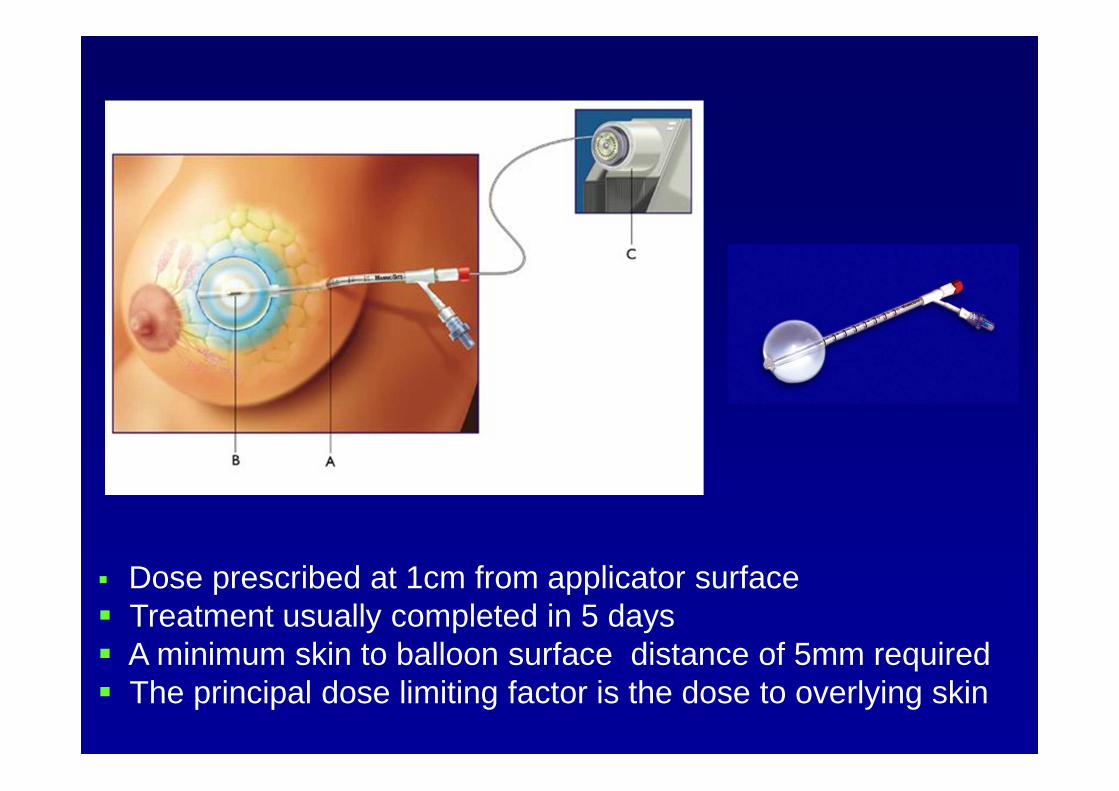
� Dose prescribed at 1cm from applicator surface� Treatment usually completed in 5 days� A minimum skin to balloon surface distance of 5mm required� The principal dose limiting factor is the dose to overlying skin

� Accompaniments : - Due to device placement-
Mild erythema, pain, drain leakage,ecchymosis.- Due to radiation therapy-
erythema, dry desquamation
� Factors limiting use of mammositeBalloon- cavity conformanceSkin – balloon cavity surface distance
31
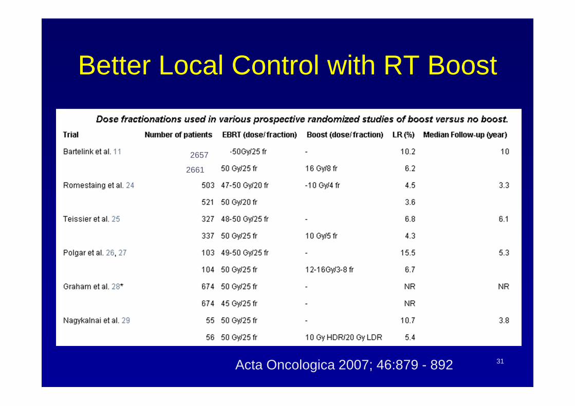
Better Local Control with RT Boost
Acta Oncologica 2007; 46:879 - 892
2657
2661
32
Delineation of Tumor Bed for Boost
� Clinical-history and patients' recollection of tumor position, clinical photographs, tattoos, surgical scar
� Mammography� Surgical clips� Ultrasonography� Computerized tomography (CT) scan� Magnetic resonance imaging (MRI) � Peroperative placement of catheters
33
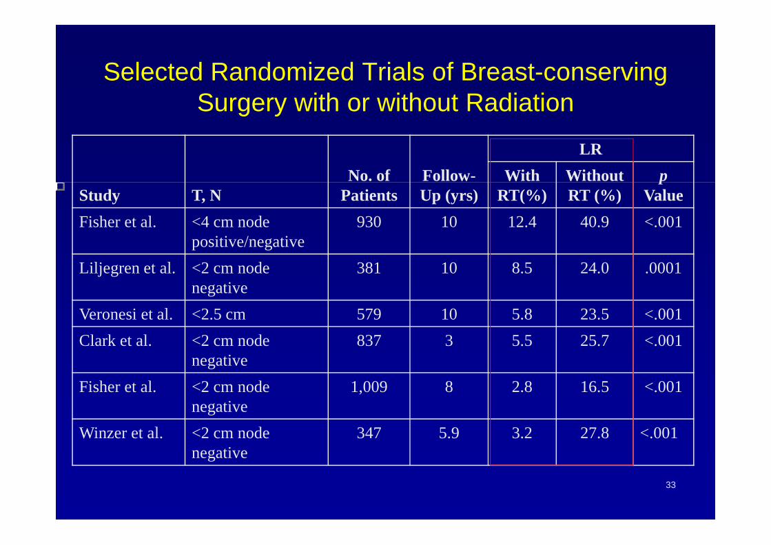
Selected Randomized Trials of Breast-conserving Surgery with or without Radiation
Study T, NNo. of
PatientsFollow-Up (yrs)
LR
With RT(%)
Without RT (%)
pValue
Fisher et al. <4 cm node positive/negative
930 10 12.4 40.9 <.001
Liljegren et al. <2 cm node negative
381 10 8.5 24.0 .0001
Veronesi et al. <2.5 cm 579 10 5.8 23.5 <.001
Clark et al. <2 cm node negative
837 3 5.5 25.7 <.001
Fisher et al. <2 cm node negative
1,009 8 2.8 16.5 <.001
Winzer et al. <2 cm node negative
347 5.9 3.2 27.8 <.001
34
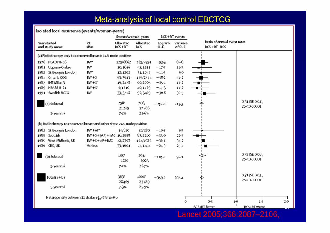
Meta-analysis of local control EBCTCG
Lancet 2005;366:2087–2106,
35
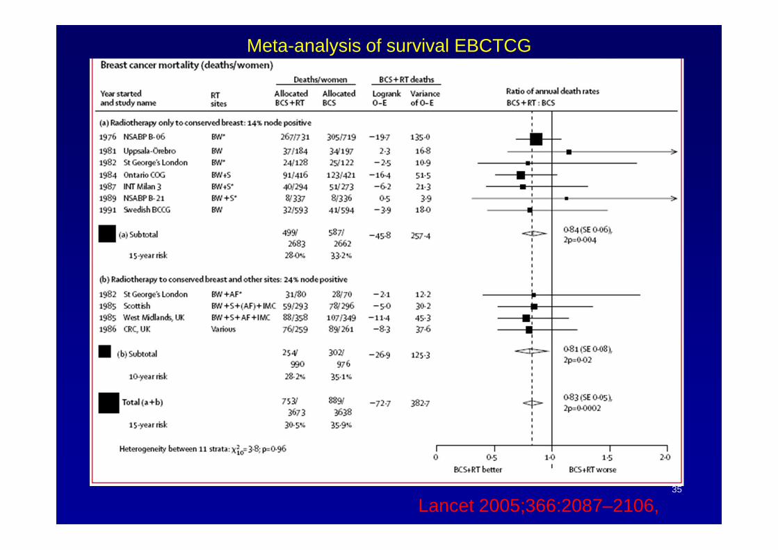
Meta-analysis of survival EBCTCG
Lancet 2005;366:2087–2106,
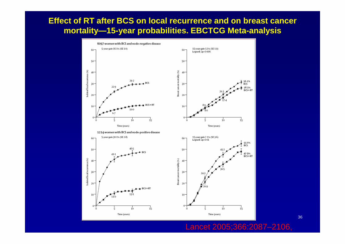
Effect of RT after BCS on local recurrence and on b reast cancer mortality—15-year probabilities. EBCTCG Meta-analysi s
36
Lancet 2005;366:2087–2106,

Attitudes and treatment outcome of breast conservat ion therapy for stage I & II breast cancer using peroperative ir idium -192
implant boost to the tumour bed.
� Surgery with peroperative implantation of iridium-192 to deliver a boost.� Whole breast irradiation was delivered 3-4 weeks after the boost. � Cosmesis was assessed at the end of 6 months from completion of therapy. � There were no locoregional failures at a median follow up of 42 months.� One patient experienced a systemic relapse.� Cosmesis was good to excellent in 80% of patients. � Breast conservation therapy using peroperative iridium-192 implant provides
excellent locoregional disease control and cosmesis.
37
AIIMS DATA
Deo SS, Mohanti BK, Shukla NK, Chawla S, Raina V, J ulka PK, Rath GK.Australas Radiol. 2001 Feb;45(1):35-8
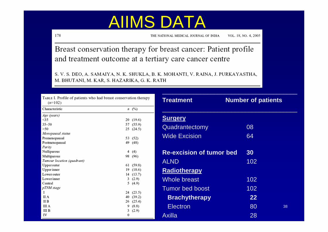
AIIMS DATA
38
_______________________________________Treatment Number of patients_______________________________________SurgeryQuadrantectomy 08Wide Excision 64
Re-excision of tumor bed 30ALND 102RadiotherapyWhole breast 102Tumor bed boost 102
Brachytherapy 22Electron 80
Axilla 28
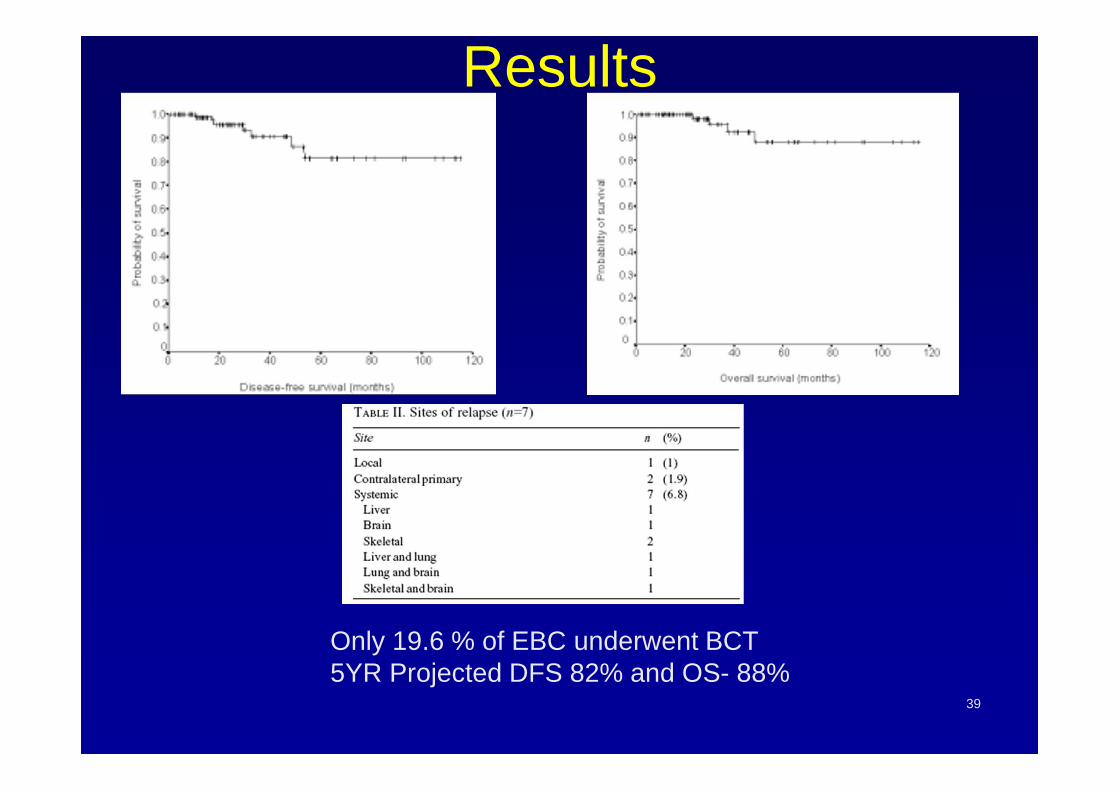
Results
39
Only 19.6 % of EBC underwent BCT5YR Projected DFS 82% and OS- 88%

Partial Breast Irradiation
Definition Delivery of larger doses/fraction of radiation to the lumpectomy cavity (plus 1-2 cm margin) after breast conserving surgery in patients with early stage breast cancer
PBI: Concept
Selected cases (low risk)� Partial breast
� Only the breast tissue adjacent to the tumor bed is irradiated
� Accelerated schedules� Dramatic reduction in duration of RT to 1-5 days
� Accelerated Partial Breast Irradiation (APBI)

PBI: Scientific rationale
� 80% of breast recurrences after BCS occur at or near the tumor bed, implicating residual tumor foci from the original index tumor
� Major effect of post-lumpectomy radiotherapy: reduce risk of recurrence in tumor bed region
� Incidence of ‘elsewhere’ failures 3-5%� Some ‘elsewhere’ failures- new primaries, unaffected by
whole breast irradiation� Whole breast radiation may not be needed in
“appropriately” selected cases

Failures Outside of the Tumor Bed in Randomized Trials Comparing Lumpectomy with/without Postop RT
Baglan KL et al. Int J Radiat Oncol Biol Phys. 2001;50:1003-11.
Surgery aloneSurgery plus RT
TrialMedian f/u
(mo)N % N %
NSABP-B06 125 17 / 636 2.7 24/629 3.8
Milan 39 4 / 273 1.5 0/294 0
Uppsala-Orebro 64 7 / 197 3.5 - -
Ontario 43 15 / 421 3.5 4/416 1.0

PBI: Potential advantages� Reduces overall treatment time
� Improves acceptability of BCT� Reduces waiting time for radiotherapy� Improves access to radiotherapy treatment machines
� Smaller treatment volumes� Large dose per fraction may be delivered without an increase in
toxicity� Normal structures like heart, lungs, contralateral breast may be
spared� Better cosmetic results (lower skin & breast parenchyma integral
dose)� Eliminate scheduling problems with systemic chemotherapy� Cost savings
� Reduces hospital visits� Reduces absence from work and associated income losses
� Improves quality of life

Patient selection� Age: Postmenopausal� T2 or less� N0� Low grade� Negative surgical margins� ER +
� Exclude � Young patients� Large tumors� N+� High grade� Multicentric� Invasive lobular histology� EIC� Positive surgical margins
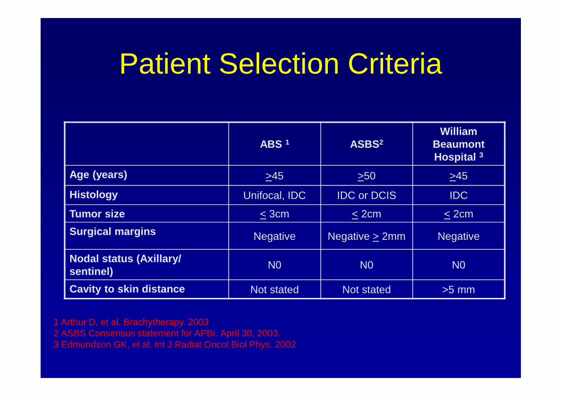
Patient Selection Criteria
ABS 1 ASBS 2William
Beaumont Hospital 3
Age (years) >45 >50 >45
Histology Unifocal, IDC IDC or DCIS IDC
Tumor size < 3cm < 2cm < 2cm
Surgical margins Negative Negative > 2mm Negative
Nodal status (Axillary/ sentinel)
N0 N0 N0
Cavity to skin distance Not stated Not stated >5 mm
1 Arthur D, et al. Brachytherapy. 20032 ASBS Consensus statement for APBI. April 30, 2003.3 Edmundson GK, el al. Int J Radiat Oncol Biol Phys. 2002

PBI: Techniques� Brachytherapy
� Interstitial Brachytherapy� Mammosite balloon brachytherapy
� Intraoperative radiotherapy� Intraoperative electrons (IOERT)� Targeted intraoperative radiotherapy (TARGIT) � Brachytherapy
� EBRT� Electrons� 3D-CRT� IMRT� Protons

� Outside of multi-institutional studies and institutional protocols, patients should be carefully selected for APBI and properly informed of the benefits and risks of this type of radiation treatment.
� The following selection criteria when considering patients for treatment with APBI:
� • Age 45 years old or greater�
• Invasive ductal carcinoma or ductal carcinoma in situ �
• Total tumor size (invasive and DCIS) less than or equal to 3 cm in size�
• Negative microscopic surgical margins of excision�
• Axillary lymph nodes/sentinel lymph node negative 48
ASBS consensus statement for APBI

� Surgeons, radiation oncologists and physicists who will be utilizing the various APBI techniques should be adequately trained to allow for optimum radiation therapy planning and treatment.
� All patients should be monitored regularly to identify adverse events as well as local recurrences.
� Continuous, long-term, outcomes-based monitoring of APBI is desirable.
49
ASBS consensus statement for APBI contd..
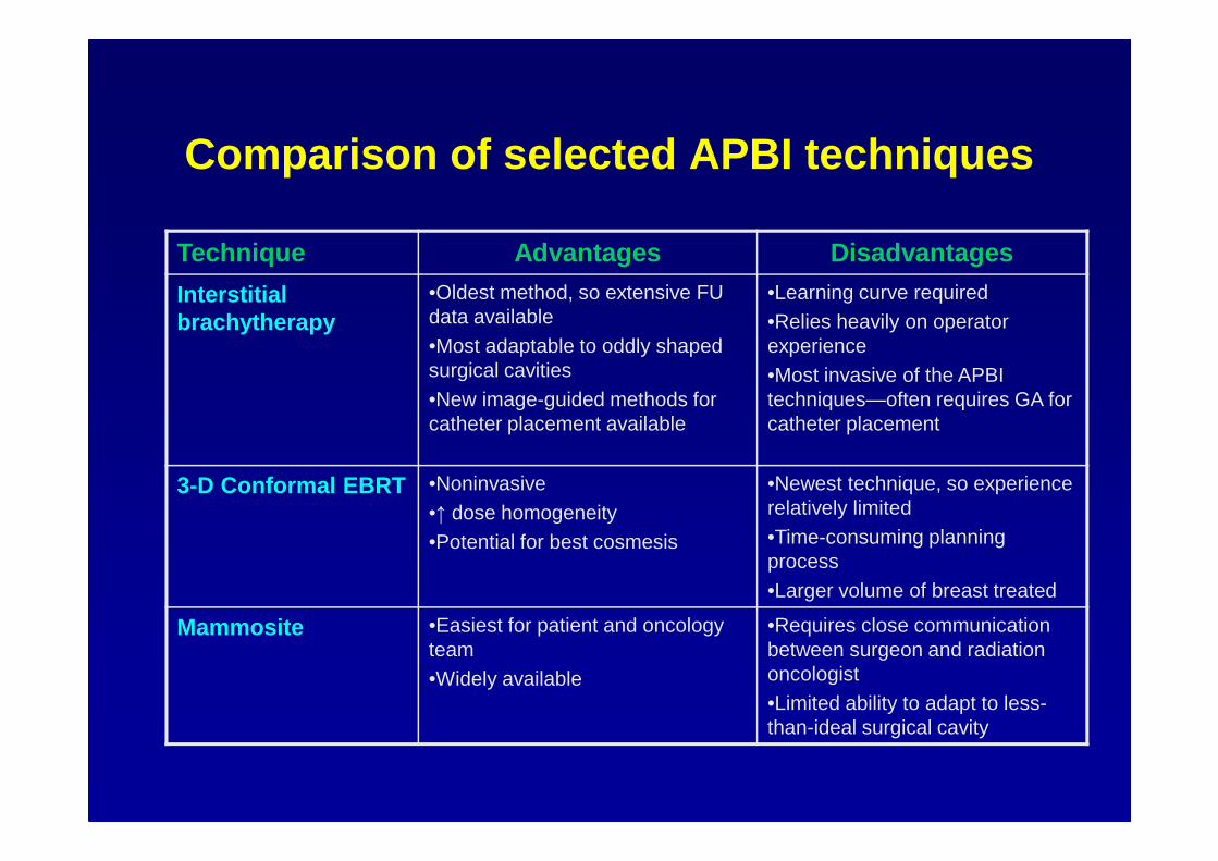
Comparison of selected APBI techniques
Technique Advantages Disadvantages
Interstitial brachytherapy
•Oldest method, so extensive FU data available•Most adaptable to oddly shaped surgical cavities•New image-guided methods for catheter placement available
•Learning curve required•Relies heavily on operator experience•Most invasive of the APBI techniques—often requires GA for catheter placement
3-D Conformal EBRT •Noninvasive•↑ dose homogeneity•Potential for best cosmesis
•Newest technique, so experience relatively limited•Time-consuming planning process•Larger volume of breast treated
Mammosite •Easiest for patient and oncology team•Widely available
•Requires close communication between surgeon and radiation oncologist•Limited ability to adapt to less-than-ideal surgical cavity
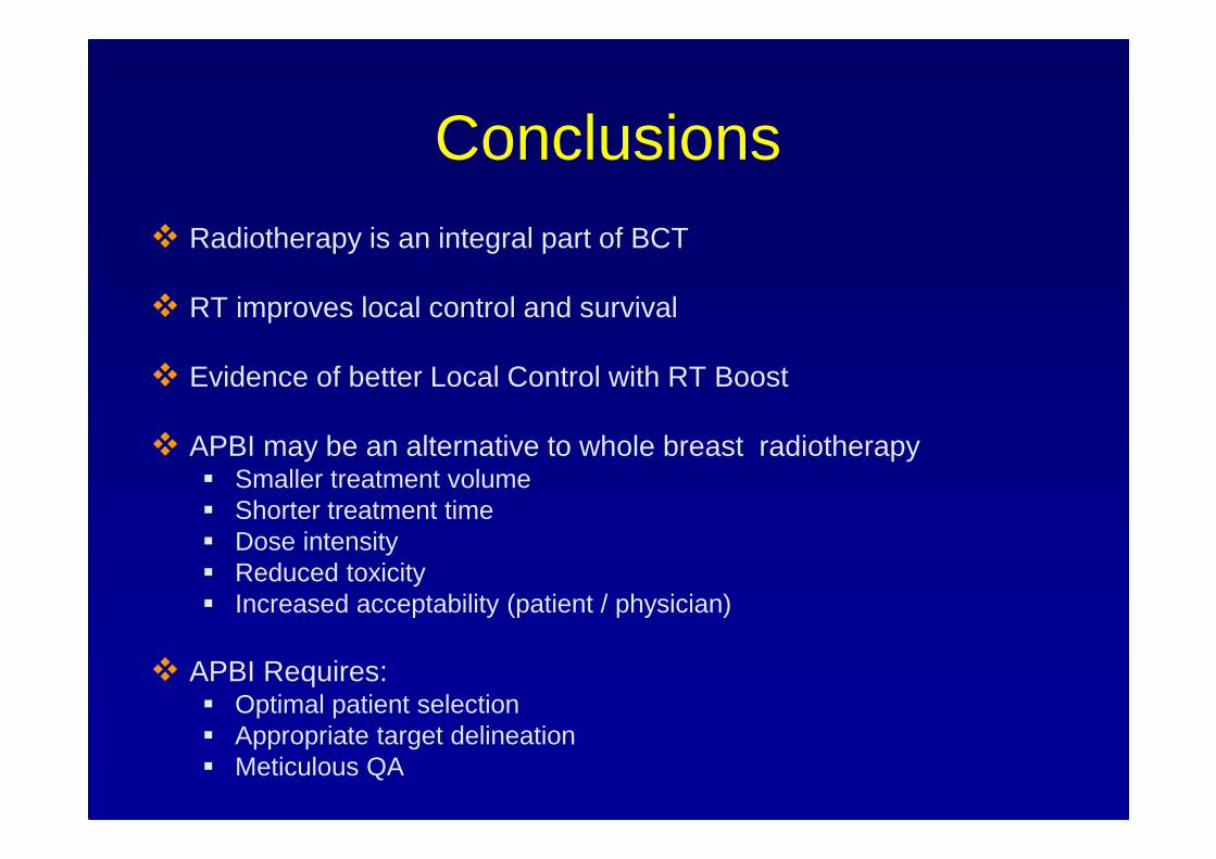
Conclusions� Radiotherapy is an integral part of BCT
� RT improves local control and survival
� Evidence of better Local Control with RT Boost
� APBI may be an alternative to whole breast radiotherapy� Smaller treatment volume� Shorter treatment time� Dose intensity� Reduced toxicity� Increased acceptability (patient / physician)
� APBI Requires:� Optimal patient selection� Appropriate target delineation� Meticulous QA

52
THANK YOU