Embed Size (px)
DESCRIPTION
Bone Histology and Skeletal Structure Review. What is bone?. Bone is living tissue that makes up the body's skeleton. There are three types of bone tissue, including the following:. Compact (dense) bone tissue. - PowerPoint PPT Presentation
Citation preview

Bone Histology and Skeletal Structure Review

What is bone?
Bone is living tissue that makes up the body's skeleton. There are three types of bone tissue, including the following:

Compact (dense) bone tissue the harder, outer tissue of bones; made up
of precise arrangements of microscopic cylindrical structures called osteons. The matrix and osteocytes of osteon are laid down in concentric rings around a central (Haversian) canal which contains blood vessels and nerves.
If you look at compact bone tissue with naked eye, it look very dense: you cannot see any cavities in it.

Spongy (cancellous) bone tissue
the sponge-like tissue inside bones. In contrast, made up of irregular latticework of thin blades of bone called trabeculae. The spaces between the trabeculae contain blood vessels and red marrow which produces blood cells.

The spaces between the trabeculae can be seen with naked eye and give spongy bone tissue its "spongy" look.
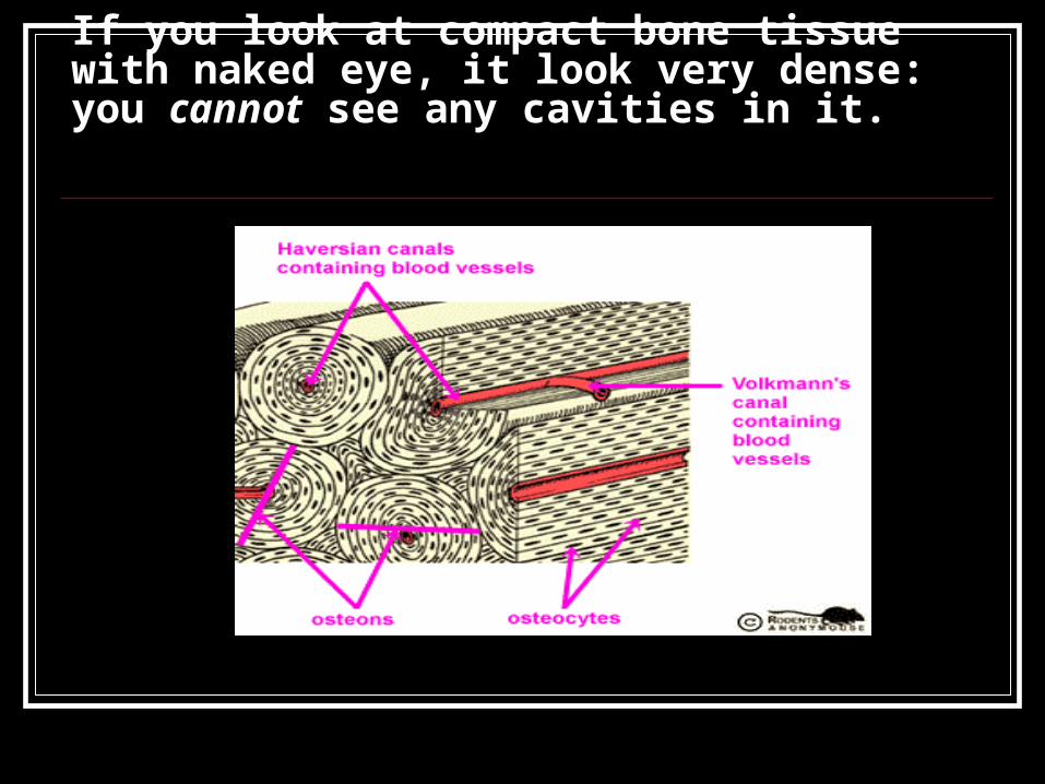
subchondral tissue
the smooth tissue at the ends of bones, which is covered with another type of tissue called cartilage. Cartilage is the specialized, gristly connective tissue that is present in adults, and the tissue from which most bones develop in children.

What are the different types of bone cells? The different types of bone cells include the following:
osteoblast - found within the bone, its function is to form the tissue and minerals that give bone its strength.
osteoclast - a very large cell formed in bone marrow, its function is to absorb and remove unwanted tissue; remodeling
osteocyte - found within the bone, its function is to help maintain bone as living tissue.
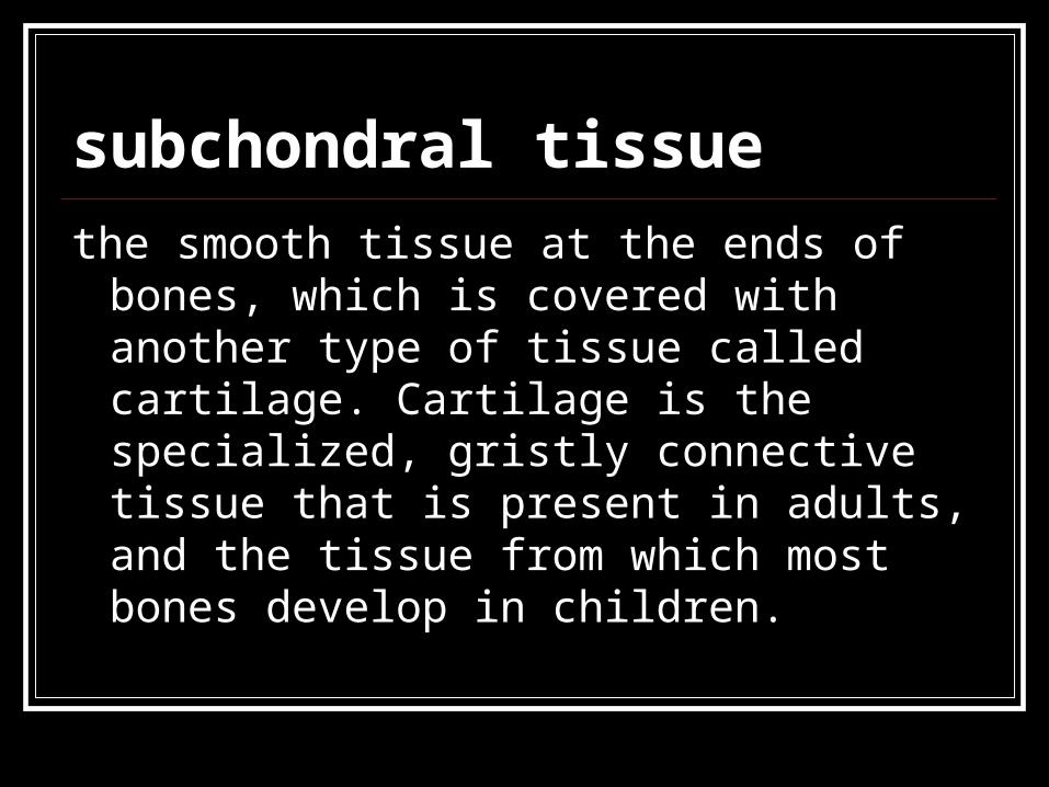
Fat cells and hematopoietic cells are found within the bone marrow.
Hematopoietic cells are those that produce blood cells.


Bone's function? shape, support, and protect body structures; aid
in body movement
serve as a site for development and storage of blood cells disorders and diseases that can affect bone.
serves as a storage site for minerals; provides the medium - marrow - for the development and storage of blood cells.

But How? Support
bones of the leg, pelvic, and vertebral column hold up the bodythe mandible supports the teethprovide support for muscles and other soft organs
Protectionbones enclose and protect the brain, spinal chord, lungs, heart, pelvic viscera, and bone marrow
Movementof course, to walk, reach, touch (limb leverage)lung ventilation depends on by movement of ribs by skeletal muscles
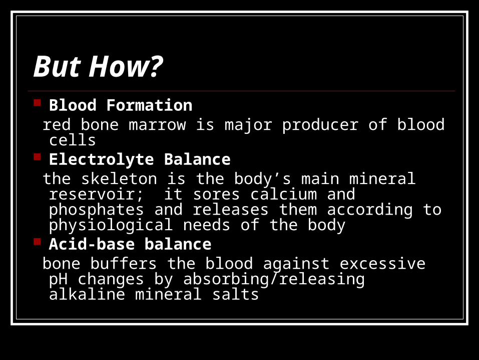
But How? Blood Formation red bone marrow is major producer of blood cells Electrolyte Balance the skeleton is the body’s main mineral reservoir; it
sores calcium and phosphates and releases them according to physiological needs of the body
Acid-base balance bone buffers the blood against excessive pH
changes by absorbing/releasing alkaline mineral salts

Types of Bone Long Bone
Example: Femur Short Bone
Example: Carpal Flat Bone
Example: Pelvic Irregular Bone
Example: facial Sesamoid Bone
Example: Patella
Skeletal Structure Tissue cartilage osseous tissue bone marrow periosteum/endosteum
The Skull
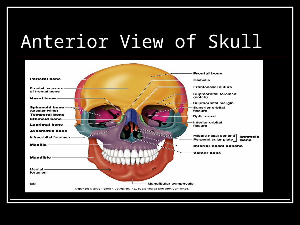
Anterior View of Skull
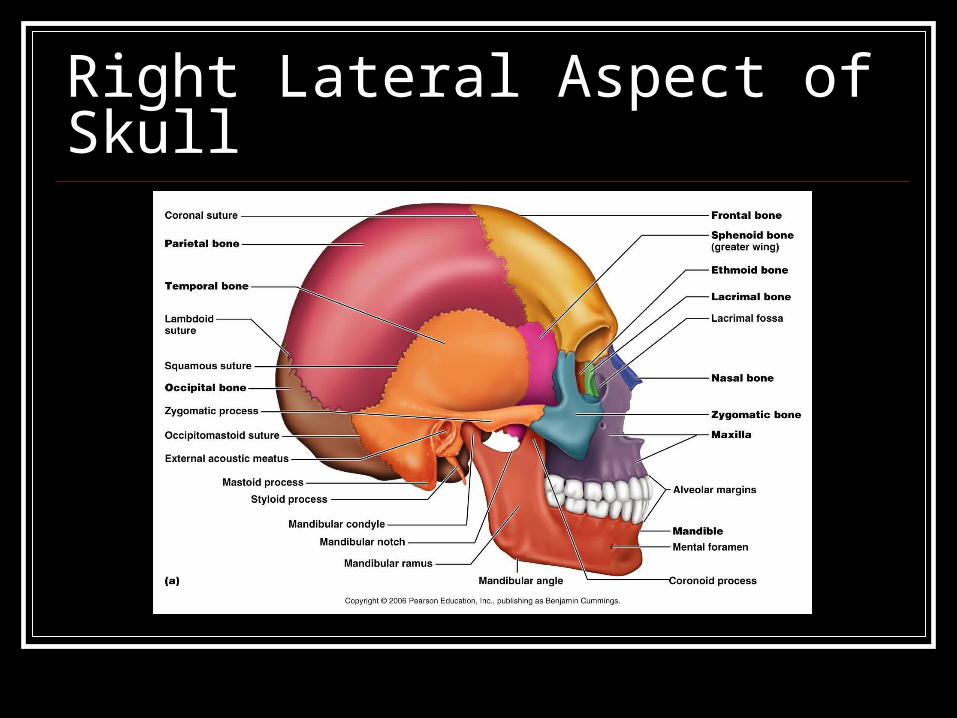
Right Lateral Aspect of Skull

Posterior View of Skull
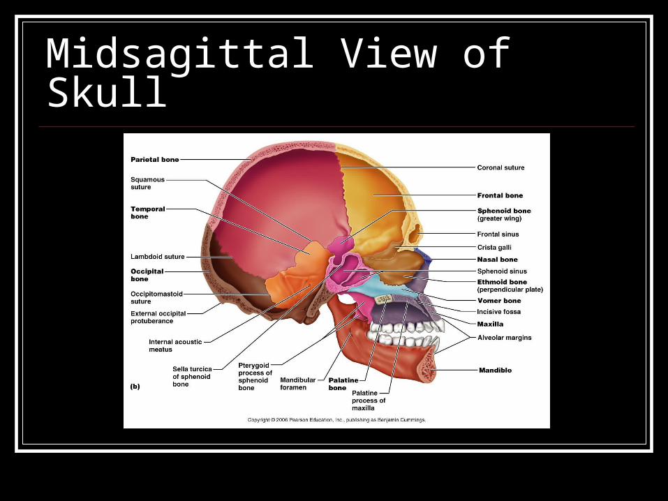
Midsagittal View of Skull

Inferior Superficial View of Skull

Superior View of Cranial Cavity
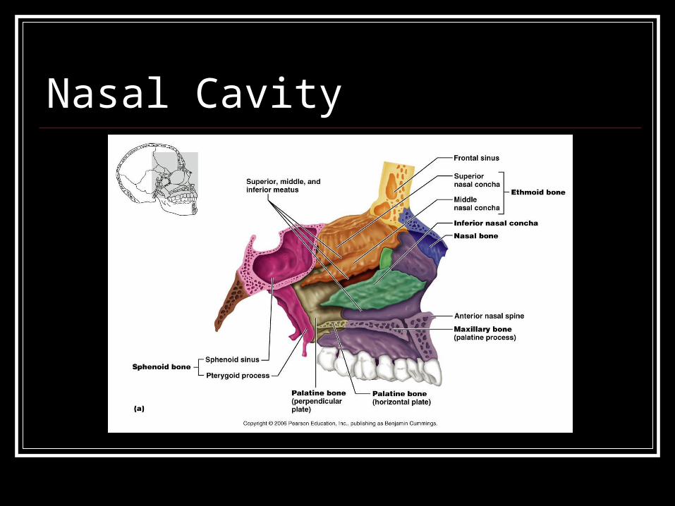
Nasal Cavity

Vertebrae, Ribs, and Sternum
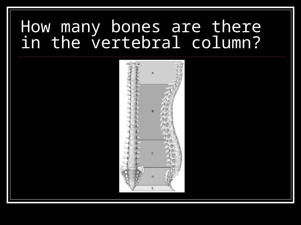
How many bones are there in the vertebral column?

Answer: 26

How regions are there in the vertebral column and what are their names?

Answer: 5 regions: cervical, thoracic, lumbar, sacral,
coccygeal

What are the name of the first two bones of the vertebral column and their functions?

Answer: Atlas and Axis the support and movement
of the skull
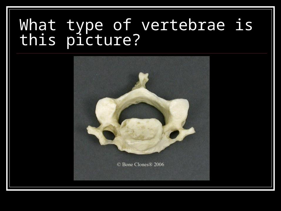
What type of vertebrae is this picture?

Answer: Cervical vertebrae
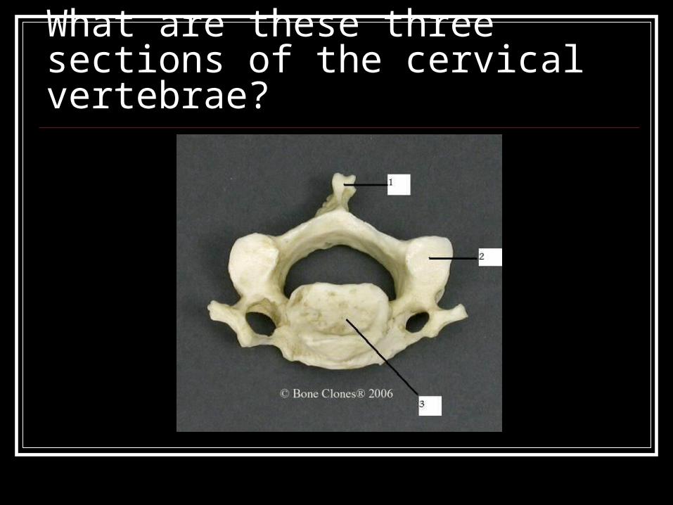
What are these three sections of the cervical vertebrae?
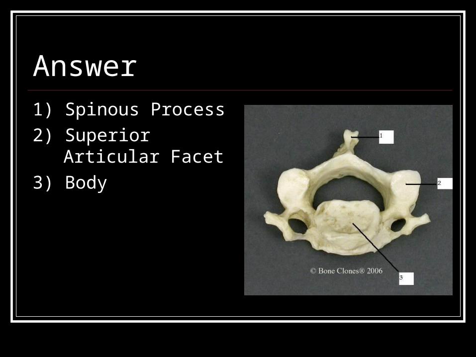
Answer1) Spinous Process
2) Superior Articular Facet
3) Body

What type of vertebrae is this picture?

Answer: Thoracic Vertebrae

What are these three sections of the thoracic vertebrae?
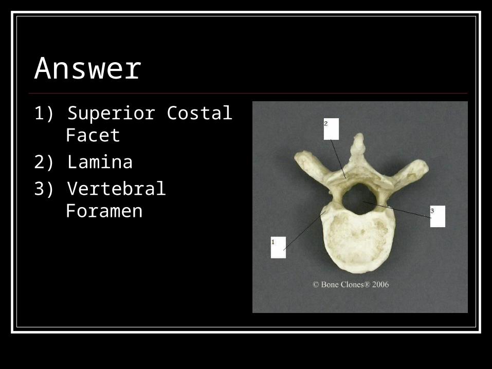
Answer1) Superior Costal Facet
2) Lamina
3) Vertebral Foramen

How many pair’s ribs does the human
body have?

Answer: 12 pairs
What are the names of the types of ribs?

Answer: True and False Ribs
What is the difference between True and
False Ribs?
Answer: True ribs are attached to the sternum.

What bone is this?

Answer Sternum
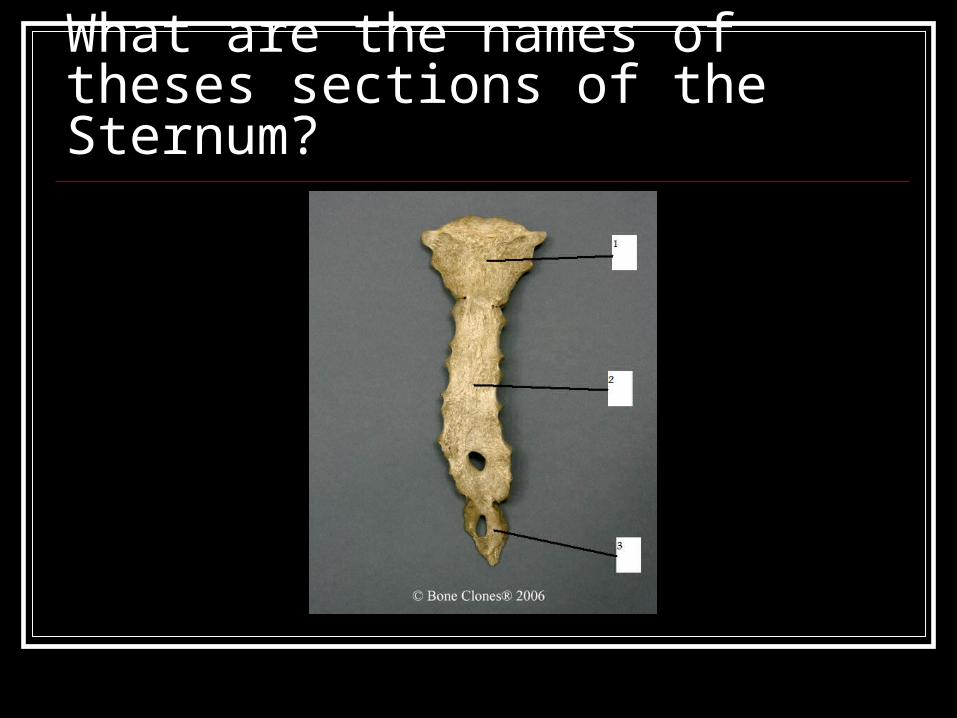
What are the names of theses sections of the Sternum?

Answer1) Manubrium
2) Body
3)Xiphoid Process
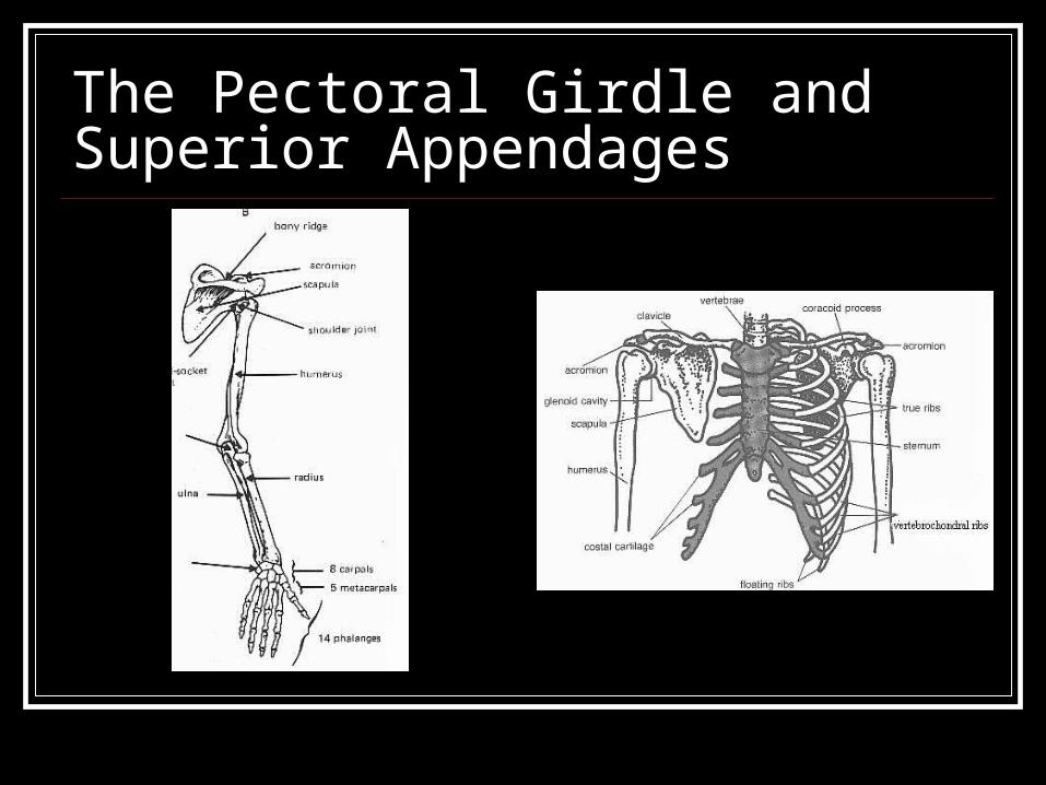
The Pectoral Girdle and Superior Appendages

Pectoral girdles are responsible for attaching the superior appendages (the arms, forearms, wrists, and hands) to the axial skeleton.
On each side of the body the pectoral girdle consists of two bones: the scapula and the clavicle.
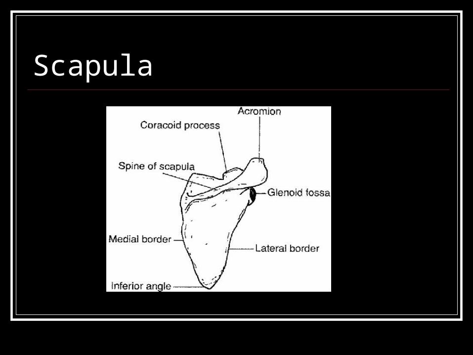
Scapula
The scapula is also known as the shoulder blade.
It is connected to the humerus by a ball and socket joint.
It is an irregular bone.
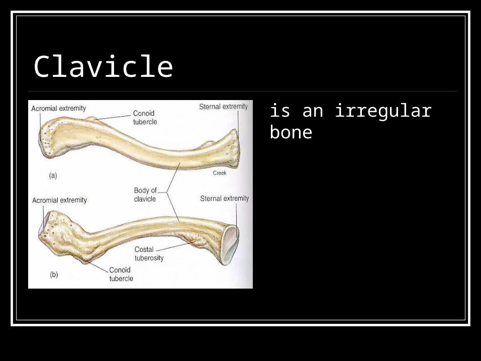
Clavicleis an irregular bone

Humerus The humerus is a long
bone.
It connects to the ulna by the elbow which is a hinge joint.
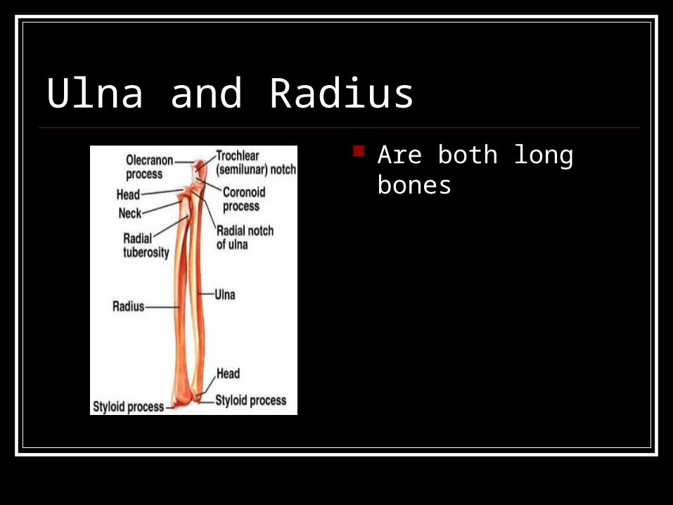
Ulna and Radius Are both long bones
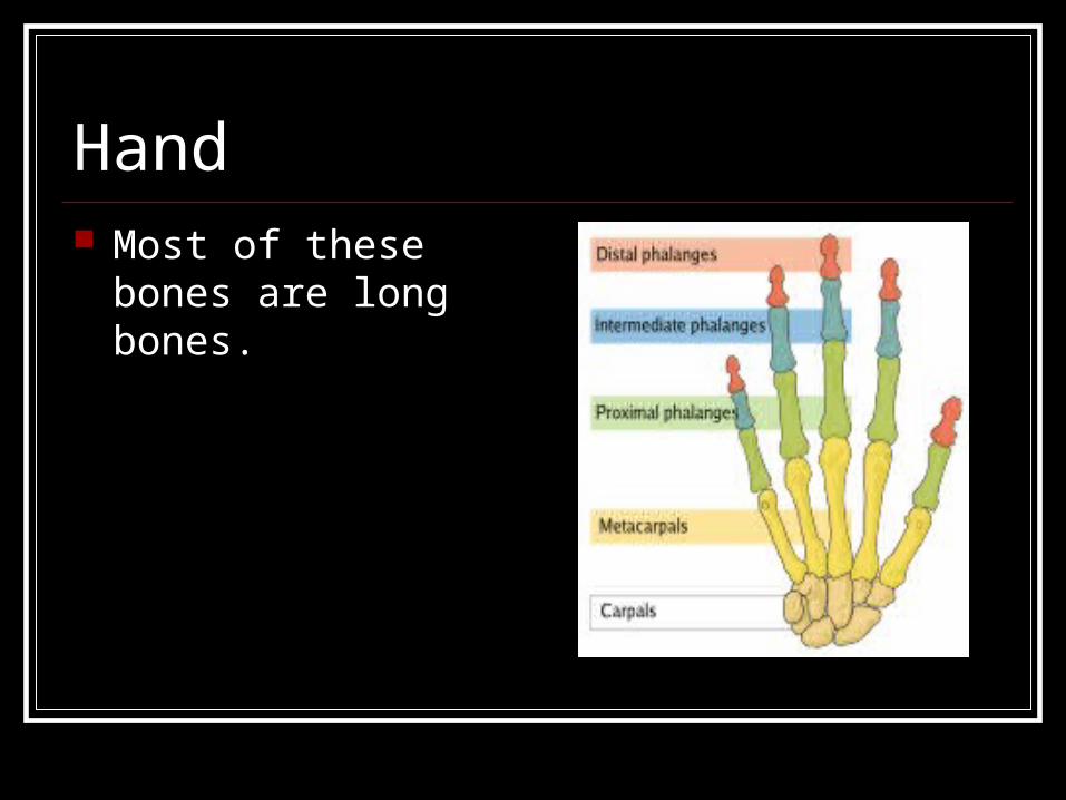
Hand Most of these bones
are long bones.
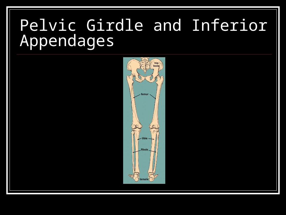
Pelvic Girdle and Inferior Appendages


The hip bone is composed of threeelements:
Which also fuse at the acetabulum (the
socket of the hip joint).
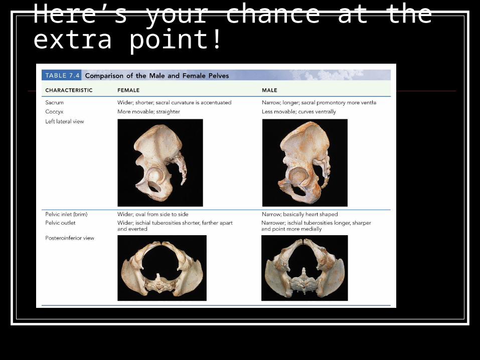
Here’s your chance at the extra point!

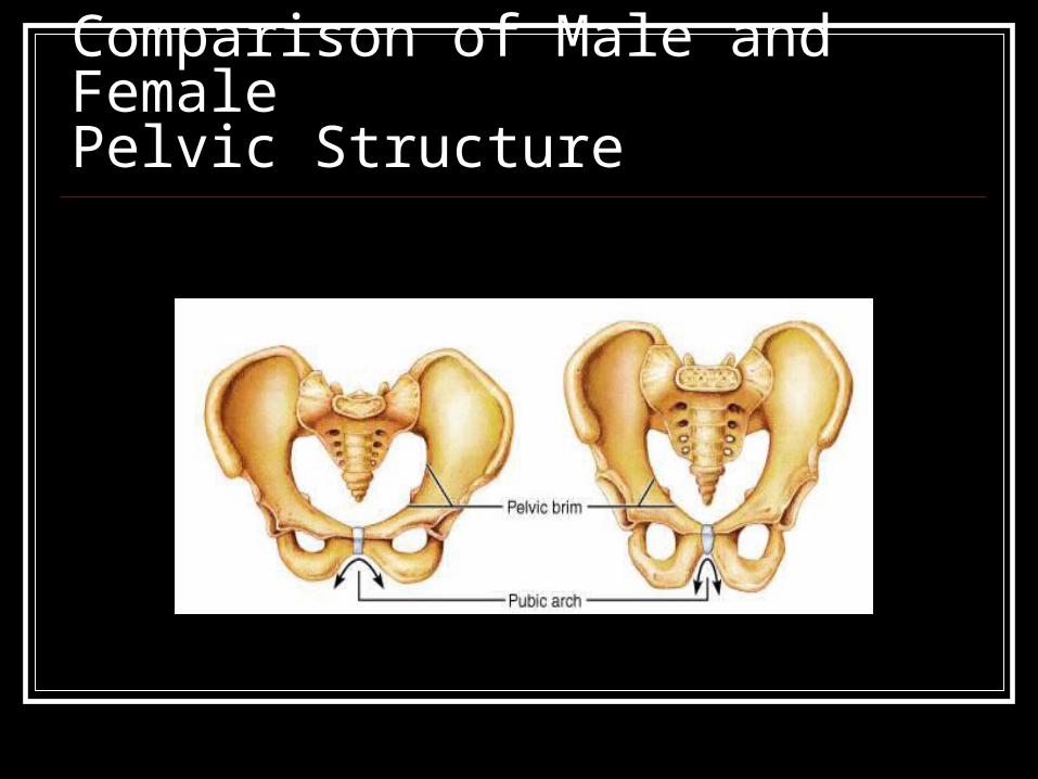
Comparison of Male and FemalePelvic Structure
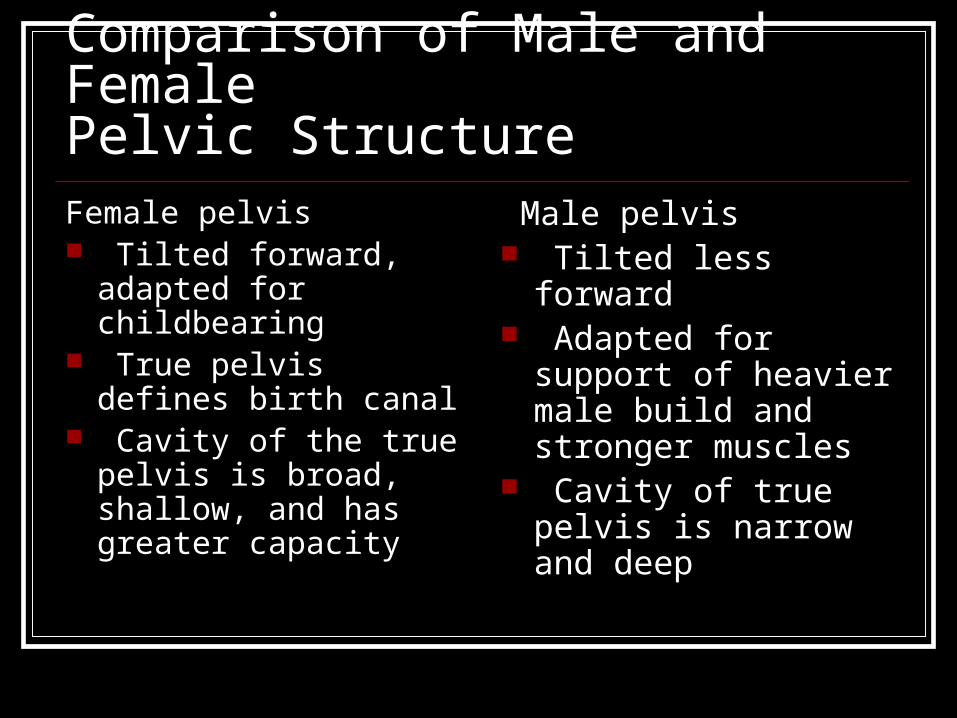
Comparison of Male and FemalePelvic StructureFemale pelvis Tilted forward,
adapted for childbearing
True pelvis defines birth canal
Cavity of the true pelvis is broad, shallow, and has greater capacity
Male pelvis Tilted less forward Adapted for support
of heavier male build and stronger muscles
Cavity of true pelvis is narrow and deep
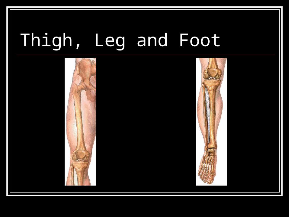
Thigh, Leg and Foot