Embed Size (px)
Citation preview
Histology of
BoneBy: - Addisu A. (MD)
IntroductionBone tissue along with other
CT like cartilage, fibrous tissue, fat, blood vessels, nerves, and hematopoietic elements form the individual bones.
Bones are the organs of the skeletal system; bone tissue is the structural component of bones.
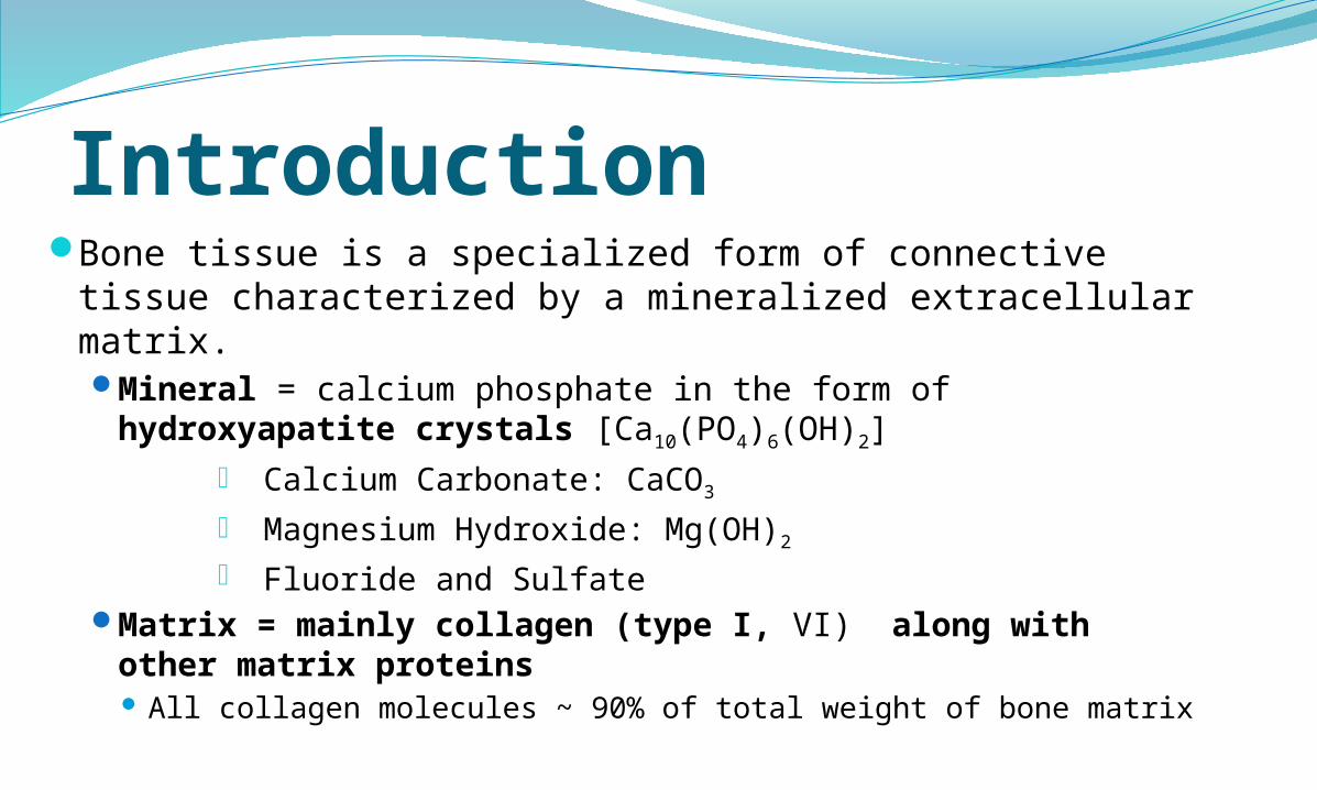
Introduction Bone tissue is a specialized form of connective tissue
characterized by a mineralized extracellular matrix.Mineral = calcium phosphate in the form of
hydroxyapatite crystals [Ca10(PO4)6(OH)2]- Calcium Carbonate: CaCO3
- Magnesium Hydroxide: Mg(OH)2
- Fluoride and SulfateMatrix = mainly collagen (type I, VI) along with
other matrix proteins All collagen molecules ~ 90% of total weight of bone matrix
Function 1) storage for elements and minerals -
homeostatic regulation of blood calcium levels
2) mechanical structures for movement and protection of viscera,
3) a home for hematopoietic tissue, and4) Storage of adipose tissue: yellow marrow
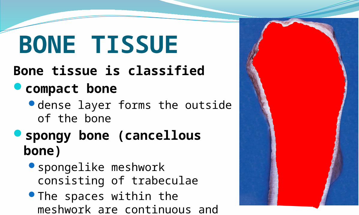
BONE TISSUEBone tissue is classified compact bone
dense layer forms the outside of the bone
spongy bone (cancellous bone)spongelike meshwork consisting of
trabeculaeThe spaces within the meshwork
are continuous and occupied by marrow and blood vessels.
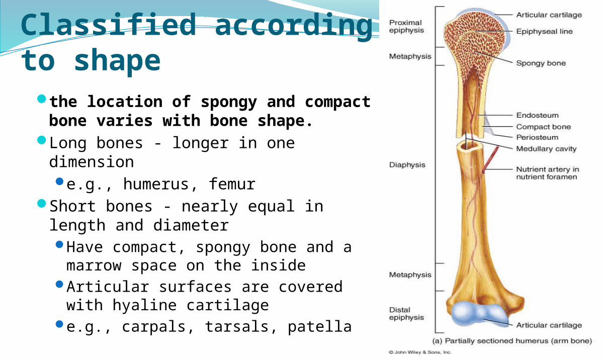
Classified according to shapethe location of spongy and compact
bone varies with bone shape.Long bones - longer in one dimension
e.g., humerus, femurShort bones - nearly equal in length
and diameterHave compact, spongy bone and a
marrow space on the insideArticular surfaces are covered with
hyaline cartilagee.g., carpals, tarsals, patella
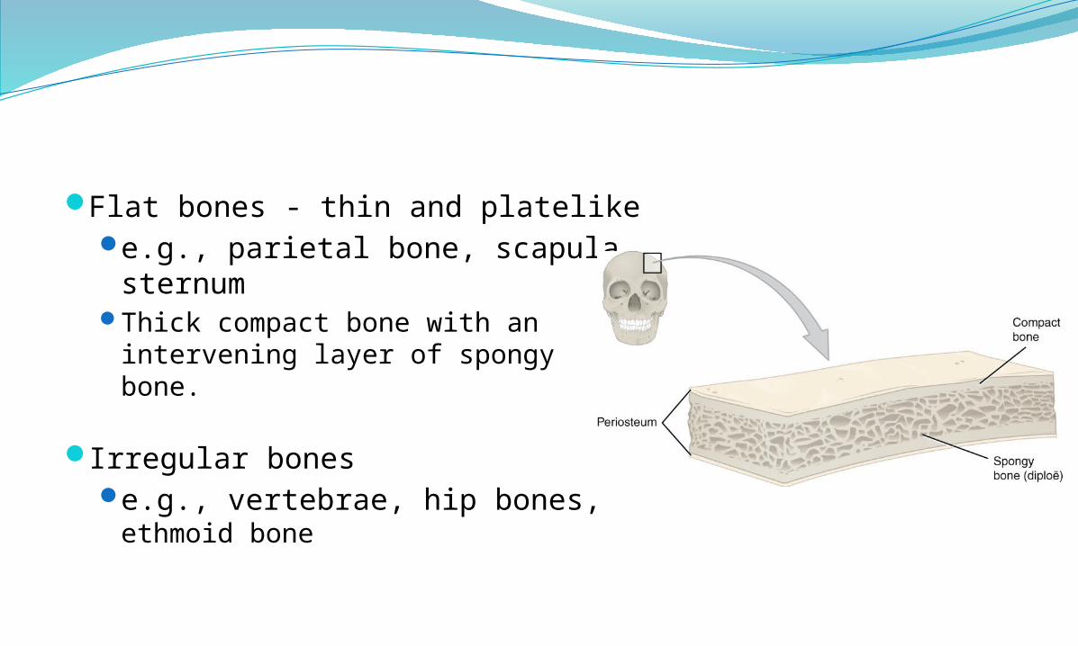
Flat bones - thin and platelikee.g., parietal bone, scapula,
sternumThick compact bone with an
intervening layer of spongy bone.
Irregular bonese.g., vertebrae, hip bones,
ethmoid bone
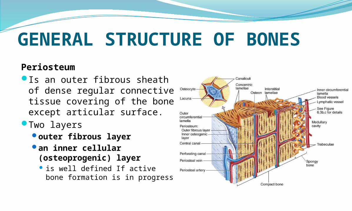
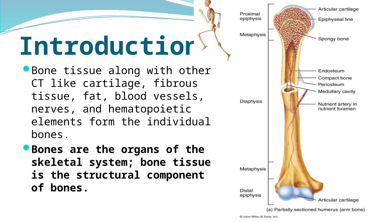
GENERAL STRUCTURE OF BONESPeriosteumIs an outer fibrous sheath of
dense regular connective tissue covering of the bone except articular surface.
Two layers outer fibrous layer an inner cellular
(osteoprogenic) layer is well defined If active bone
formation is in progress
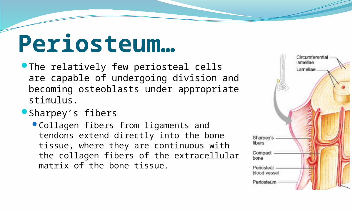
Periosteum…The relatively few periosteal cells are
capable of undergoing division and becoming osteoblasts under appropriate stimulus.
Sharpey’s fibersCollagen fibers from ligaments and tendons
extend directly into the bone tissue, where they are continuous with the collagen fibers of the extracellular matrix of the bone tissue.
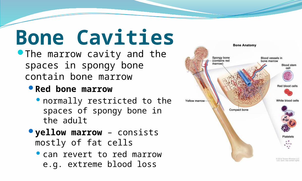
Bone CavitiesThe marrow cavity and the
spaces in spongy bone contain bone marrowRed bone marrow
normally restricted to the spaces of spongy bone in the adult
yellow marrow – consists mostly of fat cells can revert to red marrow
e.g. extreme blood loss
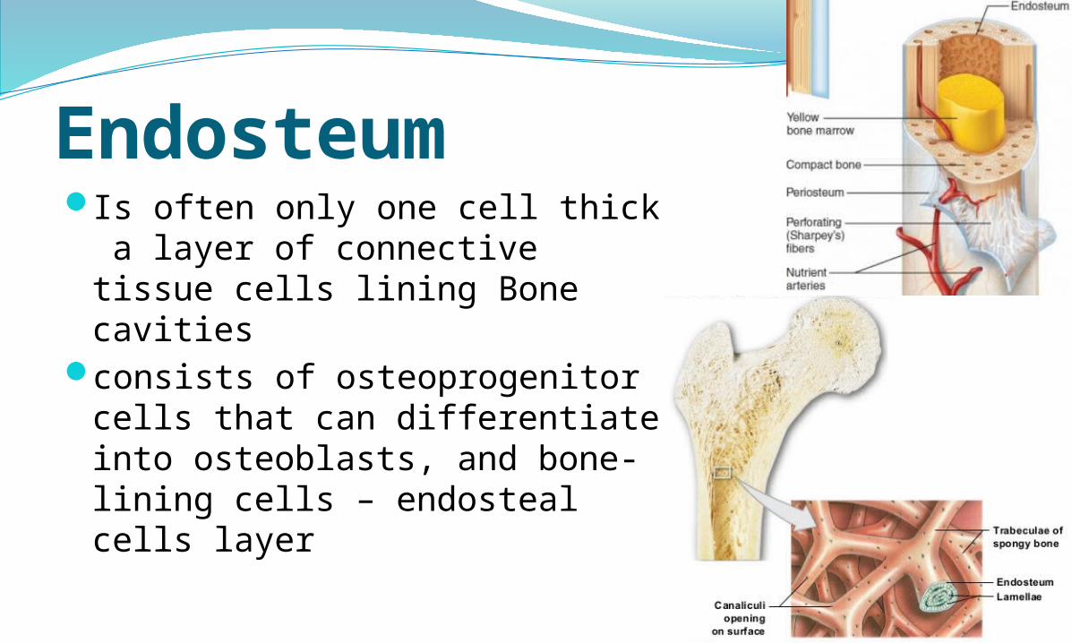
Endosteum Is often only one cell thick a
layer of connective tissue cells lining Bone cavities
consists of osteoprogenitor cells that can differentiate into osteoblasts, and bone-lining cells – endosteal cells layer
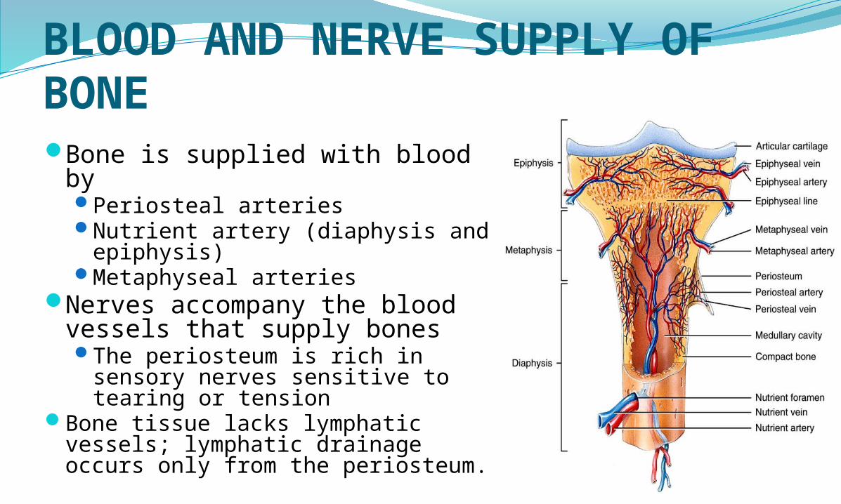
BLOOD AND NERVE SUPPLY OF BONEBone is supplied with blood by
Periosteal arteriesNutrient artery (diaphysis and
epiphysis)Metaphyseal arteries
Nerves accompany the blood vessels that supply bonesThe periosteum is rich in
sensory nerves sensitive to tearing or tension
Bone tissue lacks lymphatic vessels; lymphatic drainage occurs only from the periosteum.
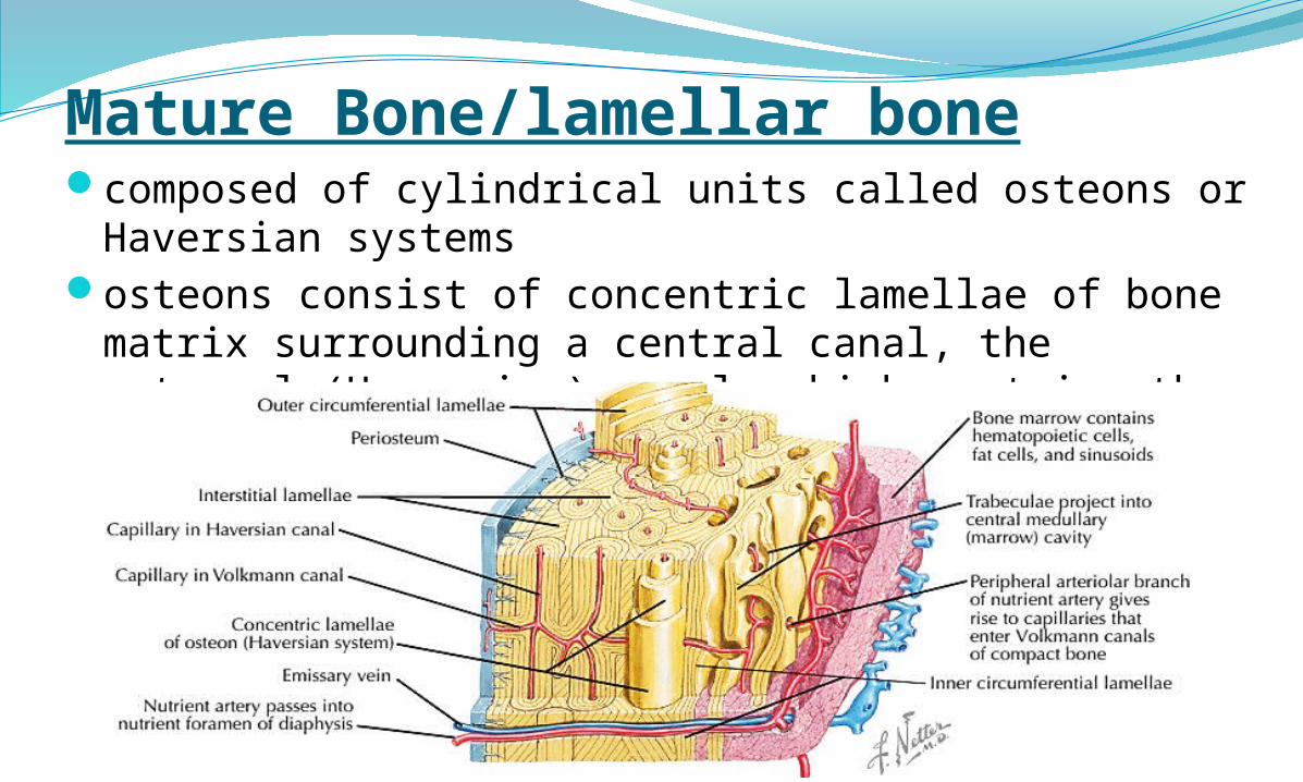
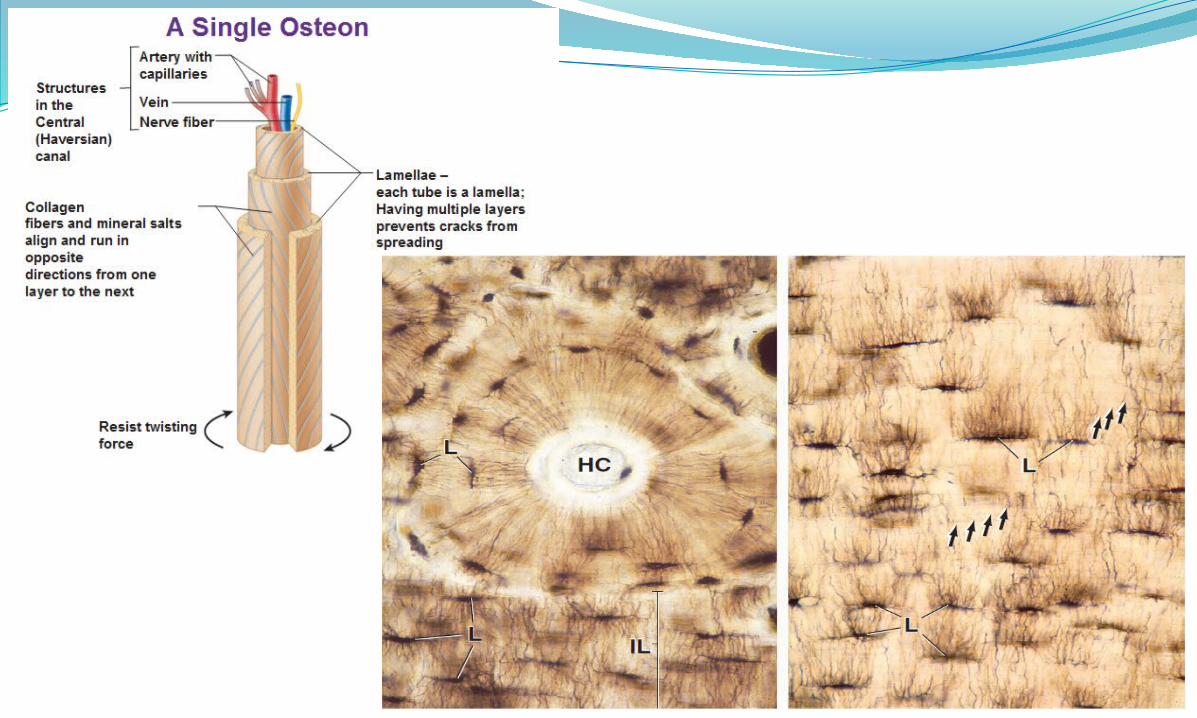
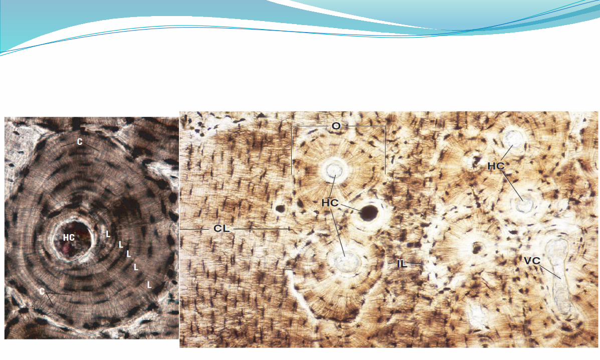
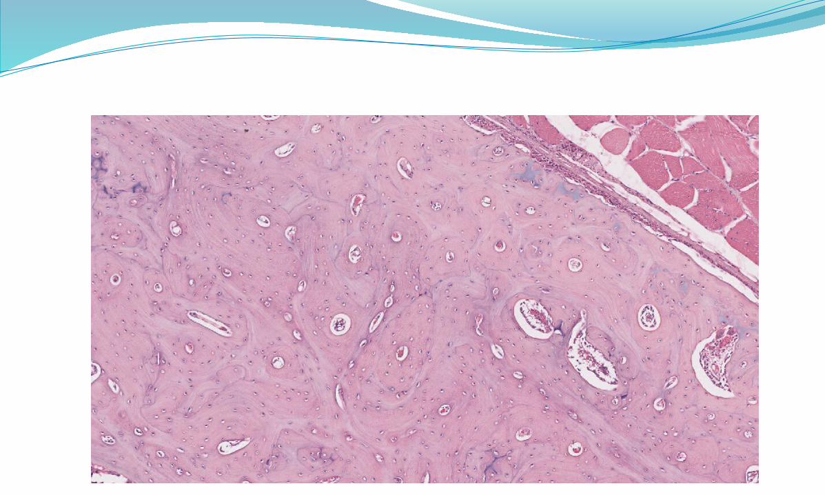
Mature Bone/lamellar bonecomposed of cylindrical units called osteons or
Haversian systemsosteons consist of concentric lamellae of bone matrix
surrounding a central canal, the osteonal (Haversian) canal, which contains the vascular and nerve supply of the osteon.
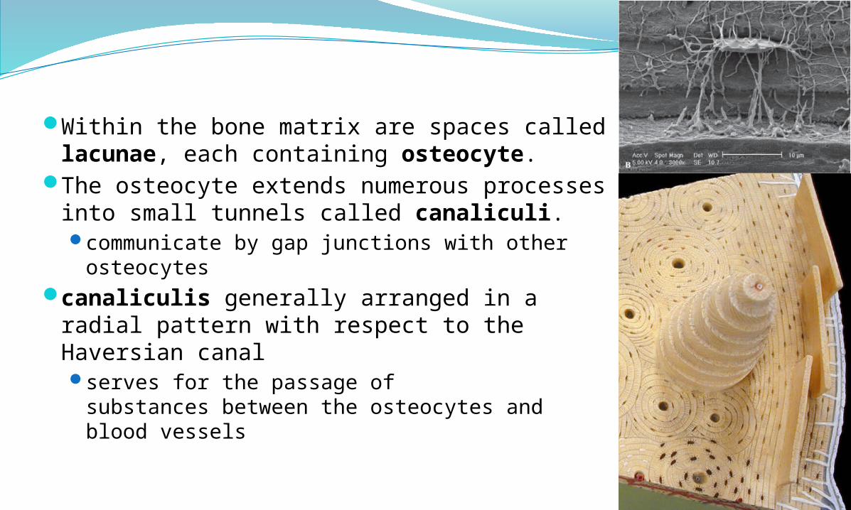
Within the bone matrix are spaces called lacunae, each containing osteocyte.
The osteocyte extends numerous processes into small tunnels called canaliculi.communicate by gap junctions with other
osteocytes canaliculis generally arranged in a radial
pattern with respect to the Haversian canalserves for the passage of
substances between the osteocytes and blood vessels
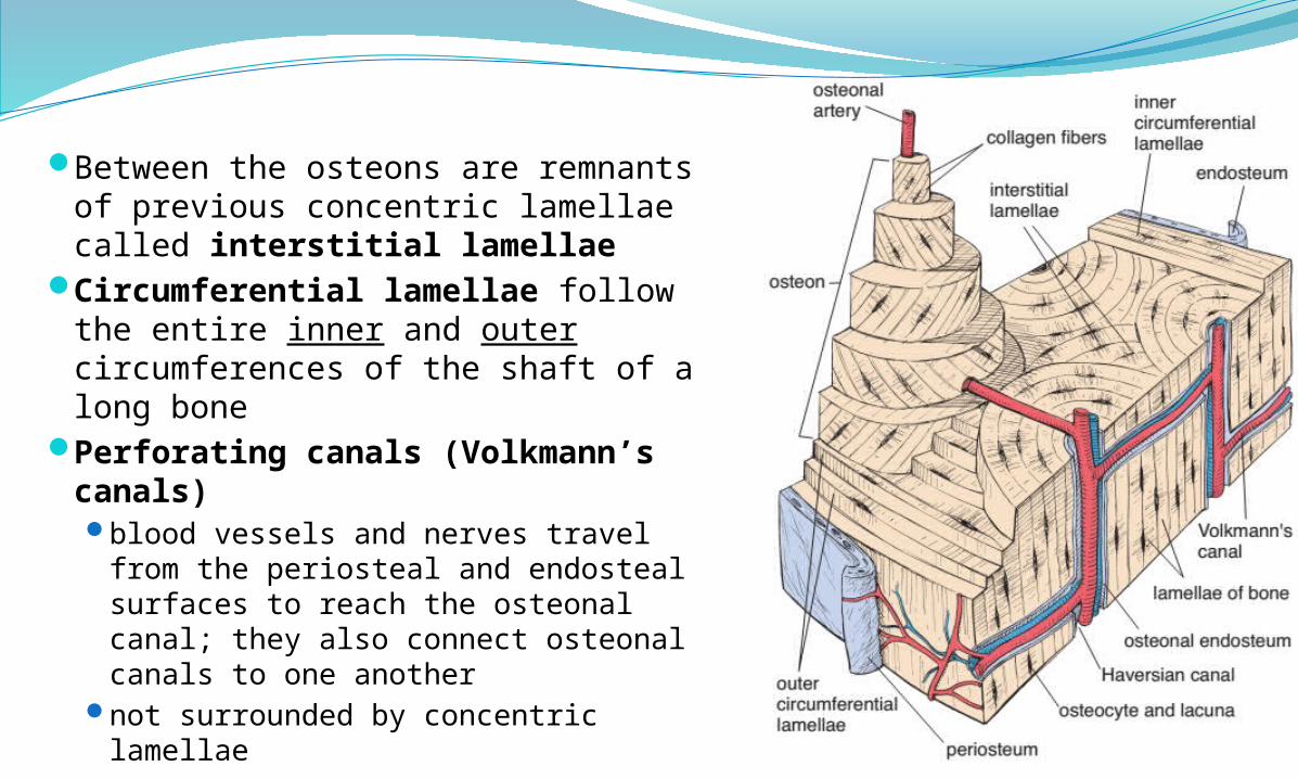
Between the osteons are remnants of previous concentric lamellae called interstitial lamellae
Circumferential lamellae follow the entire inner and outer circumferences of the shaft of a long bone
Perforating canals (Volkmann’s canals) blood vessels and nerves travel from
the periosteal and endosteal surfaces to reach the osteonal canal; they also connect osteonal canals to one another
not surrounded by concentric lamellae
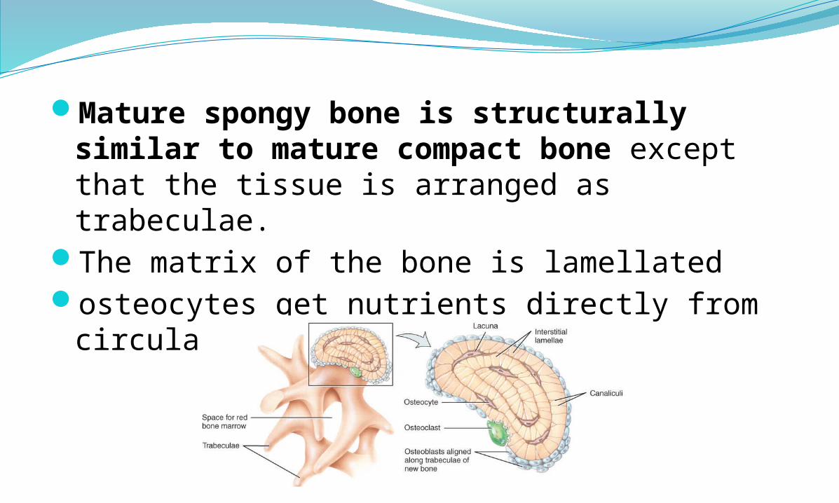
Mature spongy bone is structurally similar to mature compact bone except that the tissue is arranged as trabeculae.
The matrix of the bone is lamellatedosteocytes get nutrients directly from
circulating blood.
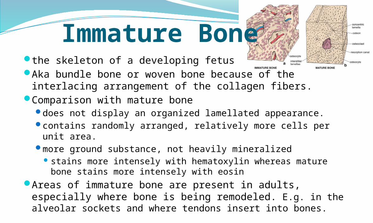
Immature Bonethe skeleton of a developing fetus Aka bundle bone or woven bone because of the interlacing
arrangement of the collagen fibers.Comparison with mature bone
does not display an organized lamellated appearance. contains randomly arranged, relatively more cells per unit
area.more ground substance, not heavily mineralized
stains more intensely with hematoxylin whereas mature bone stains more intensely with eosin
Areas of immature bone are present in adults, especially where bone is being remodeled. E.g. in the alveolar sockets and where tendons insert into bones.
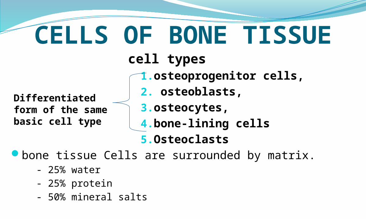
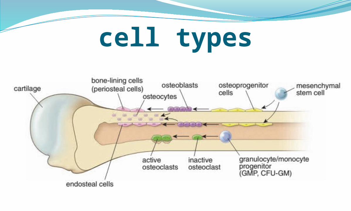
CELLS OF BONE TISSUEcell types
1.osteoprogenitor cells,2. osteoblasts, 3.osteocytes, 4.bone-lining cells5.Osteoclasts
bone tissue Cells are surrounded by matrix.- 25% water- 25% protein- 50% mineral salts
Differentiated form of the same basic cell type
cell types
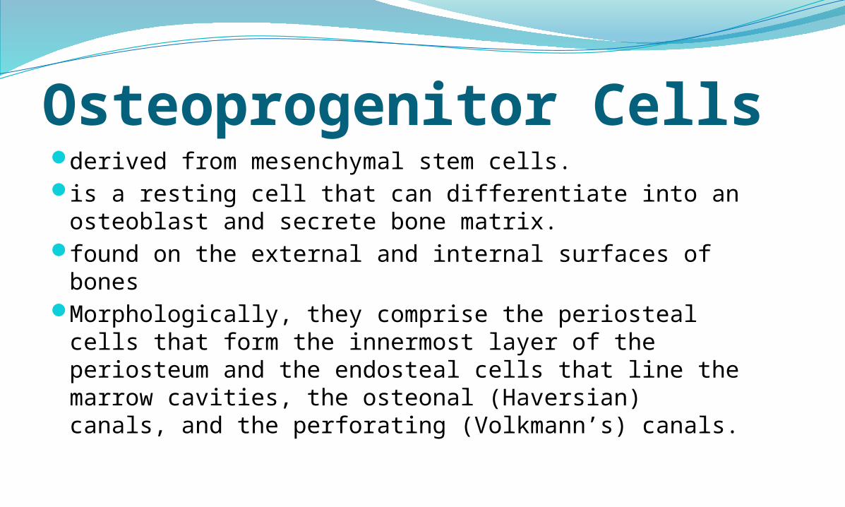
Osteoprogenitor Cellsderived from mesenchymal stem cells.is a resting cell that can differentiate into an
osteoblast and secrete bone matrix.found on the external and internal surfaces of
bonesMorphologically, they comprise the periosteal
cells that form the innermost layer of the periosteum and the endosteal cells that line the marrow cavities, the osteonal (Haversian) canals, and the perforating (Volkmann’s) canals.
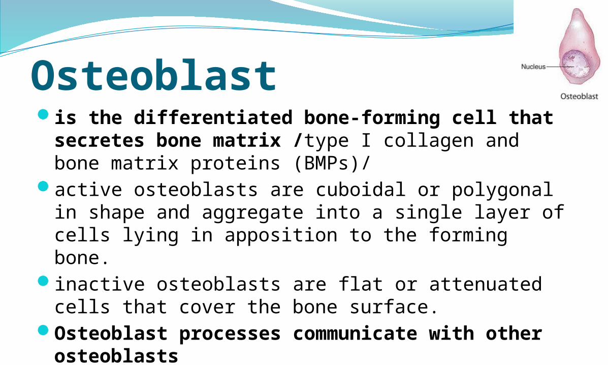
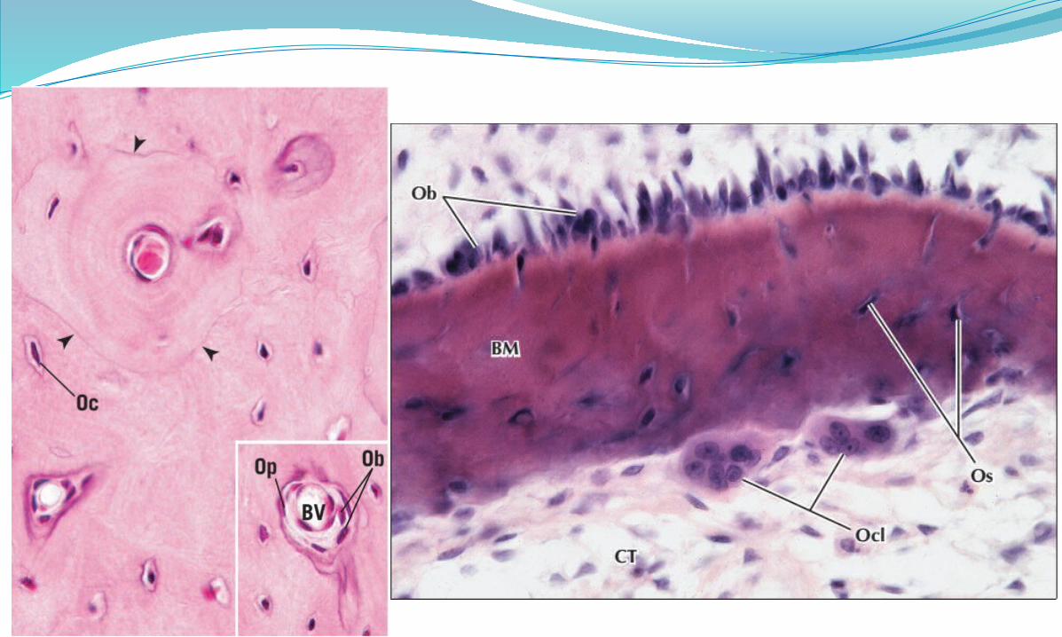
Osteoblastis the differentiated bone-forming cell that
secretes bone matrix /type I collagen and bone matrix proteins (BMPs)/
active osteoblasts are cuboidal or polygonal in shape and aggregate into a single layer of cells lying in apposition to the forming bone.
inactive osteoblasts are flat or attenuated cells that cover the bone surface.
Osteoblast processes communicate with other osteoblastsand with osteocytes by gap junctions.
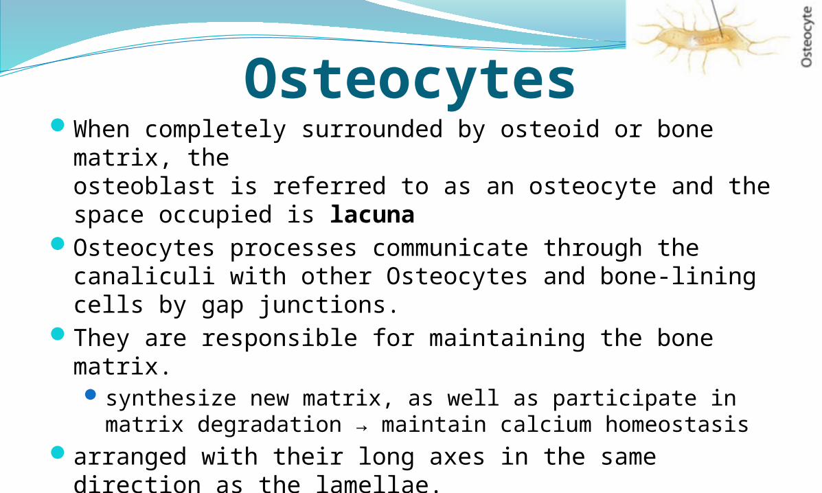
OsteocytesWhen completely surrounded by osteoid or bone matrix,
theosteoblast is referred to as an osteocyte and the space occupied is lacuna
Osteocytes processes communicate through the canaliculi with other Osteocytes and bone-lining cells by gap junctions.
They are responsible for maintaining the bone matrix.synthesize new matrix, as well as participate in matrix
degradation → maintain calcium homeostasisarranged with their long axes in the same direction as the
lamellae.a reduced load on bone initiates expression of matrix
metalloproteinases (MMP)

Bone-Lining CellsBone-lining cells are derived from osteoblasts
and cover bone that is not remodeling.layer of flat cells with attenuated cytoplasm
on external bone surfaces = periosteal cellson internal bone surfaces = endosteal cells
Cell processes contact one another and with osteocytic processes → Gap junctions
Function maintenance and nutritional support of the osteocytes
and regulate the movement of calcium and phosphate into and out of the bone.

Osteoclastsare phagocytotic cells derived from fusion of
hemopoietic progenitor cells of neutrophilic granulocyte and monocyte lineages. → multinucleated cells
are bone-resorbing cells present on bone surfaces where bone is being removed or remodeled a shallow bay called a resorption bay (Howship’s
lacuna) can be observed in the bone directly under the osteoclast.
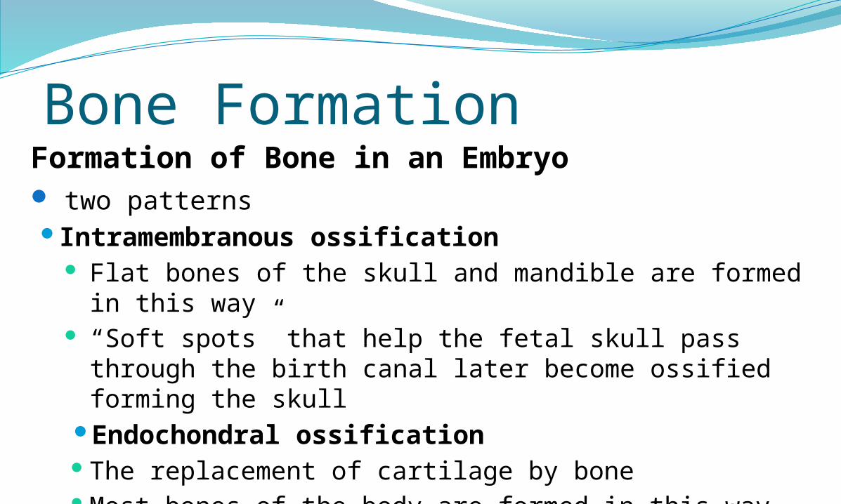
Bone FormationFormation of Bone in an Embryo two patternsIntramembranous ossification
Flat bones of the skull and mandible are formed in this way
“Soft spots” that help the fetal skull pass through the birth canal later become ossified forming the skull
Endochondral ossificationThe replacement of cartilage by boneMost bones of the body are formed in this way including
long bones
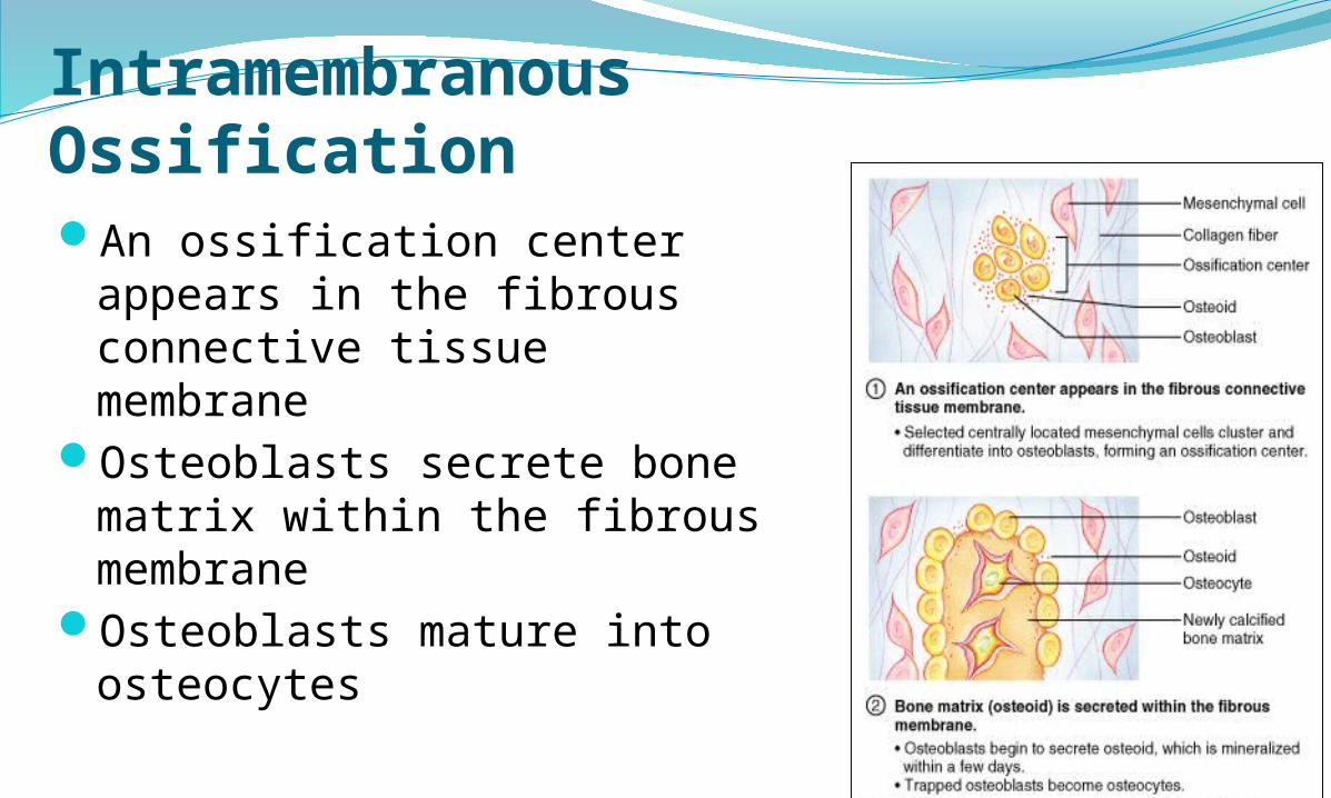
Intramembranous OssificationAn ossification center appears
in the fibrous connective tissue membrane
Osteoblasts secrete bone matrix within the fibrous membrane
Osteoblasts mature into osteocytes

Enlargingchondrocytes within
calcifying matrix
Chondrocytes at the center of the growing cartilage model enlarge and then die as the matrix calicifies.
Newly derived osteoblasts cover the shaft of the cartilage in a thin layer of bone.
Blood vessels penetrate the cartilage. New osteoblasts form a primary ossification center.
The bone of the shaft thickens, and the cartilage near each epiphysis is replaced by shafts of bone.
Blood vessels invade the epiphyses and osteo-blasts form secondary centers of ossification.
Cartilagemodel
Boneformation
Epiphysis
Diaphysis Marrowcavity
Primaryossificationcenter
Bloodvessel
Marrowcavity
Bloodvessel
Secondaryossificationcenter
Epiphysealcartilage
Articularcartilage
Replacement of hyaline cartilage with bone Most bones are formed this way (i.e. long bones).
Endochondral Ossification
Reference Histology A text and Atlas with correlated cell
and molecular Biology, Micheal H. Ross, Lippincott Williams & Wilkins, 2011 G.C.; 6th Ed
Color Atlas and Text book of Histology, Leslie P. Gartner, 2014 G.C.; 6th Ed

Thank You