Embed Size (px)
Citation preview

BioMed CentralBMC Neuroscience
ss
Open AcceResearch articleTyrosine phosphatases such as SHP-2 act in a balance with Src-family kinases in stabilization of postsynaptic clusters of acetylcholine receptorsAlain A Camilleri1, Raffaella Willmann1, Gayathri Sadasivam1, Shuo Lin2, Markus A Rüegg2, Matthias Gesemann1 and Christian Fuhrer*1Address: 1Brain Research Institute, University of Zurich, Winterthurerstrasse 190, CH-8057 Zurich, Switzerland and 2Biozentrum, University of Basel, Klingelbergstrasse 70, CH-4056 Basel, Switzerland
Email: Alain A Camilleri - [email protected]; Raffaella Willmann - [email protected]; Gayathri Sadasivam - [email protected]; Shuo Lin - [email protected]; Markus A Rüegg - [email protected]; Matthias Gesemann - [email protected]; Christian Fuhrer* - [email protected]
* Corresponding author
AbstractBackground: Development of neural networks requires that synapses are formed, eliminated andstabilized. At the neuromuscular junction (NMJ), agrin/MuSK signaling, by triggering downstream pathways,causes clustering and phosphorylation of postsynaptic acetylcholine receptors (AChRs). Postnatally, AChRaggregates are stabilized by molecular pathways that are poorly characterized. Gain or loss of function ofSrc-family kinases (SFKs) disassembles AChR clusters at adult NMJs in vivo, whereas AChR aggregatesdisperse rapidly upon withdrawal of agrin from cultured src-/-;fyn-/- myotubes. This suggests that a balancebetween protein tyrosine phosphatases (PTPs) and protein tyrosine kinases (PTKs) such as those of theSrc-family may be essential in stabilizing clusters of AChRs.
Results: We have analyzed the role of PTPs in maintenance of AChR aggregates, by adding and thenwithdrawing agrin from cultured myotubes in the presence of PTP or PTK inhibitors and quantitatingremaining AChR clusters. In wild-type myotubes, blocking PTPs with pervanadate caused enhanceddisassembly of AChR clusters after agrin withdrawal. When added at the time of agrin withdrawal, SFKinhibitors destabilized AChR aggregates but concomitant addition of pervanadate rescued cluster stability.Likewise in src-/-;fyn-/- myotubes, in which agrin-induced AChR clusters form normally but rapidlydisintegrate after agrin withdrawal, pervanadate addition stabilized AChR clusters. The PTP SHP-2, knownto be enriched at the NMJ, associated and colocalized with MuSK, and agrin increased this interaction.Specific SHP-2 knockdown by RNA interference reduced the stability of AChR clusters in wild-typemyotubes. Similarly, knockdown of SHP-2 in adult mouse soleus muscle by electroporation of RNAinterference constructs caused disassembly of pretzel-shaped AChR-rich areas in vivo. Finally, we foundthat src-/-;fyn-/- myotubes contained elevated levels of SHP-2 protein.
Conclusion: Our data are the first to show that the fine balance between PTPs and SFKs is a key aspectin stabilization of postsynaptic AChR clusters. One phosphatase that acts in this equilibrium is SHP-2. Thus,PTPs such as SHP-2 stabilize AChR clusters under normal circumstances, but when these PTPs are notbalanced by SFKs, they render clusters unstable.
Published: 2 July 2007
BMC Neuroscience 2007, 8:46 doi:10.1186/1471-2202-8-46
Received: 2 January 2007Accepted: 2 July 2007
This article is available from: http://www.biomedcentral.com/1471-2202/8/46
© 2007 Camilleri et al; licensee BioMed Central Ltd. This is an Open Access article distributed under the terms of the Creative Commons Attribution License (http://creativecommons.org/licenses/by/2.0), which permits unrestricted use, distribution, and reproduction in any medium, provided the original work is properly cited.
Page 1 of 16(page number not for citation purposes)

BMC Neuroscience 2007, 8:46 http://www.biomedcentral.com/1471-2202/8/46
BackgroundNeural networks are shaped through the formation, stabi-lization and elimination of synapses that connect neuronswith their targets. The neuromuscular junction (NMJ), amodel synapse in the peripheral nervous system, forms bythe contact of motor neurons and muscle fibers. Theseinteractions lead to a polyinnervated synapse at birth, inwhich acetylcholine receptors (AChRs) are clustered in aflat, plaque-like postsynaptic membrane. In a postnatalphase of elimination, NMJs mature and AChRs becomestabilized at the crests of postjunctional folds to form pret-zel-shaped areas, while all but one axon withdraw in aprocess in which adjacent AChRs are destabilized [1-3].Mechanisms of stabilization of AChR clusters are thusimportant for proper postnatal maturation of the NMJ,which ultimately will allow for correct nerve-evoked mus-cle contractibility.
The molecular processes that first form NMJs are wellknown. Neural agrin, by activating the kinase MuSK, playsa crucial role by triggering downstream signaling path-ways that cause clustering and tyrosine phosphorylationof AChRs, as reviewed recently [4,5]. Neural activity dis-solves receptor clusters that are not protected by localagrin deposition in the basal lamina, thereby shaping thepostsynaptic architecture [6]. In cultured myotubes, ashort pulse of agrin leads to long-lasting MuSK phospho-rylation and normal AChR clustering much later, imply-ing that, once activated, a balance of downstream proteintyrosine kinases (PTKs) and protein tyrosine phos-phatases (PTPs) keeps postsynaptic clustering mecha-nisms activated [7].
Much less is known about the molecular pathways thatmature NMJs postnatally and stabilize adult pretzel-shaped AChR clusters. Although MuSK is involved [8],these pathways differ from those in NMJ induction [9,10].Stability of AChR clusters can be modeled in culturedmyotubes, by adding and then removing agrin, and stud-ying cluster dispersal in the withdrawal phase. Despite thedifference in time scale, this method reveals many paral-lels to postnatal stabilization of the NMJ in vivo, which canbe assessed by electroporating interfering constructs intomouse soleus muscle [8,11]. Thus, the protein complexassociated with utrophin including the components dys-troglycan and dystrobrevin, and Src-family kinases (SFKs),stabilize the postsynapse and AChR clusters both in vivoand in cultured myotubes in vitro [11-16]. SFKs are acti-vated by agrin [17], interact with AChRs [18,19], andmaintain AChR β subunit phosphorylation and interac-tion of the receptor with its anchoring protein rapsyn[11]. In cultured src-/-;fyn-/- myotubes, agrin or laminininduce normal AChR clustering, but the clusters dissem-ble rapidly within a few hours after withdrawal of thesefactors [12,14]. SFKs act in a dual mechanism with choles-
terol-rich lipid microdomains related to lipid rafts, asSFKs promote normal microdomain assembly, while themicrodomains allow SFKs to act upon postsynaptic pro-teins [20]. In vivo, interfering with SFK function causespostsynaptic disintegration of adult NMJs. Interestingly,decreasing or increasing SFK activity through expressionof dominant-negative (kinase-dead) or constitutivelyactive Src both cause disassembly of AChRs aggregates invivo [11]. This raises the possibility that balanced SFKactivity is important for postsynaptic stability and suggeststhat PTPs may be involved in stabilization of AChR clus-ters as well. One PTP known to be enriched at the NMJ isthe non-receptor phosphatase SHP-2 [21]. SHP-2 canmodulate agrin/MuSK signaling and neuregulin-regulatedAChR transcription [21,22], making it a good candidatefor postsynaptic signaling processes.
We have therefore analyzed the role of PTPs, especiallySHP-2, in the stabilization of AChR clusters, and theirinterplay with SFKs. We find that phosphatases such asSHP-2 stabilize AChR clusters after withdrawal of agrinfrom cultured myotubes. SHP-2 is also necessary to stabi-lize adult AChR pretzels at NMJs in mouse soleus musclein vivo. Interestingly, in src-/-;fyn-/- myotubes SHP-2 is over-expressed, and blocking PTP activity restores AChR clusterstability completely, showing that a fine balance of PTPswith SFKs is crucial for maintaining AChR clusters.
ResultsPTP inhibition by pervanadate reduces the stability of agrin-induced AChR clustersTo address the role of the activity of PTPs in the stabiliza-tion of agrin-induced AChR clusters, we made use of thepotent PTP inhibitor sodium pervanadate [23,24] andcultured C2C12 myotubes, in which AChRs were visual-ized by treatment with fluorescent α-bungarotoxin (α-BTX). PTP inhibition did not lead to any change in thenumber of spontaneous AChR clusters observed per fieldwhen compared to the untreated control (data notshown). The stability of AChR clusters can be modeled incell culture by incubating C2C12 myotubes with agrin toinduce maximal clustering, followed by agrin withdrawal,washing the cells, incubating them in agrin-free mediumfor a number of hours, and counting clusters at the end ofthis withdrawal period [7,11,12,14]. When pervanadatewas added at the time of agrin withdrawal, significantlyless AChR clusters were observed after a 16 h withdrawalperiod than in controls lacking pervanadate (Figure 1).These data show that block of PTP activity enhances thedisintegration of pre-existing agrin-induced AChR clustersand that therefore, PTPs are required to stabilize clustersof AChRs. This is a specific process operating in cells thatwere previously exposed to agrin, since block of PTP activ-ity as such does not alter the level of spontaneous AChRclustering.
Page 2 of 16(page number not for citation purposes)

BMC Neuroscience 2007, 8:46 http://www.biomedcentral.com/1471-2202/8/46
A balance of PTP and SFK activities stabilizes AChR clusters: the instability of AChR clusters in the absence of SFKs is rescued by pervanadate treatmentSFKs are key in stabilizing AChR clusters in vivo and in cul-tured myotubes in vitro [11,12,14]. In src-/-;fyn-/- myotubes,agrin induces normal AChR clustering, but the clustersdisassemble rapidly within few hours upon agrin with-drawal [14]. We analyzed whether under these circum-stances PTPs may also influence the stability of AChRclusters. For this purpose we treated src-/-;fyn-/- myotubeswith agrin to induce maximal clustering, then withdrewagrin and added pervanadate at the same time, for a with-drawal period of 5 h. This pervanadate treatment pre-
vented AChR clusters from disappearing, such thatclusters remained completely stable during the with-drawal period (Figure 2A, B). In sharp contrast, omittingpervanadate led to disassembly of the agrin-induced clus-ters down to spontaneous levels (Figure 2A, B). Thus,block of PTP activity causes stabilization of AChR clustersin the absence of Src and Fyn. This is a specific process thatonly affects pre-existing agrin-induced clusters, becausepervanadate treatment alone did not alter the levels ofspontaneous clusters (data not shown). Moreover, whenadded together with agrin, pervanadate did not affect theextent of agrin-induced AChR clustering (data notshown).
Src and Fyn are not the only members of this family ofkinases present in myotubes. A third member of the SFKs,Yes, is present in muscle and upregulated in src-/-;fyn-/-
myotubes [14]. This led us to investigate whether theobserved destabilizing effect of PTPs was solely due to theabsence of Src and Fyn, or whether this effect would alsooccur when all members of the SFKs in muscle wereblocked. We therefore made use of the specific inhibitorPP2 to block SFKs in C2C12 myotubes [25]. We againinduced AChR clustering by agrin and then removed agrinto analyze the effect on the stability of these AChR clus-ters. We found that when added together with agrin, PP2had no effect on the level of clustering induced by agrin(data not shown), as has been previously observed [14].However, upon removal of agrin, blocking SFKs with PP2led to a two-fold decrease in the number of remainingAChR clusters, almost down to spontaneous levels (Figure2C, D). This instability was completely prevented whenPTPs were inhibited with pervanadate in the withdrawalphase (Figure 2C, D). These data confirm those from src-/-
;fyn-/- myotubes and show, firstly, that SFK activity main-tains the stability of AChR clusters in cultured wild-typemyotubes, similar to recent findings in muscle in vivo [11].Secondly, PTPs destabilize AChR clusters in the absence ofSFK activity and could indeed be the key destabilizing fac-tor for agrin-induced AChR clusters in the absence ofSFKs. Taken together, these data show that the stability ofagrin-induced AChR clusters following the withdrawal ofagrin requires a fine balance between the kinase activitiesof SFKs and the phosphatase activities of PTPs.
Protein tyrosine phosphatase SHP-2 increasingly associates with MuSK upon agrin stimulationWe wanted to identify possible PTPs that play a role in sta-bilization of AChR clusters. An example of how the activ-ity of a PTP is required in positively controlling the actionsof a kinase is the regulation of Src activity by the SH2domain-containing protein tyrosine phosphatase SHP-2[26]. SHP-2 is enriched at neuromuscular synapses andcolocalizes with AChRs in vivo [21]. In cultured myotubes,SHP-2 is a major PTP and can control the phosphoryla-
PTP inhibition by pervanadate reduces the stability of agrin-induced AChR clusters in C2C12 myotubesFigure 1PTP inhibition by pervanadate reduces the stability of agrin-induced AChR clusters in C2C12 myotubes. (A) Myotubes were treated with agrin (1 nM) for 6–8 h, after which PTPs were blocked with 20 μM pervanadate at the point of agrin withdrawal (Agrin + Withdr. + PV) for a with-drawal period of 16 h. In controls, cultures were left untreated (No Agrin), treated with agrin alone for 6–8 h (Agrin), or treated with agrin followed by withdrawal for 16 h (Agrin + Withdr.). Cells were also treated with agrin for 6–8 h + 16 h, and this produced the same amount of clusters as a 6–8 h agrin incubation (data not shown). AChRs were stained with rhodamine-α-BTX and analyzed by fluorescence microscopy. Scale bar, 40 μm. (B) The number of AChR clus-ters per field was calculated using ImageJ software, using fixed intensity thresholds (150–255) and minimum area of 100 pixels occupied by a cluster. The number of AChR clus-ters per field (400× magnification) is shown as the percent-age of clusters in agrin-treated cells (Agrin) (means ± SEM, N = 50 from four similar experiments). Phosphatase inhibition significantly decreases the number of pre-existing, agrin-induced AChR clusters following agrin removal (** p < 0.01; two-tailed paired t test).
Page 3 of 16(page number not for citation purposes)

BMC Neuroscience 2007, 8:46 http://www.biomedcentral.com/1471-2202/8/46
Page 4 of 16(page number not for citation purposes)
AChR cluster stability is rescued by PTP inhibition in src-/-;fyn-/- myotubes and PP2-treated C2C12 myotubesFigure 2AChR cluster stability is rescued by PTP inhibition in src-/-;fyn-/- myotubes and PP2-treated C2C12 myotubes. (A) PTPs were blocked with 20 μM pervanadate in src-/-;fyn-/- myotube cultures at the point of agrin withdrawal for a period of 5 h (Agrin + Withdr. + PV), following agrin treatment for 16 h. As controls, myotubes were either left untreated (No Agrin), treated with agrin for 16 h (Agrin) or treated with agrin followed by withdrawal for 5 h (Agrin + Withdr.). AChRs were stained with rhodamine-α-BTX and analyzed by fluorescence microscopy. Scale bar, 40 μm. White arrowheads indicate AChR clusters. (B) The number of AChR clusters per myotube is shown as the percentage of clusters in agrin-treated src-/-;fyn-/-
cells (Agrin) (means ± SEM; N = 15 from three similar experiments; **p < 0.01, ***p < 0.001 by two-tailed paired t test). Pervanadate restores cluster stability following agrin withdrawal in src-/-;fyn-/- myotubes. (C) The inhibitor PP2 was used to block SFK activity in C2C12 myotubes. Myotubes were treated with agrin for 6–8 h in the presence of 10 μM PP2 for the last 2 h (these 2 h were chosen to ensure that PP2 was effective at the point of agrin withdrawal). Agrin was removed and myotubes incubated with agrin-free medium in the presence of pervanadate and PP2 for 16 h (Agrin + Withdr. + PP2 + PV). In controls, cultures were treated with agrin followed by withdrawal for 16 h in the presence or absence of PP2 (PP2 was added 2 h before agrin removal) (Agrin + Withdr.; Agrin + Withdr. + PP2). AChRs were stained with rhodamine-α-BTX, analyzed by fluorescence microscopy. Scale bar, 40 μm. (D) The number of AChR clusters per field (400× magnification) was calculated, as described in Figure 1, as the percentage of agrin-treated C2C12 cells (Agrin) (not shown) (means ± SEM, N = 30 from three experiments). *** indicates significant difference to the other two bars (p < 0.001; two-tailed paired t test). Blocking SFKs with PP2 reduces the stability of pre-existing clusters in C2C12 myotubes, and pervanadate restores the stabil-ity to normal levels.

BMC Neuroscience 2007, 8:46 http://www.biomedcentral.com/1471-2202/8/46
tion level of MuSK and the extent of agrin-induced AChRclustering [22]. SHP-2 is therefore a likely candidate PTPfor playing roles in the stabilization process of AChR clus-ters.
We first determined whether SHP-2 is in a position toaffect cluster stabilization by testing its association withMuSK. We treated C2C12 myotubes with agrin to induceMuSK phosphorylation, following which we immunopre-cipitated MuSK and probed for both MuSK and SHP-2.We found that SHP-2 is co-immunoprecipitated togetherwith MuSK, also without agrin treatment, and that theassociation between the two proteins increases 2.3-foldfollowing MuSK phosphorylation by agrin (Figure 3A).
Since SHP-2 is known to colocalize with AChRs and hencepresumably also with MuSK at NMJs [21], we investigatedthis association between MuSK and SHP-2 further using aheterologous cell system. We transfected COS cells withmyc-tagged MuSK and Flag-tagged SHP-2 constructs, andvisualized MuSK and SHP-2 in the transfected cells byimmunocytochemistry. As expected, being a ubiqui-tously-expressed non-receptor PTP, SHP-2 was localizedthroughout the whole of the cell, but also along mem-brane ruffles on the plasma membrane (Figure 3B). Onthe other hand, MuSK showed more of a membrane local-ization, characteristic for a receptor tyrosine kinase (RTK).SHP-2 and MuSK colocalized within these membrane ruf-fles present along the plasma membrane (Figure 3B). Theyhowever did not cause the relocalization of one another,since singly-transfected COS cells also exhibited similardistribution patterns of the expressed proteins (data notshown).
From these results we can conclude that MuSK and SHP-2colocalize in ruffles along the plasma membrane. Theyalso associate with one another, and SHP-2 increases itsassociation to MuSK upon MuSK activation by agrin, per-haps through an interaction of the SH2 domains in SHP-2 with MuSK phosphorylation sites. The association withMuSK highlights that SHP-2 is in a position to affect AChRcluster stability.
Knockdown of SHP-2 in myotubes by vector-driven shRNA specifically reduces SHP-2 protein levelsTo address the role of SHP-2 in stabilization of AChR clus-ters, we used a knockdown approach by RNA interference(RNAi). We cloned three short-hairpin RNA (shRNA)loops into the pSUPER.gfp vector and tested their efficacyin downregulating SHP-2 expression in C2C12 myotubes.We used shRNA loops generated against different regionsof the murine SHP-2 open-reading frame (ORF), based onalready published and successfully used siRNA or shRNAsequences [27,28]. These constructs co-expressed EGFP,allowing easy identification of the successfully transfectedmyoblasts, and observation of the differentiation of thesemyoblasts into mature myotubes. Transfection efficien-cies at the myoblast level were 40–50%, which upon dif-ferentiation and fusion led to close to 100% of themyotubes expressing EGFP (see Figure 5A). By Westernblot analysis of C2C12 myotube lysates we probed for the
Protein tyrosine phosphatase SHP-2 increasingly associates with MuSK upon agrin stimulationFigure 3Protein tyrosine phosphatase SHP-2 increasingly associates with MuSK upon agrin stimulation. (A) C2C12 myotubes were treated with 0.5 nM agrin for 40 min. Following MuSK immunoprecipitation, immunoblotting for SHP-2 and MuSK was carried out, followed by reprobing for phosphorylation of MuSK. Levels of SHP-2 co-precipitated with MuSK were quantitated (normalized for the amount of precipitated MuSK) as percentage of No Agrin control. Co-immunoprecipitation of SHP-2 with MuSK indicates its spe-cific association with MuSK, which increases significantly upon agrin-induced MuSK phosphorylation (means ± SEM, from seven experiments; * p < 0.05; two-tailed unpaired t test). As a control, the MuSK antibody was omitted in the immunoprecipitation, leading to no substantial signal in the Western blots (not shown). (B)COS cells were co-trans-fected with SHP-2-Flag and MuSK-myc constructs. Cells were then fixed and stained with anti-Flag/Alexa488 and anti-myc/Alexa546 antibodies. SHP-2 localizes throughout the whole of the cytoplasm, while MuSK localization is concen-trated along the plasma membrane. White arrowheads in the overlay indicate a colocalization of MuSK and SHP-2 along membrane ruffles on the plasma membrane. Scale bar, 40 μm.
Page 5 of 16(page number not for citation purposes)
BMC Neuroscience 2007, 8:46 http://www.biomedcentral.com/1471-2202/8/46
Page 6 of 16(page number not for citation purposes)
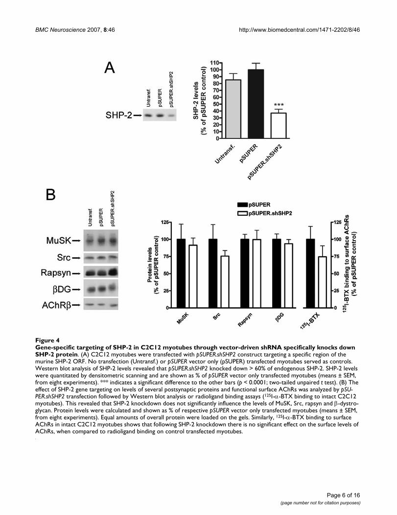
Gene-specific targeting of SHP-2 in C2C12 myotubes through vector-driven shRNA specifically knocks down SHP-2 proteinFigure 4Gene-specific targeting of SHP-2 in C2C12 myotubes through vector-driven shRNA specifically knocks down SHP-2 protein. (A) C2C12 myotubes were transfected with pSUPER.shSHP2 construct targeting a specific region of the murine SHP-2 ORF. No transfection (Untransf.) or pSUPER vector only (pSUPER) transfected myotubes served as controls. Western blot analysis of SHP-2 levels revealed that pSUPER.shSHP2 knocked down > 60% of endogenous SHP-2. SHP-2 levels were quantitated by densitometric scanning and are shown as % of pSUPER vector only transfected myotubes (means ± SEM, from eight experiments). *** indicates a significant difference to the other bars (p < 0.0001; two-tailed unpaired t test). (B) The effect of SHP-2 gene targeting on levels of several postsynaptic proteins and functional surface AChRs was analyzed by pSU-PER.shSHP2 transfection followed by Western blot analysis or radioligand binding assays (125I-α-BTX binding to intact C2C12 myotubes). This revealed that SHP-2 knockdown does not significantly influence the levels of MuSK, Src, rapsyn and β-dystro-glycan. Protein levels were calculated and shown as % of respective pSUPER vector only transfected myotubes (means ± SEM, from eight experiments). Equal amounts of overall protein were loaded on the gels. Similarly, 125I-α-BTX binding to surface AChRs in intact C2C12 myotubes shows that following SHP-2 knockdown there is no significant effect on the surface levels of AChRs, when compared to radioligand binding on control transfected myotubes.

BMC Neuroscience 2007, 8:46 http://www.biomedcentral.com/1471-2202/8/46
effective suppression of endogenous SHP-2 protein. Oneof the three constructs, termed pSUPER.shSHP2, led to astrong (> 60%) reduction of endogenous SHP-2 proteinlevels when compared to control transfected myotubes(pSUPER vector only; Figure 4A). We therefore proceededto analyze the effect of SHP-2 downregulation on otherpostsynaptic proteins. In immunoblots, levels of otherpostsynaptic proteins such as MuSK, Src, rapsyn and β-dystroglycan were not affected by SHP-2 knockdown (Fig-ure 4B). Using radioligand binding of 125I-α-BTX weprobed functional surface AChRs following transfectionwith pSUPER.shSHP2 and observed no significant effect(Figure 4B). pSUPER.shSHP2 thus allows specific andstrong knockdown of SHP-2 without interfering withother muscle proteins.
SHP-2 is required for stabilization of AChR clusters in cultured myotubesTo study the role of SHP-2 for AChR cluster stability, weused C2C12 myotubes and suppressed SHP-2 by shRNAas described above. We incubated the cells with agrin to
induce maximal clustering and withdrew the agrin toassess the stability of the clusters. In both control cells(pSUPER) and after SHP-2 knockdown (pSUPER.shSHP2),spontaneous AChR aggregates were present and agrininduced strong clustering of the receptors (Figure 5A),similar to a recent report [22]. After withdrawal of agrinfor 8 h, few clusters remained in pSUPER.shSHP2-trans-fected cells whereas more clusters were present in pSU-PER-treated cells (Figure 5A).
To quantitate the effect of SHP-2 knockdown on the sta-bility of agrin-induced AChR clusters, we normalized thenumber of AChR clusters following agrin withdrawal("Agrin + Withdr.") to the number of clusters that wereinduced by agrin ("Agrin") in pSUPER.shSHP2- and pSU-PER-transfected cells. The result shows that there is a sig-nificant decrease in the stability of AChR clustersfollowing agrin withdrawal when SHP-2 expression isknocked down by shRNA (Figure 5B). These data illustratethat SHP-2 is required for optimal stabilization of AChRclusters in cultured myotubes.
Upon knockdown of SHP-2 in C2C12 myotubes, AChR clusters are less stableFigure 5Upon knockdown of SHP-2 in C2C12 myotubes, AChR clusters are less stable. (A) pSUPER-, and pSUPER.shSHP2-transfected C2C12 myotubes were either left untreated (No Agrin), treated with agrin for 16 h (Agrin), or treated with agrin (16 h) followed by withdrawal of agrin for 8 h (Agrin+Withdr.). AChRs were identified by rhodamine-α-BTX staining and cells were analyzed by fluorescence microscopy. Scale bar, 40 μm. Transfected myotubes expressing EGFP indicated that transfec-tion efficiency at the myotube level approached 100%. The data show that agrin induces AChR clustering both in pSUPER- and pSUPER.shSHP2-transfected myotubes. After the withdrawal phase, less clusters remain in the case of SHP-2 knockdown when compared to the control. (B) The stability of clusters was quantitated by setting the number of AChR clusters in agrin-treated myotubes (Agrin) to 100%, both for pSUPER-, and pSUPER.shSHP2-transfected myotubes. Clusters after the withdrawal period (Agrin + Withdr.) were then calculated accordingly. The absence of SHP-2 renders AChR clusters significantly less stable when compared to control transfected myotubes (means ± SEM, N = 30 from four similar experiments; *p < 0.05; two-tailed unpaired t test).
Page 7 of 16(page number not for citation purposes)

BMC Neuroscience 2007, 8:46 http://www.biomedcentral.com/1471-2202/8/46
Page 8 of 16(page number not for citation purposes)
Electroporation of SHP-2 shRNA construct into soleus muscle of adult mice leads to disassembly of NMJsFigure 6Electroporation of SHP-2 shRNA construct into soleus muscle of adult mice leads to disassembly of NMJs. Adult mice soleus muscles were electroporated in vivo with a mixture of pSUPER.shSHP2 and NLS-GFP constructs (A, C), or pSUPER vector and NLS-GFP as control (B, D). After six weeks muscles were dissected, whole mounts of fibers prepared and stained with rhodamine-α-BTX and a mixture of neurofilament and synaptophysin antibodies followed by Cy5 secondary anti-body. NMJs were analyzed by confocal microscopy, whereby successfully electroporated muscle fibers were identified by the expression of NLS-GFP in myonuclei. (A) Expression of pSUPER.shSHP2 results in NMJ disassembly in GFP-positive myofibers (closed arrowhead). NMJs of GFP-negative fibers remain intact (open arrowheads). (B) Expression of control pSUPER vector and NLS-GFP has no effect on the NMJs. A three-dimensional reconstruction of a confocal image of a muscle electroporated with pSUPER.shSHP2 and NLS-GFP (C), or control pSUPER vector and NLS-GFP (D) shows details of the NMJs. (C) A 3D view from inside the muscle illustrates that expression of SHP-2 shRNA leads to disassembly of the NMJ, loss of the usual morphol-ogy and pretzel shape, and a resulting fragmented appearance of the NMJ, with the nerve becoming visible through the pretzel remnants. (D) In control electroporated muscle fibers, GFP-positive synaptic nuclei are located below the intact pretzel-shaped accumulations of AChRs and no nerve is visible through the pretzel. (E) The number of intact and disassembled NMJs is shown for control (pSUPER) vector and pSUPER.shSHP2 electroporated muscle fibers, as percentage of the total number of NMJs ana-lyzed (total of 25 endplates from 3 mice for pSUPER and 15 endplates from 5 mice for pSUPER.shSHP2). Only endplates with synapse-associated nuclei expressing NLS-GFP were analyzed. Scale bars, 30 μm in A, B; 10 μm in C, D.

BMC Neuroscience 2007, 8:46 http://www.biomedcentral.com/1471-2202/8/46
Requirement of SHP-2 for stabilization of postsynaptic AChR pretzels at the NMJ in vivoTo address the role of SHP-2 in stabilization of postsynap-tic AChRs at the NMJ in vivo, we used RNA interference insoleus muscles of adult mice as described earlier [8,11].The shRNA construct pSUPER.shSHP2, together with aconstruct expressing GFP containing a nuclear localiza-tion signal (NLS-GFP), was introduced into adult soleusmuscle by electroporation. After 6 weeks, muscles weredissected, stained as whole mount preparations withrhodamine-α-BTX and antibodies against neurofilamentand synaptophysin, and analyzed by confocal microscopyincluding 3D reconstruction.
The in vivo expression of pSUPER.shSHP2 led to the disas-sembly of adult pretzel-shaped AChR clusters (Figure 6A,closed arrowhead). This was in sharp contrast to the intactAChR pretzel structures observed at NMJs of neighboringGFP-negative myofibers (Figure 6A, open arrowheads)and of myofibers of mice electroporated with control pSU-PER vector and NLS-GFP (Figure 6B).
Disassembled NMJs of myofibers expressing SHP-2shRNA lost their typical pretzel morphology and werefragmented (example in Figure 6C) and sometimes alto-gether widened in appearance (example in Figure 6A). Inaddition, the synapse-associated myonuclei were some-times no longer tightly packed subsynaptically but associ-ated more loosely with the disassembled AChR pretzel(Figure 6C). In contrast, in myofibers electroporated withcontrol pSUPER vector, the nerve was hardly visible frominside the muscle and synaptic nuclei were clustered justbeneath the AChR pretzels (Figure 6D).
A quantitative assessment was made following the criteriadescribed earlier [11](see also Method section), by analyz-ing many NMJs from several mice, for both pSU-PER.shSHP2 and pSUPER control vector. In the control,almost all NMJs scored as intact, but in myofibers whereSHP-2 was suppressed, only a minority (less than 25%) ofall NMJs analyzed was intact and most synapses scored asdisassembled. These data show that SHP-2 is required formaintenance of the adult postsynaptic apparatus of theNMJ in vivo including typical pretzel-shaped AChRdomains and synaptic myonuclei. These results are verysimilar to the role of SHP-2 in stabilization of agrin-induced AChR clusters in myotubes in vitro.
Elevated levels of SHP-2 in the absence of Src and FynWe have observed similar destabilizing effects on AChRclusters in C2C12 myotubes upon PTP inhibition by per-vanadate, and upon knockdown of SHP-2 by shRNA (Fig-ures 1 and 5). This instability is also present in the absenceof Src and Fyn [14], or upon blocking SFKs with the inhib-itor PP2, and is rescued completely by inhibition of phos-phatase activity by pervanadate (Figure 2). Theseobservations lead to the conclusion that it is the balancebetween PTK and PTP activity that regulates the stability ofAChR clusters in myotubes. For this reason we sought tocompare the levels of SHP-2 between src-/-;fyn-/- myotubesand wild-type myotubes. We carried out Western blotanalysis of myotube lysates, and found that SHP-2 levelsare 3-fold higher in myotubes lacking Src and Fyn com-pared to wild-type myotubes (Figure 7). This finding sug-gests that these high levels of SHP-2 may cause clusterdestabilization and further strengthens our proposal thata crucial aspect for AChR cluster stabilization is the bal-ance between Src and Fyn, and PTPs such as SHP-2.
Elevated protein levels of SHP-2 in the absence of Src and FynFigure 7Elevated protein levels of SHP-2 in the absence of Src and Fyn. Western blot analysis of total SHP-2 protein levels in wild-type versus src-/-;fyn-/- myotubes reveals that in the absence of Src and Fyn there is a >3-fold increase in the protein levels of SHP-2. SHP-2 levels are shown for both wild-type and src-/-;fyn-/- myotubes; equal amounts of cellular protein were loaded on the gel. In the quantitation, SHP-2 levels are normalized for the level of AChR β subunit (*p < 0.05; two-tailed unpaired t test).
Page 9 of 16(page number not for citation purposes)

BMC Neuroscience 2007, 8:46 http://www.biomedcentral.com/1471-2202/8/46
DiscussionWe have analyzed the role of PTPs in the stabilization ofAChR clusters and report that PTPs are required for clustermaintenance and that the fine balance between PTPs andSFKs is a key aspect in this process. On one hand, block ofPTPs by pervanadate or knockdown of SHP-2 in wild-typemyotubes both rendered agrin-induced clusters less sta-ble. In vivo, SHP-2 knockdown in muscle caused disassem-bly of adult pretzel-shaped AChR-rich areas at NMJs. Onthe other hand, pervanadate restored the defective stabil-ity of clusters in myotubes lacking SFK activity, and src-/-
;fyn-/- myotubes had elevated levels of SHP-2. Thus, PTPssuch as SHP-2 stabilize AChR clusters under normal cir-cumstances, but when these PTPs are not balanced bySFKs, they render clusters unstable.
PTPs such as SHP-2 stabilize AChR clustersWe used two approaches to illustrate the role of PTPs inthe maintenance of AChR clusters: inhibiting the activityof all PTPs by pervanadate, and testing the role of a candi-date PTP, SHP-2. The stability of AChR clusters was mod-eled in cell culture, by adding and then removing agrin,and pervanadate was added at the point of agrin with-drawal to specifically test its effect on the maintenance ofpre-existing agrin-induced clusters. The withdrawal andwashing procedure removes the vast majority of agrinfrom cells, such that signaling processes in the myotubesduring the withdrawal phase do not reflect the activity ofextracellular agrin but ongoing processes within the myo-tubes [7]. When agrin is withdrawn from wild-type myo-tubes, AChR clusters slowly disintegrate over the course ofseveral hours [29]. Block of PTPs by pervanadate acceler-ated the decay of clusters, showing that PTP activity is arequirement for optimal AChR cluster stability. This was aspecific process, since pervanadate as such did not alterlevels of spontaneous AChR clustering. Rather, followingan agrin stimulation to produce maximal aggregates, PTPsact to fully maintain these clusters.
PTPs exist in many families, as receptor tyrosine phos-phatases or cytosolic non-receptor phosphatases, similarto kinase families. While several PTPs could share thefunction of maintaining AChR clusters, we identifiedSHP-2 as one PTP that plays such a role. SHP-2 has previ-ously been put into the context of the NMJ as a regulatorof AChR synthesis and MuSK activity [21], and we first fur-ther verified whether SHP-2 is located to play a postsynap-tic role. We found SHP-2 to associate with MuSK, and thisassociation to be increased (2.3-fold) by an agrin treat-ment that caused heavy MuSK tyrosine phosphorylation.In COS cells, MuSK and SHP-2 showed colocalization inmembrane ruffles at the plasma membrane, implying thatthey can interact with each other independently of mus-cle-specific linker proteins. These data show that SHP-2,
by increasingly interacting with MuSK, is positioned tocontrol postsynaptic stability of AChR clusters.
We determined the role of SHP-2 in the stabilization ofAChR clusters by an RNAi strategy, using vector-drivenshRNA and short-hairpin loops that were previously usedto knock down SHP-2 [27,28]. One of the shRNA con-structs tested (pSUPER.shSHP2) was effective in myotubeswhen using an efficient transfection protocol that allowedits expression in nearly 100% of myotubes. pSU-PER.shSHP2 massively reduced protein amounts of SHP-2without significantly interfering with other postsynapticproteins such as MuSK, Src, rapsyn and β-dystroglycan orwith levels of functional AChR at the cell surface. This spe-cificity in knockdown allowed the study of the role ofSHP-2 in AChR cluster stability.
In the absence of normal SHP-2 levels, spontaneous AChRaggregates were present and AChR clusters were inducedby agrin, but the stability of these clusters was reducedcompared to controls, showing that, similar to the resultsdescribed above with pervanadate, SHP-2 stabilizes AChRclusters. SHP-2 was also important at NMJs in vivo,because electroporation of pSUPER.shSHP2 into adultmouse soleus muscle led to disassembly of the postsyn-apse: pretzel-shaped AChR-rich areas became fragmentedand some lost their overall pretzel outline, and sometimesmyonuclei were no longer packed subsynaptically butmore loosely associated with fragmented AChR clusterremnants. Taken together, our data show that SHP-2 is aPTP that, perhaps together with other PTPs, contributes tothe maintenance of AChR clusters at NMJs in vivo and incultured myotubes in vitro.
Upon knockdown of SHP-2, fragments of AChR clusterswere visible at NMJs but not in cultured myotubes fromwhich agrin had been withdrawn. In myotubes, clusterdisappearance appears as a statistical process wherebysome clusters still exist while others have vanished and areoutside the detectable ranges used in our method. Thiscould point towards differences in cluster dynamicsbetween our in vitro and in vivo systems. Such differencescould result from one or more of several possibilities: atNMJs, the turnover of AChRs is much slower and the sub-synaptic cytoskeleton more elaborate, presumably immo-bilizing the AChR in a stronger fashion; culturedmyotubes lack innervation and the continuous presenceof basal lamina-anchored agrin as seen in vivo; and thetiming of AChR cluster disassembly is different as indi-cated by the different time scale of our in vivo vs. in vitroanalysis. However, equally likely is the possibility thatupon SHP-2 knockdown and agrin withdrawal, AChRcluster fragments do exist in cultured myotubes but can-not be visualized due to low intensity; in comparison thedensity of AChRs in pretzels is much higher in vivo. Fur-
Page 10 of 16(page number not for citation purposes)

BMC Neuroscience 2007, 8:46 http://www.biomedcentral.com/1471-2202/8/46
thermore, fragments of AChR pretzels may completelydisappear from pSUPER.shSHP2-expressing myofibersafter prolongated times.
There are several possibilities of how SHP-2 could act tomaintain clusters. The agrin-induced increase in MuSK-SHP-2 interaction is very similar to SHP-2 associationwith tyrosine phosphorylated DOK1, a member of insulinreceptor substrate protein family that binds β3. Associa-tion with DOK1 also occurs under basal conditions, andupon stimulation of DOK1 by insulin-like growth factor I(IGI-I), SHP-2-DOK1 association increases 2.7-fold. Thisassociation is important for DOK1 to present SHP-2 todownstream SHPS-1 [30,31]. In a similar fashion, SHP-2associates, via its SH2 domains, to other RTKs includingthe platelet-derived growth factor (PDGF) and epidermalgrowth factor (EGF) receptors [32]. In SHP-2, the engage-ment of the N-SH2 domain is required for its activation[33,34]. By inference, in our case, SHP-2 recruitment totyrosine phosphorylated MuSK may allow for properSHP-2 localization at the muscle membrane, and at siteswhere its phosphatase activity would be required in post-synaptic stabilization. Possible downstream targets ofrecruited SHP-2 could be MuSK itself, allowing fine-tun-ing of MuSK phosphorylation levels in a feedback mecha-nism [22] or SFKs. SHP-2 is known to interact [35] andpositively regulate Src activity, either directly or throughintermediate proteins such as PAG and Csk [26,35,36].SHP-2 could also associate with and dephosphorylate theAChR as proposed earlier [37]; the AChR is known toundergo dephosphorylation in myotubes, since pervana-date treatment rapidly causes strong AChR β phosphoryla-tion [23].
The balance does the trick: upon lack of balance by SFKs, PTPs destabilize AChR clustersWe unraveled the mechanism by which PTPs such as SHP-2 act in stabilizing postsynaptic AChR clusters by testingtheir interplay with SFKs. We used two models: src-/-;fyn-/-
myotubes, and wild-type (C2C12) myotubes treated withthe specific inhibitor PP2. In both situations, agrininduced normal AChR clusters, but upon agrin with-drawal these clusters disintegrated more rapidly than inthe parallel controls. Importantly, in both src-/-;fyn-/- myo-tubes and PP2-treated C2C12 myotubes, blocking PTPswith pervanadate after agrin induction restored stability ofthe pre-existing agrin-induced clusters. This stabilizationwas a specific process by several criteria: pervanadate didnot alter levels of spontaneous clusters; the stability, notthe formation of clusters was affected by having less SFKs;and when added together with agrin, pervanadate did notaffect formation of AChR clusters in src-/-;fyn-/- myotubes.Interestingly, src-/-;fyn-/- myotubes were found to have sub-stantially elevated levels of SHP-2 protein. The elevatedamounts may be the result of changes in SHP-2 synthesis,
turnover or degradation, and the amounts may be anattempt of src-/-;fyn-/- myotubes to produce normal SFKactivity, because SHP-2 is known to activate SFKs, at leastin other cells [26,35,36]. Along the same lines, levels ofthe SFK member Yes are upregulated in src-/-;fyn-/- myo-tubes [13].
Collectively, our data show that in the absence of normalSFK activity, PTPs no longer act to stabilize AChR clusters(as is normally the case). Rather, PTPs, when not counter-balanced by SFKs, destabilize the clusters, and blockingPTP activity restores cluster stability. In a related mecha-nism, agrin activates PTKs as well as PTPs in Xenopus mus-cle cells, and this allows formation of new AChR clustersbut at the same time causes disassembly of pre-existingspontaneous AChR hotspots. Hotspot disassembly isblocked by pervanadate, implying that agrin-activatedPTPs disassemble AChR clusters when they are not pro-tected by local agrin-activated PTKs [22].
Our findings strengthen the proposal that it is the balancebetween PTKs such as SFKs and PTPs such as SHP-2,which controls AChR cluster stability. In src-/-;fyn-/- myo-tubes, elevated SHP-2 levels could be causing the instabil-ity of AChR clusters. Using the inhibitor pervanadate onthese myotubes could bring down the phosphatase activ-ity, normalizing the PTP-PTK balance in the system, andallowing for more stable AChR clusters. On the other
Model for a balance between PTPs such as SHP-2 and kinases such as SFKs, which stabilizes postsynaptic AChR clustersFigure 8Model for a balance between PTPs such as SHP-2 and kinases such as SFKs, which stabilizes postsynap-tic AChR clusters. Process A, stabilization: MuSK-bound and activated SHP-2 could activate SFKs, leading to AChR β phosphorylation, stable AChR-rapsyn (rap) interaction and phosphorylation of cytoskeletal regulators (e.g. actin-control-ling proteins such as the SFK-substrates cortactin, p190RhoGAP or WASp). Process B, destabilization: SHP-2 may dephosphorylate AChR β and the cytoskeletal regula-tors. A balance between A and B keeps clusters intact. UGC, utrophin-glycoprotein complex: an array of proteins that sta-bilize the postsynaptic apparatus. Members of this complex interact with rapsyn (β-dystroglycan) and actin (utrophin).
Page 11 of 16(page number not for citation purposes)

BMC Neuroscience 2007, 8:46 http://www.biomedcentral.com/1471-2202/8/46
hand, in wild-type myotubes phosphatase inhibition bypervanadate reduces the phosphatase activity below nor-mal levels, tipping the balance towards increased PTKactivity, and leading to a destabilization of the system andconsequently of AChR clusters.
The balance of SFKs and SHP-2 also plays in vivo: very sim-ilar to our present findings on SHP-2, it was recentlyshown, using the same electroporation technique, that adecrease or increase in SFK activity causes disassembly ofpostsynaptic pretzel-shaped AChR clusters and often dis-placement of subsynaptic myonuclei [11]. Thus interfer-ing with SFK activity produces the same effect as reducingSHP-2 function at NMJs in vivo.
Our results describe a "phosphostat", which is required tokeep AChR clusters optimally stable, but what could bethe effector machinery that translates this phosphostatinto postsynaptic stability? Most likely it is not just a clas-sic simple mechanism where PTKs phosphorylate targetswhereas PTPs dephosphorylate them. Rather, it appears tobe a combination of common phosphorylation substratesand mutual control of activity of PTKs and PTPs. SinceSHP-2 positively controls the activity of Src kinase [26],block of SHP-2 would result in less Src activity, leading tounstable AChR clusters. This may indeed be part of the sit-uation in our C2C12 myotubes and at NMJs in vivo, wherePTP block or SHP-2 knockdown destabilizes AChR clus-ters. On the other hand, AChR β subunit phosphorylationis a factor for efficient receptor clustering and cytoskeletalanchorage, and the AChR is dephosphorylated by PTPs, asshown by its overphosphorylation due to pervanadatetreatment [23,38], also in our cultures (data not shown).With this in mind, blocking PTPs would be expected tolead to more stable AChR phosphorylation and thus tomore stable clusters. However, the fact that pervanadatetreatment increases AChR phosphorylation but destabi-lizes AChR clusters shows that phosphorylation of thereceptor itself is not sufficient to keep clusters stable.Rather, other processes govern the stability of AChR aggre-gates, and SHP-2-mediated SFK activation governing acti-vation of downstream substrates is one possibility in vitroand in vivo.
In the absence of SFKs, unbalanced PTP activity leads todephosphorylation of AChRs, as shown in the agrin with-drawal phase in src-/-;fyn-/- myotubes [11], and most likelyof other downstream substrates such as cytoskeletalorganizers. Accordingly, the strength of the overallcytoskeletal link of AChRs is reduced in src-/-;fyn-/- myo-tubes [11]. From studies on non-muscle cells, cortactin,p190RhoGAP and WASp are known direct substrates ofSFKs and involved, via direct or indirect action upon theArp2/3 complex, in the dynamics of actin filaments[39,40]. Actin reorganization, in turn, and the action of
Rac and Cdc42 (which can regulate cortactin and WASp,respectively) are important in clustering of AChRs [41-43]. Dephosphorylation of such regulators in the absenceof SFKs could be a further reason for postsynaptic instabil-ity [11], and blocking PTPs could restore the activity of theregulators to normal operating levels, thereby stabilizingclusters.
These considerations lead to a model in which, in the sta-bilization phase of postsynaptic AChR clusters, PTPs suchas SHP-2 associate with MuSK. Besides fine-tuning MuSKdephosphorylation, PTPs keep, indirectly or directly, SFKsactivated (Figure 8, process A). This activity maintainsAChR β phosphorylation, AChR-rapsyn interaction [11]and may maintain phosphorylation of critical down-stream cytoskeletal regulators at operating levels. In paral-lel, PTPs such as SHP-2 dephosphorylate AChRs and thedownstream regulators (Figure 8, process B). Under nor-mal circumstances the balance of process A and B leads toa certain level of AChR and substrate phosphorylation,keeping clusters stable. Upon block of PTPs, SFK activity iscompromised and clusters unstable. In the absence ofSFKs, process B dominates, destabilizing clusters, but PTPblock rescues stability.
Thus the effector pathway that operates between this PTP-PTK phosphostat and postsynaptic stabilization throughAChR phosphorylation and cytoskeletal intermediatescould be complex and awaits further investigation.Another level of complexity lies in the possible compen-sation between different PTPs or between PTP families,and this could explain why upon inactivation of the SHP-2 gene in muscle, no clear effect was seen at NMJs in vivo[44].
ConclusionIn summary, our data show that PTPs such as SHP-2 sta-bilize postsynaptic AChR clusters and that the fine bal-ance between PTPs and SFKs is a key aspect in this process.The data are the first, to our knowledge, to demonstrate arole for a tyrosine phosphatase in postsynaptic stabiliza-tion of a synapse in the nervous system in vivo. Previousreports concentrated on the role of PTPs in neurotransmit-ter receptor trafficking underlying synaptic plasticity [45],identified presynaptic PTP actions [46,47], or were lim-ited to in vitro cultured neurons [48].
In our experiments, pharmacological inhibition of PTPsor knockdown of SHP-2 render AChR clusters less stable,whereas PTP inhibition restores the defective stability ofclusters in myotubes lacking SFK activity. In addition, src-
/-;fyn-/- myotubes, which have unstable AChR clusters,show elevated levels of SHP-2 protein. Thus, under nor-mal circumstances PTPs such as SHP-2 stabilize AChR
Page 12 of 16(page number not for citation purposes)

BMC Neuroscience 2007, 8:46 http://www.biomedcentral.com/1471-2202/8/46
clusters; but when these PTPs they are not balanced bySFKs, then they destabilize clusters.
While the crucial role of a balance between PTPs such asSHP-2 and kinases such as SFKs for AChR cluster mainte-nance is clear from the present study, the underlyingcytoskeletal pathways and downstream targets await fur-ther investigation.
MethodsExpression of agrin and cell cultureThe agrin used in this paper is a soluble neural agrin con-struct (C-Ag12,4,8) [49] and was produced in COS cells aspreviously described [50]. C-Ag12,4,8 lacks the N-terminalhalf of agrin including the laminin binding site but caninteract with dystroglycan, activate MuSK and induce clus-tering of postsynaptic proteins in cultured myotubes, asfull-length agrin does [51,52]. Reagents for cell culturewere purchased from Invitrogen AG (Basel, Switzerland).C2C12 mouse muscle cells were propagated as myoblastson 3.5 or 6 cm (Corning), and 10 or 15 cm plastic dishes(Nunc) in Dulbecco's modified Eagle's medium (DMEM)with 4.5 g/l D-glucose and pyruvate, supplemented with20% fetal bovine serum, 0.5% chick embryo extract, 2mM glutamine and penicillin/streptomycin. After reach-ing 90–100% confluence, cells were shifted to fusionmedium containing DMEM, 5% horse serum and 2 mMglutamine and penicillin/streptomycin. Fusion of myob-lasts to generate myotubes was evident after 1 day. By 2–3 days, contracting myotubes were usually observed, andcells were used for experiments [50]. src-/-;fyn-/- myoblasts(clone DM15), and the corresponding wild-type myob-lasts (SW10) were grown in 6 and 10 cm plastic plates(Nunc) in DM growth medium and switched to DMfusion medium to form myotubes as previously described[11,14].
InhibitorsSodium pervanadate was prepared as previously described[23,53]. One part of 500 mM hydrogen peroxide wasadded to 50 parts of 10 mM sodium orthovanadate(Sigma) (pre-boiled at 100°C for 10 minutes) in modi-fied Tyrodes solution (145 mM NaCl, 5 mM KCl, 5.5 mMglucose, 40 μM CaCl2, 1 mM MgCl2, and 10 mM HEPESpH 7.4). The mixture was shaken for 10 min at room tem-perature and diluted in cell culture fusion medium todefined concentrations immediately before use. Cultureswere routinely treated with 20 μM pervanadate. Src-classkinase inhibitor PP2 [4-amino-5-(4-chlorophenyl)-7-(t-butyl)pyrazolo[3,4-d]pyrimidine] (Calbiochem) wasdiluted into cell culture medium to a final concentrationof 10 μM [14,54].
AntibodiesAntibodies against phosphotyrosine (PY20, 4G10); theAChR β subunit (mAb35); the conserved C-terminus ofSrc, Fyn, and Yes (src-CT); MuSK; rapsyn; and β-dystrogly-can were all used as previously described [17,55,56].Human anti-MuSK serum was obtained from Dr. AngelaVincent, Oxford, UK. It originates from myasthenia gravispatients who had high concentrations of anti-MuSK anti-bodies in their bloodstream. Experiments for Figure 3were carried out with this serum and also with rabbit anti-MuSK antibodies [50], producing identical results. SHP-2antibody (sc-7384; Santa Cruz Biotechnology Inc., CA,USA); Alexa Fluor® 488 anti-mouse and Alexa Fluor® 546anti-rabbit (Molecular Probes, Eugene, OR, USA); rabbitpolyclonal c-Myc antibody (sc-789; Santa Cruz Biotech-nology Inc., CA, USA); and mouse monoclonal Flag M2antibody (F3165; Sigma, Switzerland) were used as indi-cated by the suppliers.
Assay for stability of AChR clustersTo study the effect of pervanadate on the stability of agrin-induced AChR clusters, C2C12 myotubes were treatedwith agrin (1 nM) for 6–8 hours to induce clustering, andwere subsequently washed twice with fusion medium andmaintained in fusion medium lacking agrin for 16 h, inthe presence or absence of pervanadate (20 μM). Thewashing procedure was shown to be efficient in removingthe vast majority of agrin from cells, one reason presuma-bly being the lack of the laminin-binding site in C-Ag12,4,8[7]. In controls, C2C12 myotubes were treated with agrinfor 6–8 h followed by withdrawal of agrin for 16 h, myo-tubes were simply treated with agrin for 6–8 h, or cultureswere left untreated. In src-/-;fyn-/- and corresponding wild-type cells, following a 16 h treatment with agrin, cultureswere washed twice with DM fusion medium and main-tained in fusion medium for 5 hours, in the presence orabsence of pervanadate. In controls, src-/-;fyn-/- and corre-sponding wild-type cells were incubated with agrin for 16h followed by withdrawal for 5 h, incubated with agrin for16 h, or cultures were left untreated.
To study the requirement for both SFKs and PTPs for thestability of agrin-induced AChR clusters, cultures weretreated with agrin for 6–8 hours. Src-class kinase inhibitorPP2 (10 μM) was added to cultures during the last 2 hoursof agrin treatment [14,54]. Cultures were then washedtwice with fusion medium followed by a 16 h incubationin fusion medium including PP2, or PP2 and pervanad-ate.
AChR clustering assay and quantification of clustersTo study the effects of inhibitors and of SHP-2 shRNA onAChR cluster formation or stability, AChR clusters werevisualized by incubating myotube cultures grown in 3.5cm dishes with 100 nM tetramethylrhodamine-conju-
Page 13 of 16(page number not for citation purposes)

BMC Neuroscience 2007, 8:46 http://www.biomedcentral.com/1471-2202/8/46
gated α-bungarotoxin (α-BTX) (Molecular Probes) infusion medium for 1 hour at 37°C followed by fixation in3% paraformaldehyde (PFA) in potassium phosphatebuffer containing 4% sucrose for 15 minutes at room tem-perature, or in methanol for 7 minutes at -20°C [55].Myotubes were examined at 400× magnification in bothrhodamine and fluorescein channels with a fluorescencemicroscope (Axioskop II; Zeiss). Representative pictures(1344 × 1024 pixels) were taken and processed with acooled digital camera (Orcacam; Hamamatsu) and Sim-plePCI software run on a Dell Dimension 8300 personalcomputer. The exposure times for individual channelswere kept constant in all experiments.
AChR clusters were quantified using the NIH ImageJ 1.34software. Images were opened in ImageJ; the scale was setto 3.8 pixels/μm for pictures taken at 400×, and to 1.9 pix-els/μm for pictures taken at 200×. The threshold levelswere set to 150–255, depending on the general back-ground. Particles were analyzed, with the minimum parti-cle size defined at 100 pixels. Data extracted from eachpicture included minimum and maximum particle size,mean particle size, mean particle area and number of par-ticles. Particles >100 pixels approximated very closely towhat would be considered as a cluster >10 μm length asjudged by eye, and were thus taken as representative of asingle AChR cluster. AChR clusters were quantified fromapproximately 15 random fields per experiment, and themean ± SEM number of clusters per field was determined.We did several trial quantitations by both this automatedmethod and by eye, and the outcome was very similar –hence our method reports very closely what can be seen byeye (A. A. Camilleri and C. Fuhrer, unpublished observa-tions).
Immunoprecipitations and immunoblot analysisFor immunoprecipitation of MuSK, cell lysates from cellsgrown on 10 cm plates were prepared as previouslydescribed [7,50], and MuSK precipitated using humananti-MuSK sera or rabbit MuSK antibodies, followed byprotein A or G sepharose beads (GE Healthcare, Uppsala,Sweden), as previously described [7,50]. Followingimmunoprecipitations, all samples were loaded onsodium dodecyl sulfate (SDS)-polyacrylamide gels andprobed using a mixture of phosphotyrosine antibodies4G10 and PY20, and subsequently anti-MuSK or anti-SHP-2 antibodies (details above). Quantitations ofimmunoblots were done by scanning exposed films con-taining grey, nonsaturated signals with a computerizeddensitometer (HP Scanjet 5530) and using the NIHImageJ 1.34 software. Experiments were repeated seventimes, to obtain consistent results.
To analyze the effects of SHP-2 knockdown on the endog-enous levels of SHP-2 and several other postsynaptic pro-
teins, mature transfected myotubes were lysed, and equalamounts of proteins were loaded onto SDS gels. Follow-ing transfer to nitrocellulose paper, postsynaptic proteinswere probed using specific antibodies against rapsyn, β-DG, MuSK, SHP-2, AChR β subunit and Src-CT.
Expression constructs and shRNAMyc-tagged wild-type MuSK expression construct, MuSK-myc (pMuSK_myc), was a gift of Prof. H.R. Brenner(Department of Physiology, University of Basel, Switzer-land) [57]. Wild-type SHP-2 (obtained from Dr. J.L. Bixby(University of Miami School of Medicine, Miami, USA))[58] was subcloned into Bam/Not of pcDNAI-Flag toobtain a SHP-2-Flag (pcDNAI-SHP-2-Flag) construct. Theconstructs were transfected into COS cells using Fugene6(Roche, Basel, Switzerland) as described below.
pSUPER.neo+gfp vector (pSUPER) was purchased fromOligoEngine (Seattle, USA). A shRNA (short-hairpinRNA) construct against murine SHP-2 (NCBI Accessionnumber NM_011202) was generated by cloning the targetsequence (5'-gaatacggggtcatgcgtgtt-3') into the BglII/XhoIsites of the pSUPER vector (adapted from [27]) accordingto the manufacturer's recommendations.
TransfectionsTransfections of constructs into C2C12 and COS cellswere carried out using Fugene6 transfection reagent(Roche, Basel, Switzerland) according to the manufac-turer's recommendations. Optimal transfection condi-tions were established by transfecting C2C12 myoblasts at70–90% confluence. Two days later cells were induced tofuse by switching to fusion medium for 2 days. Trans-fected cells were observed by the expression of green fluo-rescence protein (GFP). Transfection efficiency on themyoblast stage was at 40–50%, and at the myotube stage(following fusion) was estimated at 80–100%. For co-transfection of COS cells with MuSK-myc and SHP-2-Flagconstructs, equal quantities of DNA were transfected.
125I-α-bungarotoxin binding assayTo measure the surface level of AChRs following SHP-2knockdown by shRNA, myotube cultures grown in 3.5 cmplates were incubated with 125I-α-BTX (Amersham Bio-sciences, Arlington Heights, IL) for 1 hour at 37°C. Cul-tures were washed twice with cold PBS supplemented with1 mM Na orthovanadate and 50 mM NaF and lysed at 4°Cin 1 ml lysis buffer [50] for 15 minutes. Cell lysate aliq-uots were taken for protein determination assays. Radio-activity from surface AChR-bound 125I-α-BTX was countedin an LKB Wallac 1282 CompuGamma counter (2 mincounting mode). Non-specific binding was determinedusing 1 μM unlabelled α-BTX (1 hour pre-incubation andduring radioactive labeling) [54]. Radioactive signals wererobust (30000–100000 cpm), and the non-specific back-
Page 14 of 16(page number not for citation purposes)

BMC Neuroscience 2007, 8:46 http://www.biomedcentral.com/1471-2202/8/46
ground was low (5–10%). Counts were normalized forthe total protein content for each sample. AChR surfacelevels following SHP-2 knockdown by shRNA were plot-ted as percentages of pSUPER transfected controls.
In vivo electroporation of soleus muscles, whole-mount preparation, and immunohistochemistryTargeting of SHP-2 in vivo was achieved by electroporatingthe shRNA construct pSUPER.shSHP2 (2 or 4 μg/μl finalconcentration) into the soleus muscle of mice, togetherwith green fluorescent protein (GFP) containing a nuclearlocalization signal (NLS-GFP; 2 μg/μl). In control mice,soleus muscles were electroporated with pSUPER vectorconstruct (2 or 4 μg/μl) together with NLS-GFP (2 μg/μl).DNA constructs were pre-mixed and injected into thesoleus muscle of adult C57BL/6 mice (8–11 weeks of age).After closing the wound, electrodes were mounted againstthe leg, and electroporation was performed as describedpreviously [8] using an ECM 830 electroporation system(BTX, Holliston, MA). The procedure for the electropora-tion, dissection, fixation, and whole mount preparationof the muscle fibers was carried out as detailed previously[8,11]. Muscle fibers were stained with polyclonal anti-neurofilament and anti-synaptophysin antibodies fol-lowed by Cy5-conjugated goat anti-rabbit antibodies tovisualize the nerve, as well as with rhodamine-α-BTX todetect AChRs, as previously described [8,11].
Confocal imaging, image processing and quantitationsConfocal microscopy was used to analyze the mounted,fixed and stained muscle fibers. A Leica SP5 laser scanningmicroscope (Leica Microsystems, Wetzlar, Germany) run-ning on a Hewlett-Packard Workstation xw6400, and a20× oil-immersion lens, with settings specific for EGFP(green), rhodamine (red) and Cy5 (blue), were used. Thethree channels were detected sequentially, adjusting thelaser power and detection windows individually for eachchannel, so as to exclude bleed-through. Confocal stackswere generated, and image processing, analysis, and three-dimensional reconstructions of confocal stacks was car-ried out using Imaris 5.0.1 software (Bitplane, Zurich,Switzerland) as previously described [8,11]. Assessmentof NMJ disassembly and quantitation were done asdetailed before [11]. Briefly, only endplates having GFP-positive synapse-associated nuclei were taken into consid-eration for quantitation, to make sure that NMJs corre-sponded to electroporated myofibers. Intact endplateswere large, compact, pretzel-shaped structures of labelledAChRs, appearing brightly stained and continuous alongtheir contours. Endplates were considered disassembledwhen the typical pretzel shape was no longer present, orendplates were crumpled and broken up into fragments.Quantitation of the number of intact or disassembledNMJs was done for each vector. A total of 25 endplatesfrom three mice for the control pSUPER vector, and 15
endplates from five mice for the pSUPER.shSHP2 vectorwere analyzed.
Authors' contributionsAAC and CF conceived the project and wrote the manu-script. AAC performed the studies of C2C12 and src-/-;fyn-
/- myotubes, RNA interference and in vivo electropora-tions, and performed all the statistical analyses. RW con-tributed to the studies on the MuSK and SHP-2association, GS to the experiments with the src-/-;fyn-/-
myotube cultures, and MG to the cloning of shRNA con-structs. SL and MAR contributed to the in vivo electropora-tions. RW and GS also contributed to the general concept.All authors read and approved the final manuscript.
AcknowledgementsWe are very grateful to all members of the Fuhrer lab for fruitful discus-sions. We thank Dr. Mathias Höchli (Laboratory of Electron Microscopy, University of Zurich) for his excellent technical assistance with the confocal microscope. This work was supported by the Eric Slack-Gyr Foundation, the Roche Research Foundation, the Zurich Neuroscience Center (ZNZ); and by grants from the Swiss National Science Foundation and the Swiss Foundation for Research on Muscle Diseases (to CF). The work in the lab-oratory of MAR is supported by the Canton Basel-Stadt, the Swiss National Science Foundation and the Swiss Foundation for Research on Muscle Dis-eases.
References1. Sanes JR, Lichtman JW: Induction, assembly, maturation and mainte-
nance of a postsynaptic apparatus. Nat Rev Neurosci 2001, 2(11):791-805.2. Lichtman JW, Colman H: Synapse elimination and indelible memory.
Neuron 2000, 25(2):269-278.3. Witzemann V: Development of the neuromuscular junction. Cell Tissue
Res 2006, 326(2):263-271.4. Strochlic L, Cartaud A, Cartaud J: The synaptic muscle-specific kinase
(MuSK) complex: New partners, new functions. Bioessays 2005,27(11):1129-1135.
5. Ngo ST, Noakes PG, Phillips WD: Neural agrin: A synaptic stabiliser. IntJ Biochem Cell Biol 2006.
6. Kummer TT, Misgeld T, Sanes JR: Assembly of the postsynaptic mem-brane at the neuromuscular junction: paradigm lost. Curr Opin Neurobiol2005.
7. Mittaud P, Camilleri AA, Willmann R, Erb-Vogtli S, Burden SJ, Fuhrer C: A sin-gle pulse of agrin triggers a pathway that acts to cluster acetylcholinereceptors. Mol Cell Biol 2004, 24(18):7841-7854.
8. Kong XC, Barzaghi P, Ruegg MA: Inhibition of synapse assembly in mam-malian muscle in vivo by RNA interference. EMBO Rep 2004,5(2):183-188.
9. Huh KH, Fuhrer C: Clustering of nicotinic acetylcholine receptors: fromthe neuromuscular junction to interneuronal synapses. Mol Neurobiol2002, 25(1):79-112.
10. Willmann R, Fuhrer C: Neuromuscular synaptogenesis: clustering ofacetylcholine receptors revisited. Cell Mol Life Sci 2002, 59(8):1296-1316.
11. Sadasivam G, Willmann R, Lin S, Erb-Vogtli S, Kong XC, Ruegg MA, Fuhrer C:Src-family kinases stabilize the neuromuscular synapse in vivo viaprotein interactions, phosphorylation, and cytoskeletal linkage ofacetylcholine receptors. J Neurosci 2005, 25(45):10479-10493.
12. Marangi PA, Wieland ST, Fuhrer C: Laminin-1 redistributes postsynapticproteins and requires rapsyn, tyrosine phosphorylation, and Src andFyn to stably cluster acetylcholine receptors. J Cell Biol 2002,157(5):883-895.
13. Wiesner A, Fuhrer C: Regulation of nicotinic acetylcholine receptors bytyrosine kinases in the peripheral and central nervous system: sameplayers, different roles. Cell Mol Life Sci 2006, 63(23):2818-2828.
14. Smith CL, Mittaud P, Prescott ED, Fuhrer C, Burden SJ: Src, Fyn, and Yes arenot required for neuromuscular synapse formation but are necessaryfor stabilization of agrin-induced clusters of acetylcholine receptors.J Neurosci 2001, 21(9):3151-3160.
15. Grady RM, Zhou H, Cunningham JM, Henry MD, Campbell KP, Sanes JR: Mat-uration and maintenance of the neuromuscular synapse: genetic evi-dence for roles of the dystrophin--glycoprotein complex. Neuron 2000,25(2):279-293.
16. Jacobson C, Cote PD, Rossi SG, Rotundo RL, Carbonetto S: The dystrogly-can complex is necessary for stabilization of acetylcholine receptorclusters at neuromuscular junctions and formation of the synapticbasement membrane. J Cell Biol 2001, 152(3):435-450.
Page 15 of 16(page number not for citation purposes)

BMC Neuroscience 2007, 8:46 http://www.biomedcentral.com/1471-2202/8/46
Publish with BioMed Central and every scientist can read your work free of charge
"BioMed Central will be the most significant development for disseminating the results of biomedical research in our lifetime."
Sir Paul Nurse, Cancer Research UK
Your research papers will be:
available free of charge to the entire biomedical community
peer reviewed and published immediately upon acceptance
cited in PubMed and archived on PubMed Central
yours — you keep the copyright
Submit your manuscript here:http://www.biomedcentral.com/info/publishing_adv.asp
BioMedcentral
17. Mittaud P, Marangi PA, Erb-Vogtli S, Fuhrer C: Agrin-induced activation ofacetylcholine receptor-bound Src family kinases requires Rapsyn andcorrelates with acetylcholine receptor clustering. J Biol Chem 2001,276(17):14505-14513.
18. Fuhrer C, Hall ZW: Functional interaction of Src family kinases with theacetylcholine receptor in C2 myotubes. J Biol Chem 1996,271(50):32474-32481.
19. Swope SL, Huganir RL: Molecular cloning of two abundant protein tyro-sine kinases in Torpedo electric organ that associate with the acetyl-choline receptor. J Biol Chem 1993, 268(33):25152-25161.
20. Willmann R, Pun S, Stallmach L, Sadasivam G, Santos AF, Caroni P, Fuhrer C:Cholesterol and lipid microdomains stabilize the postsynapse at theneuromuscular junction. Embo J 2006, 25(17):4050-4060.
21. Tanowitz M, Si J, Yu DH, Feng GS, Mei L: Regulation of neuregulin-medi-ated acetylcholine receptor synthesis by protein tyrosine phos-phatase SHP2. J Neurosci 1999, 19(21):9426-9435.
22. Madhavan R, Zhao XT, Ruegg MA, Peng HB: Tyrosine phosphatase regula-tion of MuSK-dependent acetylcholine receptor clustering. Mol CellNeurosci 2005, 28(3):403-416.
23. Wallace BG: Regulation of the interaction of nicotinic acetylcholinereceptors with the cytoskeleton by agrin-activated protein tyrosinekinase. J Cell Biol 1995, 128(6):1121-1129.
24. Pumiglia KM, Lau LF, Huang CK, Burroughs S, Feinstein MB: Activation of sig-nal transduction in platelets by the tyrosine phosphatase inhibitorpervanadate (vanadyl hydroperoxide). Biochem J 1992, 286 ( Pt2):441-449.
25. Hanke JH, Gardner JP, Dow RL, Changelian PS, Brissette WH, Weringer EJ, Pol-lok BA, Connelly PA: Discovery of a novel, potent, and Src family-selec-tive tyrosine kinase inhibitor. Study of Lck- and FynT-dependent Tcell activation. J Biol Chem 1996, 271(2):695-701.
26. Zhang SQ, Yang W, Kontaridis MI, Bivona TG, Wen G, Araki T, Luo J, Thomp-son JA, Schraven BL, Philips MR, Neel BG: Shp2 Regulates Src FamilyKinase Activity and Ras/Erk Activation by Controlling Csk Recruit-ment. Molecular Cell 2004, 13(3):341-355.
27. Kontaridis MI, Eminaga S, Fornaro M, Zito CI, Sordella R, Settleman J, BennettAM: SHP-2 positively regulates myogenesis by coupling to the RhoGTPase signaling pathway. Mol Cell Biol 2004, 24(12):5340-5352.
28. Higuchi M, Tsutsumi R, Higashi H, Hatakeyama M: Conditional gene silenc-ing utilizing the lac repressor reveals a role of SHP-2 in cagA-positiveHelicobacter pylori pathogenicity. Cancer Sci 2004, 95(5):442-447.
29. Ferns M, Deiner M, Hall Z: Agrin-induced acetylcholine receptor cluster-ing in mammalian muscle requires tyrosine phosphorylation. J Cell Biol1996, 132(5):937-944.
30. Ling Y, Maile LA, Badley-Clarke J, Clemmons DR: DOK1 mediates SHP-2binding to the alphaVbeta3 integrin and thereby regulates insulin-likegrowth factor I signaling in cultured vascular smooth muscle cells. JBiol Chem 2005, 280(5):3151-3158.
31. Ling Y, Maile LA, Clemmons DR: Tyrosine phosphorylation of the beta3-subunit of the alphaVbeta3 integrin is required for embrane associa-tion of the tyrosine phosphatase SHP-2 and its further recruitment tothe insulin-like growth factor I receptor. Mol Endocrinol 2003,17(9):1824-1833.
32. Case RD, Piccione E, Wolf G, Benett AM, Lechleider RJ, Neel BG, Shoelson SE:SH-PTP2/Syp SH2 domain binding specificity is defined by directinteractions with platelet-derived growth factor beta-receptor, epi-dermal growth factor receptor, and insulin receptor substrate-1-derived phosphopeptides. J Biol Chem 1994, 269(14):10467-10474.
33. Feng GS: Shp-2 tyrosine phosphatase: signaling one cell or many. ExpCell Res 1999, 253(1):47-54.
34. Hof P, Pluskey S, Dhe-Paganon S, Eck MJ, Shoelson SE: Crystal structure ofthe tyrosine phosphatase SHP-2. Cell 1998, 92(4):441-450.
35. Walter AO, Peng ZY, Cartwright CA: The Shp-2 tyrosine phosphataseactivates the Src tyrosine kinase by a non-enzymatic mechanism.Oncogene 1999, 18(11):1911-1920.
36. Peng ZY, Cartwright CA: Regulation of the Src tyrosine kinase and Syptyrosine phosphatase by their cellular association. Oncogene 1995,11(10):1955-1962.
37. Mei L, Si J: Tyrosine phosphorylation and synapse formation at the neu-romuscular junction. Life Sci 1995, 57(16):1459-1466.
38. Meier T, Perez GM, Wallace BG: Immobilization of nicotinic acetylcho-line receptors in mouse C2 myotubes by agrin-induced protein tyro-sine phosphorylation. J Cell Biol 1995, 131(2):441-451.
39. Chang JH, Gill S, Settleman J, Parsons SJ: c-Src regulates the simultaneousrearrangement of actin cytoskeleton, p190RhoGAP, andp120RasGAP following epidermal growth factor stimulation. J Cell Biol1995, 130(2):355-368.
40. Kaksonen M, Peng HB, Rauvala H: Association of cortactin with dynamicactin in lamellipodia and on endosomal vesicles. J Cell Sci 2000, 113 Pt24:4421-4426.
41. Weston C, Yee B, Hod E, Prives J: Agrin-induced acetylcholine receptorclustering is mediated by the small guanosine triphosphatases Racand Cdc42. J Cell Biol 2000, 150(1):205-212.
42. Weston C, Gordon C, Teressa G, Hod E, Ren XD, Prives J: Cooperative reg-ulation by Rac and Rho of agrin-induced acetylcholine receptor clus-tering in muscle cells. J Biol Chem 2003, 278(8):6450-6455.
43. Dai Z, Luo X, Xie H, Peng HB: The actin-driven movement and forma-tion of acetylcholine receptor clusters. J Cell Biol 2000, 150(6):1321-1334.
44. Dong XP, Li XM, Gao TM, Zhang EE, Feng GS, Xiong WC, Mei L: Shp2 is dis-pensable in the formation and maintenance of the neuromuscularjunction. Neurosignals 2006, 15(2):53-63.
45. Moult PR, Gladding CM, Sanderson TM, Fitzjohn SM, Bashir ZI, Molnar E,Collingridge GL: Tyrosine phosphatases regulate AMPA receptor traf-
ficking during metabotropic glutamate receptor-mediated long-termdepression. J Neurosci 2006, 26(9):2544-2554.
46. Ackley BD, Harrington RJ, Hudson ML, Williams L, Kenyon CJ, Chisholm AD,Jin Y: The two isoforms of the Caenorhabditis elegans leukocyte-com-mon antigen related receptor tyrosine phosphatase PTP-3 functionindependently in axon guidance and synapse formation. J Neurosci2005, 25(33):7517-7528.
47. Kaufmann N, DeProto J, Ranjan R, Wan H, Van Vactor D: Drosophila liprin-alpha and the receptor phosphatase Dlar control synapse morpho-genesis. Neuron 2002, 34(1):27-38.
48. Dunah AW, Hueske E, Wyszynski M, Hoogenraad CC, Jaworski J, Pak DT,Simonetta A, Liu G, Sheng M: LAR receptor protein tyrosine phos-phatases in the development and maintenance of excitatory syn-apses. Nat Neurosci 2005, 8(4):458-467.
49. Ferns MJ, Campanelli JT, Hoch W, Scheller RH, Hall Z: The ability of agrinto cluster AChRs depends on alternative splicing and on cell surfaceproteoglycans. Neuron 1993, 11(3):491-502.
50. Fuhrer C, Sugiyama JE, Taylor RG, Hall ZW: Association of muscle-specifickinase MuSK with the acetylcholine receptor in mammalian muscle.Embo J 1997, 16(16):4951-4960.
51. Gesemann M, Cavalli V, Denzer AJ, Brancaccio A, Schumacher B, Ruegg MA:Alternative splicing of agrin alters its binding to heparin, dystrogly-can, and the putative agrin receptor. Neuron 1996, 16(4):755-767.
52. Sugiyama J, Bowen DC, Hall ZW: Dystroglycan binds nerve and muscleagrin. Neuron 1994, 13(1):103-115.
53. Megeath LJ, Kirber MT, Hopf C, Hoch W, Fallon JR: Calcium-dependentmaintenance of agrin-induced postsynaptic specializations. Neuro-science 2003, 122(3):659-668.
54. Charpantier E, Wiesner A, Huh KH, Ogier R, Hoda JC, Allaman G, RaggenbassM, Feuerbach D, Bertrand D, Fuhrer C: Alpha7 neuronal nicotinic acetyl-choline receptors are negatively regulated by tyrosine phosphoryla-tion and Src-family kinases. J Neurosci 2005, 25(43):9836-9849.
55. Marangi PA, Forsayeth JR, Mittaud P, Erb-Vogtli S, Blake DJ, Moransard M,Sander A, Fuhrer C: Acetylcholine receptors are required for agrin-induced clustering of postsynaptic proteins. Embo J 2001,20(24):7060-7073.
56. Moransard M, Borges LS, Willmann R, Marangi PA, Brenner HR, Ferns MJ,Fuhrer C: Agrin regulates rapsyn interaction with surface acetylcho-line receptors, and this underlies cytoskeletal anchoring and cluster-ing. J Biol Chem 2003, 278(9):7350-7359.
57. Jones G, Moore C, Hashemolhosseini S, Brenner HR: Constitutively activeMuSK is clustered in the absence of agrin and induces ectopic postsy-naptic-like membranes in skeletal muscle fibers. J Neurosci 1999,19(9):3376-3383.
58. Chen B, Hammonds-Odie L, Perron J, Masters BA, Bixby JL: SHP-2 mediatestarget-regulated axonal termination and NGF-dependent neuritegrowth in sympathetic neurons. Dev Biol 2002, 252(2):170-187.
Page 16 of 16(page number not for citation purposes)