Embed Size (px)
Citation preview

BioMed CentralBMC Immunology
ss
Open AcceResearch articleThe orphan adapter protein SLY1 as a novel anti-apoptotic protein required for thymocyte developmentBernhard Reis1, Klaus Pfeffer1 and Sandra Beer-Hammer*1,2Address: 1Institute of Medical Microbiology and Hospital Hygiene, Heinrich-Heine-University Duesseldorf, Universitaetsstrasse 1, D-40225 Duesseldorf, Germany and 2Institute of Experimental and Clinical Pharmacology and Toxicology, Eberhard-Karls-University Tuebingen, Wilhelmstrasse 56, D-72074 Tuebingen, Germany
Email: Bernhard Reis - [email protected]; Klaus Pfeffer - [email protected]; Sandra Beer-Hammer* - [email protected]
* Corresponding author
AbstractBackground: SH3 containing Lymphocyte Protein (SLY1) is a putative adapter protein exclusivelyexpressed in lymphocytes which is involved in antigen receptor induced activation. We previouslyhave generated SLY1Δ/Δ mice harbouring a partial deletion in the N-terminal region of SLY1 whichrevealed profound immunological defects in T and B cell functions.
Results: In this study, T cell development in SLY1-/- and SLY1Δ/Δ mice was analysed ex vivo and uponcultivation with the bone marrow stromal cell line OP9. SLY1-deficient thymocytes werecompromised in inducing nutrient receptor expression and ribosomal protein S6 phosphorylation,indicating a defect in mTOR complex activation. Furthermore, SLY1 was identified as a novel anti-apoptotic protein required for developmental progression of T cell precursors to the CD4+CD8+
double-positive stage by protecting from premature programmed cell death initiation in developingCD4-CD8- double-negative thymocytes. In addition, SLY1 phosphorylation was differentiallyregulated upon Notch ligand-mediated stimulation and expression of the preTCR.
Conclusion: Thus, our results suggest a non-redundant role for SLY1 in integrating signals fromboth receptors in early T cell progenitors in the thymus.
BackgroundT cell development mainly takes place in the thymus andis characterized by defined developmental stages ulti-mately leading to the generation of mature αβ and γδ Tcells. The earliest T cell progenitors entering the thymusvia the bloodstream lack CD4 and CD8 expression andare therefore called double-negative (DN). DN thymo-cytes can be further subdivided into four successive devel-opmental stages according to the expression of CD25 (IL-2 receptor α chain) and CD44 (Pgp-1) [1,2]. The mostimmature progenitors (DN1) are assigned to the
CD44+CD25- subset. Upregulation of CD25 marks pro-gression to the DN2 stage (CD44+CD25+). Irreversiblecommitment to the T cell lineage is not established untilthe DN3 stage, which can be identified as the CD44-
CD25+ DN subset. In this cell population, rearrangementof the TCRβ- or TCRγδ genes is accomplished, leading toeither development into the αβ- or the γδ lineage. For αβT cells, a successfully generated TCRβ chain leads to for-mation of the preTCR via pairing with the preTα chainand CD3 signalling complexes. Thymocytes failing to gen-erate a preTCR undergo programmed cell death [3]. Thy-
Published: 15 July 2009
BMC Immunology 2009, 10:38 doi:10.1186/1471-2172-10-38
Received: 21 February 2009Accepted: 15 July 2009
This article is available from: http://www.biomedcentral.com/1471-2172/10/38
© 2009 Reis et al; licensee BioMed Central Ltd. This is an Open Access article distributed under the terms of the Creative Commons Attribution License (http://creativecommons.org/licenses/by/2.0), which permits unrestricted use, distribution, and reproduction in any medium, provided the original work is properly cited.
Page 1 of 14(page number not for citation purposes)

BMC Immunology 2009, 10:38 http://www.biomedcentral.com/1471-2172/10/38
mocytes otherwise destined to induce programmed celldeath are rescued from apoptosis upon preTCR genera-tion. This event is termed β-selection, leading to extensivecellular expansion and cessation of further TCRβ locusrecombination (allelic exclusion). Cells which havepassed this developmental checkpoint downregulateCD25 to become DN4 cells and then proceed to thenumerically dominant CD4and CD8 double-positive(DP) stage. Lymphocytes evading cell death are frequentlythe origin of malignant transformations [4,5]. Althoughof high clinical relevance, the regulation of pro- and anti-apoptotic processes at this specific stage of development isstill incompletely understood.
In the past years, it has been established that for commit-ment to the T cell lineage Notch signalling is essential [6-9]. In combination with the preTCR, Notch induces pro-liferation and differentiation to DP thymocytes [10,11].More recently, Notch ligand engagement was additionallyshown to be crucial for controlling progenitor cell metab-olism by activating Phosphatidyl-Inositol-Kinase-1 (PDK-1), Akt and the mammalian Target of Rapamycin (mTOR)complex [12,13]. Notch- and preTCR-dependent upregu-lation of mTOR induces expression of the nutrient recep-tors CD71 (transferrin receptor) and CD98 (amino acidtransporter). This is, together with the PDK-1- and mTOR-dependent activation of AGC-serine kinases like S6 Kinase1 and Ribosomal S6 Kinase, a key event for cell massincrease and induction of a proliferation-competent sta-tus, eventually leading to six to ten consecutive rounds ofcell division after passage of the β-selection checkpoint.
SH3 Lymphocyte Protein (SLY1) is a putative adapter pro-tein containing a SH3- and SAM-domain. It constitutesthe first described member of a family of highly homolo-gous proteins conserved in mammals whose molecularfunction is still elusive. The protein SLY1 consists of 380amino acids and is exclusively expressed in lymphocytes[14]. It has been originally found in an adhesion assayscreen using a T cell lymphoma cDNA library. SLY1 hasbeen independently identified by another group during ascreen for new serine kinase substrates [15]. There it wasshown that SLY1 is specifically phosphorylated upon Tcell receptor (TCR)-triggering or stimulation with phorbolesters at Serin27. This phosphorylation could be pre-vented by Protein Kinase C (PKC)- or PI3Kinase-inhibi-tors, however, the directly phosphorylating kinase has notbeen identified to date. Further hints for a possibleinvolvement of SLY1 in the TCR-transduction pathwaywere delivered by the immune-compromised phenotypeof mice expressing a truncated SLY1 protein. This trunca-tion resulted in aberrant subcellular localisation of theremaining protein SLY1Δ which is lacking part of thebipartite nuclar localisation sequence and the knownphosphorylation site [16]. Furthermore, it was shown thatSLY1 plays a substantial role in the development and acti-
vation of immune cells, although the underlying mecha-nism of this phenotype could not be clarified [16,17].
Two additional highly homologous proteins have beendescribed: SLY2, also termed SAMSN1/NASH1/HACS1, ismore broadly expressed in haematopoietic tissues,endothelial cells and myeloid leukemias and myelomas[18,19]. SLY2 has been implicated in inducing a plasma-cell-like phenotype when overexpressed in B cells and hasbeen linked to the inhibitory receptor PirB [20]. The SLy2locus lies in a region which is frequently disrupted intranslocation events leading to haematopoietic malignan-cies [18]. Most recent data suggest the involvement ofSLY2 as tumour suppressor in human lung cancer [21].SLY3 (SASH1) is expressed in almost all tissues and hasbeen repeatedly described as a tumour suppressor basedon its frequent downregulation in breast cancer and coloncarcinoma [22,23].
In this study, mice harbouring a complete deletion ofSLY1 protein were generated and thymocyte developmentwas subsequently analysed. Thereby, an important rolefor the orphan adapter protein SLY1 at the thymocyte DNto DP progression was identified, leading to a reduction inthymic cellularity by about 50%. SLY1-deficient thymo-cytes were compromised in inducing nutrient receptorexpression and ribosomal protein S6 phosphorylation,indicating a defect in mTOR complex activation and sug-gesting a role for SLY1 in integrating signals derived fromthe preTCR and from the Notch receptor. This defectresulted in abnormally increased deletion of precursorcells by induction of programmed cell death, yielding astrong reduction in DP thymocyte numbers. Interestingly,SLY1Δ mice expressing a protein harbouring a deletion of81 amino acids in the N-terminal region of SLY1 exhibitedan identical phenotype. Thus, a non redundant role forthis region comprising the phosphorylation site and partof the nuclear localisation sequence (NLS) could beassigned.
ResultsTo elucidate the role of SLY1, mice expressing a truncatedSLY1 protein (SLY1Δ) have been generated previously[16]. SLY1Δ/Δ mice display a severe defect in lymphocytenumbers as determined by measuring cellularity of lym-phoid organs and lymphocyte activation in a mixed lym-phocyte reaction. As the expression of a truncated proteincan display unpredictable results, the phenotype of SLY1Δ/
Δ mice may actually not reflect the precise molecular roleof SLY1 in lymphocyte development and activation.Therefore, SLY1-/--mice were generated by injection of E14embryonic stem cells carrying a targeted homologousinactivation of SLY1. Figure 1A shows the arrangement ofthe first exons of the genomic SLy1 locus and of the target-ing vector. After homologous recombination of the targetvector with the SLy1 locus, a neomycin cassette is integrated
Page 2 of 14(page number not for citation purposes)
BMC Immunology 2009, 10:38 http://www.biomedcentral.com/1471-2172/10/38
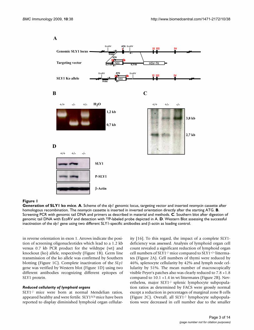
in reverse orientation in exon 1. Arrows indicate the posi-tion of screening oligonucleotides which lead to a 1.2 kbversus 0.7 kb PCR product for the wildtype (wt) andknockout (ko) allele, respectively (Figure 1B). Germ linetransmission of the ko allele was confirmed by Southernblotting (Figure 1C). Complete inactivation of the SLy1gene was verified by Western blot (Figure 1D) using twodifferent antibodies recognizing different epitopes ofSLY1 protein.
Reduced cellularity of lymphoid organsSLY1-/- mice were born at normal Mendelian ratios,appeared healthy and were fertile. SLY1Δ/Δ mice have beenreported to display diminished lymphoid organ cellular-
ity [16]. To this regard, the impact of a complete SLY1-deficiency was assessed. Analysis of lymphoid organ cellcount revealed a significant reduction of lymphoid organcell numbers of SLY1-/- mice compared to SLY1+/+ litterma-tes (Figure 2A). Cell numbers of thymi were reduced by46%, splenocyte cellularity by 42% and lymph node cel-lularity by 51%. The mean number of macroscopicallyvisible Peyer's patches also was clearly reduced to 7.8 +1.8compared to 10.1 +1.4 in wt littermates (Figure 2B). Nev-ertheless, major SLY1-/- splenic lymphocyte subpopula-tion ratios as determined by FACS were grossly normalexcept a reduction in percentages of marginal zone B cells(Figure 2C). Overall, all SLY1-/- lymphocyte subpopula-tions were decreased in cell number due to the smaller
Generation of SLY1 ko miceFigure 1Generation of SLY1 ko mice. A. Scheme of the sly1 genomic locus, targeting vector and inserted neomycin cassette after homologous recombination. The neomycin cassette is inserted in inverted orientation directly after the starting ATG. B. Screening PCR with genomic tail DNA and primers as described in material and methods. C. Southern blot after digestion of genomic tail DNA with EcoRV and detection with 32P-labeled probe depicted in A. D. Western Blot assessing the successful inactivation of the sly1 gene using two different SLY1-specific antibodies and β-actin as loading control.
Genomic SLY1 locus
Targeting vector
SLY1 Ko allele II III IV
HSV - TK
II III IV I ATG
neo Ia ATG
Ib neo
Ia Ib neo
Ia Ib
neo Ia ATG
Ib neo
Ia Ib neo
Ia Ib
0,5 kb 4,3 kb
Probe
Probe
EcoRV EcoRV
EcoRV EcoRV
A
C
-/- +/- +/+
2,7 kb
3,8 kb
B
+/+ -/- +/- H2O
1,2 kb
0,7 kb
+/+ +/- -/-
SLY1
P-SLY1
β-Actin
D
Page 3 of 14(page number not for citation purposes)

BMC Immunology 2009, 10:38 http://www.biomedcentral.com/1471-2172/10/38
organ size (Figure 2D), even though major B and T cellsubpopulation ratios were normal. In summary, thesedata are strikingly reminiscent of the previously describedphenotype of SLY1Δ/Δ mice, which also displayed a reduc-tion in cellularity of lymphoid organs, namely spleen, thy-mus, lymph nodes and Peyer's patches [16].
Delayed T cell developmentAs both partial and complete deletion of SLY1 protein hadan obviously deleterious effect on thymocyte cell numberleading to a reduction of about 50% of wt levels, SLY1-dependent thymocyte development was examined moreclosely. SLY1 is expressed in all major thymocyte subsets,as determined by RT-PCR (see Additional file 1). To gaina better understanding of the impact of the N-terminaltruncation in the SLY1 protein versus complete deletion,
SLY1Δ/Δ thymocytes were included in the following exper-iments. CD4- and CD8-staining of total thymocytesrevealed no apparent differences in percentages of DP andsingle-positive (SP) cells in the thymus (Figure 3A, leftpanel). Comparable expression of TCRβ, CD69 and CD5on DP thymocytes indicated normal TCR signalling andpositive selection events in SLY1-targeted thymocytes(Figure 3A, right panel). In line with the reduced thymo-cyte cellularity, SLY1-/- and SLY1Δ/Δ mice had markedlyreduced total numbers of CD4+ and CD8+ DP and SP thy-mocytes, only a minor decrease in DN thymocyte countswas observed (Figure 3B, left panel). These results sug-gested a partially delayed development of DN to DP thy-mocytes in the absence of a functional SLY1 protein. Inaccordance with this hypothesis, a doubling in the per-centage of DN cells in SLY1-/- and SLY1Δ/Δ thymi was
Reduced cellularity of lymphoid organs in SLY1-/--miceFigure 2Reduced cellularity of lymphoid organs in SLY1-/--mice. A. Mean cell count of thymus, spleen and pooled axial and inguinal lymph nodes was determined (n = 4). B. Reduced number of visible Peyer's patches in SLY1-/--mice. C. FACS analysis of splenic lymphocyte subpopulations. T cells were determined as Thy1.2+, B cells as B220+, marginal zone B cells as CD21highCD23- of B220+, and NK cells as CD3-CD56+ (n = 9). D. Absolute cell counts/spleen (n = 9). **p ≤ 0.01; *** p ≤ 0.001.
Thy1.2+ B220+ MZB NK 0
10
20
30
40
50
60
70
Thy1.2+ B220+ MZB NK
0
1
2
3
4
Thymus Spleen LN
0
5
10
15
20
25
**
***
**
+/+
-/-
A
Cell
co
un
t/ o
rg
an
x1
0 7
B
Nu
mb
er
of
peyer's
patc
hes
4
6
8
10
12
14
+/+ -/-
**
% o
f su
bse
t
C D
*** ***
***
+/+
-/-
Cell
cou
nt/
sp
leen
x10
7
***
*** +/+
-/-
Page 4 of 14(page number not for citation purposes)

BMC Immunology 2009, 10:38 http://www.biomedcentral.com/1471-2172/10/38
observed (Figure 3B middle and right panel). Thisprompted us to further analyse the DN subset frequenciesbased on the expression of CD25 and CD44 (Figure 3C).We observed a slight decrease in DN1, a slight increase inDN2 and DN3 ratios in SLY1-/- and SLY1Δ/Δ thymi, and aslight decrease in DN4. Therefore, SLY1 appears to berequired for developmental progression in all four thymicDN subsets and the block in development could not beassigned to a specific DN stage, providing a first hint thatSLY1 expression is especially critical in thymic DN pro-genitor cells.
Defect in proliferation and differentiation in vitroThe partial block in developmental progression of DNthymocytes lacking functional SLY1 protein indicated that
proliferation of DN thymocytes in SLY1-defective micemight be impaired. To test this hypothesis, we analysedSLY1-dependent thymocyte proliferation and differentia-tion in vitro. To this end, the bone marrow stromal cellline OP9 was used allowing in vitro differentiation of DNto DP thymocytes induced by presentation of the Notchligand Delta-like 1 (DL-1) on the surface of OP9 cells[6,24]. Congruent with published data, sorted DN thymo-cytes proliferated and upregulated CD4- and CD8-expres-sion dependent on the presence of Notch ligand DL-1after three and six days of cultivation on OP9 cells (Figure4A, B and data not shown). As expected from previouslyobserved ex vivo data, we found a markedly reducedexpansion rate of sorted SLY1-/- and SLY1Δ/Δ DN thymo-cytes upon cultivation on OP9 cells compared to wt litter-
Block in thymocyte CD4-CD8- (DN) to CD4+CD8+ (DP) differentiationFigure 3Block in thymocyte CD4-CD8- (DN) to CD4+CD8+ (DP) differentiation. A. Total thymocyte CD4- and CD8-expres-sion was determined by FACS analysis (left panel). Histograms (right panel): Surface expression of TCRβ, CD69 and CD5 on gated CD4+CD8+ thymocytes. B. Cell count of DN, DP or CD4- and CD8-single positive (SP) thymocytes (left panel). Gating (middle panel) and percentage of DN thymocytes relative to total thymocytes (right panel). C. DN thymocytes were gated as shown in B and a subset analysis according to expression of CD44 and CD25 was performed. DN1 = CD44+CD25-; DN2 = CD44+CD25+; DN3 = CD44-CD25+; DN4 = CD44-CD25-. Dot plots show representative data of three independent experi-ments (n = 3). *p ≤ 0.05; **p ≤ 0.001; *** p ≤ 0.0005.
DP SP 0
2
4
6
8
10
12
14
16
DN
Th
ym
ocyte
cou
nt
x107
0,0
0,2
0,4
0,6
0,8
1,0
1,2
1,4
1,6
+/+
-/- Δ/Δ
A
CD4+CD8+ thymocytes:
% o
f m
ax
0 100 1000 10000 1x10 5 0
20
40
60
80
100
TCRβ
% o
f m
ax
0 100 1000 10000 1x10 5 0
20
40
60
80
100
CD69
% o
f m
ax
0 100 1000 10000 1x10 5 0
20
40
60
80
100
CD5
0 100 1000 10000 1x10 5
0
100
1000
10000
1x10 5
8
2.29
3.46
84.7
0 100 1000 10000 1x10 5
0
100
1000
10000
1x10 5
10
3.73
7.86
75.3
CD4
CD
8
+/+ Δ/Δ -/-
C
DN1 DN2 DN3 DN4
% T
hy
mic
DN
su
bp
op
ula
tio
ns
0
10
20
30
40
50
60
70 +/+
-/- Δ/Δ
0 100 1000 10000 1x10 5
0
100
1000
10000
1x10 5 11.6 2.99
55.1 30.3
CD25
CD
44
B
0 100 1000 10000 1x10 5
0
100
1000
10000
1x10 5
9.15
3.66
6.56
77.9
+/+ -/- Δ/Δ
% D
N o
f to
tal
thym
ocyte
s
0
1
2
3
4
5
-/-
+/+ Δ/Δ
0 100 1000 10000 1x10 5
0
100
1000
10000
1x10 5
1.86
CD90
CD
4/C
D8
/TC
Rγδ
+/+
0 100 1000 10000 1x10 5
0
100
1000
10000
1x10 5
3.93
0 100 1000 10000 1x10 5
0
100
1000
10000
1x10 5
3.53
-/- Δ/Δ
+/+ -/- Δ/Δ
0 100 1000 10000 1x10 5
0
100
1000
10000
1x10 5 8.25 4.44
60.1 27.2 0 100 1000 10000 1x10 5
0
100
1000
10000
1x10 5 6.29 4.08
59.1 30.5
DN thymocytes:
*
******
* *
***
**
* * *
****
*
Page 5 of 14(page number not for citation purposes)

BMC Immunology 2009, 10:38 http://www.biomedcentral.com/1471-2172/10/38
mates (Figure 4B and 4C, left panel). Additionally, thedifferentiation of SLY1-/- and SLY1Δ/Δ DN thymocytes wasdiminished relative to littermate controls (Figure 4C, rightpanel). If sorted DN3 cells were used, differentiation wasequally affected, also adding IL-7 to the medium gave sim-ilar results (see Additional file 2).
Partially impaired activation of mTOR-complex in SLY1-defective DN thymocytesOne of the cellular outcomes of DN thymocytes success-fully passing the β-selection check point is the upregula-tion of metabolic activity. This is a prerequisite to copewith the increasing energy demands at this developmentalstage needed to allow for the upcoming extensive prolifer-ation and differentiation of DN to DP thymocytes. Of par-ticular importance in this context is the activation of theRapamycin-sensitive mTOR-complex eventually resultingin an increase in cell mass and subsequent induction ofcell division. A first hint for a blunted mTOR-dependentactivation in SLY1-/- and SLY1Δ/Δ thymocytes came fromthe observation of a reduced frequency of thymocyteswith increased cell size ex vivo and after OP9 DL-1 culture(see Additional file 3 and Figure 5B left panel)[25]. Sev-eral key targets of mTOR in activated DN thymocytes have
been identified so far, their activation or induced expres-sion being strictly dependent on the combined engage-ment of a successfully rearranged preTCR and Notchreceptor [11-13]. Well reported events at this develop-mental stage are the activation of S6 Kinase and upregula-tion of the nutrient receptors CD71 and CD98, all ofwhich have been shown to be Rapamycin-sensitive [13].First, mTOR activation in SLY1+/+ thymocyte T cell precur-sor populations ex vivo and after cultivation for 12 h onOP9 DL-1 cells was analysed. This was accomplished bydetermination of cell size, phosphorylation of ribosomalprotein S6, CD71 and CD98 expression as surrogatemarkers for mTOR activity (Figure 5A). In line with pub-lished data, activation of mTOR in DN thymocytes isabsolutely dependent on preTCR-expression ex vivo and invitro. Furthermore, mTOR activation strictly requiresNotch signalling (see Additional file 3) and can be inhib-ited by addition of Rapamycin in vitro. Next, this single-cell based analysis was extended to SLY1-/- and SLY1Δ/Δ
thymocytes ex vivo and after 12 h culture on OP9 DL-1cells (Figure 5B). In summary, the activation status ofmTOR in preTCR-positive DN thymocytes of SLY1-/- andSLY1Δ/Δ mice was clearly reduced compared to wt litterma-tes under both conditions tested. The activation of mTOR
SLY1-targeted DN thymocytes exhibit impaired proliferation and differentiation when cultured on OP9 DL-1 cellsFigure 4SLY1-targeted DN thymocytes exhibit impaired proliferation and differentiation when cultured on OP9 DL-1 cells. A. CD4 and CD8 FACS profiles of sorted DN thymocytes before cultivation (input, upper row), and after cultivation for six days on OP9 DL-1 (middle row) or on OP9 GFP control cells (lower row). B. DL-1-dependent expansion of DN thymo-cytes after three days culture. C. Fold expansion on day three and day six (left panel). Differentiation rate (right panel) corre-sponds to the percentage of DP cells divided by the percentage of remaining DN cells. The expansion rate was determined by counting triplicates and comparing to initial seeding numbers on day 0. Representative data of at least three independently per-formed experiments is shown. *p ≤ 0.05; **p ≤ 0.005; *** p ≤ 0.001.
Input
DL-1
Day6
GFP
Day6
0 100 1000 10000 1x10⁵
0
100
1000
10000
1x1050.44 0.12
0.07999.4
0 100 1000 10000 1x10⁵
0
100
1000
10000
1x1052.95 49.1
33.214.7
0 100 1000 10000 1x10⁵
0
100
1000
10000
1x1050.55 2.23
691.2
+/+
0 100 1000 10000 1x10⁵
0
100
1000
10000
1x1050.32 0.081
0.0299.6
0 100 1000 10000 1x10⁵
0
100
1000
10000
1x1055.34 35.1
34.824.8
0 100 1000 10000 1x10⁵
0
100
1000
10000
1x1050.31 0.37
4.1995.1
-/-
CD4
CD
8
A
Fold
exp
an
sion
0
2
4
6
8
10
12
14
16
18 +/+Δ/Δ-/-
Fold expansion on day 3
GFP DL-1
B
0 100 1000 10000 1x10⁵
0
100
1000
10000
1x1050.29 0.057
0.02299.6
0 100 1000 10000 1x10⁵
0
100
1000
10000
1x1055.62 34.8
32.227.3
0 100 1000 10000 1x10⁵
0
100
1000
10000
1x1050.28 0.83
5.6993.2
Δ/Δ
day 3 day 6
Fold
exp
an
sion
0
20
40
60
80
100DL-1 induced proliferation
+/+Δ/Δ-/-
0,0
0,2
0,4
0,6
0,8
1,0
day 3 day 6
Differentiation to CD4+CD8+
DP
to D
N r
ati
o
+/+Δ/Δ-/-
C
* *
** **
******
* *
******
Page 6 of 14(page number not for citation purposes)

BMC Immunology 2009, 10:38 http://www.biomedcentral.com/1471-2172/10/38
in the absence of Notch signalling or in preTCR-negativethymocytes was equally low (see Additional file 3 anddata not shown). These results suggest a defect in inducingmTOR-mediated activation signals in a subset of T cellprecursor cells in SLY1-/- and SLY1Δ/Δ mice in response todevelopmental needs. Moreover, we propose a defect intransmitting signals from either the preTCR or the Notchreceptor to the mTOR protein complex.
Increased apoptosis induction in SLY1-defective DN3 thymocytesA decrease in the expansion rate of proliferating cells canbe either mediated by alterations in cell cycle progressionor elevated apoptosis induction, or even both. To identifythe underlying mechanism for the observed proliferationdefect, sorted DN thymocytes were plated on OP9 DL-1
cells and cell cycle distribution as well as apoptosis induc-tion was assessed. We found an equal cell cycle distribu-tion based on DNA-staining of permeabilized cells withDAPI (Figure 6A). Yet an increase in early (Annex-inV+DAPI-) and late (AnnexinV+DAPI+) apoptosis induc-tion in SLY1-targeted thymocytes was detected (Figure6B). Therefore we hypothesized that the impaired prolif-eration can be attributed to an increased apoptosis rate inthe absence of functional SLY1 protein. PreTCR-expres-sion and Notch signalling have both been reported to playpivotal roles in protecting DN thymocytes from apoptosisat the β-selection checkpoint [10,12,26]. Interestingly,SLY1 protein has been reported to be specifically phos-phorylated after TCR crosslinking in peripheral T cells[15]. It was hence reasonable to speculate a role for SLY1downstream of the preTCR or Notch in this context. To
mTOR-signalling is diminished in developing SLY1-/- and SLY1Δ/Δ DN thymocytesFigure 5mTOR-signalling is diminished in developing SLY1-/- and SLY1Δ/Δ DN thymocytes. Cell size, activation of S6 kinase and expression levels of the nutrient receptors CD71 and CD98 were determined as surrogate markers for mTOR activity in thymocytes. Phosphorylation of ribosomal protein S6 was used as a marker of S6 Kinase activity [25]. A. SLY1+/+ DN thymo-cytes were analysed ex vivo and after 12 h culture on OP9 DL-1 cells. Thymocytes were further subdivided by gating on preTCR-negative and -positive cells. B. Activation of mTOR in preTCR-expressing SLY1+/+, SLY1-/- and SLY1Δ/Δ thymocytes was analysed ex vivo and after OP9 DL-1 culture in vitro for 12 hours. 20 nM Rapamycin was added as a control during the cul-tivation period to inhibit mTOR activation. Histograms show representative data of four independent experiments (n = 4).
Phospho-S6
0 100 1000 10000 1x10 5 0
20
40
60
80
100
% o
f Max
0 100 1000 10000 1x10 5 0
20
40
60
80
100
% o
f Max
0 100 1000 10000 1x10 5 0
20
40
60
80
100
% o
f Max
0 100 1000 10000 1x10 5 0
20
40
60
80
100 %
of M
ax
Phospho-S6
CD71
0 100 1000 10000 1x10 5 0
20
40
60
80
100
% o
f Max
0 100 1000 10000 1x10 5 0
20
40
60
80
100
% o
f Max
0 100 1000 10000 1x10 5 0
20
40
60
80
100
% o
f Max
0 100 1000 10000 1x10 5 0
20
40
60
80
100
% o
f Max
CD71
Ex vivo
12h DL-1
Cell size
0 50K 100K 150K 200K 250K 0
20
40
60
80
100
% o
f Max
0 50K 100K 150K 200K 250K 0
20
40
60
80
100
% o
f Max
0 50K 100K 150K 200K 250K 0
20
40
60
80
100
% o
f Max
0 50K 100K 150K 200K 250K 0
20
40
60
80
100
% o
f Max
Cell size
Ex vivo
12h DL-1
CD98
0
20
40
60
80
100
% o
f Max
0 100 1000 10000 1x10 5
0 100 1000 10000 1x10 5 0
20
40
60
80
100
% o
f Max
0 100 1000 10000 1x10 5 0
20
40
60
80
100
% o
f Max
0 100 1000 10000 1x10 5 0
20
40
60
80
100
% o
f Max
CD98 icTCRβ+
icTCRβ-
icTCRβ+ +Rapamycin 12h
+/+
-/-
Δ/Δ Rapamycin 12h
A
B icTCRβ+ DN thymocytes
SLY1+/+ DN thymocytes
Page 7 of 14(page number not for citation purposes)

BMC Immunology 2009, 10:38 http://www.biomedcentral.com/1471-2172/10/38
Page 8 of 14(page number not for citation purposes)
Increased apoptosis of OP9 cultured DN thymocytes in the absence of functional SLY1 proteinFigure 6Increased apoptosis of OP9 cultured DN thymocytes in the absence of functional SLY1 protein. A. Cell cycle analysis of permeabilized DN cells after 2 days of OP9 DL-1 culture using DAPI. B. AnnexinV and DAPI staining of DN thymo-cytes cultivated for 4 days on OP9 DL-1 cells. C. SLY1-targeting results in increased Caspase-3 activation independent of TCRβ-expression and Notch signalling. Sorted DN3 thymocytes were cultured on OP9 cells for 2 days and then Caspase-3 active fragment in DN cells was analysed dependent on TCRβ-expression. D. Intracellular Caspase-3 activation in DN4 cells is negligible. *p ≤ 0.05; **p ≤ 0.01; *** p ≤ 0.001.
DN3: Caspase 3 activation
C
0 100 1000 10000 1x1050
20
40
60
80
100
% o
f M
ax
% a
ctiv
e C
asp
ase
-3
0
2
4
6
8
10
12
DN4: Caspase3 activation
D
+/+
-/-
Δ/Δ
0 50K 100K 150K 200K 250K0
500
1000
1500
2000
23.4
+/+
0 50K 100K 150K 200K 250K0
500
1000
1500
2000
23.6
Δ/Δ
DAPI
Cell
cou
nt
AnnexinV+DAPI- AnnexinV+DAPI+% o
f D
N t
hy
mo
cy
tes
0
1
2
3
4
5
6
7 +/+
-/-Δ/Δ
*
***
Α Β% cycling cells of CD4-CD8- thymocytes CD4-CD8- cell death
0 100 1000 10000 1x1050
20
40
60
80
100
% o
f M
ax
0 100 1000 10000 1x1050
20
40
60
80
100
% o
f M
ax
icTCRβ- icTCRβ+
OP9 GFP
% a
ctiv
e C
asp
ase
-3
0
10
20
30
40
50
60
+/+
-/-
Δ/Δ
% a
ctiv
e C
asp
ase
-3
0
5
10
15
20
Casp-3+ Casp-3+
0 100 1000 10000 1x1050
20
40
60
80
100
% o
f M
ax
0 100 1000 10000 1x1050
20
40
60
80
100
% o
f M
ax
OP9 DL-1
+/+
% a
ctiv
e C
asp
ase
-3
0
10
20
30
40
50
60
70
% a
ctiv
e C
asp
ase
-3
0
2
4
6
8
10
12
+/+
-/-
Δ/Δ
icTCRβ- icTCRβ+
Casp-3+ Casp-3+ Casp-3+
0 50K 100K 150K 200K 250K0
500
1000
1500
24.6
-/-
-/-Δ/Δ
+/+ -/-Δ/Δ +/+ -/-Δ/Δ
+/+ -/-Δ/Δ +/+ -/-Δ/Δ
icTCRβ+
OP9 DL-1
+/+
-/-
Δ/Δ
+/+
-/-
Δ/Δ
*** ****** ***
*** ***
***
**

BMC Immunology 2009, 10:38 http://www.biomedcentral.com/1471-2172/10/38
dissect the pathways SLY1 is involved in, we cultivatedsorted DN3 cells on OP9 DL-1 and OP9 control cells todiscriminate the role of Notch signalling, and used Cas-pase-3 active fragment as an intracellular marker for apop-tosis induction (Figure 6C). Intracellular staining forTCRβ-expression served to identify cells which had suc-cessfully rearranged their TCR-β-chain and which shouldtherefore receive preTCR-derived proliferative and sur-vival signals. Intracellular TCRβ-expression was similarbetween wt cells and SLY1-targeted cells (data notshown). As anticipated, Caspase-3 activation was lowestin wt preTCR-positive DN3 thymocytes cultivated on OP9DL-1 cells, thus receiving a preTCR-derived and a Notch-derived pro-survival signal, accounting for approximately2% of Caspase-3 active fragment positive thymocytes (Fig-ure 6C, histogram overlay and diagram upper rightpanel). In this setting, which physiologically correspondsto thymocytes successfully passing β-selection, apoptosisinduction was increased 4-fold in SLY1-/- and SLY1Δ/Δ DN3thymocytes compared to wt littermate controls. In con-trast, thymocytes failing to rearrange the TCRβ-chain stayTCRβ-negative and hence are increasingly prone to pro-grammed cell death initiation. This was indeed the case:apoptosis induction in TCRβ-negative wt DN3 thymo-cytes was 20 times higher than in their TCRβ-positivecounterparts (compare wt apoptosis upper left with upperright panel). Again, the percentage of Caspase-3 positiveprogenitor cells was markedly increased (by 20%) inSLY1-/- or SLYΔ/Δ cultures. Interestingly, SLY1-/- and SLYΔ/Δ
thymocytes also displayed increased susceptibilitytowards programmed cell death initiation in the absenceof Notch signalling (lower part of Figure 6C). Yet wtTCRβ-negative DN3 thymocytes cultivated on OP9 con-trol cells were highly susceptible to apoptosis, accountingfor around 30% Caspase-3-positive cells. Though the rateof apoptotic SLY1-/- and SLY1Δ/Δ DN3 thymocytes was ele-vated roughly by 15% relative to wt controls (left part,lower panel). We also observed a more than twofoldincrease in Caspase-3 activation in the preTCR-positivepopulation of DN3 thymocytes when plated on OP9 con-trol cells in the absence of SLY1. Thus, an overall increasein programmed cell death was observed in both SLY1-/-
and SLY1Δ/Δ DN3 thymocytes. In general, TCRβ-expres-sion was the major pro-survival determinant, Notch sig-nals even were detrimental if thymocytes failed tosuccessfully rearrange their TCRβ-genes (compare left partupper and lower panel). Nevertheless, Caspase-3 activa-tion was elevated in SLY1-/- and SLY1Δ/Δ DN3 thymocytesindependent of the availability of signals from the preTCRor the Notch receptor or even both. In summary, theseresults suggest a general anti-apoptotic role of SLY1 inDN3 thymocytes. Remarkably, programmed cell death inDN4 cells was equally undetectable independent of SLY1-expression (Figure 6E). This data supports a specific rolefor SLY1 in protecting DN3 thymocytes during the β-selec-tion checkpoint. To further elucidate a possible role for
SLY1 in amplifying Notch-derived signals, a titration of γ-secretase-inhibitor (GSI) which is reported to inhibit spe-cifically Notch receptor activation on sorted DN3 SLY+/+,SLY1-/- and SLY1Δ/Δ thymocytes was performed (see Addi-tional file 4). After six days of cultivation on OP9 cells, dif-ferentiation and proliferation was assessed. Appearance ofDP cells gradually decreased with increasing GSI concen-trations likewise did the expansion rate. At 1 μM GSI, inhi-bition was almost complete, as assessed by comparing theproliferation and differentiation rate with the OP9 GFPcultivated controls. SLY1Δ/Δ and SLY1-/- thymocytes werefound to be more sensitive than SLY1+/+ thymocytes tointermediate concentrations of GSI, supporting a functionfor SLY1 in signal propagation from the Notch receptor.However, this increased sensitivity was only very weaklypronounced and could also be attributed to a defect inintegration of preTCR and Notch signals, as both signalsare required for proliferation.
Regulation of phosphorylation of SLY1 in thymocytesIt has been reported that SLY1 is phosphorylated atserine27 specifically upon TCR-stimulation in humanperipheral T-lymphocytes and not upon chemokine- orcytokine-driven stimulation [15]. This data implicated afunction of SLY1 downstream of the TCR. In murinesplenocytes, TCR-stimulation-induced phosphorylationcan also be observed, albeit there is considerable basalphosphorylation in contrast to the situation in humanlymphocytes (Figure 7A). Likewise, SLY1 phosphorylationin murine thymocytes is sensitive to inhibition by thePI3K inhibitor Ly294002 (Figure 7B). We also observedinhibition of SLY1-phosphorylation by treatment withRapamycin, which has not been previously reported inperipheral lymphocytes and might reflect a differentialregulation in thymocytes (Figure 7B). Interestingly, wedetected a high degree of basal phosphorylation of SLY1in early developing DN3 and DN4 thymocytes isolated exvivo, suggesting a requirement of the phosphorylated var-iant of SLY1 in T cell development (Figure 7C). In DN3thymocytes, the ratio of preTCR-expressing thymocytes islow and also gated preTCR-negative DN thymocytesexhibited increased apoptosis induction (Figure 6C, leftpanel), arguing against a role downstream of the preTCR.Therefore, we reasoned that SLY1 might be independentlyphosphorylated of preTCR-mediated signalling in earlythymocytes. To test this hypothesis, RAG1-/- DN thymo-cytes unable to rearrange the TCR-genes and thereforelacking preTCR-signalling were incubated over night onOP9 cells, sorted and then SLY1-phosphorylation wasdetected via Western blotting (Figure 7D, left panel).Indeed, we observed preTCR-independent phosphoryla-tion of SLY1 in DN thymocytes as anticipated and in linewith previous data. In addition, we detected an increase inSLY1 phosphorylation in RAG1-/- thymocytes stimulatedwith Notch ligand. Unexpectedly, this Notch-inducedincrease in phosphorylation was only observed in
Page 9 of 14(page number not for citation purposes)

BMC Immunology 2009, 10:38 http://www.biomedcentral.com/1471-2172/10/38
preTCR-deficient thymocytes. In preTCR-sufficientRAG1+/+ DN thymocytes, SLY1 phosphorylation wasdiminished upon Notch signalling (Figure 7D, rightpanel). This data suggests a role for SLY1 in integratingsignals derived from the preTCR and from the Notchreceptor leading to prevention of premature apoptosis ini-tiation in developing thymocytes.
DiscussionT cell development is a tightly regulated process involvingnumerous successive steps of differentiation. Of particularimportance for the maturation of αβ T cells is the success-ful rearrangement of the VDJ genes coding for the TCRβchain which then pairs with the preTα chain to initiate
preTCR signalling [1,27-29]. In the present work, induc-tion of programmed cell death in DN3 thymocytes couldbe precisely recapitulated as being principally dependenton intracellular preTCR expression, thereby supportingpreTCR-signalling as the major decisive constraint indeath versus survival decision. Furthermore, the orphanadapter protein SLY1 was identified as a novel anti-apop-totic protein required for developmental progression of Tcell precursors to the DP stage. This anti-apoptotic func-tion resulted in protection of thymocytes from prematureprogrammed cell death initiation at the DN3 thymocytestage. As a consequence, thymocyte DP number and there-fore total thymus cellularity was severely reduced in SLY1-targeted mice.
Regulation of SLY1 phosphorylation in thymocytesFigure 7Regulation of SLY1 phosphorylation in thymocytes. A. Phosphorylation of SLY1 at serine27 in peripheral splenocytes. CD90+ MACS-isolated T cells were stimulated with αCD3 (10 μg/ml) for the indicated time and analysed by Western blotting. B. Thymocytes were left unstimulated or incubated for 20' with 20 ng/ml PDBU. Where indicated, cells had been pretreated with Rapamycin (20 nM) or Ly294002 (10 μM) for 1 hour. C. DN3 and DN4 thymocytes were single-cell-sorted according to the expression of CD90, CD25 and CD44, lysed and analysed by Western blotting. D. RAG1-/- or RAG1+/+ DN3 thymocytes were plated over night on OP9 cells. DN thymocytes were then sorted by single-cell flow cytometry, lysed and analysed by Western blotting.
P-SLY1
SLY1
β-Actin
DN
3
DN
4
0‘ 5‘ 60‘
CD90+ splenocytes
P-SLY1
β-Actin
P-SLY1
β-Actin
+Rapamycin
+Ly294002
PDBU + + - +
P-Akt
P-SLY1
β-Actin
GFP DL-1
DN RAG-/-->OP9
GFP DL-1
DN RAG+/+->OP9
P-SLY1
β-Actin
A B
DC
αCD3
Ex vivo CD4-CD8-
thymocytes
Page 10 of 14(page number not for citation purposes)

BMC Immunology 2009, 10:38 http://www.biomedcentral.com/1471-2172/10/38
Haematopoietic progenitors entering the thymus via thebloodstream depend on a sustained Notch receptor-lig-and engagement to develop towards the T cell lineage[2,6,30,31]. The OP9 differentiation system can be usedto recapitulate the signals required for haematopoieticprecursor cells to undergo T cell development until the DPthymocyte stage in vitro. This differentiation system wasused to further elucidate the underlying defect responsiblefor the reduced cellularity of SLY1-/- and SLY1Δ/Δ thymi. Asanticipated from the ex vivo situation, a reduction in pro-liferation and differentiation upon OP9 culture relative tolittermate controls was found. Thus, a migration defect ofprogenitor cells with a subsequent reduction in thymicrepopulation capacity as the major cause for the resultingdefect in thymic cellularity could be excluded. Further-more, it could be clearly shown that the reduction in thy-mocyte proliferation is cell intrinsic and that the principaldefect is not influenced by differences in thymic environ-ment derived from stromal cells.
Yet Notch signalling is not only mandatory for commit-ment to the T cell lineage. Recently, it was also shown tobe crucial for inducing a replication-competent status inthymocytes once preTCR signalling is initiated [10,12,13].A prerequisite for cellular expansion is an increased meta-bolic activity including elevated expression of nutrientreceptors and protein synthesis. The activity of mTOR hasa key role in controlling this process. In peripheral T cells,growth factors have been reported to modulate Akt andmTOR activity and thereby to increase survival of T cells[32]. Growth factor withdrawal resulted in metabolic col-lapse and loss of nutrient receptor expression. In SLY1-/-
and SLY1Δ/Δ thymocytes, induced expression of the nutri-ent receptors CD71 and CD98 as well as S6 Kinase activa-tion were impaired relative to wt littermates. Thisdiminished mTOR activity correlated with a reduced cellsize increase. Therefore a defect in metabolic activity inSLY1-defective thymocytes can be inferred, leading toincreased cell death rate and concomitant reduction ofthymic cellularity.
Full mTOR activation in DN thymocytes is dependent onboth Notch- and preTCR-derived signals [10,12,13].There is limited insight into the signal integration processof both pathways in thymocytes. Current literature datasuggests that signal integration occurs at the level ofPI3Kinase activation. Notch signalling is thought todecrease expression of PTEN, the negative regulator ofPI3Kinase activation [33]. preTCR-formation is thoughtto activate Akt, probably also via PI3Kinase activation[34]. It is not precisely clear how the presence of a preTCRwithout ligand engagement leads to the activation of Akt,and whether there are also more direct interactions of theNotch pathway with the PI3Kinase pathway which are notregulated by transcription. The present work may addinsight into how the different signals are processed by
connecting them with the orphan adapter protein SLY1.The reduced frequency of SLY1-/- and SLY1Δ/Δ progenitorcells with increased metabolic activity suggests a role forSLY1 in transmitting signals from either the preTCR or theNotch receptor to mTOR, or even for integrating signalsfrom both receptors. As opposed to reports from periph-eral T lymphocytes showing that SLY1 phosphorylation isstrictly dependent on TCR-stimulation, we have shown inthis study that SLY1-phosphorylation in thymocytes isindependent of the preTCR. Interestingly, we have foundthat the phosphorylation of SLY1 at serine27 is differen-tially regulated upon Notch-stimulation dependent onthe absence or presence of preTCR-signalling. This differ-ing phosphorylation could be interpreted as a signal inte-gration process of the two converging pathwaysdownstream of the Notch receptor and downstream of thepreTCR.
When cultured on OP9 DL-1 and OP9 GFP control cells,the anti-apoptotic function of SLY1 was independent ofpreTCR- and Notch-derived signals, indicating a generalpro-survival role for SLY1 protein independent of bothreceptors. However, OP9 GFP cells have been reported toexpress mRNA for the Notch ligands jagged-1 and jagged-2, although in low amounts [6]. Therefore, it cannot beexcluded that there might still be residual Notch-signalsavailable to thymocytes cultivated on OP9 GFP controlcells. In addition, OP9 cells secrete or present otherunknown factors required by haematopoietic precursorcells. These residual signals induced by unknown factorscould therefore transmit anti-apoptotic signals which arenot properly transmitted in SLY1-defective thymocytescultivated on OP9 GFP cells compared to the situation inSLY1+/+ thymocytes. In addition, freshly isolated thymo-cytes have already been receiving Notch signals duringtheir sojourn in the thymus in vivo, and therefore theincreased apoptosis induction of SLY1-defective thymo-cytes on OP9 GFP cells compared to SLY1+/+ thymocytescould already have been initiated in the thymus in vivobefore isolation of the cells. Yet unexpected is the observa-tion that Notch signalling can even drive thymocytes intoapoptosis if they fail to generate a preTCR as observedupon OP9 culture of DN3 thymocytes in the presentwork. In contrast to this notice, Notch signalling in DN3thymocytes is thought to play a general anti-apoptoticrole by activating the Akt kinase pathway [12]. This studytherefore adds additional complexity to the effector func-tions of Notch signalling in DN3 thymocytes, implying apro-apoptotic outcome of Notch receptor activation in acertain cellular context.
As previously described, SLY1Δ/Δ mice expressing a SLY1protein harbouring a partial deletion in the N-terminalregion show impaired immune responses [16,17]. Theregion missing in SLYΔ contains part of a bipartite nuclearlocalisation sequence and the known phosphorylation
Page 11 of 14(page number not for citation purposes)

BMC Immunology 2009, 10:38 http://www.biomedcentral.com/1471-2172/10/38
site closely associated to the potential localisationsequences. As a consequence, the subcellular localisationof SLY1Δ is shifted from a nucleocytoplasmic to a strictlycytoplasmic localisation [16]. The putative protein-pro-tein interacting SH3- and SAM-domains remain intact inSLY1Δ, possibly still enabling its interference with othersignalling proteins. However, a dominant negative effectof SLY1Δ can be excluded due to the comparable pheno-type of SLY1-/- mice. One conclusion that can be drawnfrom this data is that an essential function, at least for Tcell development, of SLY1 is mediated by the amino acidsat position 20–100. An apparent role for this deletedregion could be either to regulate the correct subcellularlocalisation of the protein or to be directly required forinteraction with SLY1 binding partners by providing asequence specific recognition signal.
ConclusionIn the present work, SLY1 is described as a novel anti-apoptotic protein required for thymocyte development bypreventing DN thymocytes from premature initiation ofprogrammed cell death. SLY1-negative thymocytes exhib-ited impaired activation of the mTOR complex, support-ing an essential role of mTOR in thymocyte development.We therefore propose a role for SLY1 in signal integrationof the Notch receptor and preTCR pathways culminatingin the activation of the mTOR protein complex in devel-oping thymocytes. As the sly1 gene is located on the X-chromosome in man, its function could be either nega-tively affected in some cases of x-linked immunodefi-ciency or activating mutations could contribute tolymphoma development. In the future, the identificationof the interacting kinase and the precise molecular mech-anism by which SLY1 contributes to mTOR activation andprevention of programmed cell death will be of greatinterest.
MethodsRNA isolation and RT-PCRTotal RNA from cells was isolated using Trizol Reagent(Invitrogen) according to the manufacture's instructions.First-strand cDNA synthesis was performed using 1 μg oftotal RNA with M-MLV reverse transcriptase and oligo dTprimer (Invitrogen).
SLY1 transcripts were amplified withSLY1_exon6_forward: 5'-CCC GGA GGA TTC TGG GAAGA-3' and SLY1_exon8_reverse: 5'-GAA GTC AGT GTGGAC TCG GG-3'. The following primer sequences wereused to amplify GAPDH: (forward) 5'-CAT GTA GGC CATGAG GTC CAC CAC-3' and (reverse) 5'-TGA AGG TCGGTG TGA ACG GAT TTG GC-3'.
Gene targeting and miceGeneration of SLYΔ/Δ mice and genotyping has beendescribed previously [16]. For generation of SLY-/- mice,
E14.1 embryonic stem (ES) cells from 129/Ola mice weregrown in Dulbecco's modified Eagle's medium (Invitro-gen, Carlsbad, CA, USA) supplemented with 2 mMglutamine (Seromed, Wien, Austria), leukemia inhibitoryfactor, 100 U/ml penicillin, 100 μg/ml streptomycin (Ser-omed), 50 μM 2-mercaptoethanol (Invitrogen) and 15%heat-inactivated fetal bovine serum (Pan Biotech, Aiden-bach, Germany). Genomic fragments flanking the murinesly1 gene as well as neomycin and thymidine kinase werecloned into pBluescript (Agilent technologies, SantaClara, CA, USA) and fully sequenced. The targeting vectorwas designed in such a way that the neomycin resistancecassette was inserted in reverse orientation in exon 1 of thesly1 gene 3' of the starting ATG without deleting anyendogenous sequence. E14.1 ES cells were electroporatedwith the NotI-linearized targeting vector, and the trans-fected cells were subsequently subjected to G418 and gan-ciclovir selection. Clones carrying the correct homologousrecombination were identified by Southern blot hybridi-zation with the 5' flanking probe indicated in Figure 1after digestion of ES cell DNA with EcoRV and BamHI.Single integration was verified by probing the Southernblot with the neomycin resistance cassette. Correctly tar-geted ES cell clones were injected into C57BL/6 blasto-cysts, which were transferred into pseudopregnant fostermice. Resulting chimeric mice were backcrossed toC57BL/6 mice, and germ line transmission of the targetedallele was again confirmed by Southern blot analysis.SLY1-/- mice were backcrossed to the C57BL/6 backgroundfor six to ten generations. Wild-type littermates were usedas controls. Genotyping of SLY1 knockout mice was per-formed by PCR with the following primers: 5'-TGA CGGCAG TAG GGA TGG TAG-3' (forward); 5'-CGC CTT CTTGAC GAG TTC TTC T-3' (neo reverse); 5'-AGT GGC CTGGGG GAG ATG T-3' (Wt reverse). SLY1Δ/Δ mice were back-crossed for twelve generations.
Mice were kept according to national guidelines for ani-mal care in an SPF animal facility. All animal work wasperformed according to the guidelines of the Germannational animal care regulations. RAG1-/- mice were pur-chased from Jackson Laboratories.
Flow cytometric analysisAntibodies conjugated to fluorescein isothiocyanate, phy-coerythrin, allophycocyanin, percp, phycoerythrin-cy7,allophycocyanin-cy7 and biotin were obtained fromeither Pharmingen (San Diego, CA, USA), Southern Bio-tech (Birmingham, Alabama, USA) or eBioscience (SanDiego, CA, USA). Alexa-fluor488-conjugated anti-phos-pho-Akt and anti-phospho-S6 antibodies were obtainedfrom Cell Signalling (Danvers, MA, USA). Cells werestained with saturating concentrations of antibody inaccordance with the manufacturer's instructions. Datawere acquired with either a FACSCalibur or FACSCanto(Becton Dickinson, Franklin Lakes, NJ, USA) using Cel-
Page 12 of 14(page number not for citation purposes)

BMC Immunology 2009, 10:38 http://www.biomedcentral.com/1471-2172/10/38
lquest or DIVA (Becton Dickinson) software and wereanalysed using Flowjo software (Treestar, San Carlos, CA,USA). Viable cells were gated according to their forwardand sideward scatter profiles and DAPI (Invitrogen) exclu-sion. CD4 and CD8 DN Thy1.2+ subsets were gated by lin-eage exclusion of CD4 and CD8 DP and SP cells andTCRγδ. DN subsets were further subdivided by CD25 andCD44 expression, defining CD25-CD44+ as DN1,CD25+CD44+ as DN2, CD25+CD44- as DN3 and CD25-
CD44- as DN4, respectively. Cellular DNA content wasmeasured by DAPI staining of methanol permeabilisedcells. Phospho-S6 and phospho-Akt levels were assessedas described previously [11].
OP9 cultures and assayOP9 bone marrow stromal cells expressing Notch ligandDelta Like-1 (DL-1) and control OP9 cells [6] were a giftfrom Juan Carlos Zúñiga-Pflücker (Toronto, Canada).OP9 cells were maintained in α MEM (Invitrogen) supple-mented with 50 μM 2-mercaptoethanol, 100 U/ml peni-cillin, 1 mg/ml streptomycin and 20% heat-inactivatedFBS. Sorted DN thymocytes were co-cultured on OP9 DL-1 or OP9 control cells for the indicated time periods. DNthymocytes were obtained using an AutoMACS (MiltenyiBiotech, Bergisch-Gladbach, Germany) magnetic cellsorter by depleting CD4+ and CD8+ thymic subpopula-tions to greater than 97% purity. DN3 or DN4 cells wereobtained by subsequently either positively sorting ordepleting CD25-expressing DN3 cells, respectively. Alter-natively, DN3 cells were obtained by sorting with a FAC-SAria single cell sorter (Becton Dickinson, Heidelberg,Germany) with greater than 98% purity. On the day ofharvest thymocytes were filtered through 50 μm filters toremove OP9 cells before assessing developmental pro-gression and proliferation of T lineage cells.
Western blot analysisThymocytes or splenocytes were resuspended in lysisbuffer containing 10 mMNaF, 1 mM sodium orthovanad-ate and protease inhibitor cocktail (Sigma) on ice for15min. Lysates were separated by SDS-PAGE and transferredto nitrocellulose membranes (Perkin-Elmer, Rodgau-Jügesheim). After blocking with 5% milk in TBST, mem-branes were probed with primary antibodies and subse-quently detected using horseradish peroxidase-linkedgoat anti-mouse or anti-rabbit IgG and visualized by theenhanced chemilumescent (ECL) detection system (GEHealthcare, Munich, Germany). Membranes were rep-robed with anti-β-actin (Sigma, St. Louis, Missouri, USA).The anti-SLY1 antibody has been generated by Eurogentec(Seraing, Belgium) and has already been described [16].Anti-phospho-SLY1 antibodies also have been generatedby Eurogentec by immunizing rabbits with oligopeptide(H2N-LQR SSpS FKD FAK C-CONH2). Both antibodieshave been tested extensively for specificity (data notshown).
Statistical analysesStatistical significance of differences between wildtypeand SLY1-targeted mice were evaluated using the Studentst test. P < 0.05 was considered significant. All results arepresented as mean values ± SEM.
Authors' contributionsBR has made substantial contributions to conception anddesign of the study, acquired, analysed, and interpretedthe data, and drafted the manuscript. KP has given finalapproval of the version to be published. SB participated inthe design and coordination of the study and helped todraft the manuscript. All authors read and approved thefinal manuscript.
Additional material
Additional file 1SLY1 is expressed in all major thymocyte subsets. Semi-quantitative RT-PCR. Thymocytes were sorted according to their surface expression of CD4 and CD8. Subsequently, RNA was isolated and transcribed into cDNA. A serial 1:3 dilution of cDNA was used to amplify SLY1 or GAPDH tran-scripts with SLY1- or GAPDH-specific primers, respectively. Zipped folder containing EPS file.Click here for file[http://www.biomedcentral.com/content/supplementary/1471-2172-10-38-S1.zip]
Additional file 2Proliferation and differentiation defect of sorted SLY1-/- and SLY1Δ/Δ
DN3 thymocytes cannot be rescued by adding IL-7. A. Fold expansion of sorted DN3 thymocytes after six days of culture on OP9 DL-1 cells. If indicated, 1 ng/ml mIL-7 was included in the cultures. B. Differentiation rate of sorted DN3 thymocytes after six days of culture on OP9 DL-1 cells without IL7. C. Differentiation rate of sorted DN3 thymocytes after six days of culture on OP9 DL-1 cells with 1 ng/ml murine IL7. Data are based on triplicate analyses. The differentiation rate corresponds to the percentage of DP cells divided by the percentage of remaining DN cells. The expansion rate was determined by comparing the obtained cell number on day six to initial seeding numbers on day 0. Representative data of two independently performed experiments is shown. Zipped folder containing EPS file.Click here for file[http://www.biomedcentral.com/content/supplementary/1471-2172-10-38-S2.zip]
Additional file 3Notch- and preTCR-dependent activation of mTOR results in cell size increase and is diminished in sorted DN SLY1-/- and SLY1Δ/Δ thymo-cytes. A. mTOR activation is strictly dependent on a combined preTCR- and Notch receptor-derived signal. Cell size of DN thymocytes was ana-lysed after 12 h culture on OP9 GFP or OP DL-1 cells. Cells were gated on intracellular TCRβ-positive or -negative. B. Cell size overlay of DN SLY1+/+, SLY1-/- and SLY1Δ/Δ preTCR-positive thymocytes cultured on OP9 DL-1 cells. As a control, 20 nM Rapamycin was added during the cultivation period to inhibit mTOR activation. Zipped folder containing EPS file.Click here for file[http://www.biomedcentral.com/content/supplementary/1471-2172-10-38-S3.zip]
Page 13 of 14(page number not for citation purposes)

BMC Immunology 2009, 10:38 http://www.biomedcentral.com/1471-2172/10/38
AcknowledgementsWe thank Prof. Doreen Cantrell and David Finlay for experimental advice and Prof. Juan Carlos Zúñiga-Pflücker who generously provided the OP9 DL-1 and OP9 GFP cells. We thank Nicole Küpper and Karin Buchholz for technical assistance.
This work was supported by grants BE 2813/1-1 (to S.B.) of the Deutsche Forschungsgemeinschaft (DFG), and the Forschungskommission of the Heinrich-Heine University Duesseldorf (to S.B.).
References1. Godfrey DI, Kennedy J, Mombaerts P, Tonegawa S, Zlotnik A: Onset
of TCR-beta gene rearrangement and role of TCR-betaexpression during CD3-CD4-CD8-thymocyte differentia-tion. J Immunol 1994, 152:4783-4792.
2. Ciofani M, Zuniga-Pflucker JC: The Thymus as an Inductive Sitefor T Lymphopoiesis. Annu Rev Cell Dev Biol. 2006, 23:463-493.
3. von Boehmer H, Aifantis I, Feinberg J, Lechner O, Saint-Ruf C, WalterU, et al.: Pleiotropic changes controlled by the pre-T-cellreceptor. Curr Opin Immunol 1999, 11:135-142.
4. Strasser A, Huang DC, Vaux DL: The role of the bcl-2/ced-9 genefamily in cancer and general implications of defects in celldeath control for tumourigenesis and resistance to chemo-therapy. Biochim Biophys Acta 1997, 1333:F151-F178.
5. Cory S, Vaux DL, Strasser A, Harris AW, Adams JM: Insights fromBcl-2 and Myc: malignancy involves abrogation of apoptosisas well as sustained proliferation. Cancer Res 1999,59:1685s-1692s.
6. Schmitt TM, Zuniga-Pflucker JC: Induction of T cell developmentfrom hematopoietic progenitor cells by delta-like-1 in vitro.Immunity 2002, 17:749-756.
7. Sambandam A, Maillard I, Zediak VP, Xu L, Gerstein RM, Aster JC, etal.: Notch signaling controls the generation and differentia-tion of early T lineage progenitors. Nat Immunol 2005,6:663-670.
8. Maillard I, Fang T, Pear WS: Regulation of lymphoid develop-ment, differentiation, and function by the Notch pathway.Annu Rev Immunol 2005, 23:945-974.
9. Hayday AC, Pennington DJ: Key factors in the organized chaosof early T cell development. Nat Immunol 2007, 8:137-144.
10. Ciofani M, Schmitt TM, Ciofani A, Michie AM, Cuburu N, Aublin A, etal.: Obligatory role for cooperative signaling by pre-TCR andNotch during thymocyte differentiation. J Immunol 2004,172:5230-5239.
11. Hinton HJ, Alessi DR, Cantrell DA: The serine kinase phosphoi-nositide-dependent kinase 1 (PDK1) regulates T cell devel-opment. Nat Immunol 2004, 5:539-545.
12. Ciofani M, Zuniga-Pflucker JC: Notch promotes survival of pre-Tcells at the beta-selection checkpoint by regulating cellularmetabolism. Nat Immunol 2005, 6:881-888.
13. Kelly AP, Finlay DK, Hinton HJ, Clarke RG, Fiorini E, Radtke F, et al.:Notch-induced T cell development requires phosphoi-nositide-dependent kinase 1. EMBO J 2007, 26:3441-3450.
14. Beer S, Simins AB, Schuster A, Holzmann B: Molecular cloning andcharacterization of a novel SH3 protein (SLY) preferentially
expressed in lymphoid cells. Biochim Biophys Acta 2001,1520:89-93.
15. Astoul E, Laurence AD, Totty N, Beer S, Alexander DR, Cantrell DA:Approaches to define antigen receptor-induced serinekinase signal transduction pathways. J Biol Chem 2003,278:9267-9275.
16. Beer S, Scheikl T, Reis B, Huser N, Pfeffer K, Holzmann B: Impairedimmune responses and prolonged allograft survival in SLy1mutant mice. Mol Cell Biol 2005, 25:9646-9660.
17. Scheikl T, Reis B, Pfeffer K, Holzmann B, Beer S: Reduced NotchActivity is Associated With an Impaired Marginal Zone BCell Development and Function in SLy1 Mutant Mice. Molec-ular Immunology 2008, 46(5):969-77.
18. Claudio JO, Zhu YX, Benn SJ, Shukla AH, McGlade CJ, Falcioni N, etal.: HACS1 encodes a novel SH3-SAM adaptor protein differ-entially expressed in normal and malignant hematopoieticcells. Oncogene 2001, 20:5373-5377.
19. Uchida T, Nakao A, Nakano N, Kuramasu A, Saito H, Okumura K, etal.: Identification of Nash1, a novel protein containing anuclear localization signal, a sterile alpha motif, and an SH3domain preferentially expressed in mast cells. Biochem BiophysRes Commun 2001, 288:137-141.
20. Zhu YX, Benn S, Li ZH, Wei E, Masih-Khan E, Trieu Y, et al.: TheSH3-SAM adaptor HACS1 is up-regulated in B cell activa-tion signaling cascades. J Exp Med 2004, 200:737-747.
21. Yamada H, Yanagisawa K, Tokumaru S, Taguchi A, Nimura Y, OsadaH, et al.: Detailed characterization of a homozygouSLydeleted region corresponding to a candidate tumor suppres-sor locus at 21q11-21 in human lung cancer. Genes Chromo-somes Cancer 2008, 47:810-818.
22. Rimkus C, Martini M, Friederichs J, Rosenberg R, Doll D, Siewert JR,et al.: Prognostic significance of downregulated expression ofthe candidate tumour suppressor gene SASH1 in colon can-cer. Br J Cancer 2006, 95:1419-1423.
23. Zeller C, Hinzmann B, Seitz S, Prokoph H, Burkhard-Goettges E,Fischer J, et al.: SASH1: a candidate tumor suppressor gene onchromosome 6q24.3 is downregulated in breast cancer.Oncogene 2003, 22:2972-2983.
24. Schmitt TM, de Pooter RF, Gronski MA, Cho SK, Ohashi PS, Zuniga-Pflucker JC: Induction of T cell development and establish-ment of T cell competence from embryonic stem cells differ-entiated in vitro. Nat Immunol 2004, 5:410-417.
25. Ruvinsky I, Sharon N, Lerer T, Cohen H, Stolovich-Rain M, Nir T, etal.: Ribosomal protein S6 phosphorylation is a determinant ofcell size and glucose homeostasis. Genes Dev 2005,19:2199-2211.
26. Aifantis I, Buer J, von Boehmer H, Azogui O: Essential role of thepre-T cell receptor in allelic exclusion of the T cell receptorbeta locus. Immunity 1997, 7:601-607.
27. Dudley EC, Petrie HT, Shah LM, Owen MJ, Hayday AC: T cell recep-tor beta chain gene rearrangement and selection during thy-mocyte development in adult mice. Immunity 1994, 1:83-93.
28. Mallick CA, Dudley EC, Viney JL, Owen MJ, Hayday AC: Rearrange-ment and diversity of T cell receptor beta chain genes in thy-mocytes: a critical role for the beta chain in development.Cell 1993, 73:513-519.
29. Mombaerts P, Iacomini J, Johnson RS, Herrup K, Tonegawa S, Papaio-annou VE: RAG-1-deficient mice have no mature B and T lym-phocytes. Cell 1992, 68:869-877.
30. Pui JC, Allman D, Xu L, DeRocco S, Karnell FG, Bakkour S, et al.:Notch1 expression in early lymphopoiesis influences B ver-sus T lineage determination. Immunity 1999, 11:299-308.
31. Tanigaki K, Honjo T: Regulation of lymphocyte development byNotch signaling. Nat Immunol 2007, 8:451-456.
32. Edinger AL, Linardic CM, Chiang GG, Thompson CB, Abraham RT:Differential effects of rapamycin on mammalian target ofrapamycin signaling functions in mammalian cells. Cancer Res2003, 63:8451-8460.
33. Palomero T, Dominguez M, Ferrando AA: The role of the PTEN/AKT Pathway in NOTCH1-induced leukemia. Cell Cycle 2008,7:965-970.
34. Mao C, Tili EG, Dose M, Haks MC, Bear SE, Maroulakou I, et al.: Une-qual contribution of Akt isoforms in the double-negative todouble-positive thymocyte transition. J Immunol 2007,178:5443-5453.
Additional file 4γ-Secretase-inhibitor (GSI) titration of OP9-cultured DN3 cells. Sorted SLY1+/+, SLY1-/- and SLY1Δ/Δ DN3 thymocytes were cultured on OP9 DL-1 or OP9 GFP cells for six days before analysis. Then, CD4- and CD8-expression was assessed by FACS (left panel). The expansion rate based on initial seeding numbers was determined (right panel). *p <0.01; **p <0.001; *** p <0.0001. Zipped folder containing EPS file.Click here for file[http://www.biomedcentral.com/content/supplementary/1471-2172-10-38-S4.zip]
Page 14 of 14(page number not for citation purposes)