Embed Size (px)
Citation preview
BioMed CentralBMC Medical Imaging
ss
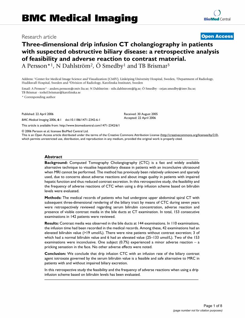
Open AcceResearch articleThree-dimensional drip infusion CT cholangiography in patients with suspected obstructive biliary disease: a retrospective analysis of feasibility and adverse reaction to contrast material. A Persson*1, N Dahlström2, Ö Smedby1 and TB Brismar3Address: 1Center for Medical Image Science and Visualization (CMIV), Linköping University Hospital, Sweden, 2Department of Radiology, Hudiksvall Hospital, Sweden and 3Division of Radiology, Karolinska Institutet, Sweden
Email: A Persson* - [email protected]; N Dahlström - [email protected]; Ö Smedby - [email protected]; TB Brismar - [email protected]
* Corresponding author
AbstractBackground: Computed Tomography Cholangiography (CTC) is a fast and widely availablealternative technique to visualise hepatobiliary disease in patients with an inconclusive ultrasoundwhen MRI cannot be performed. The method has previously been relatively unknown and sparselyused, due to concerns about adverse reactions and about image quality in patients with impairedhepatic function and thus reduced contrast excretion. In this retrospective study, the feasibility andthe frequency of adverse reactions of CTC when using a drip infusion scheme based on bilirubinlevels were evaluated.
Methods: The medical records of patients who had undergone upper abdominal spiral CT withsubsequent three-dimensional rendering of the biliary tract by means of CTC during seven yearswere retrospectively reviewed regarding serum bilirubin concentration, adverse reaction andpresence of visible contrast media in the bile ducts at CT examination. In total, 153 consecutiveexaminations in 142 patients were reviewed.
Results: Contrast media was observed in the bile ducts at 144 examinations. In 110 examinations,the infusion time had been recorded in the medical records. Among these, 42 examinations had anelevated bilirubin value (>19 umol/L). There were nine patients without contrast excretion; 3 ofwhich had a normal bilirubin value and 6 had an elevated value (25–133 umol/L). Two of the 153examinations were inconclusive. One subject (0.7%) experienced a minor adverse reaction – apricking sensation in the face. No other adverse effects were noted.
Conclusion: We conclude that drip infusion CTC with an infusion rate of the biliary contrastagent iotroxate governed by the serum bilirubin value is a feasible and safe alternative to MRC inpatients with and without impaired biliary excretion.
In this retrospective study the feasibility and the frequency of adverse reactions when using a drip infusion scheme based on bilirubin levels has been evaluated.
Published: 22 April 2006
BMC Medical Imaging 2006, 6:1 doi:10.1186/1471-2342-6-1
Received: 30 August 2005Accepted: 22 April 2006
This article is available from: http://www.biomedcentral.com/1471-2342/6/1
© 2006 Persson et al; licensee BioMed Central Ltd.This is an Open Access article distributed under the terms of the Creative Commons Attribution License (http://creativecommons.org/licenses/by/2.0), which permits unrestricted use, distribution, and reproduction in any medium, provided the original work is properly cited.
Page 1 of 8(page number not for citation purposes)
BMC Medical Imaging 2006, 6:1 http://www.biomedcentral.com/1471-2342/6/1
BackgroundFor diagnosis of hepatobiliary disease, ultrasound and MRcholangiography (MRC) are most frequently used. Endo-scopic Retrograde Cholangiography (ERC) is oftenregarded as the gold standard for visualising biliary dis-ease. The latter modality is invasive, user-dependent andmay induce pancreatitis. It should therefore not be per-formed in patients where intervention is less certain.Ultrasound, on the other hand, is easily tolerated by thepatients and cost effective. The modality is, however, user-dependent and the captured images are not easily under-stood by clinicians. MRC is superior in visualising the bil-iary system, and the images are appreciated by thesurgeons at surgical planning. It does not require any con-trast agent to visualise the bile ducts, and dilatation andgallstones in the common bile duct are easily detected [1-3]. Unfortunately, MRC cannot be performed in allpatients and hospitals due to limited availability of MRIor due to contraindications. MRC is also often inconclu-sive in patients with air in the biliary system, e.g. after pap-illotomy or liver surgery with entero-hepatic anastomoses(such as Whipple's operation and Billroth 2). Surgicalclips after cholecystectomy may also give artefacts mim-icking a ductal cancer or a stone [4,5]. An alternative non-invasive method to ultrasound and MRC is thereforerequired.
Computed Tomography Cholangiography (CTC) is a fastand widely available technique to visualise hepatobiliarydisease. Without contrast administration, multi detectorCT has been reported to have a sensitivity of 65%–88%and a specificity of 84%–97% to detect gallstones [6,7].Techniques to improve the sensitivity and specificity byadministering biliary contrast media orally [8] or intrave-nously [9,10] have been developed, but are not wide-spread. Possible explanations for infrequent use of CTCmight be the low resolution of single detector helical CTand reports of an unacceptable high number of adverseevents after injection of meglumine iotroxate [11]. Withthe development of multidetector CT, the resolution ofCTC exceeds that of MR. The number of adverse reactionswith biliary contrast media has probably diminished byinfusing the contrast media instead of injecting.
The aim of this retrospective study was to evaluate pro-longed drip infusion CT cholangiography (CTC) in
patients with suspected obstructive biliary disease withrespect to both feasibility and rate of adverse reactionsafter administration of the biliary contrast agent (iotrox-ate).
MethodsThis is a retrospective study in 142 consecutive patients(68 men and 74 women, mean age 69 years, range 24 – 95years) referred for investigation of biliary disease duringthe period from January 1996 to January 2003. Afterapproval by the ethics committee for the region, the med-ical records of all patients were retrospectively reviewedregarding bilirubin level, infusion time and adverseevents. Adverse events were defined as any signs of reac-tion to contrast media that occurred after the injection,such as anaphylaxis, urticaria and respiratory distress.
Administration of contrast mediaThe serum bilirubin concentration was measured before CTexamination using standard clinical laboratory methodsused at the hospital. 100 ml of meglumine iotroxate (Bili-scopin®, Schering AG, Berlin, Germany) 50 mg I/ml wasadministered by intravenous drip infusion. In order toallow longer infusion times, the solution volume wasincreased by dilution with isotonic sodium chloride (500ml). The infusion time was determined by the measuredbilirubin level according to a schematic protocol (Table 1).Following the guidelines from the manufacturer, the dripinfusion was started at a low infusion rate (0.5 ml/min)and increased to the desired infusion rate during the fol-lowing 3–5 minutes. The CT scan was started immediatelyafter the infusion was completed. For distension of the dis-tal duodenum, the patients ingested two glasses of drinkingwater immediately before the CT examination. To evaluate
Table 2: CT acquisition parameters
Type of scanner Collimation Pitch Increment mAs kV Number of examinations
Single Slice 1 × 5.0 mm 1.5 1 mm 200 120 103Multi-slice 4 × 2.5 mm 6 1 mm 130 120 46Multi-slice 16 × 0.75 mm varying 0.5 mm 130 120 4
Table 1: The infusion rate of iotroxate (Biliscopin®) was governed by the bilirubin level prior to the investigation. The same total amount of Iodine (5 g) was given to all patients.
Serum bilirubin Infusion time
<20 µmol/ml 40–60 min21–40 µmol/ml 1–3 hours41–99 µmol/ml 3–4 hours>100 µmol/ml 5 hours
Page 2 of 8(page number not for citation purposes)
BMC Medical Imaging 2006, 6:1 http://www.biomedcentral.com/1471-2342/6/1
compliance to the protocol, the medical records werereviewed regarding the given infusion time at the ward.
Scanning parametersPatients were scanned in the right oblique position bymeans of thin-section single-breath-hold helical CT in thecranio-caudal direction. Specific scan protocols varieddepending on the CT scanner available at the time ofexamination (Table 2). Between December 1995 andNovember 1999, 102 patients were scanned with a single-slice CT scanner (Somatom A; Siemens Medical Systems,Forcheim, Germany). From December 1999 to November2002, a 4-slice multi-detector CT scanner (Somatom Vol-ume Zoom; Siemens Medical Systems, Forcheim, Ger-many) was used in 44 exams. Between December 2002and January 2003, a 16-slice multi-detector CT scanner(Somatom Sensation16; Siemens Medical Systems,Forcheim, Germany) was used in 6 exams.
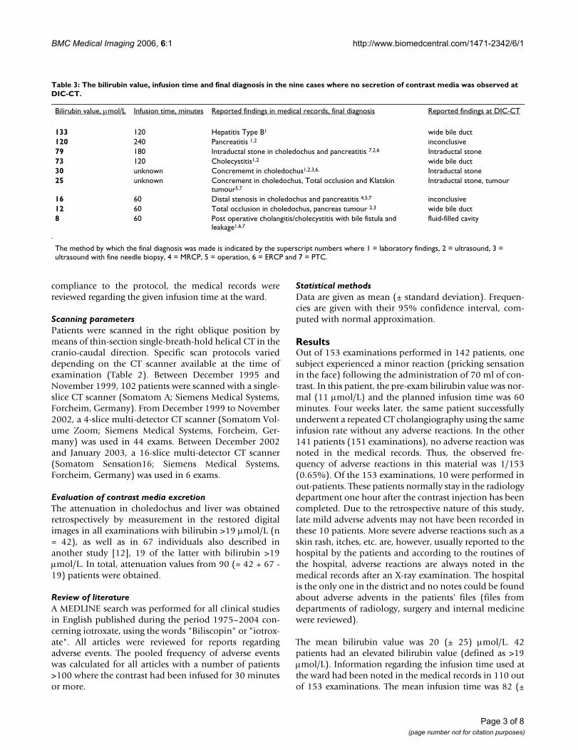
Evaluation of contrast media excretionThe attenuation in choledochus and liver was obtainedretrospectively by measurement in the restored digitalimages in all examinations with bilirubin >19 µmol/L (n= 42), as well as in 67 individuals also described inanother study [12], 19 of the latter with bilirubin >19µmol/L. In total, attenuation values from 90 (= 42 + 67 -19) patients were obtained.
Review of literatureA MEDLINE search was performed for all clinical studiesin English published during the period 1975–2004 con-cerning iotroxate, using the words "Biliscopin" or "iotrox-ate". All articles were reviewed for reports regardingadverse events. The pooled frequency of adverse eventswas calculated for all articles with a number of patients>100 where the contrast had been infused for 30 minutesor more.
Statistical methodsData are given as mean (± standard deviation). Frequen-cies are given with their 95% confidence interval, com-puted with normal approximation.
ResultsOut of 153 examinations performed in 142 patients, onesubject experienced a minor reaction (pricking sensationin the face) following the administration of 70 ml of con-trast. In this patient, the pre-exam bilirubin value was nor-mal (11 µmol/L) and the planned infusion time was 60minutes. Four weeks later, the same patient successfullyunderwent a repeated CT cholangiography using the sameinfusion rate without any adverse reactions. In the other141 patients (151 examinations), no adverse reaction wasnoted in the medical records. Thus, the observed fre-quency of adverse reactions in this material was 1/153(0.65%). Of the 153 examinations, 10 were performed inout-patients. These patients normally stay in the radiologydepartment one hour after the contrast injection has beencompleted. Due to the retrospective nature of this study,late mild adverse advents may not have been recorded inthese 10 patients. More severe adverse reactions such as askin rash, itches, etc. are, however, usually reported to thehospital by the patients and according to the routines ofthe hospital, adverse reactions are always noted in themedical records after an X-ray examination. The hospitalis the only one in the district and no notes could be foundabout adverse advents in the patients' files (files fromdepartments of radiology, surgery and internal medicinewere reviewed).
The mean bilirubin value was 20 (± 25) µmol/L. 42patients had an elevated bilirubin value (defined as >19µmol/L). Information regarding the infusion time used atthe ward had been noted in the medical records in 110 outof 153 examinations. The mean infusion time was 82 (±
Table 3: The bilirubin value, infusion time and final diagnosis in the nine cases where no secretion of contrast media was observed at DIC-CT.
Bilirubin value, µmol/L Infusion time, minutes Reported findings in medical records, final diagnosis Reported findings at DIC-CT
133 120 Hepatitis Type B1 wide bile duct120 240 Pancreatitis 1,2 inconclusive79 180 Intraductal stone in choledochus and pancreatitis 7,2,6 Intraductal stone73 120 Cholecystitis1,2 wide bile duct30 unknown Concrememt in choledochus1,2,3,6, Intraductal stone25 unknown Concrement in choledochus, Total occlusion and Klatskin
tumour5,7Intraductal stone, tumour
16 60 Distal stenosis in choledochus and pancreatitis 4,5,7 inconclusive12 60 Total occlusion in choledochus, pancreas tumour 2,3 wide bile duct8 60 Post operative cholangitis/cholecystitis with bile fistula and
leakage1,6,7fluid-filled cavity
The method by which the final diagnosis was made is indicated by the superscript numbers where 1 = laboratory findings, 2 = ultrasound, 3 = ultrasound with fine needle biopsy, 4 = MRCP, 5 = operation, 6 = ERCP and 7 = PTC.
Page 3 of 8(page number not for citation purposes)
BMC Medical Imaging 2006, 6:1 http://www.biomedcentral.com/1471-2342/6/1
Attenuation in choledochus and liver at DIC-CT as a function of serum bilirubin before the examinationFigure 2Attenuation in choledochus and liver at DIC-CT as a function of serum bilirubin before the examination.
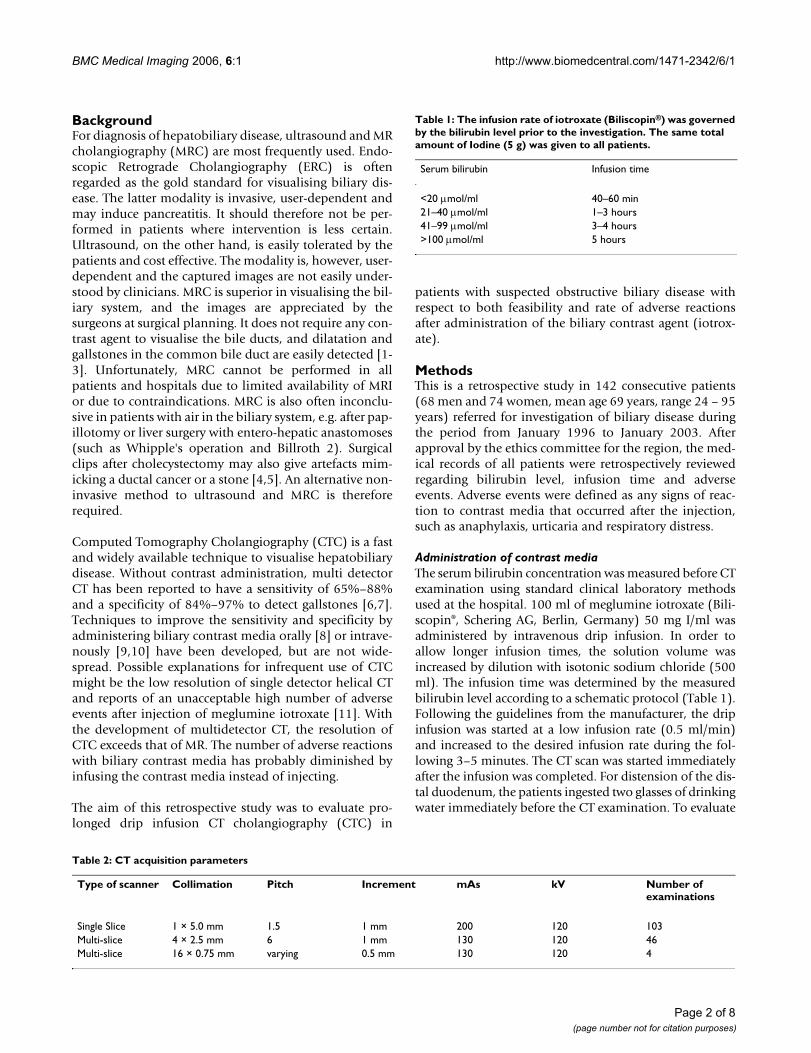
The infusion time of iotroxate (Biliscopin®) in relation to bilirubin level prior to the investigationFigure 1The infusion time of iotroxate (Biliscopin®) in relation to bilirubin level prior to the investigation. The recommended infusion times were followed in 103 out of the 110 cases (94%) where information on infusion time was found in the medical records. Cases in which the recommendations were not followed are encircled (n = 7). Unfortunately, none of the three (3/110) exam-inations with a bilirubin value >100 µmol/ml was performed according to the infusion scheme. The patient with the highest bilirubin value (159 µmol/ml) had good diagnostic excretion of contrast in the bile ducts, whereas the other two had no excre-tion.
Page 4 of 8(page number not for citation purposes)
42) minutes. Disregarding potential measurement errors of
BMC Medical Imaging 2006, 6:1 http://www.biomedcentral.com/1471-2342/6/1
at most 2 µmol/L, seven infusions (5%) had not been per-formed according to the protocol. Five of these received theinfusion too fast and 2 too slow (Fig. 1). All three patientswith a bilirubin >100 µmol/L were among the sevenpatients who did not receive the correct infusion rate. Theintended infusion rate (5 hours) could therefore not beevaluated.
Excretion of contrast media was observed in 93% (143/153) of all exams (one examination aborted due to poten-tial contrast reaction). In patients with elevated serumbilirubin (>19 µmol/L) contrast media in the bile wasobserved in 36 out of 42 patients (86%). No visible secre-tion of contrast was reported in 9 patients (Table 3). Inthree of these, the infusion protocol had not been fol-lowed, with too fast infusion (bilirubin 73–133 µmol/L).The final diagnoses in the patients with no visible secre-tion are also shown in Table 3. Three of these had occlu-sive intraductal stones, all of which were reported at theCTC. Two patients had a malignancy affecting the bileducts. One of these was reported at CTC and the othershowed signs of dilated bile ducts.
The remaining 4 patients had hepatitis, pancreatitis,cholangitis or cholecystitis.
The observed attenuation in choledochus and liver for dif-ferent serum bilirubin levels is shown in Fig. 2.
Review of literatureIn total, 42 original publications in English were found.Those with more than 100 patients and with an infusion
time of 30 minutes or more are listed in Table 4 as well asthe pooled number of adverse events (2.27%).
DiscussionWhen the bile ducts are obstructed, excretion of bile andcontrast media is decreased. It has therefore been assumedthat CT cholangiography cannot be performed in patientswith elevated serum bilirubin concentration [9,10]. In thisstudy, the infusion rate of the contrast media was adjusted tothe bilirubin value (Table 1). The aim was to keep the con-centration within the excretory capacity of the hepatocytes inorder to optimise the concentration in bile. By using thisscheme, contrast excretion into the bile ducts was observedin 93% of all exams. In patients with elevated serumbilirubin (>19 µmol/L), contrast media in the bile wasobserved in 86%. Excretion of contrast media was notedeven when the bilirubin concentration was as high as 159µmol/L (Fig. 3, 4). It has previously been recommended notto perform CTC in patients with bilirubin >50 µmol/L (3mg/dL) [14]. In this study, excretion was observed in fourout of eight patients with bilirubin >50 µmol/L. In three ofthose without contrast excretion, however, the infusion pro-tocol had not been followed (too fast infusion). Althoughabsence of contrast media in the bile ducts is more likely inpatients with greatly elevated bilirubin, CTC is not useless inthese patients – only two of the nine examinations withoutcontrast excretion were inconclusive. In the other sevencases, CTC findings could guide the referring physician toother examinations and the final diagnosis (Table 3).
The lack of excretion may also constitute valuable infor-mation. Patients without excretion are likely to haveeither a total occlusion of the main bile duct/choledochus
Table 4: Published studies on the frequency of adverse reactions at infusion of iotroxate at intravenous cholangiography. Included are all studies with at least 100 patients using an infusion time of at least 30 min. The severity of the reactions is graded as reported. The number in superscript denotes the corresponding reference.
No. of Patients Total Minor Intermediate Severe Fatal
Nilsson 198711 1 446 49 (3.4%) 41 (2.9%) 5 (0.35%) 3 (0.21%) 0Daly 198731 286 4 (1.4%) 4 (1.4%) 0 0 0Joyce 199132 100 2 (2.0%) 2 (2.0%) 0 0 0Wigmore 199333 100 0 0 0 0 0Patel 199334 113 3 (2.7%) 3 (2.7%) 0 0 0Grunshaw 199335 137 4 (2.9%) 3 (2.2%) 1 (0.7%) 0 0ASacharias 199536 1 061 11 (1.0%) 11 A (1.0%) 0 0Kwon 199816 440 2 (0.5%) 2 (0.5%) 0 0 0Kitami 200637 220 3 (1.4%) 3 (1.5%) 0 0 0Okada 200517 432 4 (0.9% 4 (0.9%) 0 0 0Hirao 200022 120 2 (1.7%) 2 (1.7%) 0 0 0BTakamatsu 200438 132 1 (0.8%) 1 (0.8%) 0 0 0Total 4587 85 65 17 3 0Frequency (95% confidence limits)
1.9% (1.5%–2.2%) 1.4% (1.1%–1.8% 0.4% (0.2%–0.5%) 0.1% (0–0.1%) 0
ANo difference was made between minor and intermediate adverse events.B The infusion time was 25–30 min. The number of complications in the article was reported by personal communication.
Page 5 of 8(page number not for citation purposes)
BMC Medical Imaging 2006, 6:1 http://www.biomedcentral.com/1471-2342/6/1
or severely impaired hepatocyte function. The bilirubinconcentration, if not already considerably elevated, islikely to increase in these patients. In this study, the lackof excretion could be explained by the final diagnosis inall patients (Table 3).
The protein-binding characteristics essential for biliarycontrast media increase the risk of adverse reactions[11,15]. In a previously published review of the literatureon the frequency of adverse reactions in examinationswith short injection time (<10 min), the pooled numberof adverse events was three times higher (16% vs. 5%)than after infusion (>30 min) of the same amount of con-trast media [11]. The frequency of adverse events ofiotroxate (Biliscopin®) at infusion has been reported to beas high as 3.4%, with a pooled frequency of 1.9% (Table4). It has been proposed that the tolerance of intravenousbiliary contrast media is improved when a slow infusiontechnique is used (up to one hour of infusion) [16,17].Our study supports this proposal, as there was only oneadverse reaction, which was mild, in 142 patients and 153examinations (0.65%).
After an inconclusive ultrasound examination, MRC has theadvantage of not exposing the patient to radiation and con-trast media. On the other hand, in many clinical situations
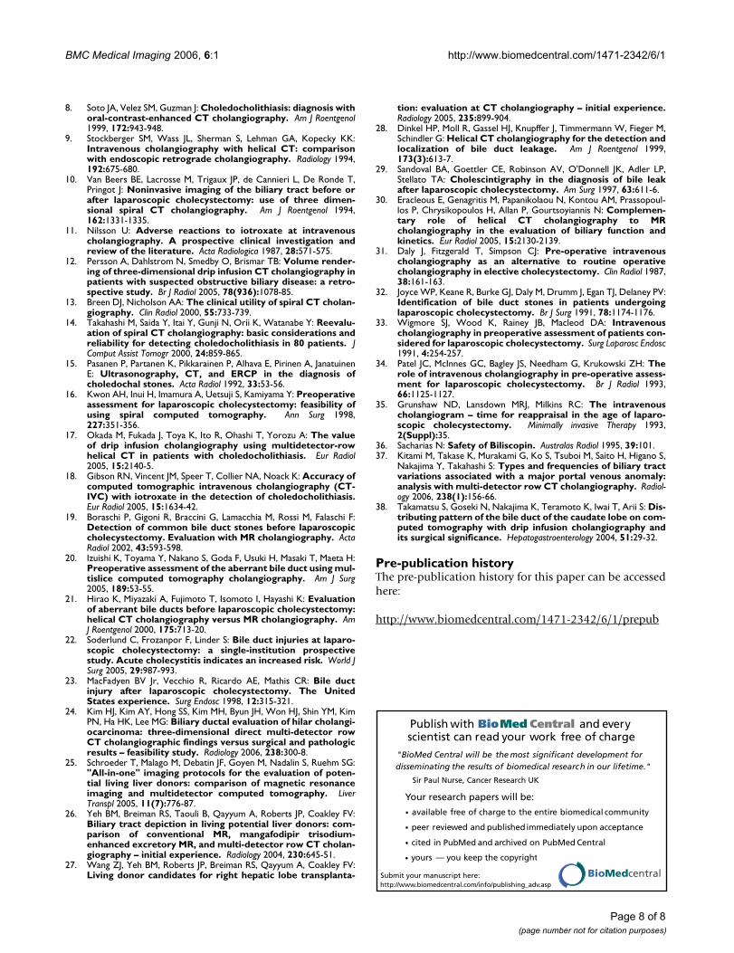
Traffic accident with a liver rupture and leakage from a small bile ductFigure 5Traffic accident with a liver rupture and leakage from a small bile duct. The DIC-CT examination led the surgeon correctly to the leaking bile duct (arrow). The diameter of the rup-tured bile duct was 1 mm.
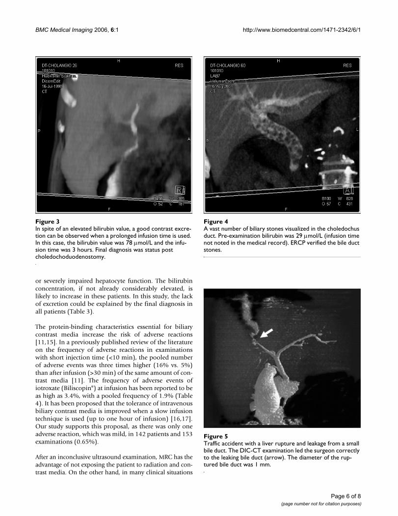
In spite of an elevated bilirubin value, a good contrast excre-tion can be observed when a prolonged infusion time is usedFigure 3In spite of an elevated bilirubin value, a good contrast excre-tion can be observed when a prolonged infusion time is used. In this case, the bilirubin value was 78 µmol/L and the infu-sion time was 3 hours. Final diagnosis was status post choledochoduodenostomy.
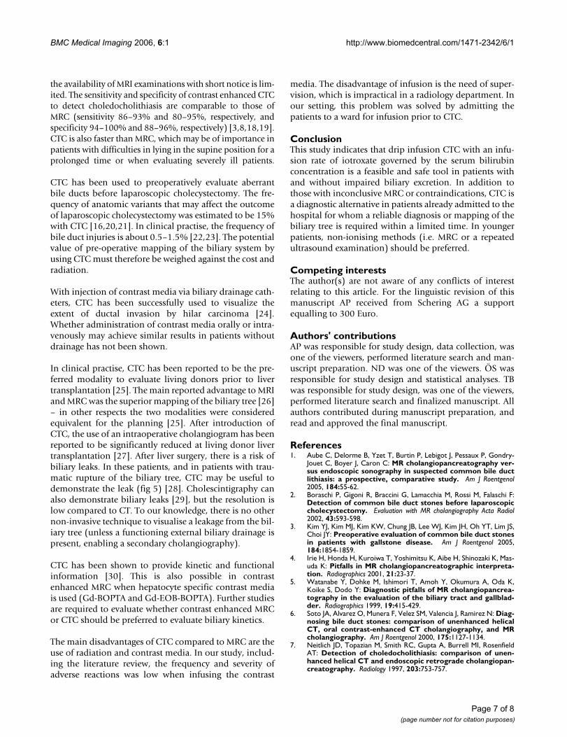
A vast number of biliary stones visualized in the choledochus ductFigure 4A vast number of biliary stones visualized in the choledochus duct. Pre-examination bilirubin was 29 µmol/L (infusion time not noted in the medical record). ERCP verified the bile duct stones.
Page 6 of 8(page number not for citation purposes)
BMC Medical Imaging 2006, 6:1 http://www.biomedcentral.com/1471-2342/6/1
the availability of MRI examinations with short notice is lim-ited. The sensitivity and specificity of contrast enhanced CTCto detect choledocholithiasis are comparable to those ofMRC (sensitivity 86–93% and 80–95%, respectively, andspecificity 94–100% and 88–96%, respectively) [3,8,18,19].CTC is also faster than MRC, which may be of importance inpatients with difficulties in lying in the supine position for aprolonged time or when evaluating severely ill patients.
CTC has been used to preoperatively evaluate aberrantbile ducts before laparoscopic cholecystectomy. The fre-quency of anatomic variants that may affect the outcomeof laparoscopic cholecystectomy was estimated to be 15%with CTC [16,20,21]. In clinical practise, the frequency ofbile duct injuries is about 0.5–1.5% [22,23]. The potentialvalue of pre-operative mapping of the biliary system byusing CTC must therefore be weighed against the cost andradiation.
With injection of contrast media via biliary drainage cath-eters, CTC has been successfully used to visualize theextent of ductal invasion by hilar carcinoma [24].Whether administration of contrast media orally or intra-venously may achieve similar results in patients withoutdrainage has not been shown.
In clinical practise, CTC has been reported to be the pre-ferred modality to evaluate living donors prior to livertransplantation [25]. The main reported advantage to MRIand MRC was the superior mapping of the biliary tree [26]– in other respects the two modalities were consideredequivalent for the planning [25]. After introduction ofCTC, the use of an intraoperative cholangiogram has beenreported to be significantly reduced at living donor livertransplantation [27]. After liver surgery, there is a risk ofbiliary leaks. In these patients, and in patients with trau-matic rupture of the biliary tree, CTC may be useful todemonstrate the leak (fig 5) [28]. Cholescintigraphy canalso demonstrate biliary leaks [29], but the resolution islow compared to CT. To our knowledge, there is no othernon-invasive technique to visualise a leakage from the bil-iary tree (unless a functioning external biliary drainage ispresent, enabling a secondary cholangiography).
CTC has been shown to provide kinetic and functionalinformation [30]. This is also possible in contrastenhanced MRC when hepatocyte specific contrast mediais used (Gd-BOPTA and Gd-EOB-BOPTA). Further studiesare required to evaluate whether contrast enhanced MRCor CTC should be preferred to evaluate biliary kinetics.
The main disadvantages of CTC compared to MRC are theuse of radiation and contrast media. In our study, includ-ing the literature review, the frequency and severity ofadverse reactions was low when infusing the contrast
media. The disadvantage of infusion is the need of super-vision, which is impractical in a radiology department. Inour setting, this problem was solved by admitting thepatients to a ward for infusion prior to CTC.
ConclusionThis study indicates that drip infusion CTC with an infu-sion rate of iotroxate governed by the serum bilirubinconcentration is a feasible and safe tool in patients withand without impaired biliary excretion. In addition tothose with inconclusive MRC or contraindications, CTC isa diagnostic alternative in patients already admitted to thehospital for whom a reliable diagnosis or mapping of thebiliary tree is required within a limited time. In youngerpatients, non-ionising methods (i.e. MRC or a repeatedultrasound examination) should be preferred.
Competing interestsThe author(s) are not aware of any conflicts of interestrelating to this article. For the linguistic revision of thismanuscript AP received from Schering AG a supportequalling to 300 Euro.
Authors' contributionsAP was responsible for study design, data collection, wasone of the viewers, performed literature search and man-uscript preparation. ND was one of the viewers. ÖS wasresponsible for study design and statistical analyses. TBwas responsible for study design, was one of the viewers,performed literature search and finalized manuscript. Allauthors contributed during manuscript preparation, andread and approved the final manuscript.
References1. Aube C, Delorme B, Yzet T, Burtin P, Lebigot J, Pessaux P, Gondry-
Jouet C, Boyer J, Caron C: MR cholangiopancreatography ver-sus endoscopic sonography in suspected common bile ductlithiasis: a prospective, comparative study. Am J Roentgenol2005, 184:55-62.
2. Boraschi P, Gigoni R, Braccini G, Lamacchia M, Rossi M, Falaschi F:Detection of common bile duct stones before laparoscopiccholecystectomy. Evaluation with MR cholangiography Acta Radiol2002, 43:593-598.
3. Kim YJ, Kim MJ, Kim KW, Chung JB, Lee WJ, Kim JH, Oh YT, Lim JS,Choi JY: Preoperative evaluation of common bile duct stonesin patients with gallstone disease. Am J Roentgenol 2005,184:1854-1859.
4. Irie H, Honda H, Kuroiwa T, Yoshimitsu K, Aibe H, Shinozaki K, Mas-uda K: Pitfalls in MR cholangiopancreatographic interpreta-tion. Radiographics 2001, 21:23-37.
5. Watanabe Y, Dohke M, Ishimori T, Amoh Y, Okumura A, Oda K,Koike S, Dodo Y: Diagnostic pitfalls of MR cholangiopancrea-tography in the evaluation of the biliary tract and gallblad-der. Radiographics 1999, 19:415-429.
6. Soto JA, Alvarez O, Munera F, Velez SM, Valencia J, Ramirez N: Diag-nosing bile duct stones: comparison of unenhanced helicalCT, oral contrast-enhanced CT cholangiography, and MRcholangiography. Am J Roentgenol 2000, 175:1127-1134.
7. Neitlich JD, Topazian M, Smith RC, Gupta A, Burrell MI, RosenfieldAT: Detection of choledocholithiasis: comparison of unen-hanced helical CT and endoscopic retrograde cholangiopan-creatography. Radiology 1997, 203:753-757.
Page 7 of 8(page number not for citation purposes)
BMC Medical Imaging 2006, 6:1 http://www.biomedcentral.com/1471-2342/6/1
Publish with BioMed Central and every scientist can read your work free of charge
"BioMed Central will be the most significant development for disseminating the results of biomedical research in our lifetime."
Sir Paul Nurse, Cancer Research UK
Your research papers will be:
available free of charge to the entire biomedical community
peer reviewed and published immediately upon acceptance
cited in PubMed and archived on PubMed Central
yours — you keep the copyright
Submit your manuscript here:http://www.biomedcentral.com/info/publishing_adv.asp
BioMedcentral
8. Soto JA, Velez SM, Guzman J: Choledocholithiasis: diagnosis withoral-contrast-enhanced CT cholangiography. Am J Roentgenol1999, 172:943-948.
9. Stockberger SM, Wass JL, Sherman S, Lehman GA, Kopecky KK:Intravenous cholangiography with helical CT: comparisonwith endoscopic retrograde cholangiography. Radiology 1994,192:675-680.
10. Van Beers BE, Lacrosse M, Trigaux JP, de Cannieri L, De Ronde T,Pringot J: Noninvasive imaging of the biliary tract before orafter laparoscopic cholecystectomy: use of three dimen-sional spiral CT cholangiography. Am J Roentgenol 1994,162:1331-1335.
11. Nilsson U: Adverse reactions to iotroxate at intravenouscholangiography. A prospective clinical investigation andreview of the literature. Acta Radiologica 1987, 28:571-575.
12. Persson A, Dahlstrom N, Smedby O, Brismar TB: Volume render-ing of three-dimensional drip infusion CT cholangiography inpatients with suspected obstructive biliary disease: a retro-spective study. Br J Radiol 2005, 78(936):1078-85.
13. Breen DJ, Nicholson AA: The clinical utility of spiral CT cholan-giography. Clin Radiol 2000, 55:733-739.
14. Takahashi M, Saida Y, Itai Y, Gunji N, Orii K, Watanabe Y: Reevalu-ation of spiral CT cholangiography: basic considerations andreliability for detecting choledocholithiasis in 80 patients. JComput Assist Tomogr 2000, 24:859-865.
15. Pasanen P, Partanen K, Pikkarainen P, Alhava E, Pirinen A, JanatuinenE: Ultrasonography, CT, and ERCP in the diagnosis ofcholedochal stones. Acta Radiol 1992, 33:53-56.
16. Kwon AH, Inui H, Imamura A, Uetsuji S, Kamiyama Y: Preoperativeassessment for laparoscopic cholecystectomy: feasibility ofusing spiral computed tomography. Ann Surg 1998,227:351-356.
17. Okada M, Fukada J, Toya K, Ito R, Ohashi T, Yorozu A: The valueof drip infusion cholangiography using multidetector-rowhelical CT in patients with choledocholithiasis. Eur Radiol2005, 15:2140-5.
18. Gibson RN, Vincent JM, Speer T, Collier NA, Noack K: Accuracy ofcomputed tomographic intravenous cholangiography (CT-IVC) with iotroxate in the detection of choledocholithiasis.Eur Radiol 2005, 15:1634-42.
19. Boraschi P, Gigoni R, Braccini G, Lamacchia M, Rossi M, Falaschi F:Detection of common bile duct stones before laparoscopiccholecystectomy. Evaluation with MR cholangiography. ActaRadiol 2002, 43:593-598.
20. Izuishi K, Toyama Y, Nakano S, Goda F, Usuki H, Masaki T, Maeta H:Preoperative assessment of the aberrant bile duct using mul-tislice computed tomography cholangiography. Am J Surg2005, 189:53-55.
21. Hirao K, Miyazaki A, Fujimoto T, Isomoto I, Hayashi K: Evaluationof aberrant bile ducts before laparoscopic cholecystectomy:helical CT cholangiography versus MR cholangiography. AmJ Roentgenol 2000, 175:713-20.
22. Soderlund C, Frozanpor F, Linder S: Bile duct injuries at laparo-scopic cholecystectomy: a single-institution prospectivestudy. Acute cholecystitis indicates an increased risk. World JSurg 2005, 29:987-993.
23. MacFadyen BV Jr, Vecchio R, Ricardo AE, Mathis CR: Bile ductinjury after laparoscopic cholecystectomy. The UnitedStates experience. Surg Endosc 1998, 12:315-321.
24. Kim HJ, Kim AY, Hong SS, Kim MH, Byun JH, Won HJ, Shin YM, KimPN, Ha HK, Lee MG: Biliary ductal evaluation of hilar cholangi-ocarcinoma: three-dimensional direct multi-detector rowCT cholangiographic findings versus surgical and pathologicresults – feasibility study. Radiology 2006, 238:300-8.
25. Schroeder T, Malago M, Debatin JF, Goyen M, Nadalin S, Ruehm SG:"All-in-one" imaging protocols for the evaluation of poten-tial living liver donors: comparison of magnetic resonanceimaging and multidetector computed tomography. LiverTranspl 2005, 11(7):776-87.
26. Yeh BM, Breiman RS, Taouli B, Qayyum A, Roberts JP, Coakley FV:Biliary tract depiction in living potential liver donors: com-parison of conventional MR, mangafodipir trisodium-enhanced excretory MR, and multi-detector row CT cholan-giography – initial experience. Radiology 2004, 230:645-51.
27. Wang ZJ, Yeh BM, Roberts JP, Breiman RS, Qayyum A, Coakley FV:Living donor candidates for right hepatic lobe transplanta-
tion: evaluation at CT cholangiography – initial experience.Radiology 2005, 235:899-904.
28. Dinkel HP, Moll R, Gassel HJ, Knupffer J, Timmermann W, Fieger M,Schindler G: Helical CT cholangiography for the detection andlocalization of bile duct leakage. Am J Roentgenol 1999,173(3):613-7.
29. Sandoval BA, Goettler CE, Robinson AV, O'Donnell JK, Adler LP,Stellato TA: Cholescintigraphy in the diagnosis of bile leakafter laparoscopic cholecystectomy. Am Surg 1997, 63:611-6.
30. Eracleous E, Genagritis M, Papanikolaou N, Kontou AM, Prassopoul-los P, Chrysikopoulos H, Allan P, Gourtsoyiannis N: Complemen-tary role of helical CT cholangiography to MRcholangiography in the evaluation of biliary function andkinetics. Eur Radiol 2005, 15:2130-2139.
31. Daly J, Fitzgerald T, Simpson CJ: Pre-operative intravenouscholangiography as an alternative to routine operativecholangiography in elective cholecystectomy. Clin Radiol 1987,38:161-163.
32. Joyce WP, Keane R, Burke GJ, Daly M, Drumm J, Egan TJ, Delaney PV:Identification of bile duct stones in patients undergoinglaparoscopic cholecystectomy. Br J Surg 1991, 78:1174-1176.
33. Wigmore SJ, Wood K, Rainey JB, Macleod DA: Intravenouscholangiography in preoperative assessment of patients con-sidered for laparoscopic cholecystectomy. Surg Laparosc Endosc1991, 4:254-257.
34. Patel JC, McInnes GC, Bagley JS, Needham G, Krukowski ZH: Therole of intravenous cholangiography in pre-operative assess-ment for laparoscopic cholecystectomy. Br J Radiol 1993,66:1125-1127.
35. Grunshaw ND, Lansdown MRJ, Milkins RC: The intravenouscholangiogram – time for reappraisal in the age of laparo-scopic cholecystectomy. Minimally invasive Therapy 1993,2(Suppl):35.
36. Sacharias N: Safety of Biliscopin. Australas Radiol 1995, 39:101.37. Kitami M, Takase K, Murakami G, Ko S, Tsuboi M, Saito H, Higano S,
Nakajima Y, Takahashi S: Types and frequencies of biliary tractvariations associated with a major portal venous anomaly:analysis with multi-detector row CT cholangiography. Radiol-ogy 2006, 238(1):156-66.
38. Takamatsu S, Goseki N, Nakajima K, Teramoto K, Iwai T, Arii S: Dis-tributing pattern of the bile duct of the caudate lobe on com-puted tomography with drip infusion cholangiography andits surgical significance. Hepatogastroenterology 2004, 51:29-32.
Pre-publication historyThe pre-publication history for this paper can be accessedhere:
http://www.biomedcentral.com/1471-2342/6/1/prepub
Page 8 of 8(page number not for citation purposes)


![BMC Medical Imaging BioMed Central - link.springer.com · BMC Medical Imaging Software Open Access Internet Image Viewer ... SPM [3], AIR [4], MRIcro [5], Brainvox [6], ... This paper](https://img.dokumen.tips/doc/110x75/5ad7e6c07f8b9af9068ccd87/bmc-medical-imaging-biomed-central-link-medical-imaging-software-open-access.jpg)